O R I G I N A L R E S E A R C H
Risk factors associated with prolonged intestinal
colonization of ESBL-producing
Enterobacteriaceae
–
a prospective cohort study
This article was published in the following Dove Press journal: Infection and Drug Resistance
Oskar Ljungquist1,2 Marcus Schönbeck1,2 Kristian Riesbeck3 Johan Tham1
1Clinical Infection Medicine, Department
of Translational Medicine, Faculty of Medicine, Lund University, Malmö SE20502, Sweden;2Department of
Infectious Disease, Helsingborg’s Hospital, Helsingborg, Region Skåne, Sweden;3Clinical Microbiology,
Department of Translational Medicine, Faculty of Medicine, Lund University, Malmö SE20502, Sweden
Background:Extended spectrumβ-lactamase-producingEnterobacteriaceae(EPE) are respon-sible for a major part of the widespread antimicrobial resistance (AMR). Increased understanding of risk factors associated with intestinal colonization of EPE is crucial to implement adequate
actions against AMR. The aim of this study was to define potential risk factors for prolonged
intestinal colonization with EPE. A secondary aim was to analyze if patients were adequately informed about being infected or colonized by antibiotic-resistant bacteria.
Methods: Patients with a positive clinical EPE culture from urine, blood or feces were recruited in a region in the south of Sweden. Selective EPE fecal cultures were obtained at least three months after the initial positive culture. Prolonged intestinal colonization was
defined as the prevalence of any EPE in the follow-up fecal sample. Risk factors for
prolonged intestinal colonization were evaluated by using a questionnaire and by retro-spective review of medical records. A univariate model and a multivariate regression analysis were performed to identify possible risk factors for intestinal EPE colonization.
Results:Out of 143 patients included in the study, 57% remained positive for EPE at the second sampling. In a multivariate regression model, urological intervention, history of EPE
infection and travel to Africa and/or Asia within 2 years were found to be significantly
associated with prolonged intestinal colonization of EPE. Before being approached by us, 50% of patients displayed inadequate knowledge of EPE infection or colonization.
Conclusion:In this prospective cohort study, urological intervention within 6 months and a history of EPE infection are independently associated with prolonged intestinal colonization with EPE. In contrast, travel to Africa and/or Asia within 2 years is associated with a decreased risk of prolonged intestinal colonization with EPE. There is room for improvement when it comes to patient information regarding EPE to decrease of spread.
Keywords: antibiotic resistance, risk of infection, one health, gram-negative bacteria, antibiotic therapy, patient information
Introduction
A growing incidence of extended spectrum β-lactamase (ESBL)-producing
Enterobacteriaceae (EPE), and consequently a rapidly increasing antimicrobial
resistance (AMR) is highly worrying.1,2 It is mainly driven by excessive
antimi-crobial use in humans and animals in addition to inadequate infection prevention
and control practices.3Humans as well as domestic and wild animals harbor EPE in
the intestine, and those bacteria are more often found in the environment as
prevalence increases.4It is crucial to study the pathogenesis, virulence and spread
of EPE in order to implement adequate preventive measures.
Correspondence: Oskar Ljungquist Clinical Infection Medicine, Department of Translational Medicine, Faculty of Medicine, Lund University, Rut Lundskogs gata 3, Malmö SE20502, Sweden Tel +46 42 406 1000
Email [email protected]
Infection and Drug Resistance
Dove
press
open access to scientific and medical research
Open Access Full Text Article
Infection and Drug Resistance downloaded from https://www.dovepress.com/ by 118.70.13.36 on 22-Aug-2020
It has previously been described thatE. colibelonging to the phylogenetic group B2 has a greater capacity to colonize the human gut compared to the other groups including A, B1
and D. These strains are associated with carriage of specific
virulence genes, such as p-fimbriae and aerobactin.5–7EPE
belonging to phylogroup B2, including the globally dissemi-nated ST131 clone, is highly virulent, and associated with
prolonged intestinal carriage of EPE.8–12 It has been
sug-gested that strain-specific virulence factors may impact the
persistence of EPE in the intestine.9
The long-term duration of intestinal colonization of EPE is unknown and only a few studies exist that study prolonged colonization.8,10,11,13,14 Most of these studies focus on molecular characterization of EPE, and only a few examine risk factors associated with prolonged colo-nization. In contrast, numerous investigations on risk fac-tors for acquisition and the short-term loss of EPE have
been published.15–23 It is at present unclear whether a
failure to detect EPE in fecal cultures from patients
pre-viously colonized with EPE reflects an absolute loss of
EPE or merely poor sensitivity of detection methods. International travel to endemic areas is a risk factor for
EPE carriage.23 The acquisition rate varies depending on
geographical regions visited, and the highest rates are
associated with Africa, the Middle East and Asia.24
However, data suggest that travel-associated EPE
coloni-zation might be short-lived.25 In addition to international
travel and infection with E. coli strains from phylogroup
B2, it has previously been demonstrated that immobiliza-tion, antibiotic consumption 4 and 12 months prior to colonization, treatment with proton-pump inhibitors and urinary catheter use are independently associated with
prolonged intestinal EPE carriage.14,26,27 Previous studies
have focused on bacterial characteristics to determine risk
factors for intestinal colonization of EPE.8,10 The main
goal of this study was to study patient characteristics and
putative risk factors for prolonged EPE carriage.
Furthermore, we wanted to investigate whether patients were adequately informed about their EPE infection/ colonization.
Materials and methods
Study design and data collection
We performed a prospective cohort study in Skåne County, a region in the south of Sweden. Inclusion of patients started in January 2016 and ended in April 2017. Patients
18 years of age or older with a verified culture of EPE in
urine, blood, feces or any other location during the
inclusion period were eligible for inclusion. Medical
records were reviewed using the software Melior
(Melior, Siemens Healthcare Services, Upplands Väsby, Sweden) for the following exclusion criteria; alcohol or substance abuse, severe psychiatric disorder,
immunosup-pressed patients (eg, immunodeficiency, ongoing cancer
treatment, neutropenia/leukopenia, treatment with TNF-α
-inhibitors), inpatient care, patients with chronic venous catheters, dementia or failure to sign informed consent and newly arrived refugees. These exclusion criteria were established as this study used the same cohort of patients as a prospective randomized clinical trial performed by the same group (not yet published).
Between July 2016 and March 2018, 2148 patients with cultures positive for EPE in the region of Skåne were screened for inclusion. Out of these, 820 adult patients with at least 1 documented positive ESBL-culture from blood, wound, cervix, urine or feces were contacted by mail and asked to submit one fecal sample for micro-biological analysis. Prolonged EPE colonization was
defined by the group as the prevalence of EPE in the
fecal culture obtained with a minimum time period of 3
months from the first positive EPE-culture. Of the 820
adult patients invited to take part in the study, 677 declined
to submit a fecal sample. Definition of variables, ie,
pos-sible risk factors, was designed prior to data collection.
The steering document containing definition of variables is
attached in theSupplementary materials.
To assess possible risk factors associated with prolonged intestinal carriage of EPE, medical and personal informa-tion was collected by sending out quesinforma-tionnaires to the
participants, and by reviewing the patients’medical records.
Some information on potential risk factors relied solely on the answers from the questionnaires, eg, travel habits, social factors, etc. The queries on the questionnaire were tested on a small number of patients before the questionnaires were distributed. Three weeks after sending out the question-naires, patients who had not responded to the questionnaires were contacted by phone and interviewed according to the same questionnaire. During the period of which the data were collected, three patients were diseased.
Study setting
The Skåne County is situated in South Sweden with a total
population of 1,324,565 (December 2016).28 All samples
from primary, secondary as well as tertiary health care suppliers in Skåne County were analyzed at Clinical Microbiology (Laboratory Medicine, Lund, Sweden).
Infection and Drug Resistance downloaded from https://www.dovepress.com/ by 118.70.13.36 on 22-Aug-2020
Laboratory methods
All samples were selectively cultivated for EPE at the Clinical Microbiology laboratory according to standard
protocols. Briefly, the sample material was plated on
URI-Select four agar plates with vancomycin (BioRad) complemented with two antimicrobial susceptibility discs containing ceftazidime (10 µg/mL, Oxoid) and merope-nem (10 µg/mL, Oxoid) followed by incubation at 37°C overnight. Sample material was also plated on chromo-genic agar plates ChromID ESBL (BioMerieux) and incu-bated as above. Colonies of presumptive EPE were subcultivated on horse blood agar (HBA) and typed to bacterial species using matrix-assisted laser desorption/
ionization time of flight (MALDI-TOF).
The phenotype of EPE was characterized by susceptibil-ity to cloxacillin (AmpC) or clavulanic acid (ESBL). All EPE were tested for susceptibility against the following antibiotics using the EUCAST methodology and breakpoints; piperacil-lin-tazobactam, cefotaxime, ceftazidime-avibactam,
ceftazi-dime, ceftolozane-tazobactam, imipenem, meropenem,
gentamicin, tobramycin, amikacin,
trimethoprim-sulfa-methoxazole, ciprofloxacin and,finally, temocillin.
Statistical analyses
Fisher’s exact test was used to perform univariate analyses.
A two-sided exact significance of ≤0.05 was considered
significant. These analyses were conducted using
dichoto-mized data. All dichotodichoto-mized data were analyzed and unevenly distributed variables were excluded for the multi-ple logistic regression after analyzing the cross-tabulation tables and the Fisher’s exact test. The variable antibiotic use was highly correlated with a history of EPE infection and was thus excluded from the multiple comparisons. The statistical importance of the risk factors was estimated using a binary multiple logistic regression model. Patients willing to submit a fecal sample and not willing to submit a fecal sample were compared with regard to baseline char-acteristics and risk factors. Due to observed differences
regarding resistance against both ciprofloxacin and
tri-methoprim/sulfamethoxazole and EPE-infected compared to EPE-colonized, statistical weighting with respect to these variables was used. A model of stepwise forward
multiple regression was used to determine the most signifi
-cant risk factors for prolonged intestinal EPE carriage. Figure 1, Tables 1, S1, S3–S5 are based on actual data,
whereas Tables 2–4andS2are based on weighted data.
Association between the potential risk factors and
pro-longed carriage was quantified with odds ratio (OR) with a
95% confidence interval (CI). A P-value of ≤0.05 was
considered statistically significant. The statistical analyses
were performed using IBM SPSS Statistics for Windows, Version 24.0. Armonk, NY: IBM Corp. Additionally, the statistical analyses were validated by a statistician using the same data.
Ethical considerations
This study was granted ethical approval from the Regional
Ethical Review Board at Lund’s Tingsrätt with reference
number 2016-304 and 2018-143. All patients included in the study provided written informed consent and this study was conducted in accordance with the Declaration of Helsinki.
Results
Characteristics of EPE and rate of
prolonged colonization
We included a total number of 143 patients in the study, and these individuals carried in total 147 strains of EPE, of
which 134 (91%) were E. coli, 12 (8%) were Klebsiella
spp. and 1 (1%) wasProteusspp. Out of the 147 strains of
EPE, 134 (91%) were characterized as having an ESBL A
phenotype (according to the Ambler classification; inhibited
by clavulanic acid), 10 (7%) as AmpC (ampicillinase C; inhibited by cloxacillin) and 3 (2%) as having both. Of the
147 strains, 24 (16%) were resistant to ciprofloxacin, 24
(16%) were resistant to trimethoprim/sulfamethoxazole and 71 (48%) were resistant to both antimicrobials. In contrast,
28 (19%) EPE were susceptible to both ciprofloxacin and
trimethoprim/sulfamethoxazole. The characteristics of EPE
strains are displayed in detail inTable S1.
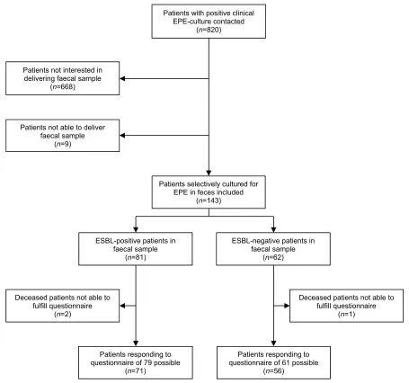
Eighty-one patients (57%) of the study cohort
remained positive for EPE in the follow-up fecal sample (Figure 1). A minority of patients (n=62; 43%) were negative for EPE in feces on follow-up. Of the 81 patients who remained positive for EPE in the follow-up fecal sample, an identical EPE was found as determined by phenotype and species when compared to the original sample. However, 19 patients (23%) had at least one anti-biotic that diverged compared to the antibiogram of the original culture.
Baseline characteristics of the study
population
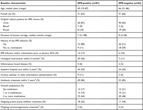
Baseline characteristics of the study population based on aggregate of medical record and questionnaires are as
out-lined in Table 1. Out of the 143 patients included in our
Infection and Drug Resistance downloaded from https://www.dovepress.com/ by 118.70.13.36 on 22-Aug-2020
study, 92 (64%) were women. The median age was 67 years
(range 19–87). The median Charlson comorbidity index
was 0 (range 0–7). At the time of inclusion (ie, submission
of fecal sample), median duration of known EPE carriage was at least 7 months in both groups. Out of the 143 patients, 106 (74%), 10 (7%) and 27 (19%) had an original culture positive for EPE in urine, blood and rectum, respec-tively. Furthermore, 116 (81%) of patients had a history of an EPE infection, whereas 27 (19%) patients were merely colonized with EPE. During the last 2 years, 70 (49%) of the 143 patients had been hospitalized and 122 (85%) had received at least one prescription of antibiotics.
The two groups, ie, EPE positive and EPE negative as determined by a follow-up fecal sample, were similar
regarding underlying medical conditions. There were no statistical differences in comorbidity as determined by
Charlson comorbidity score. Characteristics of the
patients’ comorbidity index according to this score are
presented inTable S2.
Analyses of putative risk factors for
prolonged EPE colonization
In the univariate model, urological intervention within 6
months (P=0.000), an original EPE-positive culture in
urine (P=0.009), a history of EPE infection (P=0.007)
and travel to Asia and/or Africa within two years
(P=0.020) were associated with a follow-up fecal sample
positive for EPE. Primary outcome for dichotomized data
Patients with positive clinical EPE-culture contacted
(n=820)
Patients not interested in delivering faecal sample
(n=668)
Patients not able to deliver faecal sample
(n=9)
Patients selectively cultured for EPE in feces included
(n=143)
ESBL-positive patients in faecal sample
(n=81)
ESBL-negative patients in faecal sample
(n=62)
Deceased patients not able to fulfill questionnaire
(n=2)
Deceased patients not able to fulfill questionnaire
(n=1)
Patients responding to questionnaire of 79 possible
(n=71)
Patients responding to questionnaire of 61 possible
(n=56)
Figure 1Flow chart of study patient inclusion and questionnaire distribution.
Abbreviation:EPE, extended spectrumβ-lactamase producingEnterobacteriaceae.
Infection and Drug Resistance downloaded from https://www.dovepress.com/ by 118.70.13.36 on 22-Aug-2020
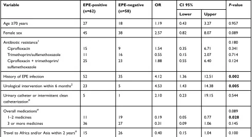
is presented in Table 2. In the multiple logistic regression
model, history of EPE infection (P=0.002, OR 4.12, 95%
CI: 1.36–12.51), urological intervention within 6 months
(P=0.005, OR 4.53, 95% CI: 1.43–14.38) and one or two
medications (P=0.028, OR 0.31, 95% CI: 0.05–0.77) were
associated with prolonged intestinal EPE colonization. Other putative risk factors for prolonged intestinal EPE carriage, ie, age over 70 years, female sex, urinary cathe-ter/intermittent clean catheterization, antibiotic resistance and travel to Africa and/or Asia within two years were not associated with prolonged intestinal EPE colonization. Secondary outcomes from a multiple logistic regression
model are displayed inTable 3.
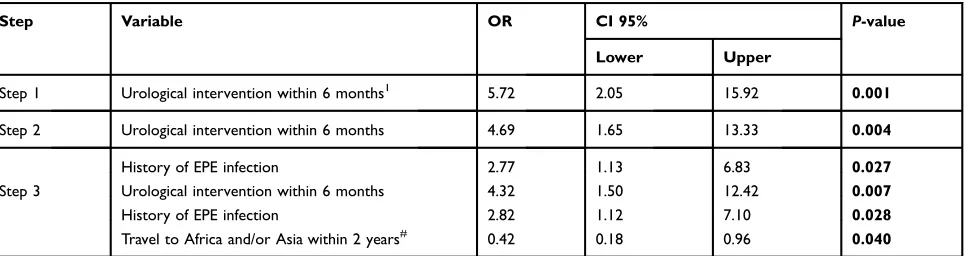
In the stepwise forward multiple regression model, the risk factor one to two medications was no longer of
sig-nificance. Urological intervention within 6 months
(P=0.007, OR 4.32, 95% CI: 1.50–12.42), history of EPE
infection (P=0.028, OR 2.82, 95% CI: 1.12–7.10) and
travel to Africa and/or Asia within the last 2 years
(P=0.04, OR 0.42, 95% CI: 0.18–0.96) remained signifi
-cant risk factors. The results of the stepwise forward
multi-ple regression model are displayed inTable 4.
Data obtained by the questionnaire
Thus, 140 patients received the questionnaire of which 127 (91%) submitted the form. The remaining 13 study sub-jects did not submit the questionnaire and could neither be reached by the investigators. The proportion of questionsanswered is displayed inTable S3.
Out of the 140 patients receiving the questionnaire, 122 (86%) answered the closed question if they had knowledge of EPE before being contacted by the study group. Sixty-one
Table 1Baseline characteristics of the study population based on medical records and questionnaires
Baseline characteristic EPE-positive (n=81) EPE-negative (n=62)
Age, median years (range) 69 (19–87) 66 (31–86)
Female sex (%) 51 (63) 41 (66)
Original culture positive for EPE, source (%)
Urine 66 (81) 40 (65)
Blood1 7 (9) 3 (5)
Rectal 8 (10) 19 (30)
Duration of known carriage, median months (range) 7 (3–108) 8 (3–56)
History of any EPE infection (%)
Yes 72 (89) 44 (71)
No, ie, colonization 9 (11) 18 (29)
EPE infection and/or colonization prior to January 2016 (%) 14 (17) 6 (10)
Urological intervention within 6 months2(%) 29 (36) 7 (11)
Inflammatory bowel disease (%) 0 (0) 2 (3)
Inpatient hospital care within 2 years3(%) 44 (54) 26 (42)
Urinary catheter or clean intermittent catheterization (%) 9 (11) 2 (3)
Antibiotic treatment within 2 years4(%) 69 (85) 53 (85)
Overall medications (%)
No medication 14 (17) 13 (21)
1 or 2 medications 15 (19) 19 (31)
3 or more medications 52 (64) 30 (48)
Ongoing proton pump inhibitor treatment (%) 18 (22) 11 (18)
Ongoing immunosuppressive treatment5(%) 4 (5) 3 (5)
Notes:All data were collected at the time of inclusion, ie, when fecal culture was obtained.1
If positive in both blood and urine, only blood was chosen.2
Minor or major invasive procedure on bladder, prostate and/or kidney and cystoscopy.3In Sweden and/or internationally.4Any systemic antibiotic.5Including any dosage of corticosteroids and/or methotrexate. All percentages are rounded up to the nearest integer.
Abbreviations:ESBL, extended spectrumβ-lactamase; EPE, extended spectrumβ-lactamase producingEnterobacteriaceae.
Infection and Drug Resistance downloaded from https://www.dovepress.com/ by 118.70.13.36 on 22-Aug-2020
patients (50%) were unaware of prior EPE infection or colo-nization. When contacted by the research group, 61 patients (50%) had knowledge of EPE infection or colonization. Of the positive cohort, 37 of 80 (45%) and in the EPE-negative cohort 24 of 61 (39%) were aware of EPE infection or colonization.
Accounting for missing data
Out of 820 patients approached in this study, only 143 (17%) were willing to submit a fecal sample. If the patients who chose not to provide a fecal sample in the study diverged from the patients included in the study, there could be a problem with the external validity of our results. To investigate this, the medical records of the patients (n=677) not willing to submit a fecal sample for the study were reviewed with regard to age, sex and Charlson comorbidity score. Also, the risk factors
significantly associated with prolonged colonization with
EPE that fell out before weighing were examined–history
of EPE infection, urological intervention within 6 months and
resistance against both ciprofloxacin and
trimethoprim/sulfa-methoxazole. These data were compared to the same
charac-teristics of the patients included in our study, andTable S4
shows the background characteristics of the groups. The med-ian age, female sex, medmed-ian Charlson comorbidity score or percentage of urological intervention within 6 months did not differ in the patients included in our study and the patients not
included. Thus, ourfindings could be generalized to the public.
Antibiotic consumption could not, however, be compared since these data relied on the submitted questionnaires. However, there was a difference in the percentage of patients
with resistance against both ciprofloxacin and trimethoprim/
sulfamethoxazole (P=0.01) and infected compared to
colo-nized (P=0.001) in the two groups. To rectify this, statistical weights were computed with regard to resistance and history of EPE infection. Computed statistical weights are presented inTable S5.
Table 2Univariate statistic model of putative risk factors for prolonged intestinal EPE carriage using Fisher’s exact test
Variable EPE-positive (n=76) EPE-negative (n=67) P-value
Age >70 years 32 19 0.115
Female sex 50 45 1.000
Escherichia coli1 73 61 0.149
ESBL phenotype A1 71 60 0.229
Bacteria resistant to Ciprofloxacin and/or Trimethoprim/Sulfamethoxazole1 62 51 0.538
Isolate in urine1 57 36 0.009
History of EPE infection 60 40 0.007
Carriage prior to January 2016 12 6 0.218
Urological intervention within 6 months2 26 6 0.000
Inflammatory bowel disease 0 2 0.218
Charlson comorbidity index >0 39 26 0.131
Inpatient hospital care within 2 years3 37 29 0.506
Urinary catheter or clean intermittent catheterization 7 1 0.066
Antibiotic use within 2 years4 60 52 1.000
Overall drug treatment >0 57 53 0.699
Proton pump inhibitor treatment 15 12 0.831
Immunosuppressive treatment5 3 3 1.000
Travel to Africa and/or Asia within 2 years# 15 26 0.020
Abdominal pain and/or altered bowel habits# 23 27 0.358
Domestic animal# 20 22 0.570
Currently working# 22 24 0.454
>1 person in household# 50 42 0.275
Known carrier of EPE in household6# 2 2 1.000
Vegetarian# 1 0 1.000
Post-secondary education# 33 32 1.000
Notes:Statistical analysis using Fisher’s exact test with exact 2-sided significance. AP-value of≤0.05 was considered significant. All data were collected originating at time of inclusion, ie, when fecal culture was collected, except1
which denotes original EPE-positive culture.2
Minor or major invasive procedure on bladder, prostate and/or kidney and cystoscopy.3
Both in Sweden and internationally.4
Any systemic antibiotic.5
Including any dosage of corticosteroids or methotrexate.6
Other than patient included in the study. All calculations are based on the weighted data. All counts are rounded to the nearest integer. Due to rounding, there are small variations in the number of patients included in each test. All percentages are rounded up to the nearest integer. #Based on the total number of questionnaire responses according toTable S3. In this article, aP-value of≤0.05 highlighted in bold was considered significant.
Abbreviations:ESBL, extended spectrumβ-lactamase; EPE, extended spectrumβ-lactamase producingEnterobacteriaceae.
Infection and Drug Resistance downloaded from https://www.dovepress.com/ by 118.70.13.36 on 22-Aug-2020
Discussion
In this prospective cohort study, factors independently associated with prolonged intestinal colonization with EPE were urological intervention within 6 months and a history of EPE infection. Travel to Africa and/or Asia
within the last 2 years was also significantly associated
with a decreased risk of becoming a long-term carrier. Furthermore, 50% of the patients in this study were not properly informed of infection/colonization of EPE.
Only a few studies exist that investigate why and in
whom prolonged EPE colonization occurs.8,10,14 This
study adds new information to the knowledge-void that
exists in thisfield of research. The strength of this study is
the prospective study design and robust methods of mea-surement. The statistical method of weighing data allowed for a somewhat sizable cohort of patients affected of EPE to be investigated. Overall, more than 90% submitted the questionnaire, either by sending it in or responding over the telephone, which is a strength in the present study. Furthermore, we show that patients affected by EPE are underinformed by health care givers, something which has not priorly been investigated in a similar setting.
Urological intervention at a maximum 6 months prior to inclusion was associated with prolonged intest-inal colonization of EPE. Apart from transrectal pros-tate biopsy, which is a common cause of nosocomial infection both with and without EPE, it is unlikely that the urological intervention itself is associated with prolonged intestinal colonization with EPE. However, antibiotic prophylaxis is common prior to urological interventions. The recommended prophylaxis according to guidelines in Skåne is one dose of either
trimetho-prim/sulfamethoxazole or ciprofloxacin prior to the
biopsy. It is a well-known fact that quinolones,
includ-ing ciprofloxacin, are drivers of antibiotic resistance.29
Importantly, EPE isolates from urological patients are
less susceptible to ciprofloxacin than isolates from
non-urological patients.30 Furthermore, fecal carriage of
EPE is common after transrectal prostate biopsy. The
presence of EPE after biopsy is associated with fl
uor-oquinolone consumption before biopsy.31 In addition,
transrectal prostate biopsy is a common cause of
antibiotic-resistant bloodstream infection, and the E.
coli clone CTX-M-15 ST131 is a major contributing
Table 3Multiple logistic regression model of putative risk factors for prolonged intestinal EPE carriage
Variable EPE-positive
(n=62)
EPE-negative (n=58)
OR CI 95% P-value
Lower Upper
Age≥70 years 27 18 1.19 0.43 3.27 0.957
Female sex 45 38 2.57 0.82 8.07 0.089
Antibiotic resistance1 0.180
Ciprofloxacin 15 9 1.54 0.35 6.71 0.341
Trimethoprim/sulfamethoxazole 11 16 0.55 0.15 2.07 0.714
Ciprofloxacin + trimethoprim/ sulfamethoxazole
25 23 1.88 0.55 6.40 0.124
History of EPE infection 52 35 4.12 1.36 12.51 0.002
Urological intervention within 6 months2 23 5 4.53 1.43 14.38 0.005
Urinary catheter or intermittent clean catheterization#
5 1 2.10 0.23 19.15 0.544
Overall medications# 0.089
1–2 medicines 11 19 0.19 0.05 0.77 0.028
3 or more medicines 36 27 0.31 0.09 1.06 0.145
Travel to Africa and/or Asia within 2 years# 15 26 0.40 0.15 1.04 0.100
Notes:Statistical analysis using binary logistic regression model.P-value≤0.05 was considered significant. Odds ratio for prolonged intestinal EPE carriage. All data were collected originating at the time of inclusion, ie, when fecal culture was collected, except1
which denotes original EPE-positive culture.2
Minor or major invasive procedure on bladder, prostate and/or kidney and cystoscopy. All calculations are based on the weighted data. All counts are rounded to the nearest integer. Due to missing data, the regression was calculated on 120 observations. All percentages are rounded up to the nearest integer.#
Information based on questionnaires. In this article, aP-value of≤0.05 highlighted in bold was considered significant.
Abbreviations:OR, odds ratio; CI, confidence interval; ESBL, extended spectrumβ-lactamase; EPE, extended spectrumβ-lactamase producingEnterobacteriaceae.
Infection and Drug Resistance downloaded from https://www.dovepress.com/ by 118.70.13.36 on 22-Aug-2020
pathogen in this context.32,33 Hence, administration of antibiotics in relation to urological interventions could explain why we found an association with prolonged intestinal colonization with EPE.
However, a more plausible explanation for the association between urological intervention and prolonged intestinal colo-nization of EPE is that cystoscopy is routinely performed in patients with recurrent UTIs. In Sweden, a nonendemic coun-try of EPE, repeated urinary cultures with EPE is an indication for urological intervention, to exclude a treatable cause for recurrent UTIs.
We suggest that this at least to a certain extent could explain the association.
When compared to mere colonization, a history of EPE infection was associated with a higher risk for prolonged intestinal colonization with EPE. This is in accordance with a previous study where colonization was associated with
clear-ance of EPE, when compared to clinical samples.18A study
from Sweden revealed that 43% of the patients still being
colonized 12 months after EPE infection.9This colonization
rate is higher compared to what Östholm-Balkhed et al and Arcilla and collaborators presented, 11.0% and 11.3%, respec-tively, found 12 months after EPE-colonization diagnosed after foreign travel.25,34In addition, Ruppé et al observed an even less frequent prevalence (4.7%) of EPE three months after
being colonized/infected abroad.15
Subsequent EPE infection in patients previously colonized
with EPE is rare.33On the other hand, recurrent infection with
EPE is common, especially within thefirst 6 months of thefirst
infection and in patients ≥65 years of age.35 Hence, it is
reasonable to assume that more virulent strains to a higher extent possess virulence factors giving them a survival
advantage, and the ability to colonize the intestine. Strains merely colonizing the intestine lack important virulence genes needed to trigger infection. This is in accordance with Ny et al who found strains of low pathogenicity in fecal samples in randomly selected Swedish citizens compared to strains causing invasive infection.36
The ST131 clone has managed to disseminate globally due to its ability to frequently colonize the intestine, its enhanced virulence and broader antibiotic resistance
com-pared to other EPE clones.37 Strains originating from this
clone are more prone to possess antibiotic resistance against
ciprofloxacin and trimethoprim/sulfamethoxazole.38We did
not find any association between EPEs with resistance
against ciprofloxacin and trimethoprim/sulfamethoxazole,
and prolonged EPE colonization.
Unfortunately, we were not able to conduct any mole-cular analysis such as whole genome sequencing (WGS) of the EPE isolates. WGS could have given us useful information on EPE characteristics. Furthermore, without WGS, multiple-locus variable number tandem repeat or
pulse-field gel electrophoresis, the strain obtained in the
fecal culture could not be determined to be exactly the same as the strain in the original culture. In fact, 23% of patients had disconcordant antibiograms when the original culture was compared to the follow-up fecal culture regarding at least one antibiotic. We believe that the main reason for this is that patients can carry many differ-ent EPEs, which can be impossible to distinguish when selecting colonies on a blood agar. A previous study, with a longer follow-up time, using MLVA showed that the majority of long-term EPE carriers had either a different
ESBL-producing species or a ESBL-producing E. coli
Table 4Stepwise forward multiple regression model of potential risk factors for prolonged intestinal EPE carriage
Step Variable OR CI 95% P-value
Lower Upper
Step 1 Urological intervention within 6 months1 5.72 2.05 15.92 0.001
Step 2 Urological intervention within 6 months 4.69 1.65 13.33 0.004
History of EPE infection 2.77 1.13 6.83 0.027
Step 3 Urological intervention within 6 months 4.32 1.50 12.42 0.007
History of EPE infection 2.82 1.12 7.10 0.028
Travel to Africa and/or Asia within 2 years# 0.42 0.18 0.96 0.040
Notes:Statistical analysis using binary logistic regression model.P-value≤0.05 was considered significant. Odds ratio for prolonged intestinal EPE carriage. All data were collected originating at the time of inclusion, ie, when fecal culture was collected.1
Minor or major invasive procedure on bladder, prostate and/or kidney and cystoscopy. All calculations are based on the weighted data. All counts are rounded to the nearest integer. Due to missing data, the regression was calculated on 120 observations. Included variables in the stepwise regression model as presented inTable 3.#
Information based on questionnaires. In this article, aP-value of≤0.05 highlighted in bold was considered significant.
Abbreviations:OR, odds ratio; CI, confidence interval; EPE, Extended spectrumβ-lactamase producingEnterobacteriaceae.
Infection and Drug Resistance downloaded from https://www.dovepress.com/ by 118.70.13.36 on 22-Aug-2020
with a different MLVA-profile in original culture
com-pared to the follow-up sample.10
Furthermore, the methodology in the Clinical
Microbiology laboratory may differ and the probability that the same strain can yield different susceptibility pat-terns increases as many antibiotics are tested. In addition, there is a slight risk that patients become colonized with new EPE strains, in which the term prolonged intestinal EPE colonization is misused in this context. This risk was estimated low by the study group in this low endemic EPE setting.
We found an association between EPE that were iso-lated from urine in original cultures, as opposed to any other anatomical location, and prolonged intestinal coloni-zation with EPE in the univariate analysis. This was,
however, not confirmed in the multivariate logistic
regres-sion model. We believe that these two risk factors, ie, history of EPE infection and EPE present in urine, are
closely connected. An EPE-positive urine culture reflects
infection or asymptomatic bacteriuria, whereas an
EPE-positive rectal culture is a marker of colonization. As previously mentioned, antibiotic consumption is clo-sely connected to a history of EPE infection and the rate of
consumption was high in both groups. We did notfind any
association between previous antibiotic consumption and prolonged intestinal colonization with EPE. This is in
accordance with several studies in non-travelers.9,10,14
However, Rogers et al found an association between anti-biotic consumption abroad and prolonged colonization of
EPE post travel.21
Traveling to high endemic areas of EPE such as Africa, Asia and Middle East is a known risk factor for EPE acquisition. In our study, we found that previous travel to EPE-high endemic areas was associated with a decreased risk of prolonged intestinal carriage of EPE. This is in accordance with ÖstholmBalkhed, who found no risk fac-tors associated with prolonged intestinal colonization with EPE, but found that diarrhea during travel and a new trip during follow-up was associated with a decreased risk of
becoming a long-term carrier.25 EPE acquisition during
travel may thus be unrelated to specific microbiota profile
but could affect the duration of colonization.39
We did not find correlation between a positive EPE
fecal sample and abdominal pain or diarrhea, the latter a
known risk factor for EPE acquisition.24 Papst et al
showed that immobilization is a risk factor for prolonged
EPE colonization, a risk factor this study did not address.14
In contrast, Alsterlund et al did not find risk factors
associated with prolonged carriage of EPE, but did not perform molecular characterization of the isolates nor
multivariable analysis of the data.40
With this study, we also wanted to investigate to what extent patients are informed about their EPE infection or colonization. Surprisingly, 50% of the patients were not adequately informed of their EPE status by their health care supplier. The Public Health Agency of Sweden states that it is mandatory for the responsible physician to provide
information regarding infection or colonization with EPE.41
This information is mandatory to prevent EPE from spread-ing, both in the community and in health care settings, as patients are obliged to disclose their EPE status when seek-ing health care. Adequate actions are needed by authorities since information is an underestimated but important tool
fighting antibiotic resistance.
A limitation with this study is that we used only one follow-up rectal culture, and it is a well-known fact that
transient negative samples occur upon EPE-colonization.8,14
It is important to note that we do not consider that a negative follow-up sample as equal to a permanent loss of EPE colonization. It could be argued, however, that a negative
EPE fecal sample reflects quantitatively less EPE shedding.
This could lead to reduced risk of spreading EPE in the community and hospital setting. If this is associated with a less risk of EPE infection is not answered in this study.
In general, when using questionnaires to collect infor-mation, response bias is a matter of concern. This was taken into consideration when composing the questionnaire. Still, some patients could have agreed to be included in the study and answer the questionnaire because they knew their EPE
status. This could be addressed by sending specific
ques-tionnaires to a sample of the patients who were invited to the study but declined to provide samples for the study.
We discovered that some questions in the questionnaire tended to be misapprehended. Furthermore, some
partici-pants did not fulfill the whole questionnaire and sample
collecting was not done immediately, which is why lack-ing memory could affect the answers. This could be coun-tered by interviewing the patients at the time of inclusion. A major limitation of this study was the low percentage of inclusion in the study, only 17% (143 of 820 patients) of the patients were willing to submit a fecal sample. A reason for this could be lack of information and implica-tions of EPE colonization. Another reason could be a perceived discomfort of submitting a fecal culture to the study group. However, as weighting was performed, our results gained validity.
Infection and Drug Resistance downloaded from https://www.dovepress.com/ by 118.70.13.36 on 22-Aug-2020
Results from this study contribute to knowledge
about the long-term carriage of EPE. We did not find
that patient characteristics such as severe underlying diseases, age, etc., are risk factors associated with pro-longed colonization of EPE. Bacterial features such as the ability to trigger an infection over colonization seem to be of greater importance. More information about colonization with EPE is crucial to implement adequate control measures, thus reducing spread of disease. It is particularly important to work preventive in a time of increasing antibiotic resistance. This is the most impor-tant reason to continue with well-designed research
within this field.
Conclusion
In this prospective cohort study, we have presented that urological intervention within 6 months and a history of EPE infection are independently associated with pro-longed intestinal colonization with EPE. Travel to Africa and/or Asia within 2 years is associated with a decreased risk of prolonged intestinal colonization with
EPE. We could not find any association between other
risk factors including prior antibiotic consumption, comorbidity, international travel, in hospital care, etc., with prolonged intestinal colonization with EPE. These
findings ultimately contribute to knowledge about EPE
pathogenesis and stimulate further research within this
field. Despite the fact that the Public Health Agency of
Sweden states that information regarding EPE is man-datory, 50% of the patients in this study were not properly informed about their EPE status.
Acknowledgments
Special thanks to Associate Professor Fredrik Resman, Professor Svante Twetman, Dr Jonas Tverring, MSc Axel Ström, MSc Fredrik Nilsson, Mrs Lena Hyllebusk and Mrs Anita Eriksson. This work was supported by grants from the Thelma Zoéga and the Stig and Ragna Gorthon foundation.
Disclosure
The authors have no conflict of interest to disclose in this
work.
References
1. World Health Organization. Antimicrobial resistance: global report on surveillance [Internet].2014Available from:https://www.who.int/dru gresistance/documents/surveillancereport/en/. Accessed August 07, 2019.
2. European Centre for Disease Prevention and Control. Antimicrobial resistance surveillance in Europe [Internet]. 2016. Available from:
https://ecdc.europa.eu/en/publications-data/antimicrobial-resistance-surveillance-europe-2016. Accessed August 07, 2019.
3. Holmes AH, Moore LS, Sundsfjord A, et al. Understanding the mechanisms and drivers of antimicrobial resistance. Lancet.
2016;387(10014):176–187. doi:10.1016/S0140-6736(15)00473-0 4. Jorgensen SB, Soraas AV, Arnesen LS, Leegaard TM, Sundsfjord A,
Jenum PA. A comparison of extended spectrum beta-lactamase pro-ducing Escherichia coli from clinical, recreational water and waste-water samples associated in time and location.PLoS One.2017;12 (10):e0186576. doi:10.1371/journal.pone.0186576
5. Nowrouzian FL, Adlerberth I, Wold AE. Enhanced persistence in the colonic microbiota of Escherichia coli strains belonging to phyloge-netic group B2: role of virulence factors and adherence to colonic cells. Microbes Infect. 2006;8(3):834–840. doi:10.1016/j.micinf. 2005.10.011
6. Nowrouzian FL, Wold AE, Adlerberth I. Escherichia coli strains belonging to phylogenetic group B2 have superior capacity to persist in the intestinal microflora of infants.J Infect Dis.2005;191(7):1078– 1083. doi:10.1086/427996
7. Johnson JR, Delavari P, Kuskowski M, Stell AL. Phylogenetic dis-tribution of extraintestinal virulence-associated traits in Escherichia coli.J Infect Dis.2001;183(1):78–88. doi:10.1086/317656 8. van Duijkeren E, Wielders CCH, Dierikx CM, et al. Long-term
carriage of extended-spectrum beta-lactamase-producing Escherichia coli and klebsiella pneumoniae in the general population in The Netherlands. Clin Infect Dis. 2018;66(9):1368–1376. doi:10.1093/ cid/cix1015
9. Titelman E, Hasan CM, Iversen A, et al. Faecal carriage of extended-spectrum beta-lactamase-producing Enterobacteriaceae is common 12 months after infection and is related to strain factors.Clin Microbiol
Infect.2014;20(8):O508–O515. doi:10.1111/1469-0691.12742
10. Jorgensen SB, Soraas A, Sundsfjord A, Liestol K, Leegaard TM, Jenum PA. Fecal carriage of extended spectrum beta-lactamase pro-ducing Escherichia coli and Klebsiella pneumoniae after urinary tract infection–a three year prospective cohort study.PLoS One.2017;12 (3):e0173510. doi:10.1371/journal.pone.0173510
11. Overdevest I, Haverkate M, Veenemans J, et al. Prolonged colonisa-tion with Escherichia coli O25: ST131 versus other extended-spec-trum beta-lactamase-producing E. coli in a long-term care facility with high endemic level of rectal colonisation, the Netherlands, 2013 to 2014. Euro Surveill. 2016;21(42). doi:10.2807/1560-7917. ES.2016.21.42.30376
12. Rodriguez-Revuelta MJ, Lopez-Cerero L, Serrano L, Luna-Lagares S, Pascual A, Rodriguez-Bano J. Duration of colonization by extended-spectrum beta-lactamase-producing Enterobacteriaceae in healthy newborns and associated risk factors: a prospective cohort study.Open Forum Infect Dis.2018;5(12):ofy312. doi:10.1093/ofid/ ofy312
13. Lohr IH, Rettedal S, Natas OB, Naseer U, Oymar K, Sundsfjord A. Long-term faecal carriage in infants and intra-household transmission of CTX-M-15-producing Klebsiella pneumoniae following a nosoco-mial outbreak. J Antimicrob Chemother. 2013;68(5):1043–1048. doi:10.1093/jac/dks502
14. Papst L, Beovic B, Seme K, Pirs M. Two-year prospective evaluation of colonization with extended-spectrum beta-lactamase-producing Enterobacteriaceae: time course and risk factors.Infect Dis (Lond).
2015;47(9):618–624. doi:10.3109/23744235.2015.1033003 15. Ruppe E, Armand-Lefevre L, Estellat C, et al. High rate of
acquisi-tion but short duraacquisi-tion of carriage of multidrug-resistant enterobac-teriaceae after travel to the tropics.Clin Infect Dis.2015;61(4):593– 600. doi:10.1093/cid/civ333
16. Jallad MA, Naoufal R, Irani J, Azar E. Extended spectrum beta-lactamase carriage state among elderly nursing home residents in Beirut.ScientificWorldJournal.2015;2015:987580.
Infection and Drug Resistance downloaded from https://www.dovepress.com/ by 118.70.13.36 on 22-Aug-2020
17. Lubbert C, Straube L, Stein C, et al. Colonization with extended-spectrum beta-lactamase-producing and carbapenemase-producing Enterobacteriaceae in international travelers returning to Germany.
Int J Med Microbiol. 2015;305(1):148–156. doi:10.1016/j.ijmm.
2014.12.001
18. Birgand G, Armand-Lefevre L, Lolom I, Ruppe E, Andremont A, Lucet JC. Duration of colonization by extended-spectrum beta-lacta-mase-producing Enterobacteriaceae after hospital discharge. Am J
Infect Control.2013;41(5):443–447. doi:10.1016/j.ajic.2012.05.015
19. Paltansing S, Vlot JA, Kraakman ME, et al. Extended-spectrum beta-lactamase-producing enterobacteriaceae among travelers from the Netherlands.Emerg Infect Dis.2013;19(8):1206–1213. doi:10.3201/ eid.1908.130257
20. Li B, Zhong Y, Fu XC, et al. Duration of stool colonization in healthy medical students with extended-spectrum-beta-lactamase-producing Escherichia coli. Antimicrob Agents Chemother. 2012;56(8):4558– 4559. doi:10.1128/AAC.00171-12
21. Rogers BA, Kennedy KJ, Sidjabat HE, Jones M, Collignon P, Paterson DL. Prolonged carriage of resistant E. coli by returned travellers: clonality, risk factors and bacterial characteristics.Eur J
Clin Microbiol Infect Dis. 2012;31(9):2413–2420. doi:10.1007/
s10096-012-1584-z
22. Zahar JR, Lanternier F, Mechai F, et al. Duration of colonisation by Enterobacteriaceae producing extended-spectrum beta-lactamase and risk factors for persistent faecal carriage. J Hosp Infect. 2010;75 (1):76–78. doi:10.1016/j.jhin.2009.11.010
23. Tangden T, Cars O, Melhus A, Lowdin E. Foreign travel is a major risk factor for colonization with Escherichia coli producing CTX-M-type extended-spectrum beta-lactamases: a prospective study with Swedish volunteers. Antimicrob Agents Chemother. 2010;54 (9):3564–3568. doi:10.1128/AAC.00220-10
24. Ruppe E, Andremont A, Armand-Lefevre L. Digestive tract coloni-zation by multidrug-resistant Enterobacteriaceae in travellers: an update. Travel Med Infect Dis. 2018;21:28–35. doi:10.1016/j. tmaid.2017.11.007
25. OstholmBalkhed A, Tarnberg M, Nilsson M, et al. Duration of travel-associated faecal colonisation with ESBL-producing Enterobacteriaceae – a one year follow-up study. PLoS One.
2018;13(10):e0205504. doi:10.1371/journal.pone.0205504
26. Morales Barroso I, Lopez-Cerero L, Navarro MD, Gutierrez-Gutierrez B, Pascual A, Rodriguez-Bano J. Intestinal colonization due to Escherichia coli ST131: risk factors and prevalence.Antimicrob Resist
Infect Control.2018;7:135. doi:10.1186/s13756-018-0427-9
27. Karanika S, Karantanos T, Arvanitis M, Grigoras C, Mylonakis E. Fecal colonization with extended-spectrum beta-lactamase-producing Enterobacteriaceae and risk factors among healthy individuals: a systematic review and metaanalysis. Clin Infect Dis. 2016;63 (3):310–318. doi:10.1093/cid/ciw283
28. Statistics Sweden. Population in the country, counties and municipa-lities on 31/ 12/16 and Population change in 2016 2016. Available from: https://www.scb.se/hitta-statistik/statistik-efter-amne/befolkn ing/befolkningens-sammansattning/befolkningsstatistik/pong/tabell-och-diagram/helarsstatistik– kommun-lan-och-riket/folkmangd-i-riket-lan-och-kommuner-31-december-och-befolkningsforandringar/. Accessed August 07, 2019.
29. Rodriguez-Bano J, Navarro MD, Romero L, et al. Epidemiology and clinical features of infections caused by extended-spectrum beta-lactamase-producing Escherichia coli in nonhospitalized patients.J
Clin Microbiol.2004;42(3):1089–1094.
doi:10.1128/jcm.42.3.1089-1094.2004
30. Bonkat G, Muller G, Braissant O, et al. Increasing prevalence of ciprofloxacin resistance in extended-spectrum-beta-lactamase-producing Escherichia coli urinary isolates. World J Urol.
2013;31(6):1427–1432. doi:10.1007/s00345-013-1031-5 31. Tukenmez Tigen E, Tandogdu Z, Ergonul O, et al. Outcomes of fecal
carriage of extended-spectrum beta-lactamase after transrectal ultra-sound-guided biopsy of the prostate. Urology. 2014;84(5):1008– 1015. doi:10.1016/j.urology.2014.04.060
32. Williamson DA, Roberts SA, Paterson DL, et al. Escherichia coli bloodstream infection after transrectal ultrasound-guided prostate biopsy: implications offluoroquinolone-resistant sequence type 131 as a major causative pathogen.Clin Infect Dis.2012;54(10):1406– 1412. doi:10.1093/cid/cis194
33. Isendahl J, Giske CG, Hammar U, et al. Temporal dynamics and risk factors for bloodstream infection with extended-spectrum beta-lactamase-producing bacteria in previously colonized indivi-duals: national population-based cohort study.
34. Arcilla MS, van Hattem JM, Haverkate MR, et al. Import and spread of extended-spectrum beta-lactamase-producing Enterobacteriaceae by international travellers (COMBAT study): a prospective, multi-centre cohort study.Lancet Infect Dis.2017;17(1):78–85. doi:10.10 16/S1473-3099(16)30319-X
35. Lindblom A, Karami N, Magnusson T, Ahren C. Subsequent infection with extended-spectrum beta-lactamase-producing Enterobacteriaceae in patients with prior infection or fecal colo-nization.Eur J Clin Microbiol Infect Dis.2018;37(8):1491–1497.
doi:10.1007/s10096-018-3275-x
36. Ny S, Lofmark S, Borjesson S, et al. Community carriage of ESBL-producing Escherichia coli is associated with strains of low patho-genicity: a Swedish nationwide study. J Antimicrob Chemother.
2017;72(2):582–588. doi:10.1093/jac/dkw419
37. Banerjee R, Johnson JR. A new clone sweeps clean: the enigmatic emergence of Escherichia coli sequence type 131. Antimicrob
Agents Chemother.2014;58(9):4997–5004. doi:10.1128/AAC.0282
4-14
38. Johnson JR, Johnston B, Clabots C, Kuskowski MA, Castanheira M. Escherichia coli sequence type ST131 as the major cause of serious multidrug-resistant E. coli infections in the United States. Clin Infect Dis. 2010;51(3):286–294. doi:10.1 086/653932
39. Leo S, Lazarevic V, Gaia N, et al. The intestinal microbiota predisposes to traveler’s diarrhea and to the carriage of multi-drug-resistant Enterobacteriaceae after traveling to tropical regions. Gut Microbes. 2019;1–11. doi:10.1080/194909 76.2018.1564431
40. Alsterlund R, Axelsson C, Olsson-Liljequist B. Long-term carriage of extended-spectrum beta-lactamase-producing Escherichia coli.
Scand J Infect Dis. 2012;44(1):51–54. doi:10.3109/00365548.
2011.592987
41. Sweden TPHAo.ESBL-producerande Tarmbakterier Kunskapsunderlag Med Förslag till Handläggning För Att Begränsa Spridningen Av
Enterobacteriaceae Med ESBL;2014. Available from:https://www.fol
k h a l s o m y n d i g h e t e n . s e / c o n t e n t a s s e t s / f 4 d f 4 2 e 7 e 6 4 3 4 1 4 ba3499a9ee1801915/esbl-producerande-tarmbakterier.pdf
Infection and Drug Resistance downloaded from https://www.dovepress.com/ by 118.70.13.36 on 22-Aug-2020
Infection and Drug Resistance
Dove
press
Publish your work in this journal
Infection and Drug Resistance is an international, peer-reviewed open-access journal that focuses on the optimal treatment of infection (bacterial, fungal and viral) and the development and institution of preventive strategies to minimize the development and spread of resis-tance. The journal is specifically concerned with the epidemiology of
antibiotic resistance and the mechanisms of resistance development and diffusion in both hospitals and the community. The manuscript manage-ment system is completely online and includes a very quick and fair peer-review system, which is all easy to use. Visit http://www.dovepress.com/ testimonials.php to read real quotes from published authors.
Submit your manuscript here:https://www.dovepress.com/infection-and-drug-resistance-journal
Infection and Drug Resistance downloaded from https://www.dovepress.com/ by 118.70.13.36 on 22-Aug-2020