UC Merced
UC Merced Electronic Theses and Dissertations
Title
Neurodevelopmental polybrominated diphenyl ether exposures
Permalink https://escholarship.org/uc/item/16k016m1 Author POSTON, ROBERT Publication Date 2019 License https://creativecommons.org/licenses/by/4.0/ 4.0 Peer reviewed|Thesis/dissertation
eScholarship.org Powered by the California Digital Library
i
UNIVERSITY OF CALIFORNIA, MERCED
Neurodevelopmental polybrominated diphenyl ether exposures
A dissertation submitted in partial satisfaction of the requirements for the degree Doctor of Philosophy
in
Quantitative and Systems Biology by
Robert Gregory Poston 2019
Committee in charge:
Professor Andy LiWang, Chair Professor Janine LaSalle Professor Xuecai Ge
ii
Copyright (©)
Portion of Chapter 1 © 2019, Robert Gregory Poston, Ramendra N. Saha, Published by Multidisciplinary Digital Publishing Institute
Portion of Chapter 2 © 2018, Robert Gregory Poston, Carissa J. Dunn, Pushpita Sarkar, Ramendra N. Saha, Published by Oxford University Press
All other chapters © 2019 Robert Gregory Poston All rights reserved
iii
The Dissertation of Robert Gregory Poston is approved, and it is acceptable in quality and form for publication on microfilm and electronically:
Professor Ramendra Saha (Advisor)
Professor Janine LaSalle
Professor Xuecai Ge
Professor Andy LiWang (Chair)
University of California, Merced 2019
iv
Table of Contents
List of Tables ... vii
List of Figures ... viii
Acknowledgements ... x
Curriculum Vita ... xi
Abstract ... xiii
Chapter 1: Introduction – Polybrominated diphenyl ethers, epigenetic regulation, and neurodevelopmental disorders ... 1
1.1 Relation of PBDEs to Human Health ... 2
1.1.1 Humans Exposure to PBDEs and Effects on Human Health ... 2
1.2 Biological mechanisms of PBDE toxicity ... 4
1.2.1 Known Mechanisms ... 4
1.2.2 Repeatedly Observed Disruption of DNA Methylation ... 6
1.2.3 Impact on Chromatin- Histone Modifications to Chromatin Remodeling ... 9
1.2.4 Other Affected Epigenetic Mechanisms (Non-coding RNAs) ... 10
1.3 Summary ... 11
Chapter 2: Persistent 6-OH-BDE-47 exposure impairs functional neuronal maturation and alters expression of neurodevelopmentally relevant chromatin remodelers ... 14
2.1 Abstract ... 14
2.2 Introduction ... 15
2.3 Materials and Methods ... 16
2.3.1 Plasmids and sub-cloning ... 16
2.3.2 Dissociated neuronal culture, RNAi and cell treatment ... 16
2.3.3 RNA extraction and gene transcription quantitation ... 16
2.3.4 Sample preparation for electrophoresis ... 17
2.3.5 Western blotting and imaging ... 17
2.3.6 Immunocytochemistry and microscopy ... 17
v
2.3.8 Statistics ... 18
2.4 Results ... 19
2.4.1 Characterization of E18 primary rat neuronal cultures ... 19
2.4.2 6-OH-BDE-47 impairs neuronal maturation and activity-induced gene transcription ... 20
2.4.3 6-OH-BDE-47 exposure impairs synaptic and extra-synaptic modules of activity-induced transcription ... 22
2.4.4 BDE-47 and its hydroxylated metabolites dysregulate expression of NDD candidate genes ... 24
2.4.5 6-OH-BDE-47 dysregulates expression of Smarcc2 (Baf170) and other BAF complex subunits ... 27
2.4.6 Role of Baf170 in neuronal maturation and activity induced Arc transcription ... 30
2.5 Discussion ... 32
Chapter 3: Specific ortho-hydroxylated brominated ethers inhibit neuronal Mek-Erk signaling and disrupt neurodevelopmental processes ... 37
3.1 Abstract ... 37
3.2 Introduction ... 38
3.3 Materials and methods ... 39
3.3.1 Primary neuronal culture and cell treatment ... 39
3.3.2 Microelectrode array recordings and data processing ... 40
3.3.3 Ligand-Protein docking simulations ... 40
3.3.4 Western blotting and imaging ... 41
3.3.5 RNA extraction and gene transcription quantitation ... 41
3.3.6 Drosophila rearing and chemical exposure ... 42
3.3.7 Fluorescence labeling and microscopy of Drosophila whole-mount brain preparations ... 42
3.3.8 Statistics ... 42
3.4 Results ... 43
3.4.1 Chronic 6-OH-BDE-47 exposure suppresses electrical activity in cortical neurons ... 43
3.4.2 6-OH-BDE-47 shares key structural features of non-ATP competitive type-III Mek1 inhibitors ... 45
3.4.3 Chronic Mek1 inhibition by PD0325901 attenuates synaptic activity ... 46
vi
3.4.4 6-OH-BDE-47 inhibits Mek-Erk signaling in vitro ... 47
3.4.5 Specific ortho-hydroxylated PBDE metabolites inhibit Mek-Erk signaling in vitro ... 51
3.4.6 Axonal guidance is dysregulated in vivo by both 6-OH-BDE-47 and the Mek inhibitor PD0325901 ... 52
3.5 Discussion ... 54
Chapter 4: Discussion and future directions–Theory of developmental PBDE exposures and subcellular disruption of membrane bound structures as a general mechanism underlying toxicity of hydroxylated PBDE metabolites ... 58
4.1 Routes of neurodevelopmental PBDE exposures: barriers to the brain ... 59
4.2 Passive diffusion: facilitation and hinderance ... 60
4.3 Active transport: thyroid hormone displacement ... 62
4.4 Summary of tissue level exposure ... 63
4.5 Cellular PBDE exposure: the importance of sub-cellular distribution ... 65
4.6 New hypotheses and future directions ... 66
References ... 68
vii
List of Tables
Table 2-1. Neurodevelopmental disorder candidate genes screened for mRNA
viii
List of Figures
Figure 1-1 Epigenetic effects of PBDE exposures ... 5 Figure 2-1 Characterization of neuronal cultures across the first week of
growth by immunocytochemistry ... 19 Figure 2-2 Chronic 6OH exposure is detrimental to neuronal maturation and
function ... 21 Figure 2-3 Effects of 6OH exposure are at the synaptic and extra-synaptic
levels and are not primarily mediated by thyroid hormone signaling ... 23 Figure 2-4 Exposure to BDE-47 and its hydroxylated metabolites
dysregulates mRNA levels of neurodevelopmental disorder candidate genes ... 26 Figure 2-5 Dysregulation of neurodevelopmental disorder candidate gene
mRNA levels by 6OH is dose- and time-of-exposure dependent ... 27 Figure 2-6 Exposure to 6OH dysregulates pre-mRNA levels of a subset of
genes affected at the mRNA level ... 28 Figure 2-7 BAF170 protein levels and mRNA levels of BAF complex subunits
are dysregulated by 6OH exposure ... 29 Figure 2-8 BAF170 contributes to the stability of BAF subunits and proper
circuit formation, but is not required for MAPK stimulated Arc transcription ... 31 Figure 3-1 Chronic 6-OH-BDE-47 exposure suppressed spontaneous
neuronal activity and alters pre-synapses ... 43 Figure 3-2 Chronic 6-OH-BDE-47 exposure impairs evoked synaptic activity ... 44 Figure 3-3 6-OH-BDE-47 shares key structural features of non-ATP
competitive type-III Mek1 inhibitors ... 45 Figure 3-4 Acute 6-OH-BDE-47 exposure inhibits activity-dependent pERK
levels ... 48 Figure 3-5 Acute 6-OH-BDE-47 exposure attenuates activity and Mek-Erk
dependent Arc transcription ... 50 Figure 3-6 Specific ortho-hydroxylated PBDE metabolites have the capacity
to inhibit neuronal Mek-Erk signaling ... 51 Figure 3-7 In vivo exposure to 6-OH-BDE-47 and the Mek inhibitor
PD0325901 disrupt axonal guidance ... 53 Figure 3-S1 Chronic PD0325901 exposure suppresses spontaneous
ix
Figure 4-1 The protective barriers in the brain ... 59 Figure 4-2 Monitoring the developing cortex in vivo ... 64
x
Acknowledgements
Chapter 2I would like to acknowledge and thank coworkers Carissa J. Dunn (Figure 2-1) and Pushpita Sarkar (Figure 2-7-B and Figure 2-8-G) for their assistance generating data in this chapter.
This work was supported by start-up funds from UC Merced, R00MH096941 award from NIMH, NIH and Hellman Fellows award to RNS.
Chapter 3
I would like to acknowledge and thank coworkers Dr. Kimberly Mulligan, Lillian Murphy, Alex Ceballos, Daniel Chu, Kaitlin Danziger, Jomari Gabriel, Chloe Welch (Figure 3-7-B, C, D), Ayna Rejepova, Mina Ghaninejad-Esfahani, and Joshua Segales (various PCRs) for their assistance generating data in this chapter, as well as Dr. Michael Colvin for advice on Ligand-Protein docking simulations.
The Drosophila portion of this chapter was supported by a NIGMS-RISE grant from the National Institute of Health (NIH) to KM (R25GM122667). The rest of the experiments were conducted under the auspices of a National Institute of Environmental Health Sciences (NIEHS), NIH grant to RNS (R01ES028738).
Chapter 4
I would like to acknowledge and thank coworker Dr. Kira Rienecker (Figure 4-2) for her assistance generating data in this chapter as well as the authors of Stolp
et al. 2013 (origin of Figure 4-1, used under Creative Commons License (link in figure legend)).
xi
Curriculum Vita
EducationUniversity of California Merced - Merced, CA(August 2015 – Fall 2019) • PhD, Quantitative and Systems Biology
• Cumulative 4.00 GPA
• Research Advisor: Dr. Ramendra Saha
• Research Topics: Developmental neurotoxicology, activity-induced gene transcription, neuroepigenetics
University of Dayton - Dayton, OH(August 2010 – May 2014) • BS, Biochemistry (awarded May 2014)
• Research Advisor: Dr. Mark Masthay
• Research Topics: Photochemistry, biophysical chemistry, carotenoids/pigment molecules Research Experience
University of California Merced– Graduate Student Researcher (May 2015 – Present) • Currently working in the lab of Dr. Ramendra Saha, a molecular neurobiology lab focused on
epigenetic regulation of gene transcription, excitation-transcription coupling, and developmental neurotoxicity– particularly pertaining to neurodevelopmental disorders.
University of Dayton – Undergraduate Research Assistant (January 2013 – May 2014) • In the lab of Dr. Mark Masthay, a biophysical chemistry lab, studied the photochemistry of
several diphenylpolyenes. These relatively small molecules were selected in an effort to model and gain insight into the photodegradation of β-carotene, a carotenoid pigment molecule and precursor of vitamin A that has been shown to impact immune response and other biological processes.
Publications(*invited for cover of issue)
• Poston RG, Murphy L, Rejepova A, Ghaninjad-Esfahani M, Segales J, Mulligan K, Saha RN; Specific ortho-hydroxylated brominated ethers inhibit neuronal MEK-ERK signaling and disrupt
neurodevelopmental processes, J Biol Chem; (2019) (In revision)
• Poston RG, Saha RN; Epigenetic effects of polybrominated diphenyl ethers on human health, Int. J. Environ. Res. Public Health; 16, 1–13 (2019) (Invited review)
• Poston RG, Dunn CJ, Sarkar P, Saha RN; Persistent 6-OH-BDE-47 exposure impairs functional neuronal maturation and alters expression of neurodevelopmentally-relevant chromatin remodelers, Environmental Epigenetics; 4:1 (2018) - (*)
• Tyssowski KM, DeStefino NR, Cho JH, Dunn CJ, Poston RG, Carty C, Jones RD, Chang SM, Romeo P, Wurzelmann MK, Ward JM, Andermann ML, Saha RN, Dudek SM, Gray JM; Different neuronal activity patterns induce different gene expression programs, Neuron; 98:1-17 (2018) - (*) • Poston RG, Masthay MB; Characterization of the role of oxygen in the photodegradation of
xii
Presentations
• Society for Neuroscience, Neuroscience 2019 Meeting– poster presentation:
Neurodevelopmental disruptions by PBDES are mediated in part by inhibition of neuronal MEK-ERK signaling. Chicago, Illinois (October 2019)
• Society for Neuroscience, Neuroscience 2019 Meeting– poster presentation: Two functionally distinct signaling cascades are required to efficiently couple neuronal activity with immediate early gene transcription. Chicago, Illinois (October 2019)
• NIEHS Epigenetics, Stem Cells, and Environmental Health Symposium– poster presentation: Persistent 6-OH-BDE-47 exposure impairs functional neuronal maturation and alters expression of neurodevelopmentally-relevant chromatin remodelers. NIEHS. Durham, North Carolina (June 2017)
Teaching Experience
University of California Merced – Teaching Assistant • Contemporary biology lab – Spring 2016, Spring 2017 • Biochemistry – Fall 2015
xiii
Abstract
Neurodevelopmental polybrominated diphenyl ether exposures
Robert Gregory Poston
Doctor of Philosophy in Quantitative and Systems Biology University of California, Merced
2019
Humans around the world are exposed to many potentially toxic compounds of both natural and anthropogenic origin. Industrial chemicals represent a large fraction of such compounds, some of which are now considered widespread environmental pollutants. One such class of compounds is polybrominated diphenyl ethers (PBDEs), a widely prevalent persistent organic pollutant spread largely by anthropogenic production and use of these chemicals as flame retardants in consumer products. The lipophilic nature of PBDEs, which are small halogenated organic molecules, allows them to accumulate in lipid-rich bodily tissues, including the brain. Due to the exposure kinetics of these compounds (detailed in Chapter 4 of this dissertation), cortical astrocytes and neurons are likely exposed in a region-specific manner. Concerningly, there is now a growing body of evidence epidemiologically linking PBDE exposure levels and incidences of behavioral deficits related to neurodevelopmental disorders (NDDs) in human children. NDDs include intellectual disability, attention deficit disorders, and autism spectrum disorders that are estimated to affect 15-20% of children in the United States. This association is supported by evidence from non-human animal studies that has established that exposures to several PBDEs affect learning. In an effort to understand how exposures to these compounds is related to observed behavioral effects, several major mechanisms have been identified, including: disruption of calcium homeostasis, interference with hormonal signaling, cellular toxicity resulting from mitochondrial disruption and the production of reactive oxygen species, and a more recently emerging focus, disruption of epigenetic mechanisms. Despite much progress, there is not a clear understanding of how molecular and cellular effects of PBDE exposure are related to observed impacts on behavior and learning. In this dissertation, I first tested the hypothesis that the effects of neurodevelopmental PBDE exposures intersect with epigenetic regulators in embryonic cortical neurons, leading to the development and testing of a mechanistic hypothesis of intracellular signaling disruption caused by ortho-hydroxylated PBDE metabolites. I close by detailing a model of neurodevelopmental PBDE exposures that I combine with novel data generated here to generate hypotheses for future study. This body of work aims to be of value in continuing to build an understanding of the effects of PBDE exposures in the developing nervous system, how these exposures are related to observed behavioral deficits in animals, and generating insight regarding the interplay between genetic and environmental risk factors in neurodevelopmental disorders.
1
Chapter 1: Introduction – Polybrominated
diphenyl ethers, epigenetic regulation, and
neurodevelopmental disorders
Across the world, humans face exposure to a vast number of industrial chemicals, whose potential for negatively impacting human health has long been a concern (Smyth 1946; Gehrmann 1946; Henry 1946). In early 2018, the United States Environmental Protection Agency (EPA) reported 30,972 active chemicals in industry out of a total of 86,071 registered in the agency’s Toxic Substances Control Act (TSCA) Chemical Substance Inventory. The European Chemicals Agency’s (ECHA) most recently updated figure from their relatively new Regulation for Registration, Evaluation, Authorization and Restriction of Chemicals (REACH) initiative reports 21,403 unique substances. China has also established a program recently—similar to Europe’s REACH regulations—that mandates new updating of China’s chemical inventory, the Inventory of Existing Chemical Substances (IECSC), which lists 45,612 substances as of 2013. As world governments attempt to define what chemicals have been produced and are in use, efficient methods to identify and evaluate compounds for safety screening are still being debated and formed. Progress is slow, with few chemicals actually being heavily regulated. In the US, the history of chemical regulation is long and convoluted, and is well reviewed elsewhere (Krimsky 2017). Presently, the EPA is in the midst of a three-tiered evaluation program designed to assess the safety of existing chemicals, with only the most dangerous chemicals likely to ever reach the eventual ‘Risk Management’ phase. It is questionable whether this type of approach is practical at all, yet meaningful change may not come soon, as it is unlikely that the country will shift the burden of proof regarding chemical safety from regulatory agencies to manufacturers (as with Europe’s REACH program). In the meantime, the vast volume and diversity of industrial chemicals we expose ourselves to continues to pose a potentially serious risk to human health. There are numerous avenues by which hazardous compounds may impact human health, perhaps the most widely recognized of which are potential for carcinogenicity, adverse effects on reproductive health, and disruption of hormonal signaling. Another exceedingly concerning endpoint for human health is nervous system toxicity, particularly during development of the brain. The developing brain is an especially vulnerable target due to the complex nature of its formation and refinement that spans prenatal and years of postnatal development. As such, neurodevelopmental toxicity induced by chemical exposures has been heavily studied (Grandjean and Landrigan 2014), but much remains unclear. In the body of work presented here, I will focus on a class of industrial chemicals that has been under heavy scrutiny for suspected neurodevelopmental toxicity: polybrominated diphenyl ethers (PBDEs).
PBDEs are a group of environmentally persistent chemicals that were first synthesized in 1871 by the German chemist Wilhelm Hoffmeister (Hofmeister
2
1871). In 1960, they were patented for use as flame retardants and subsequently have been widely used in household consumer products. Due to their environmental stability and propensity for bioaccumulation, PBDEs have since globally accumulated in the environment and in our bodies. Intriguingly, PBDEs enter the environment from natural sources in addition to industrial production. The compounds were first described in the biomedical literature as early as the 1960s—around the very beginning of their heavy anthropogenic production and use—when they were isolated from Australian marine sponges (Dysidea sp.) and found to have antimicrobial properties (Sharma and Burkholder 1967; Burkholder and Sharma 1969; Sharma et al. 1970). They have also been isolated from various red algae (Malmvärn et al. 2008; Malmvärn et al. 2005). Recently, in the case of sponges, it was demonstrated that PBDEs are actually produced by symbiotic cyanobacteria and are theorized to confer some level of microbial resistance to the host sponges, although the mechanism(s) by which the compounds are toxic to other organisms remains unknown (Agarwal et al. 2017). It is interesting to note, however, that these compounds are excreted by the cyanobacteria and subsequently accumulate in high concentrations, crystalizing in the sponge ectosomal tissues. This is perhaps how sponges avoid the compounds’ toxic effects and how they may be a defense mechanism against potential eukaryotic predators such as fish in addition to other prokaryotes (Unson et al. 1994). In the context of human health, it is unfortunate that such a class of compounds, whose natural production was likely evolutionarily driven by their toxicity, ended up becoming a flame retardant of choice for consumer products. Understanding the natural origins of PBDEs may also inform our investigation of their biological effects in humans, which is of pressing importance given another unfortunate aspect of PBDE biology—the growing evidence for their epidemiological association with neurodevelopmental disorders (NDDs). In this chapter, I will discuss known biological mechanisms affected by PBDEs, focusing on epigenetic impairments and the impacts these disruptions may have on human health, especially in the context of neurodevelopmental disorders.
1.1 Relation of PBDEs to Human Health
1.1.1 Human Exposure to PBDEs and Effects on Human Health
A major effort has been devoted to evaluating the potential risk of PBDE exposure to human health. For the sake of brevity, here I will highlight some of the major points while pointing to relevant reviews and meta-analyses of the vast number of studies that have been published on the topic. Monitoring of environmental and human levels of PBDEs in the 1990s led to a rising concern that they may pose a serious human health risk, a concern which became widely recognized by the early 2000s (Eriksson et al. 2001). At the time, the extent the of compounds’ toxicity was unclear despite increasing amounts of toxicological evidence. Following much attention in the time since, it is now clear that exposure to PBDEs is a very real concern for humans around the world as the compounds are environmentally stable and lipophilic, and thus tend to
3
bioaccumulate and also collect in households, primarily in dust. These routes of accumulation enable the most common modes of human exposure—primarily through ingestion and inhalation of dust (Frederiksen et al. 2009; Bramwell et al.
2016) and dietary intake, predominantly from seafood and dairy products (Linares et al. 2015). It is also concerning and of interest that infants and toddlers tend to have higher body burdens compared to adults when considering potential developmental toxicity (U.S. Environmental Protection Agency (EPA) 2010). This is thought to be caused by younger children having higher rates of intake from dust and household products, as well as by additional exposure to PBDEs through breastmilk.
Due to the persistent, widespread, and sometimes heavy exposure levels observed, much attention has been given to the roles of PBDEs in several major aspects of health: carcinogenicity, reproductive health, and disruption of hormonal signaling (Costa et al. 2008; Linares et al. 2015; Gorini et al. 2018). In addition to these concerning PBDE-related effects on human health, another serious worry is their neurotoxicity and potential roles in the etiology of neurodevelopmental disorders (NDDs). NDDs are a heterogeneous class of developmental disorders of the nervous system including autism spectrum disorders (ASD), attention deficit/hyperactivity disorder (ADHD), intellectual disability (ID), and schizophrenia. It is estimated that up to 15% of children in the United States are affected by such disorders (U.S. Environmental Protection Agency (EPA) 2015). Despite their growing prevalence, there remains much that is unknown regarding the etiologies of the disorders. With the continuing emergence of sequencing technologies, much effort and hope has been put into disentangling the complex origin of NDDs at the level of genetic mutation/variation, largely through whole-genome sequencing and whole-exome sequencing studies of affected individuals and their families (Jeste and Geschwind 2014; Cardoso et al. 2019). This effort has led to an understanding that complex networks of genetic mutations underlie NDDs, rather than most risk being account for by highly penetrant mutations as with Mendelian diseases (Parikshak et al. 2015; Hu et al. 2014). In addition, it has come to be increasingly appreciated that additional factors such as environmental exposures must also be accounted for to understand the complex risk interactions giving rise to NDDs (De Felice et al. 2015).
A substantial amount of work has been done, surveying the potential association of PBDE exposures with representative behavioral deficits in humans, as well as in other animal models. Animals exposed to various PBDEs during prenatal and/or postnatal periods exhibit long-lasting behavioral abnormalities, including deficiencies in motor activity and cognitive functions (Eriksson et al. 2001; Gee and Moser 2008; Branchi et al. 2002; Dufault et al.
2005). Along similar lines, epidemiological studies in human populations have reported significant associations of maternal neonatal PBDE exposure with deficits in motor behavior, Intelligence Quotient (IQ) ratings (showing intellectual
4
disability), and attention and cognitive functions in children (Herbstman and Mall 2015; Herbstman et al. 2010; Eskenazi et al. 2013; Chevrier et al. 2016; Cowell
et al. 2015; Chen et al. 2014). Recently, an expert panel identified a 70-100% probability that exposure to PBDEs contributes to IQ loss and intellectual disability that cost the EU public an estimated €9.59 billion (Bellanger et al.
2015). Among other relevant findings, a significant positive correlation has been established between concentration of PBDEs in postpartum breast milk and increased externalizing behavioral problems (Hoffman et al. 2012) (reminiscent of ADHD-like behavior in older children), as well as between PBDE concentration in peripheral or cord blood and increased risk of attention symptoms and poorer social competency scores (Gascon et al. 2011). Summarizing these and other studies, several large-scale and systematic reviews have recently been conducted, both of evidence from human (Vuong et al. 2018; Lam et al. 2017; National Academies of Sciences and Medicine 2017) and animal studies (Dorman et al. 2018). Briefly, they conclude that PBDE exposures highly correlate with externalizing behaviors and IQ in children, while BDE-47/99/209 were specifically concluded to affect learning in animal studies. There is also concern for the relationship between environmental toxins such as PBDEs and autism spectrum disorders, although the relationship is less clear, especially in human studies (Ye et al. 2017). Given the established and suspected connections between PBDE exposures and intelligence and behavioral deficits, as well as other aspects of human health, it is imperative to strive for a mechanistic understanding of PBDE toxicity at the molecular and cellular level.
1.2 Biological mechanisms of PBDE toxicity 1.2.1 Known Mechanisms
Since the rise of concern regarding PBDE toxicity, several major impacted biological mechanisms have been identified and investigated. These and other less explored effects of PBDEs have been recently reviewed (Costa et al. 2014). Briefly, major identified points of toxicity are: (1) disruption of calcium signaling— dating back to one of the earliest functional studies of PBDEs (Bussau et al. 1993) and more recently demonstrated in multiple cell types including human neuronal precursors (Dingemans et al. 2008; Gassmann et al. 2014); (2) interference with endocrine signaling—thought to be enabled by structural similarity to various hormones (Sueyoshi et al. 2014; Pacyniak et al. 2007; Wahl et al. 2010; Dingemans et al. 2011); (3) toxicity produced by mitochondrial disruptions including uncoupling of oxidative phosphorylation (van Boxtel et al. 2008) and subsequent elevated production of reactive oxygen species (ROS) which can lead to DNA damage and apoptosis. Many of the details of these known effects remain unresolved and other unidentified mechanisms are likely also affected. Additionally, much of the information on these established effects is derived from experimental exposures to individual PBDEs, while there are 209 congeners that humans are theoretically exposed to in mixtures, and these
5
compounds can be further processed to produce various metabolites. Such metabolism, thought to be initially endogenously mediated in humans by cytochrome P450 enzymes (Feo et al. 2013a; Erratico et al. 2013; Fu et al. 2016) and inherent in some other natural contexts (Agarwal et al. 2014; Agarwal et al. 2017; Malmvärn et al. 2005; Malmvärn et al. 2008), leads to the production of hydroxylated and methoxylated forms that have received less attention thus far. It has additionally been shown that these oxygenated metabolites can be further modified, producing glucuronidated and sulfated forms (Erratico et al. 2015; Cisneros et al. 2019), for which there are no data available in terms of biological effects. Addressing this lack of information regarding PBDE metabolites is important given that human exposure studies suggest that several OH-PBDEs are present in serum at concentrations similar to, and sometimes higher than,
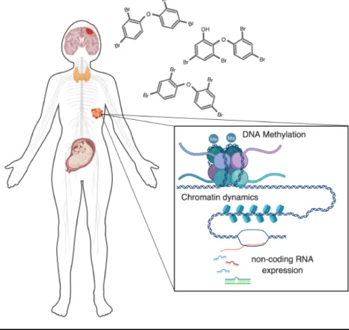
Figure 1-1. PBDE exposures affect epigenetic regulatory mechanisms at multiple levels, across multiple biological systems. There are several major aspects of human health that are of concern regarding PBDE toxicity that now have evidence for an involvement of dysregulated epigenetic regulation. These include: nervous system toxicity, disruption of thyroid hormone signaling, effects on the reproductive system (primarily on the placenta and testes), and oncogenic potential. Epigenetic components are known to be disrupted in each of these systems. In this review, we discuss these epigenetic regulators, their known modes of disruption by PBDEs, and the relationship of these disruptions to human health.
6
those of parent PBDEs (Qiu et al. 2009; Athanasiadou et al. 2008) and that OH-PBDEs appear to be more capable of competitively binding the thyroid hormone transporter transthyretin, potentially mediating increased endocrine-disruption (Meerts et al. 2000; Hamers et al. 2008; Hamers et al. 2006; Cao et al. 2010; Ren and Guo 2012). Other confounding factors include that past studies have covered a wide range of concentrations, many likely higher than environmentally relevant exposures, and much of this work has been conducted in cell culture, requiring further in vivo confirmation. Thus, while an extensive amount of work has been done, there is much that may yet be uncovered regarding biological mechanisms of PBDE toxicity.
A growing approach towards understanding PBDE toxicity has focused on PBDE-induced disruption of epigenetic regulation (Figure 1). Such mechanisms are interesting as potential PBDE targets as they would constitute a gene-environment interface for cellular disruption, wherein the effects of exposure to environmental factors interact with genomic elements, perhaps even with known genetic risk factors for human disease like NDDs (Tran and Miyake 2017). Such interactions could contribute towards explaining the elusive environmental portion of risk thought to contribute to NDD etiology. This notion is supported by the growing appreciation for the role of epigenetic mechanisms in neurodevelopment and cognition (Lasalle et al. 2013; Marshall and Bredy 2016), as well as in diseases of the nervous system, including neurodevelopmental disorders (Millan 2013; Christopher et al. 2017). Therefore, it may prove useful to understand the effects of PBDE exposures on epigenetic components, across cell and tissues types but particularly in the brain, in order to build more complete causal models towards explaining observed links to human health complications and behavioral deficits. However, compared to other widely studied mechanisms, relatively little attention has been given to epigenetic effects of PBDEs. Here, I will summarize the findings of studies conducted to date that have observed epigenetic endpoints such as DNA methylation, chromatin characteristics including modifications and remodeling of histones, and other epigenetic mechanisms such as expression of various non-coding RNAs.
1.2.2 Repeatedly Observed Disruption of DNA Methylation
One of the most commonly recognized epigenetic mechanisms is DNA methylation. Methylation commonly occurs at cytosine nucleotides positioned before a guanosine (CpG dinucleotides), resulting in a 5-methylcytosine. Given the extensive impact methylation has on transcriptional regulation (Bird 2002), it comes as little surprise that its disruption has potential impacts on human health (Bergman and Cedar 2013). An interesting example is folate deficiency’s implication in disrupted methylation during pre-natal development, although the relationship is incompletely understood (Crider et al. 2012). The relationship between PBDE exposure and DNA methylation is similarly incompletely understood. Although most studies report some correlation, they do not have a
7
clear consensus, especially in human samples, while in vitro studies more consistently report negative correlations (see references below).
Studies have assessed PBDE-exposure-dependent changes in global DNA methylation at various representative regions or at specific loci (gene promoters). Two of the most prominent examples of representative methylated regions include ALU elements and LINE1, which are common transposable repeats that can have adverse cellular impacts when de-repressed due to hypomethylation (Eden et al. 2003). Repetitive elements make up a large portion of the human genome (de Koning et al. 2011) and have high CpG frequency, contributing heavily to the global amount of DNA methylation and thereby serving as a reasonable global estimate.
One of the earliest studies on the effects of PBDE exposure on global DNA methylation in humans found a negative relationship between measured BDE-47 levels and ALU %5mC in blood samples of healthy Korean adults, while not finding significant relationships for BDE-99 or LINE1 methylation (Kim et al.
2010). Similar studies correlating PBDE levels in blood with methylation have followed. One found an inverse relationship between BDE-47 abundance and TNFα promoter methylation in cord blood samples from mother–infant pairs of the Boston Birth Cohort (Dao et al. 2015). Another reports a more complex finding in newborn cord blood samples from the CHAMACOS study, wherein significant changes in LINE1 methylation were found when considering co-exposure to DDT, DDE, and PBDEs (the direction of change depended on level of DDE or DDT co-exposure) (Huen et al. 2014).
Several groups have also examined the relationship between PBDE levels and effects on the placental epigenome. In 2016, two reports were published on effects in human placental samples. In one, the authors made PBDE, PCB, DDE measurements in villous placental tissue samples and found positive associations of PBDE levels with IGF2/H19 imprinting and methylation status (bisulfite conversion and targeted pyrosequencing) and global DNA methylation (assessed by LUMA (luminometric methylation assay)) (Kappil et al. 2016). In the other, PBDE levels in umbilical cord blood were measured from eighty human samples and correlated with placental DNA methylation levels in LINE1, NR3C1, and IGF2. BDE-66/153/209 were all found to have significant negative correlations with methylation of some of these loci (Zhao et al. 2016). Two very recent reports have also been made utilizing in vitro models of the placenta. One group exposed primary villous cytotrophoblasts (CTBs, an in vitro model of human placental development) to BDE-47 or BDE-99. They found that BDE-47 alters gene expression in a concentration-dependent manner and produced a low-level global increase in DNA methylation (assessed with HumanMethylation450 beadarray) (Robinson et al. 2019). Another group exposed human placental choriocarcinoma cells (BeWo cells) to 1μM BDE-47
8
and found reduced methylation of some CpG loci of mitochondrial biomarkers (with no differences found for 50 µMexposures) (Shan et al. 2019).
In addition to these human studies, PBDE-methylation relationships have also been investigated in model animals—mostly rodents—both in vitro and in vivo. In vitro studies have been conducted in different cell types, but consistently found negative correlations between PBDE exposure and methylation level. In the earliest of these studies, primary hippocampal neurons were exposed to various concentrations of BDE-209 for 24 hours and subsequently, a global decrease in DNA methylation was found by an antibody based ‘ELISA-like’ assay (Chen et al. 2010). Another found decreased global DNA methylation after a 10μM BDE-47 in murine N2A cells (assessed by HPLC and arbitrary primed PCR). This decrease coincided with increased adipocyte differentiation (2.5–25 µM exposures) (Bastos Sales et al. 2013). In a related effort to understand how endocrine-disrupting chemicals may be inducing adipocyte differentiation, investigators report that BDE-47 induces demethylation of several sites in the PPARγ promoter (a key adipongenic transcription factor) in COS7 and 3T3-L1 cells using Methylation-Sensitive High-Resolution Melting (MS-HRM) (Kamstra et al. 2014). Complementing these in vitro findings, in vivo studies that perinatally exposed rodents to BDE-47 reported interesting findings from offspring of various ages. These include decreased expression of LINE1 RNA (Suvorov and Takser 2011), decreased methylation of Mt-co2, L1Rn, Bdnf, and Nr3c1 (Byun et al.
2015), differentially methylated regions in sperm (Suvorov et al. 2018), and global DNA hypomethylation associated with behavioral deficits in both exposed wild-type and MeCP2-deficient female mice (Woods et al. 2012). Another study in mice assessing liver carcinoma tissue after DE-71 (a commercial mixture of PBDEs) exposure found little effect on global DNA methylation but reports a gene body methylation decrease in Tbx3 and subsequent mRNA and protein upregulation (Shimbo et al. 2017). While not directly assessing DNA methylation, a multigenerational study in zebrafish that exposed F0 animals to a PCB and PBDE mixture found disrupted behavior (hyper/hypoactivity) in F1–F4 larvae, as well as altered c-Fos expression (F1/2) and altered Dmnt3ba expression in all generations (Alfonso et al. 2019).
To our knowledge, only two reports exist that found no relationship between PBDE exposure and DNA methylation levels of any targets measured in those studies. One found no detectable decrease in methylation at the p53 promoter after 24 hours of exposure to low micromolar doses (1, 5, 10 µmol/L) of BDE-47 in human neuroblastoma cells (SH-SY5Y), although activation of the p53 pathway in general was implicated in observed effects (Zhang et al. 2013b). The other found no significant relationship between BDE-47 serum levels and global methylation as assessed by the luminometric methylation assay (LUMA) in samples from an elderly Swedish population. However, significant relationships were established for other persistent organic pollutants including PCBs and the dioxin OCDD (Lind et al. 2013). Aside from these reports, the literature suggests
9
a fairly consistent—but not necessarily linear—relationship between PBDE exposures and DNA methylation levels. It is possible that changes may vary from genomic region to region and may not always manifest an altered phenotype. Also, there is scarce evidence concerning direct cause–effect relationships between methylation changes and behavioral phenotypes. To aid in filling this gap in knowledge, it may prove useful to further refine understanding of the route by which PBDEs affect DNA methylation states—be it primarily by dysregulation of DNA methyltransferase expression, cellular metabolism, intracellular signaling pathways, etc.
1.2.3 Impact on Chromatin—Histone Modifications to Chromatin Remodeling
Other reversible chemical modifications of chromatin include modifications to histone proteins that regulate chromatin structure and instruct remodeling processes, ultimately controlling gene expression (Venkatesh and Workman 2015; Clapier et al. 2017). Studies starting as early as 2003 reported mixed results on PBDE-induced alterations of chromatin by several measures (chromosomal integrity, chromatin density and localization). Exposure of multiple bacterial strains to BDE-99 did not induce mutagenicity or a detectable increase in the number of structural chromosomal aberrations, while exposure to the PCB mixture Aroclor® 1254 did (Evandri et al. 2003). This early study explicitly stated that the possibility of PBDEs acting through epigenetic mechanisms could not be ruled out, which, in retrospect, was prudent foresight. Two subsequent studies have also reported no increase in degraded chromatin, both in sperm—the first in the sperm of mice orally exposed to BDE-209 (Tseng et al. 2006), the other in human samples of 153 men from the greater Montreal area, despite establishing a correlation between BDE-47 levels and decreased sperm concentration (Albert et al. 2018).
However, there have also been a few studies that do report chromatin disruption following PBDE exposure. One study found that 24-hr nanomolar range exposures to several PBDEs (BDE-47/99/153/183/209) induced micronuclei formation during cytokinesis in MCF-7 cells, an indicator of chromosomal damage occurrence preceding cell division (Barber et al. 2006). It has also been found that rat pups exposed to a single injection of BDE-153 at post-natal day 10 (PND10) exhibited behavioral dysfunction in a dose- and age-dependent manner one or two months later. Neurons in the CA3 region of the hippocampus of these rats were also found to be undergoing significantly increased rates of apoptosis, with chromatin condensed and localized to the nuclear membrane (Zhang et al. 2013a). Most recently, it was reported that BDE-209 exposures reduced hESC differentiation (although total induction was still greater than 90%) and also led to chromosomal copy number variants (CNVs), as well as decreased expression of DNMT1/3A (Du et al. 2018).
10
There is also some evidence specifically for PBDE-induced dysregulation of histones and histone-regulating proteins. In an effort to understand the carcinogenic potential of BDE-209, the first such study found that HEK293T cells exposed to micromolar range levels of the toxin exhibited altered expression of chromatin-regulating genes, specifically a histone gene cluster that the authors hypothesize could affect nucleosome properties (Li et al. 2014). In the same year, another group reported that exposure of the marine madaka (Oryzias melastigma) to BDE-47 led to sex-specific differential protein expression in male and female gonads, with several histone variants (H2b, H3.3, H3a, H2a) being down-regulated in male gonads (Fong et al. 2014). Another study found that exposing maize (Zea mays L.) to BDE-47, and its metabolites 6-OH-BDE-47 and 6-MeOH-BDE-47, led to elevated levels of ROS and phospho-H2AX, likely in response to DNA damage. Interestingly, the hydroxylated metabolite produced the most severe effects (Xu et al. 2015). Another study, primarily concerned with the relationship of PBDE exposure to reproductive health, exposed pregnant rats to BDE-47 from E8 to PND21. Male offspring were then assessed at PND120 for alterations in testes. It was found that exposed rats had smaller testes, decreased sperm production, and interestingly, an altered testes transcriptome and 4-fold decrease in protamine and transition gene expression (proteins responsible for histone-protamine exchange) (Khalil et al. 2017). Aside from these data that indicate potential disruptions of histone expression and nucleosome alteration, there are two studies that report PBDE-induced dysregulation of chromatin-regulating proteins. The first found that BDE-47 treatment downregulated SirT1 expression (a histone deacetylase) in the livers of mice due to NAD(+)-depletion (Zhang et al. 2015).
The potential importance of understanding the effects of PBDEs on chromatin dynamics cannot be understated given the fundamental importance of chromatin properties for regulating gene expression and thus cellular states. Going forward, it will be important for investigators to focus on identifying additional effects on chromatin while distinguishing those that are direct from indirect, hopefully allowing for elucidation of the underlying mechanism.
1.2.4 Other Affected Epigenetic Mechanisms (Non-Coding RNAs)
Non-coding RNAs—such as long non-coding RNA (lncRNA) and microRNA (miRNA)—can also act as epigenetic regulators (Rinn and Chang 2012; Cech and Steitz 2014). Various PBDE exposures have been reported to alter expression of miRNAs, and one study described effects on expression of liver lncRNAs. The earliest study assessing miRNA expression as an endpoint following PBDE exposure utilized placental samples collected from the National Children’s Study. Among other associations established for PCB and heavy metal exposures, the study reported a positive association between BDE-209 and miR-188-5p expression and an inverse association for BDE-99 and let-7c (Li et al. 2015). Another group exposed hESCs in vitro to low doses of BDE-209 (1,
11
10, 100nM) for 4 days, inducing apoptosis and downregulating pluripotency genes, particularly OCT4, in part by hypermethylation of the promoter and induction of miR-145/335 which repress OCT4. There was also generation of ROS and decreased superoxide dismutase (SOD2) expression. ROS and OCT4 effects were partially rescued by treatment with the antioxidant NAC (Du et al. 2016). An even more recent study employing human cells found that, after stimulating THP-1 macrophages with BDE-209 and LDL, there was dose-dependent repression of miRNA-21 which subsequently de-repressed toll-like receptor 4 expression (TLR4), enhancing TLR4-dependent lipid uptake (Zhi et al. 2018; Zhi et al. 2019b; Zhi et al. 2019a).
In addition to these examples in humans, two rodent studies concerning non-coding RNAs in the liver have been published. The first found that BDE-47 exposure upregulates CYP3A1 in rat liver and that this upregulation is mediated by BDE-47-induced repression of miRNA-23b, which negatively regulates CYP3A1 mRNA via a 3’ UTR binding site (Sun et al. 2016). The other study reported that conventional and gut-microbiome-depleted mice exhibit dysregulated lncRNA expression in liver tissue in response to both BDE-47 and BDE-99 exposure (Li and Cui 2018). Interestingly, BDE-47 has also been found to induce dysregulation of novel miRNAs in exposed zebrafish larvae. Of particular interest is miR-735, which may play essential roles in larval sensory development, explaining previously observed BDE-47-induced disruption of zebrafish visual perception (Zhao et al. 2017a). In the near future, a general model of PBDE-induced miRNA dysregulation may hopefully be established given the multiple intriguing examples already characterized.
1.3 Summary
Considering this growing body of work documenting epigenetic dysregulation induced by PBDE exposure, there appear to be several central lines of evidence emerging from research done in various health contexts, including: adipocyte differentiation and obesity, reproductive health—of both sperm/testes and the placenta, carcinogenicity (especially thyroid related), and negative impacts on nervous system formation and function. It is becoming clear that many, if not all, of these various aspects of human health are impacted by PBDE-induced disruption of normal epigenetic states and mechanisms.
There are fairly consistent findings of a negative relationship between PBDE levels and DNA methylation from in vitro and non-human animal studies across varied cell/tissue types and methylation detection methods. However, the data from human samples is more difficult to interpret. Studies reporting effects on global DNA methylation levels inferred from representative regions have incongruent results, and evidence of alterations to methylation in the placenta are, likewise, not in direct agreement. However, this confoundment and the fact that human studies have so far been conducted across very different populations
12
and models should only encourage further work on the topic, especially given indications from non-human animal and in vitro studies. It will be of great value if these types of studies can build on the tentatively established negative impact of PBDEs on methylation and begin to focus on understanding the mechanisms underlying the alterations, while continuing to clarify effects in human studies.
Compared to DNA methylation, the literature is sparser regarding the effects of PBDEs on other epigenetic mechanisms such as chromatin dynamics and expression of non-coding RNAs. However, some interesting ideas are beginning to emerge. While not yet well understood, PBDE-induced dysregulation of histones and chromatin regulators is an intriguing intersection for PBDEs and neurodevelopmental disorders, bolstered by the recent emergence of chromatin regulation as a major node of NDD risk (Lasalle et al. 2013; Gabriele et al. 2018). Further, it is tempting to speculate that epigenetic effects of PBDE exposure may, generally, turn out to be a point of convergence for environmental and genetic factors that contribute to NDDs. If the effects of these compounds on targets such as DNA methylation, chromatin components and regulators, and non-coding RNA expression (all of which are mechanisms known to have roles in neurodevelopment and perhaps NDD etiology) can be further explored and resolved, one or more could very well turn out to be that link. This is of pressing importance, especially for neurodevelopmental disorders considering their explosive increase in prevalence, growing evidence for the involvement of PBDEs in their etiology, and the long elusive role of environmental factors in these devastating conditions.
A major challenge for epigenetic PBDE research will be to assimilate new findings into the existing framework of PBDE toxicity that has been established from insights into other major impacted biological mechanisms. It will also be important to carefully consider nuanced aspects of exposures including tissue and sub-cellular localization, conduct more research on environmentally relevant doses and mixtures of PBDEs, further explore the prevalence and effects of their metabolites, and, to the extent that it is possible, integrate evidence generated across human and non-human studies (both in vitro and in vivo). This will be necessary in order to construct a more holistic understanding of how these compounds impact cellular states and, ultimately, phenotypic outcomes. By moving towards such an understanding with continued research, we may eventually be able to explain how and to what extent these pervasive environmental pollutants are related to the numerous human health conditions that they appear to be contributing to and may perhaps gain additional insight into the nature of PBDE-related health complications themselves.
With the emerging bodies of evidence for PBDE-induced epigenetic disruptions, epigenetic regulation (particularly chromatin remodeling) as a major node of risk in NDDs, and the growing evidence linking PBDE exposure levels with NDD-related behavioral abnormalities in humans and other animal models,
13
in my dissertation research I sought to characterize the cellular effects of PBDE exposures and related underlying molecular mechanisms in the developing brain. Specifically, I first tested the hypothesis that the effects of nanomolar range exposures to BDE-47 and its hydroxylated metabolites interact with epigenetic regulation in neurons from the developing nervous system (Chapter 2), subsequently developed and tested a mechanistic hypothesis of synaptic disruption and intracellular signaling inhibition by ortho-hydroxylated PBDE metabolites (Chapter 3) based directly on findings generated from the data presented in Chapter 2– leading to an appreciation that hydroxylated PBDE metabolites likely affect multiple levels of cellular function by interfering with molecular processes in a sub-cellular-specific manner, and finally to the generation of several hypotheses integrating these new findings with each other and with data from the diverse biological fields surrounding PBDE neurotoxicity (Chapter 4).
14
Chapter 2: Persistent 6-OH-BDE-47 exposure
impairs functional neuronal maturation and alters
expression of neurodevelopmentally-relevant
chromatin remodelers
Robert G. Poston, Carissa J. Dunn, Pushpita Sarkar, Ramendra N. Saha Environmental Epigenetics 2018
2.1 Abstract
Polybrominated diphenyl ethers (PBDEs) are a pervasive class of brominated flame retardants that are present in the environment at particularly high levels, especially in the United States. Their environmental stability, propensity for bioaccumulation, and known potential for neurotoxicity has evoked interest regarding their effects on the developing nervous system. Exposure to PBDEs has been strongly associated with neurodevelopmental disorders. However, the details of their mechanistic roles in such disorders are incompletely understood. Here, we report the effects of one of the most prevalent congeners, BDE-47, and its hydroxylated metabolites on the maturation and function of embryonic rat cortical neurons. Prolonged exposure to 6-OH-BDE-47 produces the strongest effects amongst the parent BDE-47 congener and its tested hydroxylated metabolites. These effects include: i) disruption of transcriptional responses to neuronal activity, ii) dysregulation of multiple genes associated with neurodevelopmental disorders, and intriguingly, iii) altered expression of several subunits of the developmentally-relevant BAF (Brg1-associated factors) chromatin remodeling complex, including the key subunit BAF170. Taken together, our data indicate that persistent exposure to 6-OH-BDE-47 may interfere with neurodevelopmental chromatin remodeling mechanisms and gene transcription programs, which in turn are likely to interfere with downstream processes such as synapse development and overall functional maturity of neurons. Results presented in this chapter have identified a novel aspect of 6-OH-BDE-47 toxicity and open new avenues to explore the effects of a ubiquitous environmental toxin on epigenetic regulation of neuronal maturation and function.
Keywords: neurodevelopment, BDE-47 exposure, Arc, activity-induced transcription, BAF complex
15
2.2 Introduction
An increasingly studied point of convergence that may help to provide new insight on the relationship between the various effects of PBDE exposures and adverse behavioral outcomes is epigenetic regulation, specifically of gene transcription during neurodevelopment. Epigenetic mechanisms are a major driving force of normal neurodevelopment (Lasalle et al. 2013), and many neurodevelopmental complications involve dysregulation of gene transcription (Parikshak et al. 2015; Vissers et al. 2015). Although several epigenetic components of neurodevelopment are now well known, there remains much more to be characterized (Sweatt 2013), especially in regards to how they interact with environmental challenges like PBDE exposure.
Epigenetic regulation refers to a range of processes (detailed more in the Chapter 1) including chemical modifications to DNA and histone proteins, non-coding RNA expression, and ATP-dependent remodeling of chromatin structure. The latter is of particular interest in the context of NDDs, as it has recently emerged as a major node of risk including several highly penetrant mutations associated with the disorders (Gabriele et al. 2018). ATP-dependent chromatin remodeling is a process by which regulatory proteins restructure chromatin–the complex of DNA and proteins making up chromosomes in eukaryotic cells–a process that plays a major role in facilitating the vast diversity of cellular types and function produced from an organism’s genome (Ho and Crabtree 2010). The general mechanisms of how these regulators interact with DNA and histones/nucleosomes to modify their composition and structure is thought to be largely similar. However, the various canonical chromatin remodeling protein complexes are composed of many subunits and are regulated by various interacting proteins, including cell-type specific factors (Clapier et al. 2017).
One such dynamically-regulated remodeling complex that is known to play critical roles in neurodevelopment is the BAF (Brg1-associated factors) complex (Staahl and Crabtree 2013; Sokpor et al. 2018). BAF complexes containing neuron-specific subunits are known to instruct neurodevelopment, especially the differentiation of neurons (Staahl and Crabtree 2013), as well as the regulation of synaptic formation, plasticity, and learning and memory (Vogel-Ciernia et al.
2013; Zhang et al. 2016). Interestingly, mutations in chromatin remodelers, including BAF complex subunits, have recently been identified as NDD risk factors, establishing a major node of risk in the complex network of genetic factors now associated with NDDs (further details and references in Chapter 1).
Considering this, we sought to explore the ways in which exposure to BDE-47 and its hydroxylated metabolites influence the maturation and function of neurons from the developing nervous system, and screened for possible intersections with NDD risk at the level of transcriptional dysregulation by measuring expression of NDD candidate genes representing several of the major nodes of NDD risk (Vissers et al. 2015; Iakoucheva et al. 2019)– intracellular signaling molecules, synaptic proteins, and epigenetic regulators including several BAF complex subunits.
16
2.3 Materials and Methods 2.3.1 Plasmids and sub-cloning
A commercial shRNA construct for BAF170 (CCCAAACTGCTAGGGAAATTA) was obtained from Sigma. This shRNA sequence was inserted into pLKO.1-puro (designed by RNAi consortium or TRC; obtained from Addgene) and then packaged into lentiviruses. Self-inactivating HIV lentivirus particles were produced by transfecting 293T cells with the shRNA vector, envelope (pMD2.G; Addgene), and packaging plasmids (psPAX2; Addgene) using a previously described protocol(Saha et al. 2011). The BAF170 expression construct in a lentiviral backbone was a kind gift from Dr. Trevor Archer (NIEHS, NIH)(Wade et al. 2016). BAF170 expression from this construct was validated by Western blotting.
2.3.2 Dissociated neuronal culture, RNAi and cell treatment
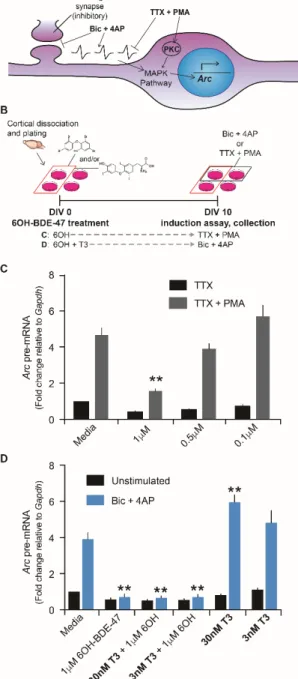
Cultures of cortical neurons were prepared from embryonic day 18 Sprague Dawley rats (UC Merced IACUC approval: AUP#13-0007 and AUP#16-0004). Dissociated cortical neurons were plated in Neurobasal medium (Invitrogen) supplemented with 25 μM glutamate (Sigma-Aldrich) and 0.5 mM L-glutamine (Sigma-Aldrich) and either B27 (Invitrogen) or NS21 and maintained in a similar medium without glutamate. NS21 was prepared in the laboratory as previously described (Chen et al. 2008). Neurons were routinely used for induction assays between 10–16 days in vitro. For infection with recombinant lentiviruses, the viral supernatant was diluted in neuronal media and cells were infected at a multiplicity of infection ranging from 2 to 5. To induce gene transcription under basal conditions using synaptic circuits, we co-treated neurons with 50μM Bicuculline (Sigma-Aldrich) and 75μM 4-Aminopyridine (Acros Organics) (Papadia et al. 2005). To induce gene transcription extra-synaptically, we blocked activity with 1μM TTX (Calbiochem) and induced the MAP-kinase pathways with 1μM phorbol 12-myristate 13-acetate (PMA; Sigma-Aldrich) (Schultz et al. 1997). BDE-47 and metabolites used to treat cultures were obtained from AccuStandard (BDE-047N, 4003N, 4004N, HBDE-4005N).
2.3.3 RNA extraction and gene transcription quantitation
Total RNA was isolated from dissociated neurons using the GeneJET RNA Purification Kit (Thermo) with an off-column DNase (Promega) digestion. cDNA was synthesized using MuLV reverse transcriptase (Promega), random primers (Promega), oligo dTs (Promega), and RNase inhibitors (Thermo Scientific). Quantitative real-time PCR (qRT-PCR) was performed to quantify mRNA levels of specific transcripts using iTaq Universal Sybr Green Supermix (BioRad) and the BIO-RAD CFX Connect realtime PCR Detection System. Pre-mRNA levels
17
were estimated as previously described (Saha et al. 2011).
2.3.4 Sample preparation for electrophoresis
Neurons were lysed in ice-cold 1X RIPA buffer (25 mM Tris, pH 7.5, 150 mM NaCl, 1% Na- deoxycholate, 0.1% SDS, 0.1% NP-40) supplemented with 1:100 protease inhibitor cocktail (Sigma-Aldrich-Aldrich). Lysed neurons were sheared by sonication (low setting; three cycles on Bioruptorâ), cell debris pelleted at
15,000 rpm for 5 minutes at 4°C, and clarified supernatant transferred to pre-chilled 1.5 mL microcentrifuge tube. Total cell extracts were denatured at 95°C, for 5 minutes, using either home-made 5X Laemmli buffer, 2X-, or 4X-Laemmli sample buffer (both from BIO-RAD).
2.3.5 Western blotting and imaging
Denatured protein samples were resolved on 4-20%- (BIO-RAD cat. no. 4568095) or 4-15%- (BIO-RAD cat. no. 456-1083) Mini PROTEAN® gels in Tris/Glycine/SDS (BIO-RAD cat. no. 1610772). Resolved proteins were transferred onto LF PVDF membrane, using the BIO-RAD TBT RTA kit and protocol (cat. no. 1704272). PVDF membranes were incubated at 4°C overnight with appropriate primary antibodies in 1X TBS-T with 0.5% BSA at 1:1000 dilution. Primary antibodies included the following antibodies: β-Actin (ThermoFisher Scientific AM4302), BAF170 (CST 12760), BAF155 (CST 11956), Brg1 (CST 49360), BAF47 (CST 91735). Next day, membranes were washed three times in 1X-TBST, probed with appropriate Alexa Fluor® secondary antibodies (Life Technologies) for 40 minutes at room temperature, washed three times with 1X TBS-T, and imaged using BIO-RAD Multiplex ChemiDocTM Imaging System.
2.3.6 Immunocytochemistry and microscopy
Antibodies for immunocytochemistry were used at dilutions between 1:100-1:500 and include the following: NeuN (Millipore ABN78), Doublecortin (CST 4604S), Nestin (Invitrogen MA1-91657). Infected neurons were washed twice with 1X ice-cold PBS (Fisher Sci). The cells were then incubated with 4% paraformaldehyde (Sigma-Aldrich) in 1X PBS for 15 minutes at room temperature and then washed twice with 1X PBS, permeabilized at room temperature for 20 minutes with 0.5% Triton X-100 (Fisher Sci), washed twice and blocked for 30 minutes with 10% goat serum (Gibco) in 1X PBS. Cells were incubated at 4°C overnight in 3% goat serum in 1X PBS with primary antibodies. Next day, primary antibody solution was removed and cells were washed thrice with 0.05% Tween (Fisher) in 1X PBS (0.05% PBS-T), and incubated with appropriate Alexa Fluor® secondary antibody (Lifetech) for 45 minutes, washed thrice with 0.05% PBS-T, cured overnight with Prolong Anti-Fade Gold with DAPI and imaged. Images were
18
captured with a Keyence BZ9000-E microscope at 40X magnification.
2.3.7 Cell viability assay
Cell viability was assessed by an MTT assay (Biotium) (Mosmann 1983) wherein mitochondrial activity is detected colorimetrically following incubation of cells with a tetrazolium salt. The assay was conducted according to manufacturer’s
instruction, except that MTT incubation time was shortened to 30 min to avoid reaching a plateau where differences in product formation would be
indistinguishable and reagent volumes were proportionally scaled up to appropriate amounts for 24-well plates.
2.3.8 Statistics
Error bars represent standard error of mean throughout this article. Statistical comparison of datasets was performed by one way ANOVA with Fisher’s LSD (Figure 2C and 6A) or by two way ANOVA with Tukey’s post hoc test (all other figures). Biological replicates are indicated throughout as N in corresponding figure legends. Biological replicates constitute cell culture preparations from independent dams.
19
2.4 Results
2.4.1 Characterization of E18 primary rat neuronal cultures
We dissected brain tissue from the pups of timed-pregnant Sprague-Dawley rats (Rattus norvegicus) on embryonic day 18 (E18) to obtain dissociated cortical neurons. Cells were then plated as monolayers in supplemented Neurobasal growth medium. The identity of these cultured cells was characterized over the first week of growth by immunocytochemistry (ICC) using antibodies raised against Nestin, DCX, and NeuN (neural progenitor, differentiating neuron, and differentiated neuronal markers, respectively (Frederiksen and McKay 1988; Gleeson et al. 1999; Mullen et al. 1992) (Fig. 1). The cultures were composed of
Figure 2-1. Characterization of neuronal cultures across the first week of growth by immunocytochemistry. Rat cortical neurons were obtained by dissecting and dissociating cortices from E18 pup brains. Cultures derived from these cells were stained using antibodies against several markers of neuronal maturation: Nestin (neuronal precursor), DCX (differentiating neurons), and NeuN (differentiated neurons). Staining was conducted at indicated time points. Scale bar = 25µm. N=3