Endemic melioidosis is caused by genetically diverse
Burkholderia pseudomallei strains. However, clonal
out-breaks (multiple cases caused by 1 strain) have occurred, such as from contaminated potable water. B. pseudomallei
is designated a group B bioterrorism agent, which neces-sitates rapidly recognizing point-source outbreaks.
Pulsed-fi eld gel electrophoresis (PFGE) and multilocus sequence typing (MLST) can identify genetically related isolates, but results take several days to obtain. We developed a sim-plifi ed 4-locus multilocus variable number tandem repeat analysis (MLVA-4) for rapid typing and compared results with PFGE and MLST for a large number of well-charac-terized B. pseudomallei isolates. MLVA-4 compared favor-ably with MLST and PFGE for the same isolates; it discrimi-nated between 65 multilocus sequence types and showed relatedness between epidemiologically linked isolates from outbreak clusters and between isolates from individual pa-tients. MLVA-4 can establish or refute that a clonal outbreak of melioidosis has occurred within 8 hours of receipt of bacterial strains.
M
elioidosis is endemic in Southeast Asia and northernAustralia (1,2). The reported incidence of
melioido-sis has been increasing within this region, and new foci and outbreaks of melioidosis are being described within this
region and in distant locations such as Brazil (3) and New
Caledonia (4). It remains unclear how much of this
expan-sion of the global distribution boundaries is from recent
spread of the causative bacterium, Burkholderia
pseudo-mallei, and how much is from unmasking of disease after
events such as the 2004 Asian tsunami (5,6). Molecular
studies have shown considerable genetic diversity within
B. pseudomallei (1,2,7). For instance, in northern Australia,
isolates from patients are generally distinct from each other
(8) unless there is a point-source outbreak, such as occurred
in 2 episodes after B. pseudomallei contamination of
com-munity water supplies (9,10).
In disease-endemic regions, melioidosis case numbers surge in the monsoonal wet season, and individual cases
are typically caused by different B. pseudomallei strains.
However, under some circumstances, a series of cases can be caused by 1 strain, indicating that a clonal or point-source outbreak has possibly occurred and an urgent pub-lic health response may be required. Because several days are required to perform the currently available molecular typing methods of ribotyping, multilocus sequence
typ-ing (MLST) and pulsed-fi eld gel electrophoresis (PFGE),
the ability to rapidly distinguish endemic infection from
a clonal outbreak has not been possible. Furthermore, B.
pseudomallei is classifi ed as a group B bioterrorism agent
by the US Centers for Disease Control and Prevention, and the implications of a possible deliberate release warrant the availability of a robust method to quickly ascertain if concomitant cases of melioidosis are caused by 1 bacterial strain.
We recently described using a BOX-PCR for rapid
typing of B. pseudomallei (11). We have now adapted
and simplifi ed multilocus variable number tandem repeat
(VNTR) analysis (MLVA) for rapid typing because this analysis potentially enables precise international strain comparisons. We have compared MLVA results on a wide
range of well-characterized B. pseudomallei isolates with
those for MLST and PFGE.
Identifi cation of Melioidosis
Outbreak by Multilocus Variable
Number Tandem Repeat Analysis
Bart J. Currie, Asha Haslem, Talima Pearson, Heidie Hornstra, Benjamin Leadem, Mark Mayo, Daniel Gal, Linda Ward, Daniel Godoy, Brian G. Spratt, and Paul Keim
Author affi liations: Menzies School of Health Research, Darwin, Northern Territory, Australia (B.J. Currie, A. Haslem, M. Mayo, D. Gal, L. Ward); Northern Territory Clinical School, Darwin (B.J. Cur-rie, L. Ward); Northern Arizona University, Flagstaff, Arizona, USA (T. Pearson, H. Hornstra, B. Leadem, P. Keim); and Imperial Col-lege, London, UK (D. Godoy, B.G. Spratt)
Methods
MLVA, PFGE, and MLST
U’Ren et al. initially described 32 VNTR loci for B.
pseudomallei that had 7–28 alleles (12). Thirty of these
VNTR markers were subsequently used for fi ne-scale
analysis of 121 isolates of B. pseudomallei (13). Various
combinations of markers were tested by MLVA; we chose 4 markers that were highly discriminatory, enabling single-run, 4-color analysis in a DNA sequencer. The 4 VNTR
loci chosen were 2341, 389, 1788, and 933 (12). Table 1
shows the PCR primers used and the repeat region
ampli-fi ed for each locus. VNTR loci 2341 and 933 are from B.
pseudomallei chromosome 1, and loci 389 and 1788 are
from chromosome 2.
PCRs contained 0.88 U HotStarTaq DNA Polymerase (QIAGEN, Hilden, Germany) per reaction, 1× PCR buffer,
1.2 M Betaine, 3 mmol/L MgCl2, 0.2 mmol/L
deoxynucle-oside triphosphates, 0.2 μM fl uorescently labeled forward
primer, 0.2 μM reverse primer, 1 μL template DNA (0.5 ng/
μL), and double-distilled water to give a volume of 11 μL
per reaction. Amplifi cations were conducted in Palm
Cy-clers (Corbett Research, Sydney, New South Wales, Aus-tralia). All PCRs underwent initial denaturation at 95°C for 5 min, then 34 cycles of 94°C for 30 s, 68°C for 30 s, and
72°C for 30 s, followed by a fi nal extension step of 72°C
for 5 min and 15°C for 3 min.
PCR products of each colored primer (FAM, NED, PET, and VIC; Table 1) were then pooled. Pooled PCR
products were diluted with 200 μL of double-distilled
wa-ter, and 1.2 μL of PCR product was added to a mixture of a
1:6 ratio of Hi-Di formamide (Applied Biosystems, Foster City, CA, USA) and GeneScan 1200 LIZ (Applied
Bio-systems) fl uorescently labeled size standard. PCR products
were then electrophoretically separated by using a 3100xl DNA Sequencer (Applied Biosystems) and analyzed by using the ABI software program GeneMapper version 3.5
(Applied Biosystems). PFGE with SpeI and MLST were
performed as described (7,14).
Data Analysis
For 4-locus multilocus VNTR analysis (MLVA-4),
GeneMapper peak fi les were imported into BioNumerics
version 4.61 (Applied Maths BVBA, Sint-Martens-Latem, Belgium). Relatedness of isolates was assessed by using a matrix of the pairwise differences of the 4 VNTR loci, with a dendrogram produced by using the unweighted pair group method with arithmetic averages (UPGMA).
For PFGE, gel images were analyzed with BioNumerics version 4.61. BioNumerics application modules used were Fingerprint Types and Comparison and Cluster Analysis modules. PFGE bands (150–700 kbp) were manually as-signed on visual inspection. PFGE dendrograms were pro-duced with Dice UPGMA with position tolerance settings of 0.5% optimization and 1.0% band position tolerance.
For MLST, alleles at each of the 7 previously described
loci (7) were assigned for each isolate by comparing
se-quences to those at the B. pseudomallei MLST website
(15). Following the standard MLST protocol, each allele
was assigned a different allele number, and the allelic
pro-fi le (string of 7 integers) was used to defi ne the sequence
type (ST) for that isolate. Allelic profi les of isolates were
imported into BioNumerics version 4.61, and relatedness of isolates was displayed as a dendrogram by using the
ma-trix of pairwise differences in the allelic profi les and
UP-GMA clustering.
B. pseudomallei Isolates
To assess the discriminatory power of MLVA-4, direct comparisons were made between the MLST dendrogram
for 65 B. pseudomallei isolates, each representing a distinct
ST, and the MLVA-4 dendrogram for these isolates. The 65 isolates were all from Australia and included human, animal, and environmental sources. There were 16 pairs of
Table 1. Characteristics of 4 VNTR loci used for identification of Burkholderia pseudomallei* VNTR loci
Characteristic 2341 1788 933 389 Color-labeled forward primer
sequence (5ƍĺ 3ƍ) FAMGGCTTCGCACC CGCCCCATTTCAGC PETGCGCGGCGAGA ACGGCAAGAACGAA NEDATGGTGGCGGC CGTCGGCGAAAACC VICGTTACAAGC GCGGGTCGGCA AGAGGCTGAAA Reverse primer sequence (5ƍĺ 3ƍ) GCACCGGGCGCGGC
GCACTCG GAGCATCGGGTGGG CGGCGCGTATTGAT GCTCGAATGGGTGT ACGAAGGGCCACGC TGATTC GCCGGTGTTGA ACGAGTGGGTG GCGTAAGC Repeat sequence (5ƍĺ 3ƍ) TTCGTGCGC GTCGTGCGATCCTG
CT
CGGCGAGGGAAA GACGAACC Minimum size, bp† (no. repeats) 111 (2) 235 (4) 171 (3) 221 (1) Maximum size, bp (no. repeats) 243 (17) 382 (13) 337 (17) 292 (10) No. alleles 16 10 13 9 No. null alleles – – 2/65 STs –
*VNTR, variable number tandem repeat; STs, sequence types. †Error range in fragment sizing is ± 3 bp.
single-locus variants (SLVs; isolates sharing identical al-leles at 6/7 loci by MLST).
To assess the ability of MLVA-4 to identify clonal clusters, direct comparisons were made between the PFGE
dendrogram for 4 defi ned clonal groups and the MLVA-4
dendrogram for these isolates. Clonal cluster I and clonal cluster II consist of 8 and 7 isolates, respectively, from the tropical Northern Territory of Australia and were
previ-ously identifi ed as clustering by PFGE (16). These 2 clonal
clusters represent geographically linked but epidemiologi-cally unrelated isolates from our prospective melioidosis studies in northern Australia. Clonal cluster III consists of 3 isolates of identical ST cultured from a detergent container implicated in an outbreak of melioidosis in Northern
Terri-tory involving 2 garage mechanics (14). Clonal cluster IV
contains 6 isolates (5 from humans, 1 from water) from an outbreak of melioidosis in a remote indigenous community in Northern Territory. The outbreak was linked to contami-nation of the unchlorinated community water supply, with
several deaths reported (10).
Finally, to assess the ability of MLVA-4 to link iso-lates from patients, we analyzed multiple isoiso-lates from 3 patients. Patient A had chronic pulmonary melioidosis,
and 5 B. pseudomallei isolates were recovered over 22
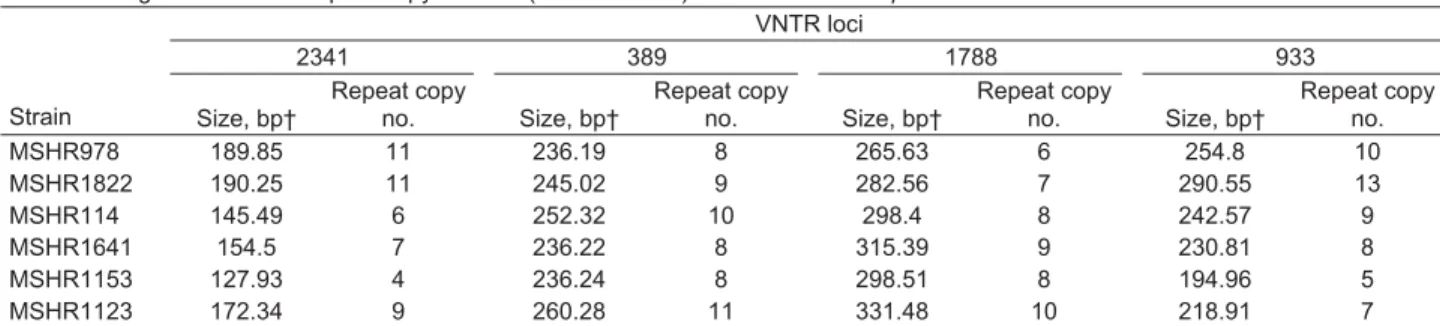
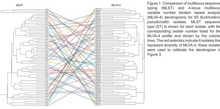
months. Patient B had chronic pulmonary melioidosis, and 7 isolates were recovered over 6 years, including 2 isolates from this patient’s water supply. Patient C died of melioi-dosis septicemia; 6 isolates were recovered over 14 days. To construct the dendrogram for these clinical isolates, we chose 6 unrelated isolates representing the diversity of Australian isolates seen with MLVA-4 (Table 2). These 6 isolates are indicated in Figure 1. This study was reviewed and approved by the Human Research Ethics Committee of the Northern Territory Department of Health and Commu-nity Services and the Menzies School of Health Research, Darwin, Northern Territory, Australia (approval 02/38).
Results
Table 1 shows size variation with calculated number of repeats and number of alleles for each of the 4 VNTR loci. Locus 933 showed null alleles for 2 of the 65 MLST STs.
Figure 1 shows the relationship between the 65 discrete MLST STs and the MLVA-4 for these isolates. MLVA-4 was able to discriminate between each ST. Relationships between STs seen on the MLST dendrogram were gener-ally not preserved with MLVA-4. This is expected because VNTRs change too rapidly and too few loci were used to compensate for homoplasy at individual loci and to provide phylogenetic content to the assay. However, strains that were closely related by MLST (SLVs) could in some cases be seen to be related by using MVLA-4 (Figure 1).
Figure 2 shows results for the 24 isolates in the clus-ter study, with 4 additional unlinked isolates, each from a different ST included for comparison. There was gener-ally excellent agreement between PFGE and MLVA-4 for each of the 4 clonal clusters. PFGE clonal clusters I (MLST ST 132) and II (ST 109), each containing epidemiologi-cally unrelated strains, also clustered on MLVA-4, with the exception of isolate MSHR1429, which by MLVA-4 was located outside its cluster group. The detergent clus-ter III (ST 123) was indistinguishable by MLVA-4, and the community outbreak strains in cluster IV (ST 125, ST 126) separated into 2 closely linked MLVA-4 patterns, 1 of which included the isolate from the community water sup-ply (MSHR491, ST 126).
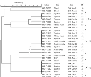
Figure 3 shows MLVA-4 results for the 3 patients. Iso-lates from patient A (ST 243) and B (ST 131) with chronic pulmonary melioidosis each had closely linked MLVA-4
results with a suggestion of fi ne-scale differentiation over
the years of infection. The 2 water supply isolates from pa-tient B were identical to 5 of her clinical isolates. The 6 clinical isolates from patient C, who had fatal melioidosis, were identical by MLVA-4, including isolates from blood and sputum.
Discussion
Ribotyping was the fi rst method widely used for
typ-ing B. pseudomallei (17), followed by PFGE. To date,
PFGE has been considered the standard method for inves-tigating potential point-source outbreaks of bacterial infec-tions. We have previously used PFGE to link case clusters
of melioidosis to water supply contamination (10) and to
Table 2. Fragment size and repeat copy number (MLVA-4 code) for 6 Burkholderia pseudomallei strains used as MLVA-4 standards* VNTR loci 2341 389 1788 933 Strain Size, bp† Repeat copy no. Size, bp† Repeat copy no. Size, bp† Repeat copy no. Size, bp† Repeat copy no. MSHR978 189.85 11 236.19 8 265.63 6 254.8 10 MSHR1822 190.25 11 245.02 9 282.56 7 290.55 13 MSHR114 145.49 6 252.32 10 298.4 8 242.57 9 MSHR1641 154.5 7 236.22 8 315.39 9 230.81 8 MSHR1153 127.93 4 236.24 8 298.51 8 194.96 5 MSHR1123 172.34 9 260.28 11 331.48 10 218.91 7
*MLVA-4, 4-locus multilocus variable number tandem repeat analysis. †Error range in fragment sizing is ± 3 bp.
contamination of a container of detergent (14). However,
such outbreaks are rare, and we have shown that, in the melioidosis-endemic region of northern Australia, case clusters during extreme weather events are usually not
ge-netically linked by PFGE (8). These clusters simply refl ect
the close association between rainfall and infection from
the diverse range of B. pseudomallei strains in soil and
surface water.
Recently, MLST has enabled new insights into
region-al and globregion-al epidemiology of melioidosis (7,16,18–20).
Although there is excellent congruence between PFGE and MLST, with PFGE and MLST providing similar results
for local epidemiologic investigations (16), MLST has the
major advantage of absolute comparative ability across
laboratories through the MLST website and unambiguous sequence type characterization.
Ribotyping and PFGE take several days to generate results, and MLST is expensive and requires sequencing and analysis capability. PCR-based typing methods have enabled more rapid availability of results. Randomly
ampli-fi ed polymorphic DNA (RAPD) analysis has been used to
analyze relationships between clinical and environmental B.
pseudomallei (21,22). However, it is not possible to make
valid comparisons of RAPD results between laboratories and sometimes even between runs in the same laboratory. Thus, despite the speed of RAPD, we no longer use it.
Analyzing bacterial genomes for VNTRs has enabled MLVA assays to be developed to differentiate among
me-Figure 1. Comparison of multilocus sequence typing (MLST) and 4-locus multilocus variable number tandem repeat analysis (MLVA-4) dendrograms for 65 Burkholderia pseudomallei isolates. MLST sequence type (ST) is shown for each isolate, with the corresponding isolate number listed for the MLVA-4 profi le and shown by the colored lines. The red asterisks indicate 6 isolates that represent diversity of MLVA-4; these isolates were used to calibrate the dendrogram in Figure 3. Clon al clust e r l Clon al clust e r ll Clon al clust e r lll Clon al clust e r lV
PFGE (Spel) MLVA-4
% Similarity % Similarity 10 0 90 80 70 60 50 40 30 20 10 10 0 90 80 70 60 50 40 MSHR1119 MSHR1179 MSHR1182 MSHR1128 MSHR0207 MSHR1031 MSHR1099 MSHR1603 MSHR465A MSHR0767 MSHR1840 MSHR0875 MSHR0719 MSHR0786 MSHR0888 MSHR1892 MSHR0910 MSHR1105 MSHR0435 MSHR0449 MSHR433A MSHR445A MSHR0343 MSHR0491 MSHR1429 MSHR1153 MSHR1839 MSHR1869 0207 (ST132) 465A (ST132) 0767 (ST132) 1031 (ST132) 1128 (ST132) 1603 (ST132) 1840 (ST132) 1099 (ST132) 0719 (ST109) 0786 (ST109) 0910 (ST109) 1105 (ST109) 1429 (ST109) 0888 (ST109) 1892 (ST109) 1869 (ST337) 1119 (ST123) 1182 (ST123) 1179 (ST123) 1153 (ST117) 433A (ST126) 445A (ST126) 0343 (ST126) 0435 (ST126) 0449 (ST126) 0491 (ST126) 0875 (ST115) 1839 (ST336)
Figure 2. Comparison of pulsed-fi eld gel electrophoresis (PFGE) and 4-locus multilocus variable number tandem repeat analysis (MLVA-4) profi les for isolates in 4 clonal groups (see text for details). Isolate number with its MLST sequence type (ST) is listed for each isolate on the PFGE profi le, with the corresponding isolate number listed for the MLVA-4 profi le. Four unrelated isolates are included for comparison: 0875 (ST115), 1869 (ST337), 1839 (ST336) and 1153 (ST117).
thicillin-resistant Staphylococcus aureus strains that are
in-distinguishable by PFGE (23) and to differentiate Neisseria
meningitidis strains with identical MLST STs (24). Liu et
al. developed the fi rst MLVA system for B. pseudomallei
(25). They selected 5 VNTR loci from the B. pseudomallei
genome to include in a multiplex PCR–based MLVA that enabled them to demonstrate extensive diversity among 32
B. pseudomallei strains obtained during an unprecedented
4-month increase in melioidosis cases in Singapore in early 2004. Their results clearly excluded a point-source out-break and suggested that the case cluster was related to the particularly high rainfall that occurred that year.
B. pseudomallei contains numerous VNTRs. Using a
32 VNTR system, U’Ren et al. showed extensive diversity
within a global B. pseudomallei isolate set (26). When 30
of these VNTR loci were used to analyze 9
epidemiologi-cally related B. pseudomallei isolate sets, fi ne-scale
diver-sity was found even among closely related strains,
includ-ing sequential isolates from persons (13). We sought to
develop a rapid and robust minimum loci B. pseudomallei
MLVA that differentiated unrelated strains and maintained the ability to link isolates from a point-source outbreak. Our approach was similar to that developed for MLVA of
N. meningitides, in which an 8-locus system was used to
look at the global epidemiology, with clustering similar to that obtained with MLST. In this system, 4 highly variable
VNTR loci were then chosen to analyze N. meningitidis
serogroup C strains collected during a meningococcal
out-break in the Netherlands (24).
Our 4-locus MLVA for B. pseudomallei separated all
65 MLST STs analyzed. In addition to being highly
dis-criminatory, the MLVA-4 had good specifi city in clustering
genetically linked B. pseudomallei strains and performed
as well as PFGE in identifying clonal clusters. In particu-lar, MVLA-4 could distinguish between epidemiologically unlinked strains that were identical by MLST and PFGE
(groups I and II; Figure 2), while isolates from confi rmed
point-source outbreaks (groups III and IV; Figure 2) were either identical or closely clustered. Similarly, multiple iso-lates from a patient with acute disease obtained over 2 weeks were all identical (patient C; Figure 3), and those recovered over a much longer period from patients with chronic
dis-ease were closely clustered but showed some diversifi cation
(patients A and B, chronic disease over years; Figure 3). Because PFGE takes >5 days to obtain results, alter-native typing methods are required to rapidly determine whether a cluster of melioidosis cases is genetically linked and therefore potentially an outbreak that requires an ur-gent public health response. We recently demonstrated that BOX-PCR can perform similarly to PFGE and MLST in
typing B. pseudomallei, with the ability to usually
discrimi-nate between unrelated isolates, while also showing
relat-edness of epidemiologically linked isolates (11). However,
although BOX-PCR can provide results within 10 hours of a laboratory receiving the bacterial strains, it is less re-producible than PFGE, and a reliable comparison of
BOX-PCR results between laboratories is not possible (27). We
found variation in BOX-PCR results when we compared results from different PCR machines in our own laboratory and band-density differentials dependent on DNA template
concentration (11).
MLVA-4 results are generally reproducible and can be
obtained quickly (24). In the initial B. pseudomallei MLVA
used to investigate the Singapore cluster, agarose gel elec-trophoresis was used to size multiplexed PCR products and
enabled analysis on the basis of the VNTR banding profi le
(25). However, use of a DNA sequencer for simultaneous
sizing of the 4 fl uorescently labeled PCR products enables
>16 isolates to be analyzed in 1 run with our MLVA-4, and results are potentially available 8 hours after receipt of bacterial strains. For related but not identical MLVA-4
patterns, we assessed the specifi city of strain clustering by
generating dendrograms that compared strains in question with 6 reference strains that represented the considerable diversity seen on MLVA-4 (Figures 1, 3). Table 2 provides fragment size and repeat copy number (MLVA-4 code) data on these 6 strains for use as standards by other labo-ratories in generating their own MLVA-4 results for their own B. pseudomallei strains, with potential for direct
com-parison of MLVA-4 results between different laboratories. Subsequently, MLST can be used to verify relatedness of strains through the MLST database.
In summary, we have developed a simplifi ed 4-locus
MLVA that compares favorably with PFGE and MLST.
% Similarity
Isolate Site Date ST
Patient A Patient B Patient C 10 0 90 80 70 60 50 40 30 20 10 0 MSHR0978 Blood 2002 Apr 2 118 MSHR1822 Blood 2004 Mar 23 335 MSHR0376 Sputum 1995 Apr 28 243 MSHR0387 Sputum 1995 Nov 10 243 MSHR0443 Sputum 1996 Jun 24 243 MSHR0338 Sputum 1994 Sep 8 243
MSHR0487 Throat swab 1997 Mar 3 243
MSHR0114 Skin 1991 Nov 25 121
MSHR1043 Sputum 2000 Jul 11 131
MSHR1218 Sputum 2001 May 8 131
MSHR1288 Throat swab 2001 Oct 16 131
MSHR1418 Sputum 2002 May 23 131
MSHR1435 Environmental 2005 Oct 3 131
MSHR1498 Environmental 2003 Jan 20 131
MSHR2408 Sputum 2006 Jun 25 131
MSHR1287 Nasal swab 2001 Oct 16 131
MSHR1459 Sputum 2002 Nov 27 131
MSHR1641 Environmental 2003 Sep 2 332
MSHR1153 CSF 2001 Mar 16 117
MSHR465A Blood 1997 Jan 13 132
MSHR465B Sputum 1997 Jan 12 132 MSHR465C Sputum 1997 Jan 16 132 MSHR465D Sputum 1997 Jan 16 132 MSHR465F Swab 1997 Jan 18 132 MSHR465J Sputum 1997 Jan 27 132 MSHR1123 Blood 2001 Mar 2 143
Figure 3. Dendrogram showing 4-locus multilocus variable number tandem repeat analysis profi les for isolates from 3 patients with melioidosis, with isolate number and multilocus sequence typing sequence type (ST) listed (see text for details). Six isolates used to calibrate the dendrogram are indicated by asterisks in Figure 1 and listed in Table 2. CSF, cerebrospinal fl uid.
This analysis can be used to recognize or exclude a point-source outbreak of melioidosis within 8 hours of receipt of
B. pseudomallei strains.
Acknowledgments
We are grateful to the microbiology laboratory staff at Royal Darwin Hospital for providing bacterial isolates.
This work was supported by project grant no. 383504 from the Australian National Health and Medical Research Coun-cil, project grant UO1AI075568 from the National Institutes of Health, and programme grant 030662 from the Wellcome Trust.
Dr Currie is head of the Tropical and Emerging Infectious Diseases Division at the Menzies School of Health Research, Dar-win, Northern Territory, Australia, and the Infectious Diseases Department at Royal Darwin Hospital, Darwin. His research in-terests focus on clinical tropical medicine and public health.
References
1. White NJ. Melioidosis. Lancet. 2003;361:1715–22. DOI: 10.1016/ S0140-6736(03)13374-0
2. Cheng AC, Currie BJ. Melioidosis: epidemiology, pathophysiol-ogy, and management. Clin Microbiol Rev. 2005;18:383–416. DOI: 10.1128/CMR.18.2.383-416.2005
3. Rolim DB. Melioidosis, northeastern Brazil. Emerg Infect Dis. 2005;11:1458–60.
4. Le Hello S, Currie BJ, Godoy D, Spratt BG, Mikulski M, Lacas-sin F, et al. Melioidosis in New Caledonia. Emerg Infect Dis. 2005;11:1607–9.
5. Currie BJ. Advances and remaining uncertainties in the epidemiolo-gy of Burkholderiapseudomallei and melioidosis. Trans R Soc Trop Med Hyg. 2008;102:225–7. DOI: 10.1016/j.trstmh.2007.11.005 6. Athan E, Allworth AM, Engler C, Bastian I, Cheng AC. Melioidosis
in tsunami survivors. Emerg Infect Dis. 2005;11:1638–9.
7. Godoy D, Randle G, Simpson AJ, Aanensen DM, Pitt TL, Kinoshita R, et al. Multilocus sequence typing and evolutionary relationships among the causative agents of melioidosis and glanders, Burk-holderiapseudomallei and Burkholderia mallei. J Clin Microbiol.
2003;41:2068–79. DOI: 10.1128/JCM.41.5.2068-2079.2003 8. Cheng AC, Jacups SP, Gal D, Mayo M, Currie BJ. Extreme weather
events and environmental contamination are associated with case-clusters of melioidosis in the Northern Territory of Australia. Int J Epidemiol. 2006;35:323–9. DOI: 10.1093/ije/dyi271
9. Inglis TJ, Garrow SC, Henderson M, Clair A, Sampson J, O’Reilly L, et al. Burkholderia pseudomallei traced to water treatment plant
in Australia. Emerg Infect Dis. 2000;6:56–9.
10. Currie BJ, Mayo M, Anstey NM, Donohoe P, Haase A, Kemp DJ. A cluster of melioidosis cases from an endemic region is clonal and is linked to the water supply using molecular typing of Burkholderia pseudomallei isolates. Am J Trop Med Hyg. 2001;65:177–9.
11. Currie BJ, Gal D, Mayo M, Ward L, Godoy D, Spratt BG, et al. Us-ing BOX-PCR to exclude a clonal outbreak of melioidosis. BMC Infect Dis. 2007;7:68. DOI: 10.1186/1471-2334-7-68
12. U’Ren JM, Schupp JM, Pearson T, Hornstra H, Friedman CL, Smith KL. et al. Tandem repeat regions within the Burkholderia pseudo-mallei genome and their application for high resolution genotyping. BMC Microbiol. 2007;7:23. DOI: 10.1186/1471-2180-7-23 13. Pearson T, U’Ren JM, Schupp JM, Allan GJ, Foster PG, Mayo
MJ, et al. VNTR analysis of selected outbreaks of Burkholderia pseudomallei in Australia. Infect Genet Evol. 2007;7:416–23. DOI:
10.1016/j.meegid.2006.12.002
14. Gal D, Mayo M, Smith-Vaughan H, Dasari P, McKinnon M, Jacups SP, et al. Contamination of hand wash detergent linked to occupation-ally acquired melioidosis. Am J Trop Med Hyg. 2004;71:360–2. 15. Multilocus sequence typing for Burkholderia pseudomallei [cited
2008 Oct 28]. Available from http://bpseudomallei.mlst.net 16. Cheng AC, Godoy D, Mayo M, Gal D, Spratt BG, Currie BJ. Isolates
of Burkholderiapseudomallei from Northern Australia are distinct
by multilocus sequence typing, but strain types do not correlate with clinical presentation. J Clin Microbiol. 2004;42:5477–83. DOI: 10.1128/JCM.42.12.5477-5483.2004
17. Sexton MM, Goebel LA, Godfrey AJ, Choawagul W, White NJ, Woods DE. Ribotype analysis of Pseudomonas pseudomallei
iso-lates. J Clin Microbiol. 1993;31:238–43.
18. Vesaratchavest M, Tumapa S, Day NP, Wuthiekanun V, Chierakul W, Holden MT, et al. Nonrandom distribution of Burkholderia pseudo-mallei clones in relation to geographical location and virulence. J Clin Microbiol. 2006;44:2553–7. DOI: 10.1128/JCM.00629-06 19. Currie BJ, Thomas AD, Godoy D, Dance DA, Cheng AC, Ward L,
et al. Australian and Thai isolates of Burkholderia pseudomallei are distinct by multilocus sequence typing: revision of a case of mis-taken identity. J Clin Microbiol. 2007;45:3828–9. DOI: 10.1128/ JCM.01590-07
20. Cheng AC, Ward L, Godoy D, Norton R, Mayo M, Gal D, et al. Ge-netic diversity of Burkholderia pseudomallei isolates in Australia. J Clin Microbiol. 2008;46:249–54. DOI: 10.1128/JCM.01725-07 21. Haase A, Smith Vaughan H, Melder A, Wood Y, Janmaat A,
Gil-fedder J, et al. Subdivision of Burkholderia pseudomallei
ribo-types into multiple ribo-types by random amplifi ed polymorphic DNA analysis provides new insights into epidemiology. J Clin Microbiol. 1995;33:1687–90.
22. Leelayuwat C, Romphruk A, Lulitanond A, Trakulsomboon S, Thamlikitkul V. Genotype analysis of Burkholderia pseudomallei
using randomly amplifi ed polymorphic DNA (RAPD): indicative of genetic differences amongst environmental and clinical isolates. Acta Trop. 2000;77:229–37. DOI: 10.1016/S0001-706X(00)00137-6 23. Tenover FC, Vaughn RR, McDougal LK, Fosheim GE, McGowan
JE Jr. Multiple-locus variable-number tandem-repeat assay analysis of methicillin-resistant Staphylococcusaureus strains. J Clin
Micro-biol. 2007;45:2215–9. DOI: 10.1128/JCM.02451-06
24. Schouls LM, van der Ende A, Damen M, van de Pol I. Multiple-locus variable-number tandem repeat analysis of Neisseria men-ingitidis yields groupings similar to those obtained by multilocus
sequence typing. J Clin Microbiol. 2006;44:1509–18. DOI: 10.1128/ JCM.44.4.1509-1518.2006
25. Liu Y, Loh JP, Aw LT, Yap EP, Lee MA, Ooi EE. Rapid molecu-lar typing of Burkholderia pseudomallei, isolated in an outbreak of melioidosis in Singapore in 2004, based on variable-number tan-dem repeats. Trans R Soc Trop Med Hyg. 2006;100:687–92. DOI: 10.1016/j.trstmh.2005.08.017
26. U’Ren JM, Hornstra H, Pearson T, Schupp JM, Leadem B, Georgia S, et al. Fine-scale genetic diversity among Burkholderia pseudo-mallei soil isolates in northeast Thailand. Appl Environ Microbiol.
2007;73:6678–81. DOI: 10.1128/AEM.00986-07
27. Coenye T, Spilker T, Martin A, LiPuma JJ. Comparative assessment of genotyping methods for epidemiologic study of Burkholderia cepacia genomovar III. J Clin Microbiol. 2002;40:3300–7. DOI: 10.1128/JCM.40.9.3300-3307.2002
Address for correspondence: Bart J. Currie, Tropical and Emerging Infectious Diseases Division, Menzies School of Health Research, Charles Darwin University, PO Box 41096, Casuarina, Darwin, Northern Territory 0811, Australia; email: [email protected]