Frontiers in Dentistry
This work is published as an open access article distributed under the terms of the Creative Commons Attribution 4.0 License
Evaluation of Dentin Adaptability of Fluoride
Varnish as a Root Canal Sealer Using Scanning
Electron Microscopy
Salma Omidi
1, Alireza Ahadian
2, Golnaz Hadidi
3, Seyed Jaber
Mousavi
4, Maryam Forghani
5*1. Department of Endodontics, Dental School, Mazandaran University of Medical Sciences, Sari, Iran 2. Private Practice, Mashhad, Iran
3. Private Practice, Ramsar, Iran
4. Department of Community Medicine, Ramsar Faculty of Medicine, Mazandaran University of Medical Sciences, Sari, Iran 5. Dental Research Center, School of Dentistry, Mashhad University of Medical Sciences, Mashhad, Iran
Article Info A B S T R A C T
Article type:
Original Article Objectives: fluoride varnish, as a novel root canal sealer, in comparison with AH-Plus sealer.This study aimed to assess the dentin adaptability of a certain type of
Materials and Methods: Twenty-four extracted single-rooted, single-canal human permanent teeth with straight and fully formed roots and no internal calcification, resorption or cracks, were selected and decoronated such that the remaining root length was 14 mm in all teeth. Root canals were prepared using the Mtwo rotary file system according to the manufacturer's instructions and filled with gutta-percha and either AH-Plus (n=12) or fluoride varnish (n=12) via the lateral compaction technique. Each root was then sectioned at 4 and 8 mm distances from the apex for evaluation under a scanning electron microscope (SEM). The gap size between the sealer and dentin was measured. Statistical analysis was performed using the Kolmogorov-Smirnov test and t-test with the significance level set at 0.05.
Results: The mean gap size was 14.407±1.402 µm and 8.342±0.694 µm in the roots obturated with AH-Plus and fluoride varnish sealers, respectively. The t-test revealed a statistically significant difference (P<0.001) in this regard between the two groups.
Conclusion: Fluoride varnish, as a root canal sealer, has a superior adaptation to dentinal canal walls compared to the AH-Plus sealer.
Keywords: AH Plus; Root Canal Filling Materials; Root Canal Sealants; Fluoride Varnish; Scanning Electron Microscopy
Article History:
Received: 21 December 2018 Accepted: 28 March 2019 Published: 15 October 2019
* Corresponding author :
Dental Research Center, School of Dentistry, Mashhad University of Medical Sciences, Mashhad, Iran
Email: [email protected]
➢ Cite this article as: Omidi S, Ahadian A, Hadidi G, Mousavi SJ, Forghani M. Evaluation of Dentin Adaptability of Fluoride Varnish as a Root Canal Sealer Using Scanning Electron Microscopy. Front Dent. 2019;16(5):335-341. doi: 10.18502/fid.v16i5.2278
INTRODUCTION
The major goal of root canal obturation is to seal the root canal system to prevent
material is, therefore, an important factor to ensure long-term stability of treatment outcome. The root filling material should be adapted to the canal walls, and the whole canal should be filled with a homogeneous mass of gutta-percha and sealer. Without a sealer, the filling material cannot adapt to the canal walls and may leave voids and gaps [2,3].
Some of the most important features of an ideal sealer include optimal adhesion, sealability, tissue compatibility, insolubility in tissue fluids, and antibacterial properties. Ideally, a root canal sealer should be able to create a strong bond between the filling material and dentinal canal walls to prevent microleakage [4].
Although several types of sealers are used in the clinical setting, they all have their limitations. Due to the different compositions of sealers, the level of adhesion of sealers to dentin or gutta-percha is expected to vary among different sealer types. AH-Plus (Dentsply DeTrey, Konstanz, Germany) is one of the most popular sealers due to its ideal properties, including high dentin adhesion and optimal sealability [5,6].
Fluoride varnishes have several applications including prevention of dental caries [7,8], application as an intracanal medicament [9], treatment of dentin hypersensitivity [10], and filling the root canals of deciduous teeth in combination with calcium hydroxide and zinc oxide [11]. Fluoride decreases the permeability of dentin by blocking the dentinal tubules through the production of calcium fluoride (CaF2) and its deposition over the tubules. In addition, the natural resins in the composition of fluoride varnishes could create an additional protective barrier on the dentin surface [12].
Recently, two studies used fluoride varnish as a root canal sealer and reported satisfactory results concerning its sealing properties compared to AH 26 and AH-Plus [13,14]. Duofluoride XII (FGM, Joinville, SC, Brazil) is a dual fluoride varnish with proven efficacy for prevention of dental caries [15].
To the best of the authors’ knowledge, no
previous study has examined the dentin adaptability of fluoride varnish when used as
a root canal sealer. This study aimed to measure the gap size between dentinal canal walls and fluoride varnish used as a sealer compared to the application of AH-Plus sealer using scanning electron microscopy (SEM).
MATERIALS AND METHODS
This in-vitro study has been approved by the Human Research Ethics Committee of Mazandaran University of Medical Sciences, Mazandaran, Iran (IR.MAZUMS.REC.94.2064). Twenty-four extracted rooted, single-canal human permanent teeth with straight, fully formed roots and no internal calcification, resorption or cracks were selected based on clinical and radiographic examinations.
Preparation of teeth:
After using each file, the canal was rinsed with 1 ml of 5.25% NaOCl.
To remove the smear layer, 17% ethylenediaminetetraacetic acid (EDTA; CinaBartar Co., Tehran, Iran) and 5.25% NaOCl were used according to the Crumpton’s
method [16]. Accordingly, 1 ml of 17% EDTA solution was left in the canal for one minute, and then, 3 ml of 5.25% NaOCl solution was used for irrigation. A final rinse with 3 ml of distilled water was also performed. This was followed by drying the canals with paper points (Meta Biomed Co. Ltd., Chungcheongbuk-do, Korea). The samples were then randomly divided into two groups of 12 roots each:
Group 1- Gutta-percha (Meta Biomed Co. Ltd., Chungcheongbuk-do, Korea) was used in combination with AH-Plus sealer (Dentsply DeTrey, Konstanz, Germany) to fill the canals after mixing the sealer according to the manufacturer's instructions.
Group 2- Gutta-percha was used along with a fluoride varnish (Doufluoride XII, FGM, Joinville, SC, Brazil) to fill the canals. The fluoride varnish was prepared according to the manufacturer's instructions.
The lateral compaction technique was used to fill the canals in all samples. First, the canal walls were coated with the sealer using a finger spreader (Dentsply Maillefer, Ballaigues, Switzerland). A #40 master gutta-percha cone with 0.02 taper (Meta Biomed Co. Ltd., Chungcheongbuk-do, Korea) was dipped in the sealer and introduced into the canal to the appropriate working length. Then, the lateral cones were placed into the canals until the spreader could not reach the coronal third of the root canal.
The remaining gutta-percha was cut using a heat carrier (Dentsply Maillefer, Ballaigues, Switzerland). Coronal sealing for all samples was performed using a 2- to 3-mm-thick temporary restorative material (Coltosol; Ariadent, Tehran, Iran). Then, a radiograph was taken to confirm the quality of the obturation. Proper obturation was defined as the presence of a dense filling without voids. Next, all teeth were incubated at 37°C with 100% humidity for 72 hours. Afterwards, the
roots were transversely sectioned at 4 and 8 mm distances from the apex with a 200-µm-thick diamond disc (CNC; Nemo Fanavaran Pars, Mashhad, Iran) mounted on a slow-speed handpiece under water coolant. By doing so, a section with a 4-mm height from the middle third of each root was dissected for examination under an SEM.
Microscopic examination:
The coronal surfaces of the specimens were ground. The specimens were then sonicated for 7 minutes in distilled water to remove the smear layer from the exposed dentinal surfaces [16]. The coronal side of the specimens was coated with a thin gold-palladium film, and they were then mounted on the aluminum stub of the sputter coater (SC7620, Emitech, UK) operating at 20 milliamperes (mA) for 3 minutes for sputter-coating. Finally, the specimens were examined under an SEM (LEO1450VP, Zeiss, Oberkochen, Germany) with a 205-nm resolution at 20 kilovoltages (kV). A single operator performed all the above-mentioned procedures for standardization.
The specimens were first examined at low magnification (x150) to have an inclusive view of the entire root area. Subsequently, the root canal cross-sections were hypothetically divided into four equal quadrants. The greatest and the smallest gap sizes between the sealer and root canal dentinal wall were measured in each quadrant at x1000 and x2000 magnifications and recorded. Ultimately, the mean minimum gap size and the mean maximum gap size were calculated for each sample. The gap size was measured in
micrometers (μm) using the calibrated
measurement instrument of the microscope. The samples were observed by two trained endodontists who were blinded to the group allocation of specimens (AH-Plus or fluoride varnish). The ideal percentage of inter-observer agreement was considered to be over 95%. In case of a lower percentage, a consensus was obtained through inter-observer discussion.
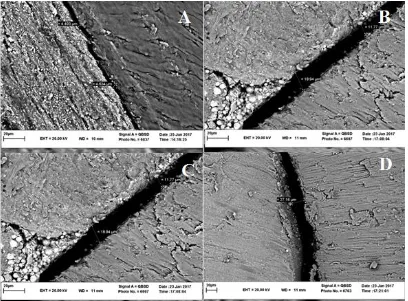
Fig. 1: Scanning electron microscopic micrograph showing the dentin-sealer interface. (A) and (B) indicate the largest and the smallest gaps between the fluoride varnish and dentinal wall, respectively. The largest and the smallest gaps between AH-Plus and dentinal wall are marked as (C) and (D), respectively.
compare the two groups. The significance level was set at P<0.05.
RESULTS
The normal distribution of data was assessed using the Kolmogorov-Smirnov and Shapiro-Wilk tests. The data in both groups had a normal distribution (P>0.05). Mean (standard deviation) gap sizes were calculated as 14.407 (1.402) µm with a range of 12.19-16.369 µm for AH-Plus and 8.342 (0.699) µm ranging from 7.562-9.933 µm for fluoride varnish (N=12 in both groups). The AH-Plus group showed significantly greater gap sizes between the sealer and dentinal walls as compared to the fluoride varnish group (P<0.001). The microscopic images are demonstrated in Figure 1.
DISCUSSION
The present study examined the dentin adaptability of fluoride varnish (Duofluoride
XII) as a root canal sealer. The results showed that this type of sealer has superior dentin adaptability in comparison with AH-Plus sealer. AH-plus sealer is known as the gold standard sealer due to its ideal properties, including good adhesion to dentin and sealability [17].
Some methods, including gap size measurement [18], bond strength measurement [19,20], and calculation of tubular penetration rate [21,22] are used to evaluate the sealer penetration ability and its adaptation to root canal dentinal walls. The gap size measurement approach was the method of choice in the current study.
during root canal filling using the lateral compaction technique [23].
Undetected gaps in some areas of root canal walls are a potential adverse occurrence, which might confound the findings of the studies investigating the gap size at certain cross-sections of the root. To overcome this problem in the current study, each cross-section was divided into four quadrants to enhance the accuracy of gap detection [18]. In the present study, an SEM was used to evaluate the dentin adaptability of AH-Plus versus the fluoride varnish. This microscope allows the examiner to observe the fine relevant details regarding the integrity and surface appearance of the sealer. In addition, the adaptation of the sealer to the dentin can be evaluated under higher magnifications, allowing for observation and measurement of the gap(s) formed between the sealer and the canal wall [24].
Gap sizes are often reported at x2000 magnification. In a microscopic study, Steier et al [25] observed that an increase in magnification from x150 to x1000 had no significant effect on the detection of a greater number of gaps. However, in the current study, x150 magnification was first used to obtain an overview followed by x2000 magnification for enhanced accuracy.
The primary characteristics emphasized by Grossman et al [26] concerning an ideal sealer include optimal adhesion quality and sealing properties. Fluoride varnish has shown satisfying adhesion and durability on the dentin surface [12]. Its application as a root canal sealer indicated favorable results in studies conducted by Parirokh et al [13] and Rao et al [14]. In the current study, the mean
gap size was 14.407±1.0402 μm in the
AH-Plus group and 8.342±0.694 μm in the
fluoride varnish group. At the time of conducting this study, no similar study seemed to be available to compare our results regarding gap size following the application of fluoride varnish as a root canal sealer. The results of some previous SEM studies showed that AH-Plus has proper dentinal adaptation [18,21,25]. Steier et al [25] evaluated the sealer-dentin interface of AH-Plus and
RealSeal. According to their findings, only 21% to 30% of the cases had a visible gap [25]. Similarly, Balguerie et al [21] found no gaps between AH-Plus sealer and dentin in most of the transverse sections of the teeth. In a similar study, Mohammadian et al [18] evaluated the dentin-sealer interfaces of BC sealer, AH-Plus, and Dorifill using an SEM at x300 magnification. They found that AH-Plus
had a mean gap size of 5.63±2.56 μm in the
middle section of the teeth [18]. Accordingly, resin-based sealers seem to have an appropriate adaptation to the canal for two reasons, namely mechanical retention [27,28] and no polymerization shrinkage [29].
CONCLUSION
The present study indicated that the examined fluoride varnish has better dentin adaptability than AH-Plus sealer. Further vitro and in-vivo studies are required to examine other properties of this varnish.
ACKNOWLEDGMENTS
This paper was based on a thesis supported by a grant from the Research Council of Mazandaran University of Medical Sciences, Sari, Iran.
CONFLICT OF INTEREST STATEMENT None declared.
REFERENCES
1. Sjogren U, Figdor D, Persson S, Sundqvist G. Influence of infection at the time of root filling on the outcome of endodontic treatment of teeth with apical periodontitis. Int Endod J. 1997 Sep;30(5):297-306.
2. Li GH, Niu LN, Zhang W, Olsen M, De-Deus G, Eid AA, et al. Ability of new obturation materials to improve the seal of the root canal system: a review. Acta Biomater. 2014 Mar;10(3):1050-1063.
3. ØRSTAVIK D. Materials used for root canal obturation: technical, biological and clinical testing. Endod Topics. 2005 Nov;12(1):25-38.
4. Grossman LI, Oliet S, Del Rio CE. Endodontic Practice. Lea & Febiger, Philadelphia, PA, USA, 1988:179-227.
5. Tyagi S, Mishra P, Tyagi P. Evolution of root canal sealers: An insight story. Eur J Gen Dent. 2013 Aug;2(3):199-218.
6. Sevimay S, Dalat D. Evaluation of penetration and adaptation of three different sealers: a SEM study. J Oral Rehabil. 2003 Sep;30(9):951-5.
7. Agouropoulos A, Twetman S, Pandis N, Kavvadia K, Papagiannoulis L. Caries-preventive effectiveness of fluoride varnish as adjunct to oral health promotion and supervised tooth brushing in preschool children: a double-blind randomized controlled trial. J Dent. 2014 Oct;42(10):1277-83.
8. Fontana M, Gonzalez-Cabezas C, Haider A, Stookey GK. Inhibition of secondary caries lesion progression using fluoride varnish. Caries Res. 2002 Mar-Apr;36(2):129-35.
9. Omidi S, Iomee M, Eshaghi F, Ahanjan M, Cherati JY. Antibacterial effect of stannous fluoride (SnF) 0.63% as an intracanal medicament against Enterococcus faecalis: An in vitro study. Ann Dent Spec. 2018 Jan-Mar:6(1):35-39.
10 Lochaiwatana Y, Poolthong S, Hirata I, Okazaki M, Swasdison S, Vongsavan N. The synthesis and characterization of a novel potassium chloride-fluoridated hydroxyapatite varnish for treating dentin hypersensitivity. Dent Mater J. 2015;34(1):31-40.
11. Chawla HS, Setia S, Gupta N, Gauba K, Goyal A. Evaluation of a mixture of zinc oxide, calcium hydroxide, and sodium fluoride as a new root canal filling material for primary teeth. J Indian Soc Pedod Prev Dent. 2008 Jun;26(2):53-8.
12. Ritter AV, de L Dias W, Miguez P, Caplan DJ, Swift EJ Jr. Treating cervical dentin hypersensitivity with fluoride varnish: a randomized clinical study. J Am Dent Assoc. 2006 Jul;137(7):1013-20; quiz 1029.
13. Parirokh M, Talebizad M, Forghani FR, Haghdoost AA, Asgary S, Eghbal MJ, et al. Fluoride varnish as root canal sealer: a scanning electron microscopy and bacterial penetration study. Iran Endod J. 2015 Winter;10(1):64-8.
14. Rao DG, Trivedi MV, Havale R, Shrutha SP. New fluoride MI Varnish as root canal sealer: An in vitro analysis of bacterial leakage. J Indian Soc Pedod Prev Dent. 2016 Oct-Dec;34(4):359-63.
15. Olympio KP, Cardoso VE, Bijella MF, Pessan JP, Delbem AC, Buzalaf MA. Urinary fluoride output in children following the use of a dual-fluoride varnish formulation. J Appl Oral Sci. 2009 May-Jun;17(3):179-83.
17. Silva EJ, Perez R, Valentim RM, Belladonna FG, De-Deus GA, Lima IC, et al. Dissolution, dislocation and dimensional changes of endodontic sealers after a solubility challenge: a micro-CT approach. Int Endod J. 2017 Apr;50(4):407-14.
18. Mohammadian F, Farahanimastary F, Dibaji F, Kharazifard MJ. Scanning Electron Microscopic Evaluation of the Sealer-Dentine Interface of Three Sealers. Iran Endod J. 2017 Winter;12(1):38-42.
19. Forough Reyhani M, Ghasemi N, Rahimi S, Salem Milani A, Mokhtari H, Shakouie S, et al. Push-Out Bond Strength of Dorifill, Epiphany and MTA-Fillapex Sealers to Root Canal Dentin with and without Smear Layer. Iran Endod J. 2014 Fall;9(4):246-50. 20. Carvalho CN, Martinelli JR, Bauer J, Haapasalo M, Shen Y, Bradaschia-Correa V, et al. Micropush-out dentine bond strength of a new gutta-percha and niobium phosphate glass composite. Int Endod J. 2015 May;48(5):451-9.
21. Balguerie E, van der Sluis L, Vallaeys K, Gurgel-Georgelin M, Diemer F. Sealer penetration and adaptation in the dentinal tubules: a scanning electron microscopic study. J Endod. 2011 Nov;37(11):1576-9. 22. Shokouhinejad N, Sabeti M, Gorjestani H, Saghiri MA, Lotfi M, Hoseini A. Penetration of Epiphany, Epiphany self-etch, and AH Plus into dentinal tubules: a scanning electron microscopy study. J Endod. 2011 Sep;37(9):1316-9.
23. Vassiliadis LP, Sklavounos SA, Stavrianos CK. Depth of penetration and appearance of Grossman sealer in the dentinal tubules: an in vivo study. J Endod. 1994
Aug;20(8):373-6.
24. Mamootil K, Messer HH. Penetration of dentinal tubules by endodontic sealer cements in extracted teeth and in vivo. Int Endod J. 2007 Nov;40(11):873-81.
25. Steier L, de Figueiredo JAP, Belli S. Comparison of the interface dentin-endodontic sealer using two SEM magnifications. Rev Odonto Ciênc. 2010 Dec;25(3):296-9.
26. Grossman LI, Shepard LI, Pearson LA. Roentgenologic and clinical evaluation of endodontically treated teeth. Oral Surg Oral Med Oral Pathol. 1964 Mar;17:368-74.
27. Barbizam JV, Trope M, Tanomaru-Filho M, Teixeira EC, Teixeira FB. Bond strength of different endodontic sealers to dentin: push-out test. J Appl Oral Sci. 2011 Nov-Dec;19(6):644-7.
28. Sagsen B, Ustun Y, Demirbuga S, Pala K. Push-out bond strength of two new calcium silicate-based endodontic sealers to root canal dentine. Int Endod J. 2011 Dec;44(12):1088-91.
29. Marciano MA, Guimaraes BM, Ordinola-Zapata R, Bramante CM, Cavenago BC, Garcia RB, et al. Physical properties and interfacial adaptation of three epoxy resin-based sealers. J Endod. 2011 Oct;37(10):1417-21.
30. Wu MK, de Gee AJ, Wesselink PR. Effect of tubule orientation in the cavity wall on the seal of dental filling materials: an in vitro study. Int Endod J. 1998 Sep;31(5):326-32.