R E S E A R C H P A P E R
Open Access
A novel framework for cross-spectral iris
matching
Mohammed A. M. Abdullah
1,2*, Satnam S. Dlay
1, Wai L. Woo
1and Jonathon A. Chambers
1Abstract
Previous work on iris recognition focused on either visible light (VL), near-infrared (NIR) imaging, or their fusion. However, limited numbers of works have investigated cross-spectral matching or compared the iris biometric performance under both VL and NIR spectrum using unregistered iris images taken from the same subject. To the best of our knowledge, this is the first work that proposes a framework for cross-spectral iris matching using unregistered iris images. To this end, three descriptors are proposed namely, Gabor-difference of Gaussian (G-DoG), Gabor-binarized statistical image feature (G-BSIF), and Gabor-multi-scale Weberface (G-MSW) to achieve robust cross-spectral iris matching. In addition, we explore the differences in iris recognition performance across the VL and NIR spectra. The experiments are carried out on the UTIRIS database which contains iris images acquired with both VL and NIR spectra for the same subject. Experimental and comparison results demonstrate that the proposed framework achieves state-of-the-art cross-spectral matching. In addition, the results indicate that the VL and NIR images provide complementary features for the iris pattern and their fusion improves notably the recognition performance.
Keywords: Iris recognition, Cross-spectral matching, Multi-spectral recognition, Photometric normalization, Score fusion
1 Introduction
Among the various traits used for human identification, the iris pattern has gained an increasing amount of atten-tion for its accuracy, reliability, and noninvasive charac-teristics. In addition, iris patterns possess a high degree of randomness and uniqueness which is true even between identical twins, and the iris remains constantly stable throughout an adult’s life [1, 2].
The initial pioneering work on iris recognition, which is the basis of many functioning commercial systems, was conducted by Daugman [1]. The performance of iris recognition systems is impressive as demonstrated by Daugman [3] who reported false acceptance rates of only 10−6 on a study of 200 billion cross-comparisons. Additionally, the potential of iris biometrics has also been affirmed with 1.2 trillion comparison by tests carried out by the National Institute of Standards and Technol-ogy (NIST) which confirmed that iris biometrics has the
*Correspondence: [email protected]
1ComS2IP Group, School of Electrical and Electronic Engineering, Newcastle
University, England, UK
2Department of Computer and Information Engineering, Ninevah University,
Nineveh, Iraq
best balance between accuracy, template size, and speed compared to other biometric traits [4].
Iris recognition technology nowadays is widely deployed in various large-scale applications such as the border crossing system in the United Arab Emirates, Mexico national ID program, and the Unique Identification Authority of India (UIDAI) project [5]. As a case in point, more than one billion residents have been enrolled in the UIDAI project where about 1015 all-to-all check opera-tions are carried out daily for identity de-duplication using iris biometrics as the main modality [5, 6].
Nearly all currently deployed iris recognition systems operate predominately in the near-infrared (NIR) spec-trum capturing images at 800–900 nm wavelength. This is because there are fewer reflections coming from the cornea and the dark pigmented irides look clearer under the NIR light. In addition, external factors such as shadows and diffuse reflections become less under NIR light [7, 8].
The color of the irides is governed by the congruity of two molecules: eumelanin (black/brown) and pheome-lanin (red/yellow). Dark pigmented irides have a high concentration of eumelanin. As the latter deeply absorbs
visible light (VL), stromal features of the iris are only revealed under NIR and they become hidden in VL so the information related to the texture is revealed rather than the pigmentation. On the other hand, pheomelanin is dominant in light-pigmented irides. Capturing such irides under NIR light eliminates most of the rich pheomelanin information because the chromophore of the human iris is only visible under VL [8, 9]. Consequently, capturing iris images under different light conditions reveals different textural information.
Research in VL iris recognition has been gaining more attention in recent years due to the interest in iris recog-nition at a distance [10, 11]. In addition, competitions such as the Noisy Iris Challenge Evaluation (NICE) [12] and the Mobile Iris Challenge Evaluation [13] focus on the processing of VL iris images. This attention to visible wavelength-based iris recognition is boosted by several factors such as (1) visible range cameras can acquire images from long distance and they are cheaper than NIR cameras and (2) surveillance systems work in the visible range by capturing images of the body, face, and iris which could be used later for authentication [14].
Since both VL and NIR iris recognition systems are now widely deployed, studying the performance difference of iris recognition systems exploiting NIR and VL images is important because it gives insight into the essential fea-tures in each wavelength which in turn helps to develop a robust automatic identification system. On the other hand, cross-spectral iris recognition is essential in secu-rity applications when matching images from different lighting conditions is desired.
In this paper, we therefore propose a method for cross-spectral iris images matching. To the best of our knowl-edge, this attempt is amongst the first in the literature to investigate the problem of VL to NIR iris recognition (and vice versa) dealing with unregistered iris images belong-ing to the same subject. In addition, we investigate the difference in iris recognition performance with NIR and VL imaging. In particular, we investigate iris performance in each channel (red, green, blue, and NIR) and the fea-sibility of cross-channel authentication (i.e., NIR vs. VL). Furthermore, enhancing the iris recognition performance with multi-channel fusion is attained.
In summary, the main contributions of the paper are as follows:
• A novel framework for cross-spectral iris recognition capable of matching unregistered iris images captured under different lighting conditions
• Filling the gap in multi-spectral iris recognition by exploring the performance difference in iris biometrics under NIR and VL imaging
• Boosting iris recognition performance with multi-channel fusion
The rest of this paper is organized as follows: related works are given in Section 2. The proposed framework for cross-spectral iris matching is explained in Section 3. Section 4 presents the experimental results and the discussion while Section 5 concludes this paper.
2 Related work
Iris recognition technology has witnessed a rapid devel-opment over the last decade driven by its wide applica-tions in the world. At the outset, Daugman [1] proposed the first working iris recognition system which has been adopted later by several commercial companies such as IBM, Irdian, and Oki. In this work, the integro-differential operator is applied for iris segmentation and the 2D Gabor filters are utilized for feature extraction while the Ham-ming distance scores serve as a comparator. The second algorithm is due to Wildes [15] who applied the Hough transform for localizing the iris and the Laplacian pyramid to encode the iris pattern. However, this algorithm has a high computational demand.
Another interesting approach was proposed by Sun and Tan [2] exploiting ordinal measures for iris feature rep-resentation. Unlike the traditional approaches that use quantitative values, the ordinal measure focuses on qual-itative values to represent features. The multi-lobe differ-ential filters have been applied for iris feature extraction to generate a 128-byte ordinal code for each iris image. Then, the error rates have been calculated based on the mea-sured Hamming distances between two ordinal templates of the same class.
In Ngo et al. [18], a multi-spectral iris recognition sys-tem was implemented which employed eight wavelengths ranges from 405 to 1550 nm. The results on a database of 392 iris images showed that the best performance was achieved with a wavelength of 800 nm. Cross-spectral experiment results demonstrated that the performance degraded with larger wavelength difference. Ross et al. [19] explored the performance of iris recognition in wave-lengths beyond 900 nm. In their experiments, they inves-tigated the possibility of observing different iris structures under different wavelengths and the potential of perform-ing multi-spectral fusion for enhancperform-ing iris recognition performance. Similarly, Ives et al. [20] examined the per-formance of iris recognition under a wide range of wave-lengths between 405 and 1070 nm. The study suggests that illumination wavelength has a significant effect on iris recognition performance. Hosseini et al. [8] proposed a feature extraction method for iris images taken under VL using a shape analysis method. Potential improvement in recognition performance was reported when combining features from both NIR and VL iris images taken from the same subject.
Recently, Alonso-Fernandez et al. [21] conducted com-parisons on the iris and periocular modalities and their fusion under NIR and VL imaging. However, the images were not taken from the same subjects as the experiments were carried out on different databases (three databases contained close-up NIR images, and two others contained VL images). Unfortunately, this may not give an accurate indication about the iris performance as the images do not belong to the same subject. In [22], the authors suggested enhancing iris recognition performance in non-frontal images through multi-spectral fusion of iris pattern and scleral texture. Since the scleral texture is better seen in VL and the iris pattern is observed in NIR, multi-spectral fusion could improve the overall performance.
In terms of cross-spectral iris matching, the authors in [14] proposed an adaptive method to predict the NIR channel image from VL iris images using neural networks. Similarly, Burge and Monaco [23, 24] pro-posed a model to predict NIR iris images using features derived from the color and structure of the visible light iris images. Although the aforementioned approaches ([14, 23, 24]) achieved good results, their methods require the iris images to be fully registered. Unfortunately, this is not applicable in reality because it is very difficult to capture registered iris images from the same subject simultaneously.
In our previous work [25], we explored the differences in iris recognition performance across the VL and NIR spec-tra. In addition, we investigated the possibility of cross-channel matching between the VL and NIR imaging. The cross-spectral matching turns out to be challenging with an equal error rate (EER) larger than 27 %. Lately, Ramaiah
and Kumar [26] emphasized the need for cross-spectral iris recognition and introduced a database of registered iris images and conducted experiments on iris recogni-tion performance under both NIR and VL. This database is not available yet. The results of cross-spectral match-ing achieved an EER larger than 34 % which confirms the challenge of cross-spectral matching. The authors con-cluded their paper by: “it is reasonable to argue that cross-spectral iris matching seriously degrades the iris matching accuracy”.
3 Proposed cross-spectral iris matching framework
Matching across iris images captured in VL and NIR is a challenging task because there are considerable differences among such images pertaining to different wavelength bands. Although, the appearance of different spectrum iris images looks different, the structure is the same as they belong to the same person. Therefore, we exploited various photometric normalization techniques and descriptors to alleviate these differences. In this con-text, we employed the Binarized Statistical Image Features (BSIF) descriptor [27], DoG filtering in addition to a collection of the photometric normalization techniques available from the INface Toolbox1 [28, 29]: adaptive single scale retinex, non-local means, wavelet based nor-malization, homomorphic filtering, multi-scale quotient, Tan and Triggs normalization, and multi-scale Weberface (MSW).
Among these illumination techniques and descriptors, the DoG, BSIF, and MSW are noticed to reduce the iris cross-spectral variations. These models are described in the next subsections.
3.1 Difference of Gaussian (DoG)
The DoG is a feature enhancement technique which depends on the difference of Gaussians filter to generate a normalized image by acting as a bandpass filter. This is achieved by subtracting two blurred versions of the original images from each other [30]. The blurred ver-sionsG(x,y)are obtained by convolving the original image
I(x,y)with two Gaussian kernels having differing standard deviations as shown in Eq. (1):
D(x,y|σ0,σ1)=
G(x,y|σ0)−G(x,y|σ1)
∗I(x,y), (1)
where * is the convolution operator andσrepresents the Gaussian kernel function which is defined as
G(x,y|σ)= √ 1
2πσ2e
−(x2+y2)/2σ2
(2)
is able to alleviate the illumination variation and alias-ing. As there are variations in the frequency between VL and NIR images, the DoG filter is efficient because it suppresses these variations and alleviates noise and aliasing which paves the way for a better cross-spectral matching [30].
3.2 Binarized statistical image features (BSIF)
The BSIF [27] have been employed due to their abil-ity to tolerate image degradation such as rotation and blurring [27]. Generally speaking, feature extraction methods usually filter the images with a set of lin-ear filters then quantize the response of such filters. In this context, BSIF filters are learned by exploit-ing the statistics of natural images rather than usexploit-ing manually built filters. This has resulted in promising results for classifying the texture in different biometric traits [31, 32].
For an image patch X of size l × l pixels and a lin-ear filter Wi of the same size, the filter response si is obtained by
si=
Wi(u,v)X(u,v)=wTi x. (3)
The binarized feature bi is obtained based on the response values by settingbi=1 ifsi>0 andbi=0 oth-erwise. The filters are learned from natural images using independent component analysis by maximizing the sta-tistical independence of si. Two parameters control the BSIF descriptor: the number of the filters (lengthnof the bit string) and the size of the filterl. In our approach, we used the default set of the filters2which were learned from 5000 patches. Empirical results demonstrated that a filter size of 7×7 with 8 bits gives the best results.
3.3 Multi-scale Weberfaces (MSW)
Inspired by Weber’s law which states that the ratio of the increment threshold to the background intensity is a constant [33], the authors in [34] showed that the ratio between local intensity of a pixel and its surround-ing variations is constant. Hence, in [34], the face image is represented by its reflectance and the illumination factor is normalized and removed using the Weberface model. Following this, we applied the Weberface model to the iris images to remove the illumination variations that result from the differences between the VL and NIR imaging, thus making the iris images illumination invariant.
Following the works of [28, 29], the Weberface algo-rithm has been applied with three scales using the follow-ing values: σ = [1 0.75 0.5], Neighbor =[9 25 49] and alfa=[2 0.2 0.02]. The steps of the Weberface algorithm are listed in Algorithm 1.
Algorithm 1:Weberface normalization
Input: Iris imageI
Output: Weber iris imageW
1: Smooth the image using Gaussian filter
2: I=I∗G(X,Y,σ)
3: For each pixel in the image
sum=
(PixelIntensity−NeighborValues)/PixelIntensity;
Wi=arctan(alfa×sum);
4: AssignWito the pixel in the Weber iris image.
3.4 Proposed scheme
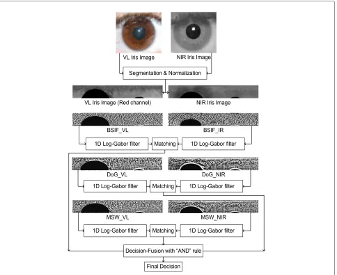
The variations in iris appearance due to different sensors, spectral bands, and illumination variations are believed to significantly degrade the iris recognition performance. To overcome these artifacts, a robust method should be carefully designed. Extensive experiments demonstrated that using one of the aforementioned methods alone is not sufficient to achieve an acceptable iris recognition performance with EER>17 %. Therefore, we propose to integrate the Gabor filter with these methods in addition to decision level fusion to achieve a robust cross-spectral iris recognition. Also, using the phase information of the Gabor filter rather than amplitude is known to result in robustness to different variations such as illumination variations, imaging contrast, and camera gain [7]. Hence, we propose to integrate the 1D log-Gabor filter [35] with DoG, BSIF, and MSW to produce the G-DoG, G-BSIF, and G-MSW (where G stands for Gabor) in addition to decision level fusion to achieve a robust cross-spectral iris recognition. The block diagram of the proposed frame-work is depicted in Fig. 1.
Unlike previous works [14, 23, 24] in which they require fully registered iris images and learn models that lack the ability of generalization, our framework does not require any training and works on unregistered iris images. This combination along with its decision level fusion achieved encouraging results as illustrated in the next section.
4 Results and discussion
In this work, our aim is to ascertain true cross-spectral iris matching using images taken from the same subject under the VL and NIR spectra. In addition, we investigate the iris biometric performance under different imaging conditions and the fusion of VL+NIR images to boost the recognition performance. The recognition performance is measured with the EER and the receiver operating characteristic (ROC) curves.
4.1 Database
Fig. 1Block diagram of the proposed cross-spectral matching framework
two sessions with 1540 images; the first session was cap-tured under VL while the second session was capcap-tured under NIR. Each session has 770 images taken from the left and right eye of 79 subjects where each subject has an average of five iris images.
4.2 Pre-processing and feature extraction
Typically, an iris recognition system operates by extract-ing and comparextract-ing the pattern of the iris in the eye image. These operations involve four main steps namely, image acquisition, iris segmentation, normalization, fea-ture extraction, and matching [7].
The UTIRIS database includes two types of iris images, half of which are captured in the NIR spectrum while the other half are captured under the VL spectrum. The VL session contains images in the sRGB color space which then are decomposed to the red, green, and blue chan-nels. To segment the iris in the eye image, the circular
Hough transform (CHT) is applied because the images used in our experiments were captured under a controlled environment so they can be segmented with circular approaches [36, 37].
It is noticed that the red channel gives the best seg-mentation results because the pupil region in this channel contains the smallest amount of reflection as shown in Figs. 2 and 3. The images in the VL session were down-sampled by two in each dimension to obtain the same size as the images in the NIR session. The segmented iris images are normalized with a resolution of 60×450 using the rubber sheet method [7].
Fig. 2Green-yellow iris image decomposed intored,green,blue, andgrayscalewith the NIR counterpart
4.3 NIR vs. VL performance
For feature extraction, the normalized iris image is con-volved with the 1D log-Gabor filter to extract the features where the output of the filter is phase quantized to four levels to form the binary iris vector [35].
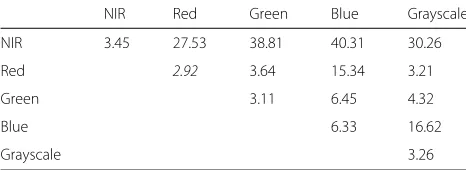
We carried out experiments on each channel (i.e., NIR, red, green, and blue) and measured the performance using ROC and EER. Figure 4 and Table 1 illustrates the EER and ROC curves for each channel. It can be seen that the best performance is achieved under the red channel with EER= 2.92 % followed by the green chan-nel with EER = 3.11 % and the grayscale channel with EER = 3.26 % while the blue channel achieved worse results with EER = 6.33 %. It is also noticed that NIR
images did not give the best performance for this database (EER=3.45 %).
This is in agreement with our results where the red channel images achieved better results than the NIR images as most of the iris images in the UTIRIS database are light pigmented. Figure 5 shows the distribution of the irides color in the UTIRIS database.
4.4 Light-eyed vs. dark-eyed
As mentioned before, capturing iris images under NIR light eliminates most of the rich melanin information because the chromophore of the human iris is only visible under VL [8, 9]. Therefore, light-pigmented irides exhibit more information under visible light. Figure 2 shows a
Fig. 4The performance of the iris recognition underred,green,blue, and NIR spectra
green-yellow iris image captured under NIR and VL. It can be seen that the red channel reveals more information than the NIR image. So, intuitively, the recognition perfor-mance would be better for such images in the VL rather than the NIR spectrum.
On the contrary, with dark-pigmented irides, stromal features of the iris are only revealed under NIR and they become hidden in VL so the information related to the texture is revealed rather than the pigmentation as shown in Fig. 2. Therefore, the recognition performance for the dark-pigmented irides would give better results if the images were captured under NIR spectrum.
4.5 Cross-spectral experiments
Cross-spectral study is important because it shows the feasibility of performing iris recognition in several secu-rity applications such as information forensics, secusecu-rity surveillance, and hazard assessment. Typically a person’s iris images are captured under NIR but most of the secu-rity cameras operate in the VL spectrum. Hence, NIR vs. VL matching is desired.
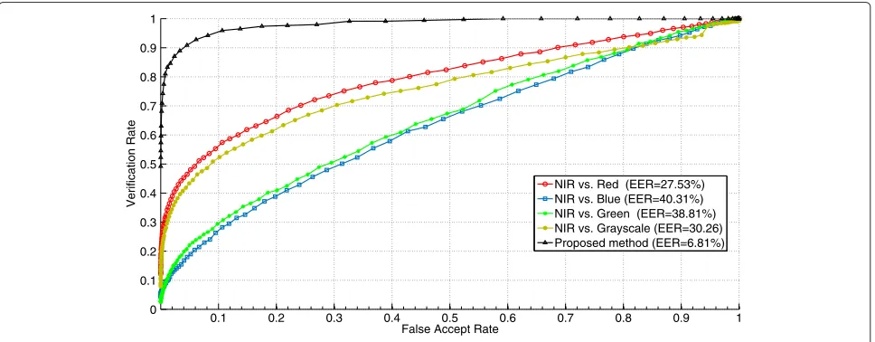
In this context, we carried out these comparisons using the traditional 1D log-Gabor filter: NIR vs. red, NIR vs. green, and NIR vs. blue. Figure 6 depicts the ROC curves
Table 1EER (%) of different channels comparison on the UTIRIS database
NIR Red Green Blue Grayscale
NIR 3.45 27.53 38.81 40.31 30.26
Red 2.92 3.64 15.34 3.21
Green 3.11 6.45 4.32
Blue 6.33 16.62
Grayscale 3.26
of these comparisons. According to Fig. 6, the green and blue channels resulted in bad performance due to the big gap in the electromagnetic spectrum between these channels and the NIR spectrum.
On the contrary, the red channel gave the best per-formance compared to the green and blue channels. This can be attributed to the small gap in the wave-length of the red channel (780 nm) compared to the NIR (850 nm). Therefore, the comparisons of red vs. NIR is considered as the baseline for cross-spectral match-ing. Table 1 shows the EER of cross-channel matching experiments.
4.5.1 Cross-spectral matching
Cross-spectral performance turned out to be a challeng-ing task with EER> 27 % which is attributable to match-ing unregistered iris images from different spectral bands. Hence, to achieve an efficient cross-spectral matching, adequate transformations before the feature extraction are needed.
Different feature enhancement techniques are employed, out of which the DoG, MWS, and BSIF recorded the best results as shown in Table 2. Therefore, our proposed framework, which is depicted in Fig. 1, is based on these descriptors.
Fig. 5The color distributions of the irides of the 79 subjects in the UTIRIS database
4.5.2 Cross-spectral fusion
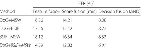
To further enhance the performance of cross-spectral matching, the fusion of the G-DoG, G-BSIF, and G-MSW is considered. Different fusion methods are investigated namely, feature fusion, score fusion and decision fusion, out of which the decision fusion is observed to be the most effective.
Table 3 shows the performance of different fusion strategies for cross-spectral matching in terms of EER. Feature fusion resulted in poor results where the EER var-ied from 14 to 18 %. Score level fusion with minimum rule achieved better results. On the other hand, AND rule decision level fusion achieved the best results with EER = 6.81 %.
A low false accept rate (FAR) is preferred to achieve a secure biometric system. To enhance the performance of our system and reduce the FAR, a fusion at the deci-sion level is performed. Thus, the conjunction “AND” rule
is used to combine the decisions from the DoG, G-BSIF, and G-MSW. This means that a false accept can only happen when all the previous descriptors produce a false accept [39].
LetPD(FA),PS(FA), andPM(FA)represent the proba-bility of a false accept using G-DoG, G-BSIF, and G-MSW, respectively. Similarly,PD(FR),PS(FR), andPM(FR) rep-resent the probability of a false reject. Therefore, the com-bined probability of a false acceptPC(FA)is the product of the three probabilities of the descriptors:
PC(FA)=PD(FA).PS(FA).PM(FA). (4)
On the other hand, the combined probability of a false rejectPC(FR)can be expressed as the complement of the probability that none of the descriptors produce a false reject:
Table 2Experiments on different descriptors for cross-spectral matching
Method EER (%)
Baseline 27.53
LBP (different combinations) [40] >28
Adaptive single scale retinex 25.56
Non-local means normalization 27.49
Wavelet-based normalization 28.65
Homomorphic filtering 29.07
Multi-scale self quotient 26.99
Tan and Triggs normalization 23.43
DoG 19.51
MSW 18.91
BSIF 20.64
PC(FR)=(PD(FR).PS(FR).PM(FR)),
=(1−(1−PD(FR))(1−PS(FR))(1−PM(FR))),
=PD(FR)+PS(FR)+PM(FR)
+PD(FR).PS(FR)+PD(FR).PM(FR)
+PS(FR).PM(FR)+PD(FR).PS(FR).PM(FR). (5)
It can be seen from the previous equations that the joint probability of false rejection increases while the joint probability of false acceptance decreases when using the AND conjunction rule.
All the previous descriptors (DoG, BSIF, and G-MSW) are considered as local descriptors. It can be argued that the fusion of local and global features could enhance the performance further. We wish to remark that fusing the local and global features would require further stages to augment the resultant global and local scores as they will be in different range/type [40]. Such stages will increase the complexity of the cross-spectral framework. We have carefully designed the proposed framework so that all three descriptors (G-DoG, G-BSIF, and G-MSW)
Table 3Experiments on different fusion strategies for cross-spectral matching
EER (%)a
Method Feature fusion Score fusion (min) Decision fusion (AND)
DoG+MSW 16.56 14.21 8.08
DoG+BSIF 17.56 15.42 8.77
BSIF+MSW 18.12 16.34 8.33
DoG+BSIF+MSW14.59 12.83 6.81
aThe cross-spectral comparison results in this table are obtained by taking the
minimum of comparison scores in each class
generate homogenous scores (binary template). There-fore, a single comparator (Hamming distance) can be quickly used for score matching.
4.6 Multi-spectral iris recognition
The VL and NIR images in the UTIRIB database are not registered. Therefore, they provide different iris tex-ture information. The cross-channel comparisons demon-strated that red and NIR channels are the most suitable candidates for fusion as they gave the lowest EER com-pared to other channels as shown in Figs. 4 and 6, so it is common sense to fuse them in order to boost the recog-nition performance. Score level fusion is adopted in this paper due to its efficiency and low complexity [41]. Hence, we combined the matching scores (Hamming distances) from both the red and NIR images using sum rule-based fusion with equal weights to generate a single matching score. After that, the recognition performance is evaluated again with the ROC curves and EER.
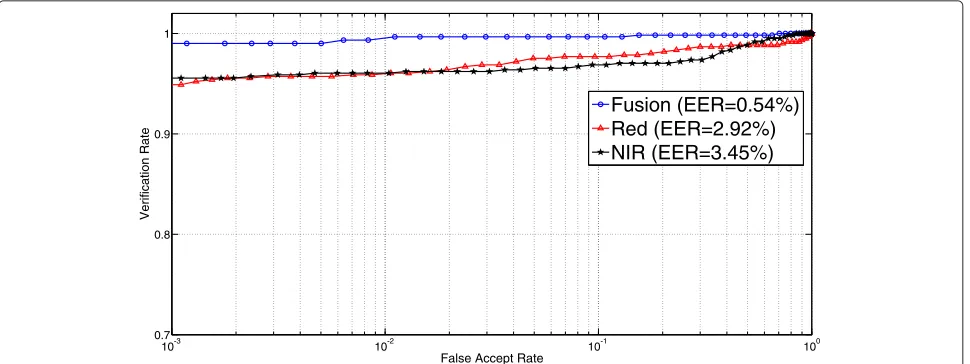
It is evident from Fig. 7 that such fusion is useful to the iris biometric as there is a significant improvement in the recognition performance after the fusion with EER of only 0.54 % compared to 2.92 and 3.45 % before the fusion.
4.7 Comparisons with related work
Although the previous works [14, 23, 24] reported good results in terms of cross-spectral iris matching, it must be noted that these works have adopted fully registered iris images and learn models that lack the ability of general-ization.
In the works of [25, 42], the results of cross-spectral matching on unregistered iris images were reported. How-ever, no models were proposed to enhance the cross-spectral iris matching. Table 4 shows the comparison results of the aforementioned works compared to our method.
4.8 Processing time
All experiments were conducted on a 3.2-GHz core i5 PC with 8 GB of RAM under the Matlab environment. The proposed framework consists of four main descrip-tors namely, BSIF, DoG, MSW, and 1D log-Gabor filter. The processing times of the 1D log-Gabor filter, BSIF, and DoG descriptors are 10, 20, and 70 ms, respectively, while the MSW processing times is 330 ms. Therefore, the total computations time of the proposed method is less than half a second which implies its suitability for real time applications.
5 Conclusions
Fig. 7ROC curves showing the iris recognition performance before and after fusing the information of the red and NIR channel
iris recognition performance under different imaging channels: red, green, blue, and NIR. The experiments were carried out on the UTIRIS database, and the performance of the iris biometric was measured.
We drew the following conclusions from the results. According to Table 2, among a variety of descriptors, the difference of Gaussian (DoG), BSIF, and multi-scale Weberface (MSW) were found to give good cross-spectral performance after integrating them with the 1D log-Gabor filter. Table 4 and Fig. 6 showed a significant improvement in the cross-spectral matching performance using the proposed framework.
In terms of multi-spectral iris performance, Fig. 4 showed that the red channel achieved better performance compared to other channels or the NIR imaging. This can be attributed to the large number of the light-pigmented irides in the UTIRIS database. It was also noticed from Fig. 6 that the performance of the iris recognition varied as a function of the difference in wavelength among the image channels. Fusion of the iris images from the red and NIR channels notably improved the recognition per-formance. The results implied that both the VL and NIR imaging were important to form a robust iris recognition system as they provided complementary features for the iris pattern.
Table 4Cross-spectral matching comparison with different methods
Method Database EER (%)
Wild et al. [42] UTIRIS 33–55
Ramaiah and Kumar [26] Private 34.01
Abdullah et al. [25] UTIRIS 27.46
Proposed UTIRIS 6.81
Endnotes
1http://luks.fe.uni-lj.si/sl/osebje/vitomir/face_tools/
INFace/
2http://www.ee.oulu.fi/~jkannala/bsif/bsif.html
Acknowledgements
The first author would like to thank the Ministry of Higher Education and Scientific Research (MoHESR) in Iraq for supporting this work.
Authors’ contributions
MA carried out the design of the iris cross-spectral matching framework, performed the experiments, and drafted the manuscript. SD participated in the design of the framework and helped in drafting the manuscript. WW participated in the comparison experiments and helped in drafting the manuscript. JC guided the work, supervised the experimental design, and helped in drafting the manuscript. All authors read and approved the final manuscript.
Competing interests
The authors declare that they have no competing interests.
Received: 8 April 2016 Accepted: 14 October 2016
References
1. Daugman J (1993) High confidence visual recognition of persons by a test of statistical independence. Pattern Anal Mach Intell IEEE Trans 15(11):1148–1161
2. Sun Z, Tan T (2009) Ordinal measures for iris recognition. IEEE Trans Pattern Anal Mach Intell 31(12):2211–2226
3. Daugman J (2006) Probing the uniqueness and randomness of IrisCodes: results from 200 billion iris pair comparisons. Proc IEEE 94(11):1927–1935 4. Grother PJ, Quinn GW, Matey JR, Ngan ML, Salamon WJ, Fiumara GP,
Watson CI (2012) IREX III: performance of iris identification algorithms, Report, National Institute of Standards and Technology
5. Jain AK, Nandakumar K, Ross A (2016) 50 Years of biometric research: accomplishments, challenges, and opportunities. Pattern Recognit Lett 79:80–105
7. Daugman J (2004) How iris recognition works. IEEE Trans Circ Syst Video Technol 14(1):21–30
8. Hosseini MS, Araabi BN, Soltanian-Zadeh H (2010) Pigment melanin: pattern for iris recognition. IEEE Trans Instrum Meas 59(4):792–804 9. Meredith P, Sarna T (2006) The physical and chemical properties of
eumelanin. Pigment Cell Res 19(6):572–594
10. Dong W, Sun Z, Tan T (2009) A design of iris recognition system at a distance. In: Chinese Conference on Pattern Recognition, (CCPR 2009). IEEE, Nanjing. pp 1–5. http://ieeexplore.ieee.org/document/5344030/ 11. Proenca H, Filipe S, Santos R, Oliveira J, Alexandre LA (2010) The UBIRIS.v2:
a database of visible wavelength iris images captured on-the-move and at-a-distance. IEEE Trans Pattern Anal Mach Intell 32(8):1529–1535 12. Bowyer KW (2012) The results of the NICE.II iris biometrics competition.
Pattern Recognit Lett 33(8):965–969
13. De Marsico M, Nappi M, Riccio D, Wechsler H (2015) Mobile iris challenge evaluation (MICHE)-I, biometric iris dataset and protocols. Pattern Recognit Lett 57(0):17–23
14. Jinyu Z, Nicolo F, Schmid NA (2010) Cross spectral iris matching based on predictive image mapping. In: Fourth IEEE International Conference on Biometrics: Theory Applications and Systems (BTAS’10). IEEE, Washington D.C. pp 1–5
15. Wildes RP (1997) Iris recognition: an emerging biometric technology. Proc IEEE 85(9):1348–1363
16. Boyce C, Ross A, Monaco M, Hornak L, Xin L (2006) Multispectral iris analysis: a preliminary study. In: Computer Vision and Pattern Recognition Workshop. IEEE, New York. pp 51–51. doi:10.1109/CVPRW.2006.141. http://www.cse.msu.edu/~rossarun/pubs/RossMSIris_CVPRW06.pdf. Accessed Sept 2016
17. Tajbakhsh N, Araabi BN, Soltanianzadeh H (2008) Feature fusion as a practical solution toward noncooperative iris recognition. In: 11th International Conference on Information Fusion. IEEE, Cologne. pp 1–7
18. Ngo HT, Ives RW, Matey JR, Dormo J, Rhoads M, Choi D (2009) Design and implementation of a multispectral iris capture system. In: Asilomar Conference on Signals, Systems and Computers. IEEE, Pacific Grove. pp 380–384
19. Ross A, Pasula R, Hornak L (2009) Exploring multispectral iris recognition beyond 900 nm. In: IEEE 3rd International Conference on Biometrics: Theory, Applications, and Systems. (BTAS’09). IEEE, Washington D.C. pp 1–8
20. Ives RW, Ngo HT, Winchell SD, Matey JR (2012) Preliminary evaluation of multispectral iris imagery. In: IET Conference on Image Processing (IPR 2012). IET, London. pp 1–5
21. Alonso-Fernandez F, Mikaelyan A, Bigun J (2015) Comparison and fusion of multiple iris and periocular matchers using near-infrared and visible images. In: 2015 International Workshop on Biometrics and Forensics (IWBF). IEEE, Gjøvik. pp 1–6
22. Crihalmeanu SG, Ross AA (2016) Multispectral Ocular Biometrics. In: Bourlai T (ed). Face Recognition Across the Imaging Spectrum. Springer International Publishing, Cham. pp 355–380. ISBN:978-3-319-28501-6. doi:10.1007/978-3-319-28501-6_15
23. Burge MJ, Monaco MK (2009) Multispectral iris fusion for enhancement, interoperability, and cross wavelength matching. In: Proceeding of SPIE 7334, Algorithms and Technologies for Multispectral, Hyperspectral, and Ultraspectral Imagery XV, Vol. 7334. SPIE. pp 73341D–1–73341D–8. doi:10.1117/12.819058. http://spie.org/Publications/Proceedings/Paper/ 10.1117/12.819058. Accessed Sept 2016
24. Burge M, Monaco M (2013) Multispectral iris fusion and cross-spectrum matching. Springer, London. pp 171–181
25. Abdullah MAM, Chambers JA, Woo WL, Dlay SS (2015) Iris biometric: is the near-infrared spectrum always the best? In: 3rd Asian Conference on Pattern Recognition (ACPR2015). IEEE, Kuala Lumpur. pp 816–819. doi:10.1109/ACPR.2015.7486616, http://ieeexplore.ieee.org/document/ 7486616/. Accessed Sept 2016
26. Ramaiah NP, Kumar A (2016) Advancing Cross-Spectral Iris Recognition Research Using Bi-Spectral Imaging. In: Singh R, Vatsa M, Majumdar A, Kumar A (eds). Machine Intelligence and Signal Processing. Springer India, New Delhi. pp 1–10. ISBN:978-81-322-2625-3. doi:10.1007/978-81-322-2625-3_1.
27. Kannala J, Rahtu E (2012) BSIF: binarized statistical image features. In: 21st International Conference on Pattern Recognition (ICPR). IEEE, Tsukuba Science City. pp 1363–1366
28. Štruc V, Pavesic N (2009) Gabor-based kernel partial-least-squares discrimination features for face recognition. Informatica 20(1):115–138 29. Štruc V, Pavesic N (2011) Photometric Normalization Techniques for
Illumination Invariance. Advances in Face Image Analysis: Techniques and Technologies. IGI Global. pp 279–300
30. Tan X, Triggs B (2010) Enhanced local texture feature sets for face recognition under difficult lighting conditions. IEEE Trans Image Process 19(6):1635–1650
31. Arashloo SR, Kittler J (2014) Class-specific kernel fusion of multiple descriptors for face verification using multiscale binarised statistical image features. IEEE Trans Inf Forensic Secur 9(12):2100–2109 32. Li X, Bu W, Wu X (2015) Palmprint Liveness Detection by Combining
Binarized Statistical Image Features and Image Quality Assessment. In: Yang J, Yang J, Sun Z, Shan S, Zheng W, Feng J (eds). Biometric Recognition: 10th Chinese Conference, CCBR 2015, Tianjin, China, November 13-15, 2015, Proceedings. Springer International Publishing, Cham. pp 275–283. ISBN:978-3-319-25417-3. doi:10.1007/978-3-319-25417-3_33
33. Jain AK (1989) Fundamentals of digital image processing. Prentice-Hall, Inc., Upper Saddle River
34. Wang B, Li W, Yang W, Liao Q (2011) Illumination normalization based on Weber’s law with application to face recognition. IEEE Signal Process Lett 18(8):462–465
35. Masek L, Kovesi P (2003) MATLAB source code for a biometric identification system based on iris patterns
36. Abdullah MAM, Dlay SS, Woo WL, Chambers JA (2016) Robust iris segmentation method based on a new active contour force with a noncircular normalization. IEEE Trans Syst Man Cybernet Syst PP(99):1–14. doi:10.1109/TSMC.2016.2562500. http://ieeexplore.ieee.org/document/ 7473859/. Accessed Sept 2016
37. Abdullah MAM, Dlay SS, Woo WL (2014) Fast and accurate pupil isolation based on morphology and active contour. In: The 4thInternational conference on Signal, Image Processing and Applications, Vol. 4. IACSIT, Nottingham. pp 418–420
38. Raja KB, Raghavendra R, Vemuri VK, Busch C (2015) Smartphone based visible iris recognition using deep sparse filtering. Pattern Recogn Lett 57:33–42
39. Maltoni D, Maio D, Jain A, Prabhakar S (2003) Multimodal biometric systems. Springer, New York. pp 233–255
40. Fang Y, Tan T, Wang Y (2002) Fusion of global and local features for face verification. In: 16th International Conference on Pattern Recognition, Vol. 2. IEEE, Quebec City Vol. 2. pp 382–385
41. He M, Horng S-J, Fan P, Run R-S, Chen R-J, Lai J-L, Khan MK, Sentosa KO Performance evaluation of score level fusion in multimodal biometric systems. Pattern Recognit 43(5):1789–1800
42. Wild P, Radu P, Ferryman J (2015) On fusion for multispectral iris recognition. In: 2015 International Conference on Biometrics (ICB). IEEE, Phuket. pp 31–37