Kumar et al. World Journal of Engineering Research and Technology
MULTI-MODEL VERSATILE THRESHOLD VECTOR ZONE (VTVZ)
VESSEL BLOCKAGE SEGMENTATION TECHNIQUE FOR DETECT
THE BLOCKAGE IN CORONARY HEART IMAGES
P. Rajesh Kumar*1 and K. Murugesan2 1
Faculty of Electronics and Communication Engineering, Tagore Institute of Engineering and
Technology, Salem, India.
2
Professor, Department of Electronics and Communication Engineering, SMK Fomra
Institute of Technology, Chennai, India.
Article Received on 06/03/2018 Article Revised on 27/03/2018 Article Accepted on 17/04/2018
ABSTRACT
Coronary heart issue has been one of the critical dangers to human
health. Coronary angiography is prepared as the best quality level for
the appraisal of coronary heart disease. Be that as it may, once in a
while the images are difficult to visually comprehend due to the
intersection and covering of vessels in the angiogram. Also due to the
low balance of the image with the setting and the little blood vessels,
the blurred and fuzzy background of coronary heart image, another blood vessel
segmentation strategy utilizing Versatile Threshold Vector Zone (VTVZ) morphology is
introduced in this work. Firstly angiogram image is preprocessed utilizing an Adaptive
temporal filter which improves the differentiation of a picture. At that point, a close
morphological task is being used for vessel segmentation. In the wake of thresholding, the
blood vessels of a coronary angiogram are removed.In this work, an adaptive temporal filter
has been connected to enter pictures for noise removal took after by versatile threshold vector
zone segmentation procedure utilizing enlargement morphology for segmentation of vessel
blockage identification. At that point, the blood vessel segmentation process incorporates
numerical morphological opening, linearization and noise extraction. The proposed strategy
demonstrates the specificity of 99.75%, a sensitivity of 99.98% and an Accuracy of 99.99%.
World Journal of Engineering Research and Technology
WJERT
www.wjert.org
SJIF Impact Factor: 5.218*Corresponding Author P. Rajesh Kumar
Faculty of Electronics and Communication
It clarifies that the proposed design is fit for identify the vessel blockage region from
coronary heart pictures and its capability to help the cardiologists in the clinical practice.
KEYWORDS: Angiogram, coronary, morphology, vessel, VTVZ, IPB. 1. INTRODUCTION
Correct appraisal, particularly accurate perception and evaluation of blood vessels reflected in
X-ray angiograms or angiography pictures, assumes a considerable part of various clinical
strategies. For different restorative demonstrative undertakings, it is essential to quantify the
vessel width, reflectivity, tortuosity, and abnormal stretching. For instance, identifying the
event of vessels of a specific width may uncover the indications of stenosis. Reviewing of
stenosis is of significance to analyze the seriousness of vascular ailment and to decide the
treatment therapy. Besides, arranging and performing neurosurgical methodology requires a
correct understanding of blood vessels and their branches, which display extreme
changeability.In arranging, they give data on where the blood is drawn and depleted, to
separate between the sustaining and transgressing vessel. Indeed, even as transgressing
vessels should be saved, the bolstering ones are shut explicitly through the conduit in
interventional neuroradiology, for example, the cerebrum arteriovenous deformity treatment.
Amid surgery, the vessels serve to give points of interest and rules to the injury.
So, exactness in the route and limitation of clinical techniques is dictated by how minute and
unpretentious the vascular data. Despite the fact that it is feasible for medicinal specialists to
outline vessels, a manual depiction of the vasculature winds up repetitive or even impossible
when the quantity of vessels in an image is extensive or when countless is obtained.Along
these lines, the advancement of programmed and exact vessel-tree recreation from
angiograms is very desirable. However, it has turned out to be a testing assignment. The
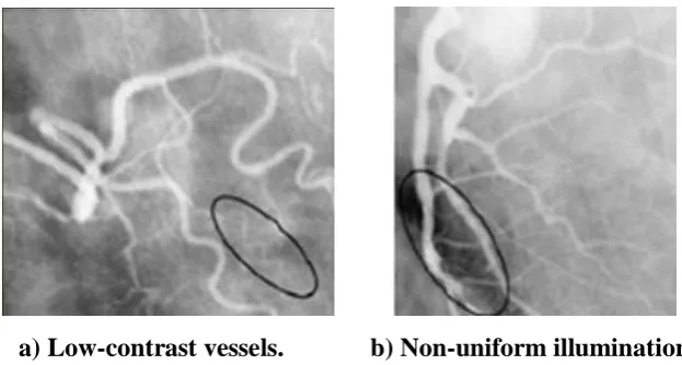
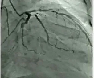
a) Low-contrast vessels. b) Non-uniform illumination. Fig. 1: Angiography images with typical hindrances.
Thick vessels have more difference to noise proportions when compared with little-limited
ones, because of the solid nearness of blood and complexity specialists in the vessels in
Figure 1a. Non-uniform brightening, as appeared in Figure 1b,is one of the major sources of
angiography image degradation and is also a hindrance for accurate reconstruction, because it
is likely to make an individual vessel break into several segments. In this work partitioned the
image processing techniques utilized as a part of coronary angiography into two principle
classifications: (1) vessel improvement (2) vessel segmentation. These classes are
additionally isolated into subcategories. As indicated by the modern programmed PC helped
methods are still a long way from giving an exact precise spatial representation of the vessel
tree.
In the remaining part of the work, Section II describes the research background. Section III
familiarizes our proposed vessel blockage detection method, and Section IV organizes the
simulation results of the proposed system. In chapter V presents conclusion and paths to
future work.
2. LITERATURE SURVEY
There is a lot of literature available for the techniques, proposed for vessel extraction from
coronary angiogram images Coronary Artery Disease (CAD) is a standout amongst the fatal
ailment for human beings,[1] which has been identified as one of the real reasons for death
overall.[2] In blood vessels, stenosis happens because of plaque gathering in the internal
dividers of the courses. Consequently, these vessels turn out to be thin after a particular
period.[3,4] To analyze CAD, medicinal experts perform X-ray angiography as a "best quality
disadvantage of X-ray angiography is that 3D structures are covered in the anticipated 2D
images.[8,11] This 3D to 2D transformation brings about the loss of angiographic images. This
misfortune additionally causes the foreshortening and covering of vasculatures.
Subsequently, the anatomical structure of the coronary conduits is hard to distinguish, and
maladies are hard to evaluate.
In,[12] connected image combination strategy for the discovery of Coronary Artery vessels in
Coronary Angiography. Two different methodology images were consolidated utilizing pixel
image combination procedure to upgrade the inside territory of the coronary heart images.
In[13] built up a system to recognize and analyze the coronary illness utilizing encourage
forward back spread neural networks. The creators accomplished 85% acknowledgment rate
for their proposed algorithm. In[14] proposed an algorithm for perceiving the coronary hallway
malady using picture taking care of frameworks. The makers achieved affectability around
91% and specificity around 92% for their proposed algorithm over the game plan of 475
cardio images. The proposed algorithm was attempted on both myocardial perfusion imaging
and tomographic angiography. In[15] used Support Vector Machine (SVM) classifier to detect
the coronary heart disease. This work diagnosed the coronary heart disease into low, medium,
high and severe using angiogram images. The authors achieved 90% recall rate, 82.143%
precision, 86.793% F-measure and 60% Overall accuracy. In[16] proposed minimum
path-based area growing algorithm for the detection of coronary vessels in angiogram images. The
authors used orientated profile symmetry information technique as the extracted features for
the detection process of vessels in coronary images. In[17] proposed a framework to detect and
segment coronary vessels in coronary images using Multi-scale Texture Dictionary
algorithm. The texture highlights were removed from the coronary heart pictures to recognize
the vessels in a mechanized way.These works deal with different strategies for approaching
the segmentation task, including image-driven algorithms was proposed in,[18,19] and,[20]
probabilistic atlases is discussed in[21] and.[22]
In[23] proposed a methodology for the classification of Coronary Heart Disease Patients from
Non-coronary Heart Disease. The prediction training set accuracy was 96.86%, and testing
set accuracy was 78.18%. The authors achieved cross-validation prediction accuracy of
92.67%. It used decimation-free directional filter bank technique to enhance the vessels in
to the image by the block-by-block manner and energy level was computed for each block.
The pixels were replaced by their calculated energy values which produced enhanced images.
In[24] proposes an automated method of blockage segmentation from coronary angiogram
images using Adaptive Neuro Fuzzy Inference System (ANFIS) classifier the proposed
method achieves 95.9% sensitivity, 99.9% specificity and 99.9% accuracy for blockage
vessel pixel detection. In[25] proposes an automated method of blockage segmentation from
coronary angiogram images using Coactive Adaptive Neuro Fuzzy Inference System
(CANFIS) classifier.The CANFIS method achieves 99.76% sensitivity, 99.9% specificity and
99.9% accuracy for blockage vessel pixel detection.
It is found that the system has an issue of vessel blockage detection in all the above
techniques. So another moved strategy has projected in this research. This work reveals how
to discover the determination of vessel blockage detection in various methods. In this work to
instead propose a novel request strategy that consolidations are gathering correctly than past
procedures
3. MATERIALS AND METHOD
Lots of researchers have been performed for the segmentation of normal and strange tissues
in coronary angiogram heart pictures. Segmentation of medical image is a testing assignment
because of the multifaceted nature of the images, and additionally to the absence of models of
the life systems that satisfactorily catch the conceivable disfigurements in each structure.
Heart tissue is an extraordinarily complex structure, and its segmentation is a basic advance
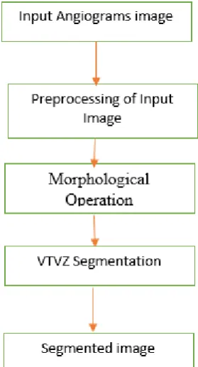
for our proposed technique. In our proposed technique comprises of four stages specifically
preprocessing, segmentation, feature extraction, and classification. The proposed procedure
Fig 2: Block Diagram of proposed System. 3.1 Pre-Processing Using Adaptive Temporal Filter
Pre-processing systems, for the most part, went for the improvement of the image without
adjusting the data contained in an image. In this work execute a pre-processing technique for
the change of coronary heart image without modifying image substance and make reasonable
for additionally processing. The pre-processing method comprises of deciding the pixels
outside the aperture that are neighbors to pixels inside the opening and supplanting every one
of their esteems with the mean estimation of their neighbors inside the aperture. In this work,
an adaptive temporal filter is utilized for preprocessing.In adaptive temporal filtering, the
result of the image is weighted with an aggregate of information pixels. The estimation of a
filter sub-image is alluded as coefficients, instead of pixels. Adaptive filtering term is the
filtering tasks that are performed straightforwardly on the pixels of an image. The filter is a
cover of weights masterminded in a rectangular example. The procedure is one of sliding the
mask and performing increase and collect activity on the pixels secured by the mask.
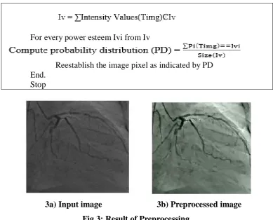
3.1.1 Adaptive Temporal Filtering Algorithm Steps Methodology: Adaptive Temporal Filter Info: Coronary heart image
Output: Preprocessed image Pimg. Begin
Catch Image Img.
Preprocess the image Img.
For every power esteem Ivi from Iv
Reestablish the image pixel as indicated by PD End.
Stop
3a) Input image 3b) Preprocessed image Fig 3: Result of Preprocessing.
The above figure 2 indicates result correlation of input and preprocessed image utilizing
adaptive temporal filtering. When contrasted with Input image the preprocessed image
demonstrates the better view without commotion. The above-talked algorithm plays out the
histogram leveling of the information image caught through the gadget and enhances the
nature of information image acquired.
3.2 Versatile Threshold Vector Zone (VTVZ) Vessel Blockage Segmentation
The preprocessed image is switched and prepared by the morphological activity. A nearby
morphological operation is utilized for the segmentation reason. In this work, adaptable
threshold Vector Zone Segmentation procedure is being used for blood vessel location. The
nearby morphological methodology is a procedure that morphological expansion took after
by erosion.The essential factor or component which is essential for closing activity is
organizing segment. Two sorts of arranging components are available, initial one is level
organizing component and the second one is non-level organizing component. The VTVZ
Segmentation relies upon the pixel esteem, choice level and features of the angiogram image.
The VTVZ has been utilized to expand the perception of differentiation of the low and high
3.2.1 Algorithm
Step1: Resampling of the angiogram image with the motivation behind conveying its measurement to the measurement of the coronary image.
Step2: Geometric and radiometric redress of the info images. Step3: Contrast and diagram adjustment of the information images. Step4: VTVZ segmentation.
Step5: Converting of the acquired angiogram image ([ANF]_MOD) into the HSV shading model ([HSV]_MUL).
Step6: Altering the R, G and B outfits of the heart image into HSV instruments ([HSV_MUL).
Step7: The subsequent stage of development of another image is substitution the brilliance part V of a [HSV]_MUL image with the splendor segment of [HSV]_MOD and changing over the outcome from the HSV shading model to the RGB shading model.
Step8: Decorrelation of the new heart image as per the picked algorithm:
Step9: A pyramidal deterioration with the given wavelet premise to the given disintegration level (L) of the shine channel (X)
Step 10: A pyramidal deterioration with the given wavelet premise to the given disintegration level (L) of the coronary heart image
Where
= approximating wavelet components of the new vessel blockage image.
= detailing wavelet components of the new vessel blockage image.
In the wake of segmentation, the blood vessels of a coronary angiogram are removed. To
express the usage procedure of the proposed strategy unmistakably, the outline of the
proposed system for the segmentation of angiograms appears in figure 4
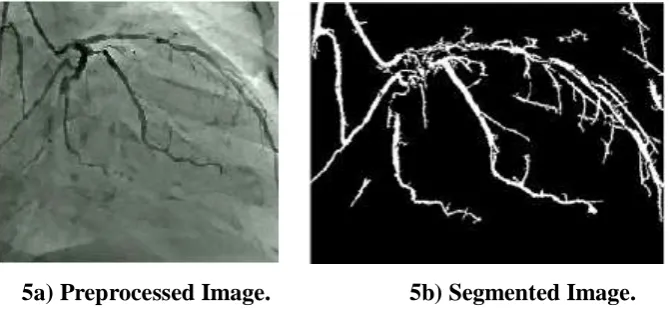
Fig. 4: Morphological Processing Steps. 3.2.2 Morphological Processing Steps
Step 1: The input coronary angiogram image is perused.
Step 2: For preprocessing Top morphological Hat task is done on the info image.
Step 3: Morphological Closing task, i.e., Dilation and erosion are connected to the image with the circle formed organizing components.
5a) Preprocessed Image. 5b) Segmented Image. Fig. 5: Segmentation Result.
Figure 4 shows the segmentation result utilizing VTVZ method. The segmented outcomes
make the centroid in light of the potential estimation of the data focuses.as contrasted with
other customary innovation the VTVZ segmentation gives a superior precision.
3.3 Feature Extraction: Diagonis Zone Unsupervised Vector
After segmentation, the Feature esteems are separated given the closeness measure of every
pixel arrangement at various zones. The inspiration for utilizing numerous zones (inside,
limit, and encompassing lesion) and the picked features depends on the way that the
encompassing shortcoming attributes. The fault is comprised of a thick improving edge; the
fault border is characterized as unpredictable or poorly marked edges.
The feature is separating the coronary heart images and recognizing the fault as the focal
zone; the proposed framework will isolate the divided lesion area vector (V0) into two ROIs
(where V0= V1 V2). V1 is the region of the fault and characterized as a zone where over
70% of the fault pixels has situated inside the board. Something else, the image has
considered a fault region (V2).
3.3.1 Algorithm
Input: Segmented Feature Values (SFv), segmented lesion (SFV0).
Procedure
By utilizing diagnose zone unsupervised vector 12 features were removed for proposed
strategy, few of them are portrayed as takes after.
3.3.2 Energy
The vitality is the measure of consistency between the pixels run = [0, 1].The consistency of
the pixel has communicated
3.3.3 Contrast
Contrast is the measure of the distinction in luminance to make protest recognizable.
3.3.4 Correlation
Correlation is the measure of the connection between the neighbor pixels.Range = [-1, 1].
3.3.5 Homogeneity
The homogeneity measures of the closeness of the component appropriation in finding zone
unsupervised vector. Range= [0,1]
Where I, j are pixel and p (I, j) is the pixel esteem. Along these lines to arrange the image as
ordinary or unusual, the middle of the images are gained, preprocessed, the element
extricated by utilizing diagnosis zone unsupervised vector strategy.
3.4 Intensive Pragmatic Blossoms Classification
The classifier is utilized to order blockage influenced zone composes from unique images.
For straightforwardness, solid augmented blossom classifier is used here. It takes set of
pictures and predicts for each information image has a place with which of the two classes of
tissue influenced and without a controlled image. The motivation behind IPB is to make
angiogram images to isolate two classes with a most extreme hole between them. In our
proposed framework, an output of segmentation is given as a contribution to concentrated
businesslike blooms classifier which takes preparing information, testing information and
gathering data which groups the given input image.
Algorithm
Input: Input Image Output: Class S Start
Area S= Perform vector threshold based absorption Estimation
Image Img = Preprocess Image from datasets (Adaptive Temporal Filter) Image Img = segmentation Image (Versatile Super threshold Vector Zone) KSDF = Intensive Pragmatic Blossoms classification
If KSDF<Th1> then Area A = S
Else KSDF <Th2> then Area B = S
Else if KSDF <Thn> then Area N = S
The classification strategy initially plays out the vector-based vessel blockage image
estimation and afterward catches the image to preprocess and segment the image. At that
point, the procedure processes the classification algorithm to arrange the outcomes given
beneath.
6a) Input Image.
6b) Segmented Image.
6c) Blockage Identified Image. Fig. 6: Classification Result.
The above figure 6 demonstrates the grouping consequences of blockage recognizable proof
in the proposed framework. The execution of the grouping has assessed concerning order
accompanying conventions.TP (True Positive) =Positive units named positive. True Negative
(TN) = Negative examples delegated negative. FP (False Positive) = Negative examples
delegated positive. FN (False Negative) = Positive examples named negative.
Precision: It is the proportion of number of positive examples accurately ordered to the aggregate number of tests in a class.
Recall: It is the proportion of some positive examples effectively arranged to the aggregate number of tests named positive.
F-measure: It is the consonant mean of accuracy and review given by the condition underneath.
Accuracy: It is the aggregate number of tests accurately ordered to the aggregate number of tests delegated given by the condition beneath.
The fault classification is performed using both VTVZ segmentation and diagnosis zone
unsupervised vector feature extraction technique. The method first performs the region based
coronary image estimation and then captures the image to preprocess and segment the image.
Then the method computes the classification algorithm to classify the fault area.
4. RESULTS AND DISCUSSION
The proposed algorithm is tried on York University open access dataset which contains 7000
typical coronary heart pictures and 980 anomalous coronary heart pictures. In this work, two
separate datasets are built from this open access dataset as preparing and testing by
extricating 350 typical and 300 unusual coronary pictures from this open access dataset.
MATLAB R2017a is utilized as a part of this work to reenact the proposed calculation, and
Intel Pentium Core-2 Duo 2.4GHz with 2 GB RAM is utilized as the equipment particulars.
Predictive Value (NPV) and Accuracy (Acc) are utilized to assess the execution of the
proposed technique.
Though TR_PO is True Positive which speaks to the accurately distinguished blockage vessel
pixel, TR_NE is the True Negative which speaks to the recognized efficiently non-blockage
vessel pixel, FA_PO speaks to the dishonestly identified blockage vessel pixel, and FA_NE
speaks to the erroneously distinguished non-blockage vessel pixel. The possibility table is
built by relating TR_PO, TR_NE, FA_PO, and FA_NE, as delineated in Table 1 utilizing
adaptable threshold vector zone vessel blockage segmentation.
Table 1: Performance results of the proposed methodology.
The morphological parameters as border, zone, width, and stature, are assessed on the
portioned tumor zone in cervical pictures. The border computation of the portioned growth
zone in a cervical picture depends on the span 'r,' and the morphological parameter for the
computation of edge is given by the accompanying condition as,
Table 2: Morphological analysis.
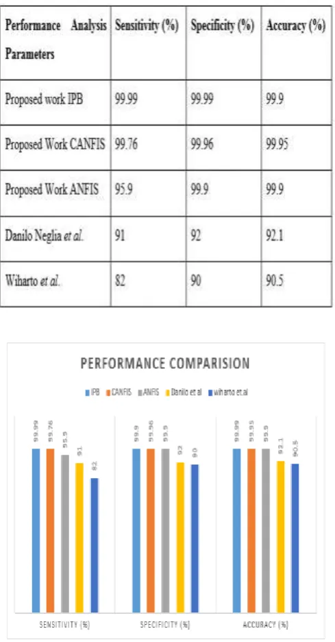
Table 3: Performance comparison of proposed and conventional methodologies.
Table 3 and figure 7 demonstrates the correlations of the proposed system for blockage vessel
location in coronary heart pictures with traditional strategies. The proposed IPB strategy
accomplishes 99.99% affectability, 99.99% specificity and 99.99% precision for blockage
vessel pixel discovery. The customary techniques as (of) Danilo Neglia et al. (2015)
accomplished 91% affectability and 92% specificity while of Wiharto et al. (2015) got 82 %
affectability, 90 % specificity. It is seen from the table that the proposed blockage vessel
pixel discovery technique is better than the customary strategies.
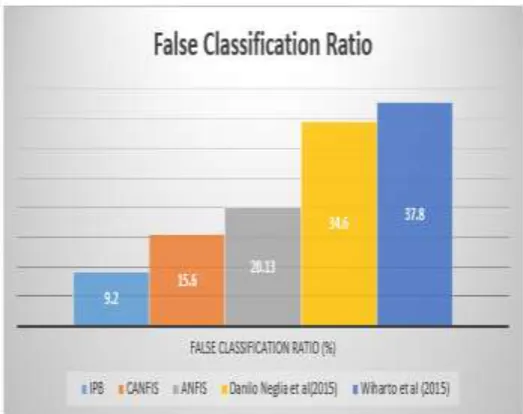
Figure 8: Comparison of various methods in a false ratio.
Figure 8 demonstrates the examination of results obtained from various strategies for false
classification. When contrasted with all systems the proposed methodology has less mean
rate which is inconsequential.
Figure 9 demonstrates the correlation of different strategies on time multifaceted nature, and
it portrays that the proposed strategy has less time complexity than others. From the above
examination played out, the proposed technique has been assessed with different parameters
and has delivered helpful outcomes with all elements of optical properties and order.
5. CONCLUSION
The vessel blockage image segmentation has been discussed in this work. In this work, an
adaptive temporal filter is associated with smooth the picture by emptying the commotion.
Adaptable threshold clustering technique is used from the preprocessed picture, where the
gathering of objects is performed. In this work, computer-aided automatic detection of
blockage in coronary heart images using IPB and compared with ANFIS and CANFIS
classifier is proposed. As compared to other conventional technique the proposed IPB
technique produces an advantageous result. The spatial domain coronary heart image is
converted into a multi-resolution image using temporal filter transform after preprocessing
the image. Then, features are extracted from this multiresolution image and these features are
trained and classify using IPB classifier to detect the coronary heart image which contains
blockage. The proposed method achieves 99.98% sensitivity, 99.975% specificity and
99.99% accuracy for blockage pixel detection.
REFERENCES
1. M. G. Tsipouras, T. P. Exarchos, D. I. Fotiadis, A. P. Kotsia, K. V. Vakalis, and K. K.
Naka et al., “Automated diagnosis of coronary artery disease based on data mining and fuzzy modeling,” IEEE Trans. Inf. Technol. Biomed., 2008; 12: 447–458.
2. S. Josephson, S. Bryant, H. Mak, S. Johnston, W. Dillon, and W. Smith, “Evaluation of
carotid stenosis using CT angiography in the initial evaluation of stroke and TIA,”
Neurology, 2004; 63: 457–460.
3. G. T. Lau, L. J. Ridley, M. C. Schieb, D. B. Brieger, S. B. Freedman, and L. A. Wong et
al., “Coronary artery stenoses: Detection with calcium scoring, CT angiography, and both
methods combined 1,” Radiology, 2005; 235: 415–422.
4. M. A. Cordeiro, A. C. Lardo, M. S. Brito, M. A. R. Neto, M. H. Siqueira, and J. R. Parga
et al., “CT angiography in highly calcified arteries: 2D manual vs. modified automated
5. A. C. Dumay, J. Reiber, and J. Gerbrands, “Determination of optimal angiographic
viewing angles: Basic principles and evaluation study,” IEEE Trans. Med. Imag., 1994;
13: 13–24.
6. S. Mavrogeni, G. Papadopoulos, M. Douskou, S. Kaklis, I. Seimenis, and P. Baras et al.,
“Magnetic resonance angiography isequivalent to X-Ray coronary angiography for the evaluation of coronary arteries in Kawasaki disease,” J. Amer. College Cardiol., 2004;
43: 649–652.
7. U. Solzbach, U. Oser, M. Rombach, H. Wollschläger, and H. Just, “Optimum
angiographic visualization of coronary segments using computer-aided 3D-reconstruction
from biplane views,” Comput. Biomed. Res., 1994; 27: 178–198.
8. Y. Sato, T. Araki, M. Hanayama, H. Naito, and S. Tamura, “A viewpoint determination
system for stenosis diagnosis and quantification in coronary angiographic image
acquisition,” IEEE Trans. Med. Imag., 1998; 17: 121–137.
9. S. Sakamoto, Y. Kiura, M. Shibukawa, S. Ohba, K. Arita, and K. Kurisu, “Subtracted 3D
CT angiography for evaluation of internal carotid artery aneurysms: Comparison with
conventional digital subtraction angiography,” Amer. J. Neuroradiol., 2006; 27:
1332–1337.
10.G. Tipper, “Detection and evaluation of intracranial aneurysms with 16-row multislice
CT angiography,” Clinical Radiol., 2005; 60: 565–572.
11.D. E. Hyde, A. J. Fox, I. Gulka, P. Kalapos, D. H. Lee, and D. M. Pelz et al., “Internal
carotid artery stenosis measurement comparison of 3D computed rotational angiography
and conventional digital subtraction angiography,” Stroke, 2004; 35: 2776–2781.
12.Julien Plessis, KarineWarinFresse, Zachary Cahouch, ThibautManigold. Value of Image
Fusion in Coronary Angiography for the Detection of Coronary Artery Bypass Grafts.
Journal of the American Heart Association, 2016; 5(6): 245–256.
13.Ebenezer ObaloluwaOlaniyi, OyebadeKayodeOyedotun. Heart Diseases Diagnosis Using
Neural Networks Arbitration. International Journal of Intelligent Systems and
Applications, 2015; 12: 75-82.
14.Danilo Neglia, Daniele Rovai, Chiara Caselli, MikkoPietila. Detection of Significant
Coronary Artery Disease by Noninvasive Anatomical and Functional Imaging.
Cardiovascular Imaging, 2015; 8(3): 1–10.
15.Wiharto, HariKusnanto, Herianto. Performance Analysis of Multiclass Support Vector
16.Qing Cao, Yang Chen, Guanyu Yang, Christine Toumoulin, HuazhongShu, LiminLuo.
Coronary vessel extraction method using an improved minimum path-based area
growing. 6th International Conference on Biomedical Engineering and Informatics, 2013:
127 – 131.
17.Ali Zifan, Brian E. Chapman Automatic Detection of Coronary Vessels Using Multi-scale
Texture Dictionaries IEEE Second International Conference on Healthcare Informatics,
Imaging, and Systems Biology, 2012; 1–15.
18.S Huang, J Liu, LC Lee, SK Venkatesh, LLS Teo, C Au, WL Nowinski, An image-based
comprehensive approach for automatic segmentation of left ventricle from cardiac short
axis cine MRI images. J. Dig. Imaging, 2010: 1–11.
19.AB Redwood, JJ Richard, A Robb, Semiautomatic segmentation of the heart from CT
images based on intensity and morphological features. In Proc SPIE, 2005; 5747:
1373–1719
20.JP Morin, C Desrosiers, L Duong, Image segmentation using random-walks on the
histogram. In. Proc. SPIE, 2012; 83(14): 1–8.
21.M Lorenzo-Valdes, GI Sanchez-Ortiz, AG Elkington, RH Mohiaddin, D Rueckert,
Segmentation of 4D cardiac MR images using a probabilistic atlas and the EM algorithm.
Med. Image Anal, 2004; 8: 255–265.
22.Isgum, Multi-Atlas-based segmentation with local decision fusion – Application to
cardiac and aortic segmentation in CT scans. IEEE Trans. Med. Imaging, 2009; 28(7):
1000–1010.
23.Hongzong, S, Tao, W, Xiaojun, Y, Huanxiang, L, Zhide, H, Mancang, L, BoTao, F.
Support Vector Machines Classification for Discriminating Coronary Heart Disease
Patients from Non-coronary Heart Disease. West Indian Medical Journal, 2007;
56(5): 451–457.
24.P. Rajesh Kumar and K. Murugesan “An Automated Screening System for Vessel
Blockage Segmentation in Coronary Angiogram Images Using ANFIS Classifier” Int. J.
Bio-Inspired Computation, 2017.
25.Rajesh Kumar, P & Murugesan, K. “Computer Aided Segmentation of Blockages in
Coronary Heart Images Using Canfis Classifier”, Wireless Personal Communications,