Needle-Entry Angle for Lumbar Puncture in Children
as Determined by Using Ultrasonography
WHAT’S KNOWN ON THIS SUBJECT: Ultrasound has been used to characterize the depth and width of the interspinous space. A study by Abo et al found that the maximum width of the interspinous space is in the sitting position with flexed hips.
WHAT THIS STUDY ADDS: This study further characterizes the interspinous space by describing the optimal angle at which to point the needle. We found that the optimal angle for performing a lumbar puncture is⬃50° in infants and 60° in children aged 1 to 12 years.
abstract
OBJECTIVE:The purpose of this study was to evaluate the angle for performing lumbar punctures in children aged 0 to 12 years. We hy-pothesized that the angle changes for different stages of development.
METHODS:Children aged 0 to 12 years who presented to the Yale–New Haven Children’s Hospital at a low-acuity triage level, in need of a lumbar puncture, their accompanying siblings and authors’ children were eligible for the study. Subjects in 3 age groups were recruited and grouped as follows: group 1, 0 to 12 months; group 2, 12 to 36 months; and group 3, 3 to 12 years. Ultrasound images of the L3-L4 and L4-L5 lumbar space were taken with subjects in the lateral recumbent and sitting positions. The angle from the interspinous space to the skin was measured.
RESULTS:Thirty-six subjects were included. The mean angles in the lateral recumbent and sitting positions were group 1, 47.8° (SD: 8.2) and 51.1° (SD: 8.5), respectively; group 2, 58.8° (SD: 6.8) and 59.6° (SD: 5.5); and group 3, 60.5° (SD: 6.6) and 61.9° (SD: 4.0). The results of group 1 were significantly different from those of groups 2 or 3 in both posi-tions (lateral recumbentP⫽.00526 and 0.00160; sittingP⫽.0499 and .00282).
CONCLUSIONS:The angle for lumbar puncture was more acute for infants than for older children in this study. Future studies should assess the difference in success rates of lumbar punctures when cli-nicians have knowledge of these angles. Pediatrics 2011;127: e921–e926
AUTHORS:Rebecca Elaine Bruccoleri, MDaand Lei Chen,
MDb
aDepartment of Emergency Medicine, Yale–New Haven Medical Center, New Haven, Connecticut; andbSection of Pediatric Emergency Medicine, Department of Pediatrics, Yale School of Medicine, New Haven, Connecticut
KEY WORDS
lumbar puncture, ultrasound, angle, interspinous space
ABBREVIATION
LP—lumbar puncture
Dr Bruccoleri acquired data, performed the statistical analysis with the assistance of Dr Menjoge (as noted in the
Acknowledgments), and drafted and edited the manuscript, and Dr Chen conceived the idea for the study, obtained institutional review board approval, acquired data, and revised and edited the manuscript. Drs Bruccoleri and Chen both approved the article that was submitted.
www.pediatrics.org/cgi/doi/10.1542/peds.2010-2511 doi:10.1542/peds.2010-2511
Accepted for publication Jan 7, 2011
Address correspondence to Rebecca Elaine Bruccoleri, MD, Department of Emergency Medicine, Yale-New Haven Medical Center, 20 York Street, New Haven, CT 06510. E-mail: rebecca. [email protected]
PEDIATRICS (ISSN Numbers: Print, 0031-4005; Online, 1098-4275). Copyright © 2011 by the American Academy of Pediatrics
FINANCIAL DISCLOSURE:Dr Bruccoleri owns 16 of 394.6 million outstanding shares in Stryker, which is involved in making ultrasound equipment. Dr Chen has indicated he has no financial relationships relevant to this article to disclose.
Funded by the National Institutes of Health (NIH).
sessment of patients with suspected sepsis and meningitis. It involves in-serting a needle into the interspinous space of the lower lumbar vertebrae into the subarachnoid space to collect a sample of cerebrospinal fluid. If the needle is not inserted along the inter-spinous space, it will encounter bone and have to be withdrawn and redi-rected. An LP is considered successful when a sufficient amount of nonbloody cerebrospinal fluid is collected. Failed attempts include those that contain peripheral blood or no spinal fluid at all. Success rates of LPs vary, with re-ported rates ranging from 45% to 54.2%.1,2This procedure is especially challenging in infants because the tar-get space is small.3 Furthermore, re-spiratory status can be compromised with positioning.4,5It is also difficult in obese patients, as the bony landmarks are often obscured by subcutaneous fat. Repeated attempts of the poten-tially painful procedure are often re-quired and have been shown to in-crease the incidence of postdural puncture headache in adult patients.6 Failed LPs can lead to unnecessary hospitalizations and prolonged use of antibiotics.7Although there have been prospective studies concerning the variables associated with successful LPs (eg, resident experience, patient position, draping), knowledge of the angle of the LP needle in relation to the skin may improve the success rates of this difficult procedure.8,9
Ultrasound has been used to guide LPs. Specifically, it has been used to mea-sure the depth to the epidural space, with high correlation to actual val-ues.10,11In addition, it has been shown to be useful in pediatrics in guiding LPs.12However, musculoskeletal appli-cations using ultrasound in a clinical setting is a relatively new field in
pedi-the width of pedi-the interspinous space was the largest in the sitting position with the hips flexed. Using ultrasound to place and identify epidural cathe-ters has been addressed in pediatric anesthesia literature.13,14 A study by Ferre et al15used static ultrasound im-ages in adults to identify landmarks but did not describe the angles ob-tained in their images. However, to the best of our knowledge no studies have determined the relationship of the an-gle of needle entry in children to pa-tient factors such as age. This knowl-edge could provide clinicians with further guidance in deciding how to orient the needle. The goal of our study was to determine and explore the ef-fect of position and age on the angle between the interspinous spaces and the skin. We hypothesized that the an-gle changes in different stages of development.
METHODS
This was a prospective observational study of a convenience sample of chil-dren aged 0 to 12 years. Informed con-sent was obtained through a written form in English and verbal explanation of the risks and benefits of the study. The study protocol was approved by the Yale Human Investigation
Commit-months; and 3 to 12 years. An LP was not required as part of the subject’s treatment plan.
Inclusion criteria were all patients who were categorized as triage level III or IV at the Pediatric Emergency De-partment of Yale–New Haven Chil-dren’s Hospital and children who re-quired an LP as part of their treatment plan. There are no specific criteria for triage level III or IV, but patients’ condi-tions are considered to be nonurgent. In addition, siblings of patients and children of authors were also eligible. Exclusion criteria included the inability of the parents to speak or read Eng-lish, any wounds or obvious trauma to the lumbar spine, skin breakdown, rash in the region of the L2 to S1 verte-brae, or if the subject’s clinical condi-tion did not allow for the patient to as-sume the lateral recumbent or sitting position.
After written signed consent was ob-tained, the subject’s birth date, date of study, weight, and gender were re-corded. Ultrasound images were ob-tained using a SonoSite 180 (Sonosite, Inc, Bothell, WA) ultrasound with an L38 high-frequency linear probe using fre-quencies of 8 to 12 MHz. Static images were taken in both the lateral recumbent
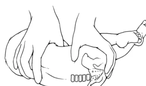
FIGURE 1
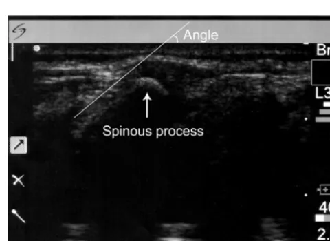
and sitting positions (Figs 1 and 2). Staff or parents assisted in maintaining the subject’s position as if the patient were actually to receive an LP. The legs were positioned with the hips flexed. The probe was covered with ultra-sound gel and placed between the pos-terior superior iliac crests along the vertebrae that correlate to the L3-L4 and L4-L5 interspinous spaces. The pa-tient was then positioned in the alter-nate position, and images were taken again. Each static image was printed and a line with a ruler was drawn from the skin to the interspinous space along the spinous process. Given that the skin was flush with the probe, the outer edge of the image was consid-ered the skin and the horizontal line of the protractor was aligned with this edge. The angle between the spinous process and the skin was then mea-sured (Figs 3 and 4).
The mean, median, and SD statistics were computed by using Excel 2007 (Microsoft, Redmond, WA) and R 2.8.1 (R Foundation for Statistical Comput-ing, Vienna, Austria).16 Because the sample sizes were small, the nonpara-metric tests of the Kruskal-Wallis test and Wilcoxon rank tests were used.
These tests were performed by using the statistical program R 2.8.1. Linear and power regressions were gener-ated by using Excel 2003 and 2007 editions.
RESULTS
Thirty-seven subjects were enrolled in the study from June 16, 2006, until De-cember 29, 2007. Of these patients, 36 had sufficient data that could be in-cluded in the study. One subject’s data were lost because of lack of disk space in the ultrasound machine. Table 1 presents the characteristics of the study subjects. Because of a lack of pa-tient cooperation, 4 papa-tients in the 0- to
FIGURE 2 Sitting position.
FIGURE 3
Angle measured during the study.
FIGURE 4
Sample image showing spinous processes.
12-month age group could only be im-aged in the lateral recumbent position; the remaining 9 were imaged in both positions. The ligamentum flavum was identified in all subjects except for 1. Regardless, the slope of the spinous process in relation to the skin was used to guide the lines drawn to mea-sure the angle.
For all subjects, the mean angle was 55.3° (SD: 9.2) for the lateral recum-bent position and 58.0° (SD: 7.5) for the sitting position. The mean angles in the lateral recumbent position were as fol-lows: group 1, 47.8° (SD: 8.2); group 2, 58.8° (SD: 6.8); and group 3, 60.5° (SD: 6.6) (Fig 5). The mean angles for the sitting position were as follows: group 1, 51.1° (SD: 8.5); group 2, 59.6° (SD: 5.5); and group 3, 61.9° (SD: 4.0) (Fig 6).
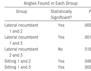
There were significant difference be-tween groups 1 and 2 and groups 1 and 3 and no significant difference be-tween groups 2 and 3 in both positions (Table 2.) We also compared the angles between the lateral recumbent posi-tion and the sitting posiposi-tion to deter-mine if there was a significant differ-ence between the angles in each
FIGURE 5
Angle versus age in the lateral recumbent position.
FIGURE 6
Angle versus age in the sitting position.
TABLE 2 Statistical Significance Between Angles Found in Each Group Group Statistically
Significant?
P
Lateral recumbent 1 and 2
Yes .00526
Lateral recumbent 1 and 3
Yes .00160
Lateral recumbent 2 and 3
No .510
Sitting 1 and 2 Yes .0499 Sitting 1 and 3 Yes .00282 Sitting 2 and 3 No .391
Male Female Age,
Mean (SD), d
Weight, Mean (SD), kg
Age, Mean (SD), d
Weight, Mean (SD), kg
1 0–12 mo 13 9 4 117 (107) 5.7 (2.3) 122 (110) 6.1 (2.5)
2 12–36 mo 10 8 2 722 (233) 12.4 (1.6) 722 (233) 12.4 (1.6)
3 3–12 y 13 6 7 2210 (778) 22.5 (6.9) 2281 (861) 24.3 (9.1)
position. Using the Wilcoxon rank tests, the comparison between the positions for groups 1, 2, and 3 showed no signif-icant differences (group 1: P ⫽ 1; group 2: P⫽ .610; and group 3:P⫽
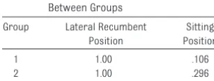
.657). Overall, there was no significant difference between all subjects in ei-ther position (P ⫽.565). These com-parisons found that the angle did not significantly change regardless of whether the patient was in the lateral recumbent or the sitting position. The regression analyses for angle versus weight resulted in power equations for the best fit curves withR2⫽0.418 and 0.258 for the lateral recumbent and sit-ting positions, respectively. In terms of gender and angle, there were no sig-nificant differences between girls and boys in either position in all individual groups (Table 3).
DISCUSSION
In this pilot study, we reported statisti-cally significant differences in the angle of needle placement in infants compared with older children. Fur-thermore, this difference applied to both common positions used for per-forming LPs. This study adds to the cur-rent literature in studying factors that could improve success rates of LPs. Considering that there are numerous pediatric LPs performed annually, it is conceivable that the results of this study could lead to increased success rates of LPs in children. Other factors that have been researched in the suc-cess rates of LPs and entering the epi-dural space (in the case of anesthesia) include the size and design of the nee-dle, advancing the needle with the sty-let in place, use of local anesthetic, and
patient movement.8,17,18 In 1996, Kokki and Hendolin17 found higher success rates in spinal anesthesia when using a 25-gauge needle instead of a 29-gauge needle as a successful spi-nal blockade. Furthermore, although Kokki et al18found no difference in the success rates of a spinal block using cutting point or pencil point needles, there was a significant difference (P⫽
.002) with the need for repeated skin puncture. They found that the cutting point needle required less repeated punctures. Nigrovic et al9 found that young age (defined as ⬍3 months), less physician experience with LPs, lack of anesthetic, patient movement, and advancement of the needle with a stylet in place were risk factors for un-successful or traumatic LPs (red blood cell count ⬎ 500 cells per L). Of those, anesthetic use, patient move-ment, and advancement of the needle without the stylet in place are the most modifiable.
In terms of the ultrasound and radio-graphic literature, in 1 study the au-thors derived an equation for calculat-ing the depth on the basis of the length of the needle inserted, with the angle of the insertion measured with a ster-ile protractor.19 However, this study did not report the angles measured. Our study adds to this characterization by giving a mean angle to place the needle and further guidance for clini-cians performing LPs. Although the use of ultrasound in pediatric emergency departments is increasing and meth-ods for using ultrasound to perform LPs have been described for adults, our study may help the clinician with-out access to ultrasound for guided procedures by providing the mean an-gles of needle insertion based on pa-tient ages.20–22
Limitations of our study include the small sample size and the fact that the study did not show how the an-gles found in static images affected
success rates. Furthermore, the study was performed with only 1 in-vestigator (Dr Bruccoleri) measur-ing the angles; therefore, we were unable to test interrater reliability. Last, we did not evaluate subject height and its relationship to the an-gle of the interspinous space. There have been studies using height and weight together to predict needle depth to the interspinous space.23,24 Although we found a poor correla-tion of needle angle to weight, there could be a relationship to height and needle angle or height and weight and needle angle.
Future study designs may include ran-domized controlled trials to study the success of LPs with providers who are given the knowledge of the angles in this study and providers who are not. Another randomized control trial de-sign would be to study the success rates of LPs between clinicians who are given still ultrasound images of a patient’s lumbar space and those who are not. In addition, further studies could explore whether the child’s abil-ity to walk upright affects the angle of the interspinous space.
CONCLUSIONS
The angle for LP as determined by ultra-sonography in this study was ⬃50° in infants and 60° in children aged 1 to 12 years in both the lateral recumbent and sitting positions. The differences in these angles between ages were statistically and clinically significant. Further studies need to be performed to determine if the results of this anatomic study lead to im-proved outcomes of LPs.
ACKNOWLEDGMENTS
This study was funded by the Depart-ment of Pediatrics at Yale School of Medicine. The Office of Student Re-search funded the Summer ReRe-search Fellowship program during 2006. This work was supported in part by grant KL2 RR024138 from the Clinical TABLE 3 PValues of Gender Comparisons
Between Groups Group Lateral Recumbent
Position
Sitting Position
1 1.00 .106
2 1.00 .296
3 .277 .222
the National Institutes of Health and NIH Roadmap for Medical Research.
Pediatric Emergency Department of Yale–New Haven Children’s Hospital
gency Department for use of the SonoSite 180 machine.
REFERENCES
1. Pinheiro JM, Furdon S, Ochoa LF. Role of lo-cal anesthesia during lumbar puncture in neonates.Pediatrics. 1993;91(2):379 –382 2. Schreiner RL, Kleiman MB. Incidence and
ef-fect of traumatic lumbar puncture in the neonate.Dev Med Child Neurol. 1979;21(4): 483– 487
3. Abo A, Chen L, Johnston P, Santucci K. Posi-tioning for lumbar puncture in children evaluated by bedside ultrasound. Pediat-rics. 2010;125(5). Available at: www. pediatrics.org/cgi/content/full/125/5/e1149 4. Gleason CA, Martin RJ, Anderson JV, Carlo
WA, Sanniti KJ, Fanaroff AA. Optimal position for a spinal tap in preterm infants. Pediat-rics. 1983;71(1):31–35
5. Weisman LE, Merenstein GB, Steenbarger JR. The effect of lumbar puncture position in sick neonates.Am J Dis Child. 1983; 137(11):1077–1079
6. Seeberger MD, Kaufmann M, Staender S, Schneider M, Scheidegger D. Repeated du-ral punctures increase the incidence of postdural puncture headache.Anesth Analg. 1996;82(2):302–305
7. Mazor SS, McNulty JE, Roosevelt GE. Inter-pretation of traumatic lumbar punctures: who can go home [published correction ap-pears inPediatrics.2003;111(5):1127]? Pe-diatrics. 2003;111(3):525–528
8. Baxter AL, Fisher RG, Burke BL, Goldblatt SS, Isaacman DJ, Lawson ML. Local anesthetic and stylet styles: factors associated with resident lumbar puncture success. Pediat-rics. 2006;117(3):876 – 881
9. Nigrovic LE, Kuppermann N, Neuman MI. Risk factors for traumatic or unsuccessful lumbar punctures in children.Ann Emerg Med. 2007;49(6):762–771
10. Currie JM. Measurement of the depth to the extradural space using ultrasound.Br J An-aesth. 1984;56(4):345–347
11. Cork RC, Kryc JJ, Vaughan RW. Ultrasonic localization of the lumbar epidural space.
Anesthesiology. 1980;52(6):513–516 12. Coley BD, Shiels WE II, Hogan MJ. Diagnostic
and interventional ultrasonography in neo-natal and infant lumbar puncture.Pediatr Radiol. 2001;31(6):399 – 402
13. Rapp HJ, Folger A, Grau T. Ultrasound-guided epidural catheter insertion in chil-dren. Anesth Analg. 2005;101(2):333–339, table of contents
14. Tsui BC, Suresh S. Ultrasound imaging for regional anesthesia in infants, children, and adolescents: a review of current litera-ture and its application in the practice of neuraxial blocks. Anesthesiology. 2010; 112(3):719 –728
15. Ferre RM, Sweeney TW, Strout TD. Ultra-sound identification of landmarks preced-ing lumbar puncture: a pilot study.Emerg Med J. 2009;26(4):276 –277
16. R Development Core Team.R: A Language and Environment for Statistical Computing. Vienna, Austria: R Foundation for Statistical Computing; 2009, 2010. Available at: www.r-project.org
17. Kokki H, Hendolin H. Comparison of 25 G and 29 G Quincke spinal needles in paediatric
day case surgery: a prospective random-ized study of the puncture characteristics, success rate and postoperative complaints.
Paediatr Anaesth. 1996;6(2):115–119 18. Kokki H, Heikkinen M, Turunen M, Vanamo K,
Hendolin H. Needle design does not affect the success rate of spinal anaesthesia or the incidence of postpuncture complica-tions in children.Acta Anaesthesiol Scand. 2000;44(2):210 –213
19. Kil HK, Cho JE, Kim WO, Koo BN, Han SW, Kim JY. Prepuncture ultrasound-measured distance: an accurate reflection of epidural depth in infants and small children.Reg Anesth Pain Med. 2007;32(2):102–106 20. Chen L, Baker MD. Novel applications of
ultrasound in pediatric emergency medi-cine. Pediatr Emerg Care. 2007;23(2): 115–123
21. Murphy M, Nagdev A. Focus On: Ultrasound Guided Lumbar Puncture.ACEP News. 2007; Sept:23–25
22. Nomura JT, et al. A Randomized Controlled Trial of Ultrasound-Assisted Lumbar Punc-ture. J Ultrasound Med. 2007;26(10): 1341–1348
23. Chong SY, Chong LA, Ariffin H. Accurate pre-diction of the needle depth required for suc-cessful lumbar puncture.Am J Emerg Med. 2010;28(5):603– 606
DOI: 10.1542/peds.2010-2511 originally published online March 28, 2011;
2011;127;e921
Pediatrics
Rebecca Elaine Bruccoleri and Lei Chen
Ultrasonography
Needle-Entry Angle for Lumbar Puncture in Children as Determined by Using
Services
Updated Information &
http://pediatrics.aappublications.org/content/127/4/e921 including high resolution figures, can be found at:
References
http://pediatrics.aappublications.org/content/127/4/e921#BIBL This article cites 21 articles, 7 of which you can access for free at:
Subspecialty Collections
sub
http://www.aappublications.org/cgi/collection/emergency_medicine_
Emergency Medicine
following collection(s):
This article, along with others on similar topics, appears in the
Permissions & Licensing
http://www.aappublications.org/site/misc/Permissions.xhtml in its entirety can be found online at:
Information about reproducing this article in parts (figures, tables) or
Reprints
DOI: 10.1542/peds.2010-2511 originally published online March 28, 2011;
2011;127;e921
Pediatrics
http://pediatrics.aappublications.org/content/127/4/e921
located on the World Wide Web at:
The online version of this article, along with updated information and services, is
by the American Academy of Pediatrics. All rights reserved. Print ISSN: 1073-0397.