http://www.sciencepublishinggroup.com/j/ajcbe doi: 10.11648/j.ajcbe.20180202.12
ISSN: 2639-9970 (Print); ISSN: 2639-9989 (Online)
The Role of Binding Pocket Amino Acid Residues in
Substrate Specificity Towards Xanthine Oxidase Enzyme
Temesgen Nurlign Chekol
Department of Chemistry, College of Natural and Computational Science, Wolikite University, Wolikite, Ethiopia
Email address:
To cite this article:
Temesgen Nurlign Chekol. The Role of Binding Pocket Amino Acid Residues in Substrate Specificity Towards Xanthine Oxidase Enzyme. American Journal of Chemical and Biochemical Engineering. Vol. 2, No. 2, 2018, pp. 27-49. doi: 10.11648/j.ajcbe.20180202.12
Received: October 26, 2018; Accepted: December 3, 2018; Published: January 3, 2019
Abstract:
Xanthine oxidase is one of the most useful molybdenum containing enzymes, which catalyzes a wide range of purine derivative heterocyclic substrates. In order for the interaction between the reactants to take place, the substrates are expected to enter the binding pocket and attain a proper orientation with the help of binding pocket amino acid residues. Therefore, the study is mainly focused to understand the role of binding pocket amino acid residues in providing the substrates proper orientation for the nucleophilic reaction to take place. The binding pocket amino acids residues in particular, Glu802 andArg880 were proposed to create a hydrogen bonding microenvironment and modulate the near attack conformation (NAC) in
the presence of substrates. In order to probe the behavior of the substrates, inside the binding pocket, the electronic structure calculations were performed. Moreover, the activation of the active site was proposed to take place after the acidic proton is abstracted from the HOeq by [bmXOR]-Glu1261. The Oxyanion of the active site is responsible for the nucleophilic attack on
the deficient carbon center of the given substrates. In general, the purpose of the study is to relate the roles of amino acid residues in the reactivities of enzyme catalyzed reactions and to determine the most favorable path way during the activation of the active site by Glu1261.
Keywords:
Xanthine Oxidase, Amino Acid Residues, Proper Orientation, Active Site, Substrate, Catalysis1. Introduction
1.1. Survey of Molybdo-Enzymes
In a natural environment, most enzymes are distributed within living cells either in the cytoplasm or membrane. Although most enzymes are proteins, some of them require the presence of additional non-protein components, such as cofactors. The presence of cofactors such as metals and/or biomolecules may have purely structural or functional roles or the cofactor may possess both roles. One of the interesting metals that serves as a cofactor in the chemistry of life, although less commonly occurring, is the molybdenum (Mo) ion. The presence of Mo in the active sites of some proteins represent a group of proteins known as ‘molybdo-proteins’ or ‘molybdo-enzymes’ [1-3]. Molybdo-enzymes are important classes of enzymes, found in several organisms [1, 2, 3] such as microorganisms [1, 3], plants [4-6], animals [1, 6] as well as human beings [1-3]. Molybdenum with atomic number 42 is found in the 5th row of the periodic table, and the 2nd row
of the transition metals, below chromium and above Tungsten of the d-block elements [1]. Molybdenum is abundant in the earth of aquatic environment in the form of molybdate (MoO4-2) anion and its common ore is found in
the form of molybdenite (MoS2) [1, 2]. The most common
oxidation states of molybdenum lie between +4 to +6, which can form various types of complexes with inorganic or organic ligands. It has been known that molybdenum is an essential micro nutrient for plants, animals and micro-organisms) [1, 2] Molybdenum is very much abundant in soil of aquatic environment in the form of molybdate (MoO4-2)
anion) [1, 2], which is the only form of molybdenum available for plants and bacteria) [1, 2]. Molybdenum as a metal is inert in the redox reactions or biological processes, and it requires pyranopterin cofactor to give the active Moco cofactor [1]. In other words, the addition of pyranopterin cofactor to the molybdenum metal promotes its chemical reactivity in the redox reactions [1, 5].
(Moco) in which molybdenum metal is ligated by a dithiolene side chain to a pyranopterin ring [6-8]. The pterin cofactor has been proposed to participate in assisting the transfer of electrons from or to the active site containing molybdenum metal. Generally, the role of Moco is to position Mo correctly within the active site, control the redox behavior of the enzymes, allow the enzyme to gain its biological activity, and participate with its pterin ring system in the electron transfer to or from the molybdenum atom [7].
In enzymes, molybdenum is found in two forms, as an integral component of the multinuclear metal center of nitrogenases and mononuclear enzymes. In mononuclear enzymes, molybdenum is part of the active sites of a much more diverse group of enzymes that in general function catalytically to transfer an oxygen atom either to or from a physiological acceptor/donor molecule [1]. It is on the basis of this commonly encountered aspect of catalysis that these enzymes are frequently referred to as oxotransferases [1, 9, 10], although no mechanistic connotation is intended in using the term. Similarly, the vast majority of these enzymes possess a “Mo=O unit” in their active sites and are often referred to as oxomolybdenum enzymes [1, 7]. Neither of these terms is strictly applicable to the entire class of enzymes, however, as some (polysulfide reductase, for example, and possibly formate dehydrogenase) do not catalyze oxygen atom transfer, and others do not possess a “Mo=O unit” [1, 6]. Enzymes that possess molybdenum in their active sites catalyze biological processes that are essential to the organisms, indeed neither plants nor animals can survive without molybdenum [1, 8].
Xanthine oxidoreductase enzymes have been isolated from a wide range of organisms, such as from bacteria [8, 11] to man [1, 11], and catalyze the hydroxylation of a wide variety of purine [2, 3, 12], pyrimidine [6, 7, 13], pterin [3, 4], and aldehyde [1, 4, 7] substrates. Xanthine oxidoreductase belongs to xanthine oxidase family enzymes, a family that encompasses a wide variety of enzymes that have similar arrangements and composition of redox centers [14, 15, 16].
1.2. Physiology and Biochemistry of Xanthine Oxidase Family Enzymes
The history of the xanthine oxidoreductase enzymes is believed to date back to the 19th century, when W. Spitzer (1899) recognized that XOR enzyme catalyzed the oxidation of hypoxanthine to xanthine and then to uric acid” [1, 14, 15]. However, XOR was discovered in 1902 by an Austrian biochemist, Franz Schardinger who described the reaction for the reduction of methyl blue in the presence of aldehyde (formaldehyde) with fresh milk [1, 4, 5]. The enzyme was initially considered as Schardinger enzyme [4, 9]. This XOR enzyme was later isolated from cow’s milk, purified, and studied by Malcolm Dixon and SylvaThurlow in 1920’s [1, 4]. The Schradinger enzyme was suggested by V. H. Booth (1938) to be referred to as xanthine oxidase [1].
The XOR enzymes are known to catalyze the final two steps of purine metabolism by converting hypoxanthine to xanthine and xanthine to uric acid [10, 11, 12]. That means,
in purine metabolism, the final two steps are catalyzed by XOR to convert hypoxanthine into xanthine and then xanthine into uric acid. However, in vitro substrates of XOR enzymes are very broad [1, 10, 11], ranging from purines [1, 3, 17] to simple aldehydes [3, 11, 18] and pteridine derivatives [1, 4, 14]. The enzyme is now known as the prototypical member of the family of proteins known as
“molybdenum hydroxylases” which catalyze the
hydroxylation of a carbon center by a fundamentally different mechanism than the monoxygenase enzymes. Example, cytochrome-P450 class of enzymes. Monoxygenase enzymes are known to utilize water rather than oxygen molecule as the source of oxygen and generating rather than consuming reducing equivalents [11-13]. Although the mammalian xanthine oxidoreductase enzymes are synthesized in the form of xanthine dehydrogenase (XDH), the XDH form of XOR enzyme is readily converted into the xanthine oxidase (XO) form by sulfhydryl oxidation or limited proteolysis [6, 7, 12]. In mammalian organs, the highest level of XOR activity is expressed in liver [3]. The presence of xanthine oxidoreductase enzymes in the liver can be used as a marker for a hepatic damage, through the XOR enzyme circulating in the blood [3]. However, the most observable disease in humans is the deposition of uric acid, known to be responsible for gouty conditions. This condition is more pronounced in the joints, through the deposition of sodium urate crystals [3, 6]. In the ischaemia-reperfusion injury hypothesis, during the course of reperfusion, XOR uses dissolved oxygen and hypoxanthine to generate O2- and H2O2
[2, 3, 19]. On the other hand, during the course of ischaemia, the catabolism of cellular ATP to hypoxanthine is believed to create a potential gradient and pumps ions across the membrane. In turn, the dissipation of ions is believed to cause a buildup of calcium concentration that can initiate the proteolytic conversion of the dehydrogenase into oxidase form of the enzyme [6, 7, 11]. The proteolytic conversion of the enzyme and deposition of xanthine in various tissues may well lead to a multiple organ failure syndrome and inherited XDH deficiency (Xanthinuria) [2, 7, 12]. Xanthinuria, involving abnormalities of these enzymes, is believed to be caused by the deficiency of XDH [3, 10, 11], XOR, and AOR [1, 5, 7], or Moco [2, 3, 6].
The structures of xanthine oxidoreductase enzymes:
According to the X-ray crystallographic data, xanthine oxidoreductase enzymes were obtained from different sources, for example, xanthine oxidase from fresh un-pasteurized mammalian milk (Bos Taurus) [11]. The bmXOR enzyme is a homo-dimer that has a molecular mass of about 300KDa [1, 4, 20]. The smallest (S) subunits (20KDa each, consisting pairs of (Fe2S2) clusters are connected by a long
segment to the medium (M) subunits (40KDa each, consisting two FAD cofactors). Similarly, the structure of the
reduction of the enzyme (at Mo IV). The iron-sulfur contribution to oxidized enzyme includes maxima at 420 nm and 470 nm, while that of the flavin consists of the typical maxima at 360 nm and 450 nm [4, 7, 21]. The two iron-sulfur clusters exhibit indistinguishable UV/Visible and CD spectral changes on reduction, [3, 7, 11]. They are readily distinguished on the basis of their EPR signals in the reduced
state [11, 22, 23]. The secondary coordination sphere of the reductive half reaction active site is defined by the amino acid residues that are located in the binding pockets (Figure 3). One of the most important active site residues which is conserved in all XO family enzymes is the Glutamic acid, Glu1261 (in bm XOR, that corresponds to Glu730 in RcXDH,
Glu869 in DgAOR, and Glu763 in Cu/Mo-CODH) [1, 4, 5].
Figure 1. The secondary coordination sphere for the (a) bmXOR (PDB accession code of 1V97) [24]. The position of the substrate (xanthine) inside the binding pocket (PDB accession code of 3EUB) [4] is shown in (b). The inset is adapted from reference [4] to show the position of substrates (such as xanthine) inside the binding pocket.
The other amino acid residues that are important in the substrate recognition in the binding pockets and providing proper orientation for the substrates are Glu802, which
corresponds to Glu232 in RcXDH, and Arg880 (in bm XOR,
which corresponds to Arg310 in RcXDH and Arg501 in
DgAOR) (Figure 6) [14, 16, 25]. As described by many workers, [11, 14, 16], these amino acid residues are expected to create a hydrogen bonding micro-environment and modulate the near attack conformation (NAC) in the presence of heterocyclic and small molecules. Therefore, the role of these binding pocket amino acid residues is to provide the proper orientation of substrates for the nucleophilic reaction to occur and the recognition of the active site in the binding pockets as well as stabilizing the transition states of the given complex [4, 16].
Finally, in XO family enzymes, the substrate access channel is directed towards the HOeq and S-terminals of XOR
enzymes [4, 10]. That means, the substrate access channel is formed by the binding pocket amino acid residues that allows the passage of substrates towards the active site. The substrate access channel is sometimes referred to as solvent access channel which is dominated by the amino acid residues that are responsible to accommodate the ring structures of heterocyclic substrates [4, 16]. Generally, the roles of amino acid residues are important in providing the substrate proper orientation in order for a nucleophilic reaction to take [4, 11, 16]. Furthermore, the amino acid residues are also capable of stabilizing the emerging charges from the interaction sites at the transition state [4, 16].
Orientation of Substrates inside the binding pocket: The secondary coordination sphere of the reductive half reaction active site is defined by the amino acid residues that are
located in the binding pockets (Figure 1, a). The binding pocket amino acid residues are expected to assist the near attack conformation (NAC) of the reactants, by promoting a hydrogen bonding micro-environment.
Glutamic acid (Glu1261) is the active site amino acid
residue that is conserved in all XO family enzymes [10, 18]. The other binding pocket amino acid residues that are believed to play important role in substrate recognition are Glu802, which corresponds to Glu232 in RcXDH, and Arg880
(in bm XOR, which corresponds to Arg310 in RcXDH and
Arg501 in DgAOR) [11, 15, 18]. These binding pocket amino
acid residues are proposed to provide proper orientation for the substrates. Amongst all the amino acid residues which play a vital role for the catalytic reaction to take place, the two amino acids (Glu802 and Arg888) are by far the most
important amino acids in providing substrates proper orientation during the NAC. As described by many workers, [4, 18], these amino acid residues are expected to create a hydrogen bonding micro-environment and modulate the near attack conformation (NAC) in the presence of heterocyclic and small molecules. Therefore, the binding pocket amino acid residues that play important role in substrate recognition are: Glu802, (corresponds to Glu232 in RcXDH), Arg880 (in
bm XOR corresponds to Arg310 in RcXDH and Arg501 in
DgAOR) [11, 15, 18].
Arg880 are the most important in providing substrates proper
orientation during the NAC. These amino acid residues are expected to create a hydrogen bonding microenvironments and modulate the NAC in the presence of heterocyclic and small molecules. The role of these amino acid residues are in providing proper orientation of substrates for the nucleophilic reaction, creating a hydrogen bonding microenvironments and stabilizing the transition state of the given complex.
Activation of the Active Site for nucleophilic Reaction: In addition to the amino acids involved in substrate recognition, one of the most important active site residues which is conserved in all XO family enzymes, is the Glutamic acid, Glu1261 (in bm XOR, (Figure 1, a)” [11, 14]. The amino acid
residue is located trans to the apical oxo ligand and in a very close proximity to the Mo ion and the HOeq terminal.
Because of its unique position, the [XOR]-Glu1261 amino acid
residue plays an important role in promoting the nucleophilicity of the HOeq ligand at the electron deficient
carbon center [1, 4]. The most important role of Glu1261 has
been suggested to serve as an active site base in assisting the nucleophilic attack during the initial stage of catalysis for hydroxylation of substrates. The active site amino acid residue (XOR)-Glu1261 is located trans to the apical oxo
ligand in close proximity to the Mo (VI) and HOeq terminal
[1, 8, 11]. This amino acid residue is capable of abstracting the acidic hydrogen from the HOeq terminal to
(XOR)-Glu1261. This role of Glu1261 as an active site base is to make a
nucleophilic attack on the deficient carbon center of the substrate. The deprotonation of HOH may be suggested to take
place before, during, or after the nucleophilic reaction (the nucleophilic attack by the equatorial Oxyanion (O-) on the activated/deficient substrate (Figure 2).
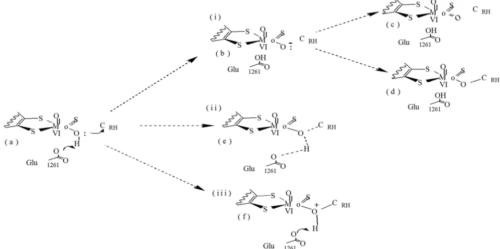
Figure 2. A hypothetical scheme for the de-protonation of Ö¯Mo bound HOH and transfer of HOH from OHMo to (XOR)-Glu1261. The de-protonation of HOH is
proposed to take place before (i-b), during (ii-e), and after (iii-f) the nucleophilic reaction took place.
If the deprotonation of the acidic hydrogen takes place before the nucleophilic attack (route i, b), the unstable negatively charged oxygen (O-) may dissociate to form an unrealistic equatorial oxo ligand (MoVI=Oeq) (Figure 2,
structure c); that means, the oxo ligand may be formed before the O- anion interacts with substrate (Figure 2, a). On the other hand, if a nucleophilic attack takes place after the deprotonation of HOH (Figure 2, f); the positive charge
density on the oxygen center (HO+Mo) may create a high energy barrier that may prevent the formation of the tetrahedral species. However, the concerted formation of OMo-CRH bond (Figure 2, e) is more favorable since the
abstraction of an acidic proton by (XOR)-Glu1261 is expected
to neutralize the charge on the nucleophile (HOMo). The abstraction of an acidic proton by (XOR)-Glu1261 is expected
to lower the activation barrier for the formation of a
tetrahedral Michaelis-Menten type activated state complexes. A mechanistic implication of lowering the activation energy is in stabilizing the tetrahedral activated state complex and influencing the events taking place such as: the cleavage of CRH-HRH bond, transfer of HRH to SMo, and allocation of the
two electrons on the MoVI center.
Given that Glu1261 is restrictly conserved, the aldehyde
oxidase is likely to operate in principally the same way, and base-assisted catalysis has been demonstrated in the reaction of the bovine xanthine oxidoreductase with an aldehyde substrate [4, 19]. Such a role for this Glutamic acid had been explicitly proposed for DgAOR” [11, 19]. In XOR enzymes, Glu802 and Arg880 have catalytic roles that are specific to
enzymes and their substrates since Glu802 and Arg880 amino
acid residues are not conserved in AOR enzymes. The catalytic role of the conserved Gln767 in the xanthine oxidase
utilizing enzymes is not well understood although its mutation to Glutamic acid results in a 10-fold decrease in the rate of enzyme reduction [1], but this residue is not conserved in the aldehyde oxidases in which it contributes to the rate of acceleration in XOR enzymes.
2. Materials and Methods
2.1. Materials
The structures of interest (such as purine derivatives, the active site, and binding pocket amino acids) were sketched using ChemDraw Ultra 2003, version 8.0 (Cambridge software corporation, Cambridge, MA. U.S.A.). These structures were also sketched using GaussView 3.0 (Gaussian, Inc., Pittersburgh, PA. U.S.A.) Software package. This software was also used to develop the input geometries, calculate the bond distances, as well as visualize the
optimized geometries and frontier orbitals. The input geometries prepared using GaussView 3.0 software program were optimized using Gaussian 03W (2003), version 6.0 (Gaussian, Inc, Pittersburgh PA, USA) software package, on Dell Optiplex780 model computer, 2011 (Dell, Inc; Wilhie Sdh Bhd; Penang, Malaysia). In addition, AOMix 2011/2012, 6.6 (Centre for Catalysis Research and Innovation, University of Ottawa, Ottawa, Canada) software package was used to determine the composition of atomic orbitals.
2.2. Methods
2.2.1. The Role of Glu1261 During The Initial Stage of
Catalysis
Optimization of the active site in the presence of substrate and Glu1261: The geometry of the active site in the presence of substrate and binding pocket amino acid (Glu1261) was
Figure 3. A hypothetical Scheme for the initial stage of catalysis, when HOH was abstracted from OHeq by [bmXOR]-Glu1261. The abstraction of HOH is
proposed to take place before (I -b), during (II - e), and after (III - f) the nucleophilic reaction took place. The upper panel shows the proposed mechanism in the presence of xanthine and Glu1261. The lower panel represents, the actual geometries optimized for the structures shown in the upper panel. The illustrations
(lower panel) were prepared with the application of GaussView software program.
The events taking place during the initial stage of catalysis mediated by XOR enzyme in the presence of substrates are the abstraction of acidic hydrogen by Glu1261, nucleophilic
attack on the electron deficient carbon, transfer of HRH from
CRH to Mo=S and transfer of 2e’s to Mo VI
center.
Characterization of the optimized structures: The
Mulliken atomic charges and the total energies were computed from the output files of the optimized structures. The total energies for the optimized geometries were plotted to compare the stabilities of the respective structures. The molecular orbital analyses for the constituent chemical fragments were performed using AOMix software package. The percentage compositions of different molecular
fragments were generated using AOMIX software package.
2.2.2. The Role of Binding Pocket Amino Acid Residues
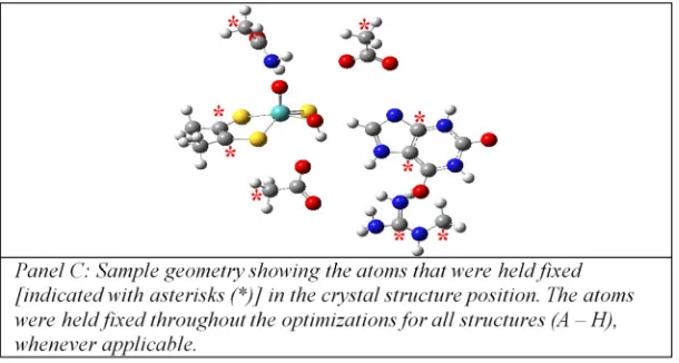
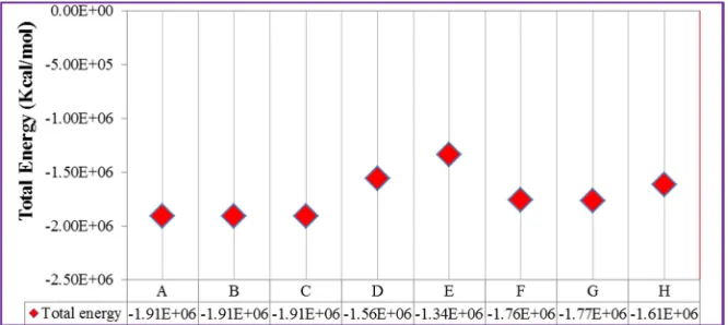
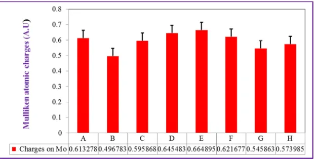
Figure 4. The primary and secondary coordination spheres (active site binding pocket) for the bmXOR (PDB accession code of 1V97) enzymes [24]. The position of the substrate (xanthine) inside the binding pocket (PDB accession code of 3EUB) was adapted from reference [24]. The description of the geometries are as follows: Structure (A) is the active site binding pocket in the presence of xanthine, during the near attack conformation, Structure (B) is the active site binding pocket during the formation of Michaelis-Menten type complex, Structure (C) is the active site binding pocket in the absence of xanthine, Structure (D) is the near attack conformation (NAC) in the absence of binding pocket amino acids, Structure (E) is the active site binding pocket during NAC in the absence of Arg880, Structure (F) is the active site binding pocket during NAC in the absence of Glu802, Structure (G) is the active site binding pocket
during NAC in the absence of Glu802 and Arg880, and Structure (H) is the active site binding pocket during NAC in the absence of Gln767 and Glu1261.
Characterization of the optimized geometries: The
Mulliken atomic charges and the total energies were computed from the output files of the optimized structures. The total energies of the optimized structures were compiled and compared against the energies obtained from the structure optimized in the absence of substrate (Figure 4, Structure C). The molecular orbital analyses for the constituent chemical fragments were performed using AOMix software package in order to generate the percentage contributions of different molecular fragments.
Definition of keywords used in Gaussian job [26, 27]: The key word “B3LYP” was used to describe Beck’s
three-parameter exchange functional combined with the Lee, Yang, and parr’s –correlation functional [26, 27]. The key word ”gen” was used to provide a separate basis set input section and specify an alternate density fitting basis set [26, 27]. The key word “#P” was used to describe additional output generated, which included messages at the beginning and end of each link giving assorted machine department information. The key word ”opt” was used to describe the geometry optimization to be performed. The key word “pop” was applied to control the outputs of molecular orbitals and orbital energies. Finally, the key word “geom=connectivity” was used to indicate the source of input files.
3. Results
3.1 The Role of Glutamic Acid, Glu1261 in the Initial Stage of Catalysis
Figure 6. The Mulliken atomic charges on equatorial oxygen terminal atom of the active site. The data was obtained from geometry optimization performed on the structures described in Figure 3.
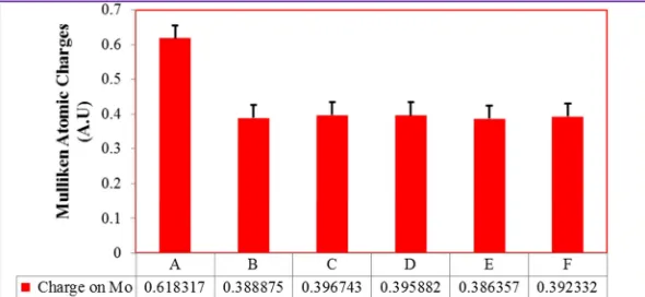
Figure 7. The Mulliken atomic charges on Mo (VI) ion of the active site The data was obtained from geometry optimization performed on the structures
described in Figure 3.
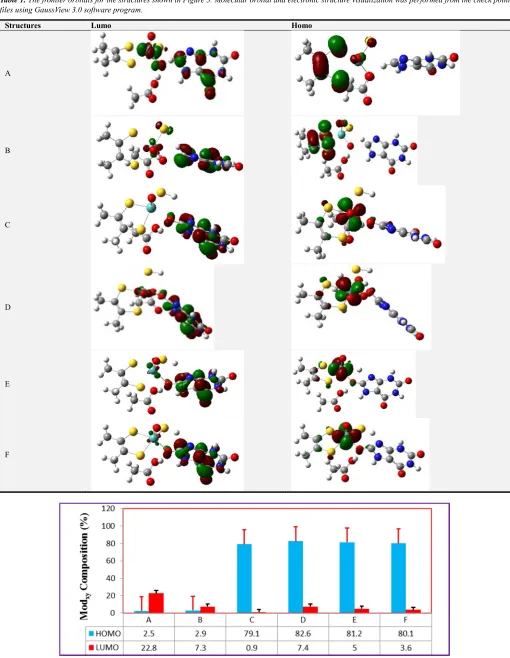
Table 1. The frontier orbitals for the structures shown in Figure 3. Molecular orbital and electronic structure visualization was performed from the check point files using GaussView 3.0 software program.
Structures Lumo Homo
A
B
C
D
E
F
Figure 9. The percent composition for Modxy, in the HOMO and LUMO molecular orbitals, obtained from the structures shown in (Figure 3). The data were
Figure 10. The percent compositions for Oeq, in the HOMO and LUMO molecular orbitals, obtained from the structures shown in Figure 3. The data were developed using AOMix software package.
Figure 11. The percent composition for the proton bound to Oeq, in the HOMO and LUMO molecular orbitals, obtained from the structures shown in Figure 3. The data were developed using AOMix software package.
Figure 12. The composition Glu1261-Og, in the HOMO and LUMO molecular orbitals, obtained from the structures shown in Figure 3. The data were
developed using AOMix software package.
3.2. The Role of Binding Pocket Amino Acid Residues
Figure 14. The Mulliken atomic charge profiles of the terminal atoms of the active site and the interaction site C of xanthine as a substrate in the binding pocket amino acid residues during the near attack conformation at different geometries (as indicated in Figure 4).
Figure 15. The raw data for Mulliken atomic charges, on (C8) position of xanthine as the substrate of the active in the presence of the binding pocket amino
acid residues during the near attack conformation at different geometries (as indicated in Figure 4).
Figure 16. The raw data for Mulliken atomic charges on equatorial oxygen terminal atom of the active site during the interaction with C8 position of xanthine
as a substrate of the active in the presence of the binding pocket amino acid residues during the near attack conformation at different geometries (as indicated in Figure 4).
Figure 17. The raw data for Mulliken atomic charges on equatorial oxygen terminal atom of the active site during the interaction with C8 position of xanthine
Table 2. The bond distances measured between the selected atoms of the substrate and the amino acid residues such as Glu802 and Arg880. The data was
obtained for Structures (A and B) shown in Figure 4. The bond distances were measured on the geometries before and after optimization.
Before Optimization After Optimization
OGlu802 H3xan (A) = 1.2018Å OGlu802 H3xan (A) = 1.2007Å
H1Arg880 O6xan (A) = 3.5767Å H1Arg880 O6xan (A) = 3.5688Å
H2Arg880 O6xan (A) = 2.5501Å H2Arg880 O6xan (A) = 2.5412Å
OGlu802 H3xan (B) = 2.1935Å OGlu802 H3xan (B) = 2.1927Å
H1Arg880 O6xan (B) = 4.9796Å H1Arg880 O6xan (B) = 4.9462Å
H2Arg880 O6xan (B) = 4.0225Å H2Arg880 O6xan (B) = 3.9960Å
Figure 18. Mulliken charges on the selected atoms of the substrate and the amino acid residues such as Glu802 and Arg880. The data was obtained for Structure
(A) shown in Figure 4.
4. Discussion
Xanthine oxidase is one of the most useful molybdenum containing enzymes which catalyze a wide range of substrate during the catalyze reactions. The objective of this research was to describe the role of the binding pocket amino acid residues during the binding stage catalysis. The structures of free substrates and the geometry of the active site in the presence of substrate and binding pocket amino acid residues were constructed using Gauss View 3.0 software package. All geometry optimizations were performed using Gaussian 03W 6.0 software package by applying a density functional theory (DFT) method to generate several parameters such as total energy, Mulliken atomic charges, orbitals and percent composition. The Mulliken atomic charges and the total energies were compiled from the output files to characterize the stability of the optimized structures. In this section, the role of the binding pocket amino acid residues will be discussed.
Nishino and his coworkers (2010) [14] have recently compared the effects of mutation of Glu802 and Arg880 for
bovine milk enzyme on the steady-state kinetics of both xanthine and hypoxanthine [14]. The kinetic parameter Kcat/ Km for xanthine during the reaction for the formation of the
tetrahedral Michael Menten type intermediate was shown to decrease from 2.17×10⁶ M-1s-1 to 1.80×10⁴ M-1s-1 upon mutation of Glu802 to Glutamine [7], 14]. This is in a good
agreement with the energy of the optimized structure of xanthine in the binding pockets during the near attack conformation was shown to decrease from -1.9096 ×10⁶to -1.7663 ×10⁶Kcal/mol (as shown in Table A3) and also the charge of the interaction site of xanthine was shown to increase from 0.138698 to 0.167737 which was resulting from the increase in charge of equatorial oxygen from -0.61069 to -0.63709 (as shown in Figure 14). Similarly, the mutation of Arg880 to methionine reduces Kcat to below the
detectable limit in steady-state experiment of the wild type enzyme activity, indicating that Kcat/ Km xanthine is affected by the mutation of Arg880 which is greater than is
seen with the mutation of Glu802, and no greater than 2×10³
M-1s-1 [7, 14, 16]. This indicates the energy of the optimized structure of xanthine in the binding pockets during the near attack conformation was shown to decrease and the charge was shown to increase slightly. In the case of hypoxanthine, mutation of Arg880 to methionine reduces Kcat/ Km for
hypoxanthine by three orders of magnitude (from 1.67×10⁷ M-1s-1 to 1.56×10⁴ M-1s-1), while mutation of Glu802 to
Glutamine yields no detectable activity which is again reflecting a reduction in Kcat by at least some order of magnitude than is seen with the mutation of Arg880 and the
value for Kcat/ Km hypoxanthine is no greater than 1×10³ M
-1
s-1 [14]. From this steady-state experiment, we can generalize that the most anticipated factor that affect the reactivity of substrates towards xanthine oxidases is the formation of hydrogen bonding microenvironments with the binding pocket amino acid residues during catalysis. The reactivity of xanthine in enzyme catalyzed reaction is affected by the mutation of Glu802 to the greater extent than
that of Arg880, indicating that the amino acid residue, Glu802
(its energy = -1.7663 ×10⁶ Kcal/mol as shown in Figure 13) and its charge = 0.167737 as shown in Figure 14)) plays a greater role in the formation of hydrogen bonding microenvironments with xanthine than Arg880 does during
catalysis (with energy = -1.7557 ×10⁶ Kcal/mol and charge = 0.138812) (shown in Figure 13 and in Figure 14)), and the reverse is true in the case of hypoxanthine. Moreover, the kinetic results indicating that negative charge stabilization by Arg880 is more important for hydroxylation of xanthine, while
the tautomerization is facilitated by Glu802 is more important
for hydroxylation of hypoxanthine.
reaction of purine with xanthine oxidase is slow compared with the physiological substrates (such as hypoxanthine and xanthine) (as shown in Table A.5). On the other hand, the lowest value of Km (6.90µM) for hypoxanthine reflects a high affinity of xanthine oxidase enzyme towards this substrate because a low concentration of substrate is needed to half- saturate the enzyme [1, 12].
In general, Argenine (Arg880) in xanthine oxidase enzyme
is involved in transition state stabilization by compensation of negative charge accumulation on the C6=O oxygen of
xanthine in the course of hydroxylation at C8, and the
orientation we see here indicates that this is also likely to be in the case for hydroxylation of hypoxanthine at C2, since the
C6 carbonyl is again oriented toward the Arg880. Similarly, we
have suggested that Glu802 in xanthine oxidase enzyme is
involved in proton tautomerization of xanthine in the course of hydroxylation at C8. Arg880 amino acid residue of bovine
enzyme plays an important role in catalysis, contributing approximately 4.5Kcal/ mol for the transition state stabilization to increase the rate of the reaction with xanthine as substrate [14]. This means that, Arg880 could lower the
activation energy for the reaction by stabilizing the negative charge accumulation on the heterocycle through an electrostatic interaction with C6 carbonyl oxygen of substrate.
This in turn implies that substrate binds in an orientation opposite to that seen in the structure of reduced enzyme in complex with xanthine. Effective substrates of purine derivatives of the wild type enzyme, possibly binding in an orientation similar to xanthine and making use the catalytic contribution of Arg880 to rate acceleration, were strongly
affected by mutation of Arg880. On the other hand, those that
react slowly with wild type enzyme (in particular 2-hydroxy-6-methyl purine) have functional groups at C6 position is
occupied by methyl group that prevented interaction with Arg880. Therefore, one can conclude that the poor substrates
bound in an inverted orientation to that of xanthine, responsible for both their low reactivity towards the wild -type enzyme and relative insensitivity to the loss of the active site amino acid residue Arg880 [4, 14].
Figure 19. The orientation of xanthine and 2-hydroxy-6-methylpurine inside the active site of xanthine oxidase enzyme with the conserved binding pocket amino acid residues. Both xanthine and HMP bound to the Molybdenum center through the same interaction site at C8.
As shown in Figure 19, HMP is oriented in such a way that the C2=O oxygen interacts with Arg880 and the C6 CH3 group
points away from it. This orientation is more reasonable for the low reactivity of 2-hydroxy-6-methyl purine in bovine milk enzyme. Here, we have suggested that HMP binds in an inverted orientation to that seen with xanthine and for this reason is unable to utilize Arg880 effectively for the transition
state stabilization. This account for the low reactivity of substrates such as HMP and 6-methylpurine with wild type enzyme and at the same time their relative insensitivity to mutation of the active site amino acid residue, Arg880 to
methionine “ [4]. As shown in Figure 19, the carbonyl oxygen of xanthine could form hydrogen bonding microenvironments with the H donor NH2 group of Arg880
binding pocket amino acid residue. However, the substituent group CH3 in HMP could not form hydrogen bonding
microenvironments with Arg880. Both xanthine and
2-hydroxy-6-methylpurine are able to bind with the equatorial hydroxyl terminal of the active site of xanthine oxidase enzyme through their C8 interaction site as indicated in
Figure 19. The anticipated factor that affects the reactivity of xanthine and HMP substrates in xanthine oxidase enzyme is the proper utilization of the conserved binding pocket amino acid residues during the initial stage of catalysis. During the near attack conformation, the substrates are modulated in a proper orientation to react with the active site of the enzyme through its equatorial hydroxyl terminal and to accommodate properly inside the binding pocket amino acid residues which align at the top and at the bottom side of the enzyme by forming hydrogen bonds [14-16]. However, the methyl group for 2-hydroxy-6-methylpurine at C6 position could prevent
the electrostatic interaction with Arg880, which cannot form
hydrogen bonding microenvironments through methyl group and thus it gives less stable product than that of xanthine. This is mainly because HMP is unable to stabilize the negative charges accumulated on the heterocycle through an electrostatic interaction of C6 carbonyl oxygen with Arg880 in
which the negatively charged carbonyl oxygen is a hydrogen acceptor and the NH2 group of Arg880 is a hydrogen donor
[15, 16]. As a summary, the orientation of substrates in the active site of xanthine oxidoreductase plays a critical role in determining the catalytic effectiveness of Glu802 and Arg880 in
hydroxylation of xanthine at its C8 position. Properly
oriented with N7 towards the Glu802 and C6=O towards the
Arg880, the reaction proceeds much more rapid than when the
substrate binds in the inverted orientation as indicated in the case of HMP. The amino acid residue, Arg880 was found to
contribute 4.5Kcal/mol to the transition state stabilization via electrostatic interaction with substrate during catalysis [4]. The proposed interaction of this residue with the transition state stabilization suggests a means of discriminating good substrates from those that are more slowly hydroxylated.
The Role of Binding Pocket Amino Acid Residues
In this section, the role of binding pocket amino acid residues in enzyme catalyzed reaction will be discussed. Thus, in this section the role of Glu1261 in initial stage of
catalysis and the role of Glu802 and Arg880 amino acid
residues will be discussed.
Glutamic acid (Glu1261) is thought to act as a general base,
deprotonating the Mo-OH of the molybdenum center to facilitate the nucleophilic attack on the substrate. In other words, it was proposed that the role of the Glutamic acid (Glu1261) in activating the equatorial hydroxyl terminal in
which the nucleophilic attack is expected to take place after the acidic proton is abstracted from the equatorial hydroxyl terminal. A mutagenic study is important to identify the role of an amino acid residue, in particular, Glu1261 in recognition
and activating substrates during the initial stage of catalysis [1, 3, 5]. The active site amino acid residue (Glu1261) could be
used as a target of investigation since it is one of the most important active site amino acid residues located in the secondary coordination sphere [3, 5, 19].
In the initial stage of catalysis as shown in (Figure 8), the abstraction of the acidic proton from the equatorial hydroxyl terminal by [bmXOR]-Glu1261, is expected to take place in
one of the three routes (such as before the abstraction of the equatorial acidic proton from the equatorial hydroxyl terminal) as shown in Figure 3 which leads to the formation of Structure (B), which further leads to the formation of either Structure (C or D) due to the unstable Oxyanion on Oeq, during the abstraction of the equatorial acidic proton
from the equatorial hydroxyl terminal which leads to the formation of Structure (E) and after the abstraction of the equatorial acidic proton which leads to the formation of Structure (F). The nucleophilic attack of the equatorial Oxyanion (O-) on the substrate is proposed to be initiated by the immediate transfer of the acidic hydrogen (HOH) from the
HOeq to (XOR)-Glu1261. The deprotonation of HOH may be
suggested to take place before, during, or after the nucleophilic reaction (the nucleophilic attack by the equatorial Oxyanion (O-) on the activated /deficient substrate (Figure 3). Although, the sequence of deprotonation is not well understood the stepwise deprotonation of HOH (Figure 3
(a)) is proposed to take place either before (shown in route I (Structure B)), during (shown in route II (Structure E)) or after the nucleophilic attack (Figure 3 route III (Structure F)). If the deprotonation takes place before the nucleophilic attack (route I (Structure B)), the unstable negatively charged oxygen (O-) may dissociate to form an unrealistic equatorial oxo ligand (MoVI=Oeq) (Figure 3 structure (C)). That means,
the oxo ligand may be formed before the O- anion interacts with substrate (Figure 3 (a)). Alternatively, the O-Mo may undergo a nucleophilic attack on the substrate carbon to form OMo-CRH bond (indicated in Figure 3 (Structure D)) [1, 7,
11]. In this case, the OMo-CRH bond may be formed before the
likely formation of an equatorial oxo ligand (MoVI=Oeq). On
the other hand, if a nucleophilic attack takes place after the deprotonation of HOH (Figure 3 (Structure F)); the positive
charge density on the oxygen center (HO+Mo) may create a
high energy barrier that may prevent the formation of the tetrahedral species. However, the concerted formation of OMo-CRH bond (Figure 3 (Structure E)) is more favorable
since the abstraction of an acidic proton by (XOR)-Glu1261 is
expected to neutralize the charge on the nucleophile (HOMo).
The energy released during the formation of the Structures varies from Structure (A) through Structure (D) (Figure 5). That is, the energy released increases from Structure (A) through Structure (D) during the initial stage of catalysis (from ∆E= -1481111.805 to ∆E= -1481140.485). However, the energy released for (B) and for (C) is almost identical during the formation of the Structures. In addition, the Structure formed during the near attack conformation (A) is energetically unfavorable (∆E= -1481111.805 Kcal/mol) whereas the Structure (D) is energetically favorable (∆E= -1481140.485). In other words, the lowest possible minimum amount of energy was attained during the formation of Structure (D). This indicates that Structure (D) is the most stable one. From this, we can suggest that Oxyanion is responsible for the nucleophilic attack of the deficient carbon atom other than the hydroxyl group itself (OH). Therefore, the most favorable pathway in the initial stage of catalysis is the reaction that occurs after the abstraction of the equatorial acidic proton of the active site. This is because Oxyanion cannot form the stable Structure when it forms a double bond with Molybdenum metal. It is evident that the stability of Structure (B) possessing Oxyanion and Structure (C) possessing double bond with Molybdenum is the same (∆E= -1481106.485Kcal/mol).
This also confirmed that Oxyanion is unstable to exist and make a nucleophilic attack on the substrate carbon rather than forming a double bond with Molybdenum metal. Thus, the most favorable reaction path way in the initial stage of catalysis occurs after the abstraction of the equatorial acidic proton of the active site. Therefore, in the most favorable path way, Glu1261 is expected to act as a
general base which is assumed to be followed by the nucleophilic attack of the Oxyanion on the carbon center of the substrate to yield the Structure. And hence, Glu1261
plays a great role in the activation of the active site by abstracting the acidic proton as well as in decreasing the activation barrier for the Structure formation during the initial stage of catalysis. As shown in Figure 6, the partial charges on the Oeq for the Structures shown in Figure 3 (A,
B, C, D, E, and F) were given as ∆qc = -0.722622, ∆qc = -0.499669, ∆qc = -0.496599, ∆qc = -0.504629, ∆qc = -0.507978, and ∆qc = -0.502671, respectively. From these partial charges, we can suggest that Oeq for Structure (E) is a good nucleophile (∆qc = -0.507978) to attack the deficient substrate carbon more readily than the remaining Structures. This shows that the Oxyanion is more susceptible to attack the deficient substrate carbon during the formation of structure D rather than any other structures. Similarly, the partial charges on the Mo atom for structures (A, B, C, D, E, and F) (Figure 4) were ∆qc= 0.618317, ∆qc = 0.388875,
that much more charges were flown to the metal center from the substrate carbon of Structure (E). This situation is a direct consequence of the highest accumulation of negative partial charge on the Oeq for Structure (E). In
addition, the partial charges on the Cs Structures (A, B, C, D, E, and F) (Table A.2) were ∆qc= 0.182387, ∆qc = 0.480415, ∆qc = 0.475512, ∆qc = 0.483265, ∆qc = 0.487586, and ∆qc = 0.483945, respectively. The highest positive charge developed on the carbon atom for Structure (E) indicates that the substrate carbon is more susceptible for the nucleophic reaction to take place. This supports the ideas explained earlier in the cases of the partial charges developed on Oeq and Mo metal. Therefore, all these suggestions showed that Structure (E) could be the most stable Structure followed by route II (shown in Figure 3 (E)). As shown in Table (A4), the % Contribution of Cs-HOMO and Mo-LUMO for the Structures (A, B, C, D, E, and F) were % Cs-HOMO = 0.02 and % Mo-LUMO = 22.8; % HOMO = 0.42 and % Mo-LUMO = 7.3; % Cs-HOMO = 0.46 and % Mo-LUMO = 0.90; % Cs-Cs-HOMO = 0.47 and % Mo-LUMO = 7.4; % Cs-HOMO = 0.41 and % LUMO = 5.0; and % Cs-HOMO = 0.46 and % Mo-LUMO = 3.6, respectively. In this case, the % Contributions of Cs-HOMO and Mo-LUMO are highest for Structure (D). This indicates that the interaction between substrate carbon HOMO and the metal center LUMO is strong. Therefore, we can suggest that Structure (D) is considered as the most stable Structure.
Finally, the amino acid residue, Glu1261 is proposed to play
an important role in promoting the nucleophilicity of the HOeq ligand of the active site. That is, Glu1261 is in a close
proximity to the equatorial hydroxyl group, HOeq capable of
abstracting hydrogen from HOeq which causes the equatorial
oxygen develops a negative charge (Oxyanion) for the nucleophilic attack on the deficient substrate carbon. That means, the Oxyanion is responsible for the nucleophilic attack of the deficient carbon atom other than the hydroxyl group itself (OH). Therefore, we can conclude that the role of Glu1261 is enhancing the nucleophilicity of HOeq ligand as it
acts as a Lewis base to accept a proton from the HOeq ligand
as well as stabilizing the tetrahedral Michaelis-Menten type intermediate at the transition state.
The Role of Glu802 And Arg880 Amino Acid Residues The secondary coordination sphere (particularly the binding pocket amino acids, Glu802 and Arg880) are important
in substrate recognition and orientation. The amino acid residues are expected to create a hydrogen bonding microenvironment and modulate the near attack conformation (NAC), in the presence of heterocyclic and small molecules. Once the substrates are modulated to attain the proper orientation, the activation of the HOeq terminal is
proposed to take place after the HOH is abstracted from the
HOeq terminal, by [bmXOR]-Glu1261.
Figure 20. The position of the substrate (xanthine) inside the binding pockets during the near attack conformation. The roles of binding pocket amino acid residues are to create the hydrogen bonding microenvironments and modulate the near attack conformation (NAC), in the presence of xanthine as a substrate as well as in the formation of the tetrahedral Michaelis- Menten type intermediate.
The amino acid residues that are important in the substrate recognition in the binding pockets and providing proper orientation for the substrates are Glu802 and Arg880 (in bm
XOR [4, 15]. As described by many scholars, [4], these amino acid residues are expected to create a hydrogen bonding micro-environment and modulate the near attack conformation (NAC) in the presence of heterocyclic and small molecules. Therefore, the role of these binding pocket amino acid residues is providing the proper substrates orientation for the nucleophilic reaction to occur and the recognition of the active site in the binding pockets as well as stabilizing the transition states of the given tetrahedral Michaelis-Menten type intermediate entering to the transition state.
Generally, in order for the interaction between the reactants (substrate and enzyme) to take place, the substrates are expected to enter the binding pocket amino acid residues and attain a proper orientation to bind through their favorable interaction sites with the enzyme active site. Therefore, the catalytic transformations of the substrates (CRH) to their
products (O-CCR) are expected to begin with the substrate
pocket amino acid residues. Therefore, amino acid residues are expected to stabilize the Structures by creating hydrogen bonding microenvironments. In contrast, the Structures which involve no amino acid residues are generally energetically less favorable due to the absence of hydrogen bonding microenvironments. The Structures optimized in the absence of Arg880 and in the presence of other amino acid
residues is less stable than those that which proceeds without Glu802 and in the presence of other binding pocket amino acid
residues.
From this, we can say that Argenine has more profound effect in stabilizing the complexes through the formation of hydrogen bonding microenvironments than Glutamate (Glu802) which is responsible for giving the proper orientation
of substrate by creating hydrogen bonding
microenvironments.
As shown in Table A3, the partial charges on the substrate carbon, Cs for the Structures such as A (with binding amino acids), E (without amino acids), F (without Arg880), G
(without Glu802), H (without Glu802 and Arg880) were given as
∆qc= 0.138698, ∆qc=0.664895, ∆qc = 0.149312, ∆qc = 0.0.138812 and ∆qc= 0.167737, respectively. Similarly, the partial charges on Mo metal for the Structures (A, E, F, G and H) were given as ∆qc= 0.613278, ∆qc= 0.664895, ∆qc = 0.621677, ∆qc = 0.545863 and ∆qc = 0.573985, respectively. In the reductive half reaction, the flow of charges is from the substrate carbon to the metal center of the active site. The variation in partial charges on the Molybdenum metal of the active site (Table A3) indicates that the tendency of the metal to accept the partial charges from the substrate carbon during interaction. This effect is due to the role of the binding pocket amino acid residues in activating the active site and providing the substrate proper orientation for the nucleophilic reaction to occur. For instance, the partial charge for Molybdenum decreases from (∆qc = 0.664895 to ∆qc = 0.545863). This shows that the flow of partial charges to Molybdenum metal center from the substrate is enhanced by the amino acid residues such as Glu1261, Glu802 and Arg880.
Similarly, the partial charge on the substrate carbon increases from (∆qc= 0.149312 to ∆qc = 0.167737) but in the other case, the partial charge decreases from (∆qc = 0.149312 to
∆qc = 0.098687). Thus, the increase in partial charge on the substrate carbon suggests that the tendency in which the flow of partial charges from the substrate carbon to the metal center of the active site is promoted by the amino acid residues. However, the decrease in partial charge shows that the flow of this partial charge from the substrate carbon to the metal center of the active site is hindered. This is because the absence of an important amino acid residue, in particular, Glu1261 which activates the active site. These amino acid
residues promote the flow of partial charges to the metal center of the active site.
In general, Glu802 and Arg880 binding pocket amino acid
residues are important to create hydrogen bonding microenvironments and modulate the near attack conformation. In addition to this, they provide the substrate proper orientation for the nucleophilic attack to take place.
On the other hand, the absence of both Glu802 and Arg880
amino acid residues lead to less stable optimized Structures than those in the absence of Glu1261 and Gln767 during the
near attack conformation. This effect suggests that Glu802
and Arg880 amino acid residues play a great role in the
reactivity of the active site with the substrate and in stabilizing the transition state. As a conclusion, the removal of Glu802, which likely forms a hydrogen bonding
interaction with the substrate causes the activity loss of an enzyme whereas the removal of Arg880 results in a great
loss of activity, possibly due to loss of transition state stabilization rather than substrate binding. The site-directed mutagenesis of Glu1261, the residue proposed to deprotonate
the hydroxyl group shows a pronounced effect in decreasing an enzyme activity.
The % Contribution of Cs-HOMO and Mo-LUMO for Structures during the near attack conformation were % Cs-HOMO (for A) = 16.14 and % Mo-LUMO (for A) = 1.87, % Cs-HOMO (for E) = 20.74 and % Mo-LUMO (for E) = 0.24, % Cs-HOMO (for F) = 16.50 and % Mo-LUMO (for F) = 5.58, % Cs-HOMO (for G) = 17.13 and % Mo-LUMO (for G) = 0.22, % Cs-HOMO (for H) = 10.20 and % Mo-LUMO (for H) = 2.86. These HOMO-LUMO data were obtained from AOMIX output files. Here, we see that the % Contribution for Cs-HOMO and % contribution for Mo-LUMO vary for each respective Structure. This variation clearly suggests that the binding pocket amino acid residues play a great role for the charge transfer from the substrate carbon HOMO to the metal center LUMO. For instance, the % Contribution for Cs-HOMO (for E without amino acid residues) = 20.74 and % Contribution for Mo-LUMO (for E without the amino acid residues) = 0.24 were obtained in the absence of the amino acid residues whereas the % Contribution for Cs-HOMO (for A with amino acid residues) = 16.14 and % Contribution for Mo-LUMO (for A with the amino acid residues) = 1.87. In this case, the percent Contribution of Cs-HOMO decreases from (20.74 to 16.14) whereas the percent Contribution of Mo-LUMO increases from (0.24 to 1.87). From this, we can suggest that the flow of partial charges from the substrate carbon HOMO orbital to the metal center LUMO orbital is promoted by the binding pocket amino acid residues. Generally, we can conclude that the flow of partial charges from Cs-HOMO to Mo-LUMO is not possible unless the active site is activated by Glu1261. This
clearly shows that the role of Glu1261 is activating the active
site for the nucleophilic attack to take place. Therefore, the amino acid residues play a great role in promoting the flow of partial charges from the substrate carbon HOMO orbital to the metal center LUMO orbital.
As shown in Table 2, we clearly show that the bond distances measured between the selected atoms of the substrate and the amino acid residues before optimization are longer than those that of the bond distances measured after optimization. That is, after optimization, the bond distances between the oxygen atom of Glu802 and hydrogen atom of
xanthine and the hydrogen atoms of Arg880 and oxygen atom
between the selected atoms of the amino acid residues and the substrate showed that the formation of hydrogen bonding microenvironments between the oxygen atom of Glu802 and
the hydrogen atom of xanthine as well as between the hydrogen atoms of Arg880 and the oxygen atom of xanthine.
Therefore, we can suggest that the role of the binding pocket amino acid residues is to create the hydrogen bonding microenvironments with the substrate.
Finally, the roles of amino acid residues are important in providing the substrate proper orientation in order for a nucleophilic reaction to take place by creating a hydrogen bonding microenvironments. They are also important for the recognition of substrates in the binding pockets of the substrate access channel [4, 11]. Furthermore, the amino acid residues are also capable of stabilizing the emerging charges from the interaction sites at the transition state [1]. Thus, the enzyme-substrate complex containing a negative charge could be interacted with the nearby amino acid residues in order to neutralize the negative charge by forming the hydrogen bonding microenvironments. Therefore, the complex could attain its stability at the transition state [4, 16].
5. Conclusions
The physiological importance of XO in areas of human health and the oxidative degradation of organic metabolites in the cell emphasizes the need to understand the mechanism of XO activity. Substrates can be used to aid in the interpretation of enzyme activity and the determination of active site electronic Structure as a result, a detailed electronic Structure description of the enzyme active sites and the substrates in the binding pocket amino acid residues can be explained.
In addition, the involvement of the binding pocket amino acid residues for the near attack conformation affects the interaction of the substrates with the active site. As a result, the rate at which the substrates interacted with the active site of xanthine oxidase enzyme is proposed to be affected. In order for the interaction between the reactants (substrate and enzyme) to take place, the substrates are proposed to enter the binding pocket amino acid residues and attain a proper orientation to bind through their favorable interaction sites with the enzyme active site. Therefore, the catalytic transformations of the substrates to their products are expected to begin with the substrate binding stage and formation of the tetrahedral Michaelis-Menten type intermediate.
In general, Oxyanion is unstable to exist and make a nucleophilic attack on the substrate carbon rather than forming a double bond with Molybdenum metal. Thus, the most favorable reaction path way in the initial stage of catalysis occurs after the abstraction of the equatorial acidic proton of the active site. Therefore, in the most favorable path way, Glu1261 is expected to act as a general base which is
assumed to be followed by the nucleophilic attack of the Oxyanion on the carbon center of the substrate to yield the
Structure. And hence, Glu1261 plays a great role in the
activation of the active site by abstracting the acidic proton as well as in decreasing the activation barrier for the Structure formation during the initial stage of catalysis. Furthermore, the flow of partial charges to Molybdenum metal center from the substrate is enhanced by the amino acid residues such as Glu1261, Glu802 and Arg880. Similarly, the partial charge on the
substrate carbon increases from (∆qc= 0.149312 to ∆qc = 0.167737) but in the other case, the partial charge decreases from (∆qc = 0.149312 to ∆qc = 0.098687). Thus, the increase in partial charge on the substrate carbon suggests that the tendency in which the flow of partial charges from the substrate carbon to the metal center of the active site is promoted by the amino acid residues. However, the decrease in partial charge shows that the flow of this partial charge from the substrate carbon to the metal center of the active site is hindered. This is because the absence of an important amino acid residue, in particular, Glu1261 which activates the
active site. These amino acid residues promote the flow of partial charges to the metal center of the active site. In general, Glu802 and Arg880 binding pocket amino acid
residues are important to create hydrogen bonding microenvironments and modulate the near attack conformation. In addition to this, they provide the substrate proper orientation for the nucleophilic attack to take place. On the other hand, the absence of both Glu802 and Arg880
amino acid residues lead to less stable optimized Structures than those in the absence of Glu1261 and Gln767 during the
near attack conformation. This effect suggests that Glu802 and
Arg880 amino acid residues play a great role in the reactivity
of the active site with the substrate and in stabilizing the transition state. As a conclusion, the removal of Glu802,
which likely forms a hydrogen bonding interaction with the substrate causes the activity loss of an enzyme whereas the removal of Arg880 results in a great loss of activity, possibly
due to loss of transition state stabilization rather than substrate binding.
amino acid residues) = 1.87. In this case, the percent Contribution of Cs-HOMO decreases from (20.74 to 16.14) whereas the percent Contribution of Mo-LUMO increases from (0.24 to 1.87). From this, we can suggest that the flow of partial charges from the substrate carbon HOMO orbital to the metal center LUMO orbital is promoted by the binding pocket amino acid residues. Generally, we can conclude that the flow of partial charges from Cs-HOMO to Mo-LUMO is not possible unless the active site is activated by Glu1261. This
clearly shows that the role of Glu1261 is activating the active
site for the nucleophilic attack to take place. Therefore, the amino acid residues play a great role in promoting the flow of partial charges from the substrate carbon HOMO orbital to the metal center LUMO orbital.
Finally, we clearly show that the bond distances measured between the selected atoms of the substrate and the amino acid residues before optimization are longer than those that of the bond distances measured after optimization. That is, after optimization, the bond distances between the oxygen atom of Glu802 and hydrogen atom of xanthine and the
hydrogen atoms of Arg880 and oxygen atom of xanthine get
nearer each other. These closer distances between the selected atoms of the amino acid residues and the substrate showed that the formation of hydrogen bonding microenvironments between the oxygen atom of Glu802 and
the hydrogen atom of xanthine as well as between the hydrogen atoms of Arg880 and the oxygen atom of xanthine.
Therefore, we can suggest that the role of the binding pocket amino acid residues is to create the hydrogen bonding microenvironments with the substrate.
Acknowledgements
I sincerely thank Dr. Abebe Berhane (PhD) for his undeserved help in fulfilling different Softwares for this computational work. I would like to extend my deepest gratitude to Dr. Feleke Wedeyes, the President of Arbaminch University, for his support in building the Chemistry Department’s Post Graduate Computation Center.
Appendix
Appendix I: The Raw Data Used to Characterize the Role of Glu1261 in Activating the HOeq Terminal of the Active Site,
During the Initial Stage of Catalysis
Table A1. The raw data for the total energies obtained from the geometry optimization carried out for the structures shown in Figure 3. The structures in Figure 3 were optimized in the presence of xanthine, formaldehyde, Formamide, and Lumazine.
Substrates Structures Total Energies
(HF) (kcal/mol)
Xanthine A -2.3602999E+03 -1.4811118E+06
B -2.3602914E+03 -1.4811065E+06
C -2.3602914E+03 -1.4811065E+06
D -2.3603456E+03 -1.4811405E+06
E -2.3603249E+03 -1.4811275E+06
F -2.3602751E+03 -1.4810962E+06
Formaldehyde A -1.9123397E+03 -1.2000123E+06
B -1.9123876E+03 -1.2000423E+06
C -1.9123331E+03 -1.2000082E+06
D -1.9123875E+03 -1.2000423E+06
E -1.9123850E+03 -1.2000407E+06
F -1.9123881E+03 -1.2000426E+06
Formamide A -1.9677517E+03 -1.2347839E+06
B -1.9677626E+03 -1.2347907E+06
C -1.9677626E+03 -1.2347907E+06
D -1.9677631E+03 -1.2347910E+06
E -1.9677616E+03 -1.2347901E+06
F -1.9677613E+03 -1.2347899E+06
Lumazine A -2.3970496E+03 -1.5041726E+06
B -2.3970151E+03 -1.5041510E+06
C -2.3970151E+03 -1.5041510E+06
D -2.3970151E+03 -1.5041510E+06
E -2.3969291E+03 -1.5040970E+06
F -2.3970250E+03 -1.5041571E+06
Table A2. The raw data for Mulliken atomic charges, on selected atoms shown in the lower panel, obtained from the geometry optimization of the structures shown in Figure 3. The abbreviations are described using the Figure shown in the lower panel.
Structures Csub Oeq Mo Ster HOH
A 0.182387 -0.722622 0.618317 -0.550249 0.324961
Structures Csub Oeq Mo Ster HOH
C 0.475512 -0.496599 0.396743 -0.354925 0.295306
D 0.483265 -0.504629 0.395882 -0.358718 0.307825
E 0.487586 -0.507978 0.386357 -0.357248 0.319904
F 0.483945 -0.502671 0.392332 -0.356946 0.305982
The abbreviations shown are Csub is the substrate carbon, Oeq is the equatorial oxygen, Mo is Molybdenum metal, Oap is
apical oxo, Ster is terminal sulfur and HOH is the acidic proton on the equatorial hydroxyl group of the active site.
Table A3. The raw data for Mulliken atomic charges obtained, from geometry optimization, for the structures shown in Figure 4.
Charges
Structures Mo Oap Ster Oeq HOH Csub
A 0.613278 -0.460893 -0.43924 -0.61069 0.269492 0.138698
B 0.496783 -0.513739 -0.31333 -0.6766 0.333404 0.124495
C 0.595868 -0.442693 -0.45508 -0.5783 0.279607
D 0.664895 -0.469122 -0.39262 -0.61952 0.27021 0.149312
E 0.621677 -0.516063 -0.44677 -0.60993 0.29327 0.138812
F 0.545863 -0.524684 -0.43898 -0.63709 0.282556 0.167737
G 0.573985 -0.544869 -0.43534 -0.63551 0.292285 0.160017
H 0.573985 -0.544869 -0.43534 -0.63551 0.292285 0.160017
Table A4. The raw data for the elemental percent composition (% C), on selected atoms, for the structures optimized as shown in Figure 3. The elemental composition was collected for the frontier orbitals, using AOMix software program.
Structures Frontier Orbitals % C
Mo Oeq HOH Og Csub
A LUMO+1 30.9 6.6 0.2 0.1 0.28
LUMO 22.8 7.2 0.2 0.1 19.90
HOMO 2.5 0.6 0 0.1 0.02
HOMO-1 3.6 5.8 0 0.2 0.12
B LUMO+1 47.8 13.5 0.1 0 0.27
LUMO 7.3 0.4 0 0 11.41
HOMO 2.9 0.9 0 0.2 0.42
HOMO-1 2.3 6.8 0 0.3 0.06
C LUMO+1 65.6 0.9 0 0.2 0.25
LUMO 0.9 4.3 0.2 0 11.36
HOMO 79.1 1.8 0 0 0.46
HOMO-1 3.5 0.2 0.1 0.1 0.06
D LUMO+1 61.3 1.5 -0.2 0 0.22
LUMO 7.4 2.5 0 0 11.38
HOMO 82.6 1.5 0 0 0.47
HOMO-1 3.8 0.1 0 0.1 0.05
E LUMO+1 17.2 0.2 -0.1 0 0.23
LUMO 5 3 0 0 11.70
HOMO 81.2 1.1 0 0 0.41
HOMO-1 4.7 0.2 0 0.3 0.06
F LUMO+1 0.4 0.1 0.5 0 0.22
LUMO 3.6 3.9 15.7 0.2 11.24
HOMO 80.1 1.7 1 1.8 0.46
![Figure 1. The secondary coordination sphere for the (a) bmXOR (PDB accession code of 1V97) [24]](https://thumb-us.123doks.com/thumbv2/123dok_us/9528996.1482086/3.595.53.545.165.335/figure-secondary-coordination-sphere-bmxor-pdb-accession-code.webp)