Advances and challenges in targeting FGFR signalling in cancer
Irina S. Babina1 and Nicholas C. Turner1, 2
1 Breast Cancer Now Research Centre, Institute of Cancer Research, London, SW3
6JB, UK.
2 Breast Unit, The Royal Marsden Hospital, Fulham Road, London, SW3 6JJ, UK.
Corresponding author:
Abstract
Fibroblast growth factors (FGFs) and their receptors (FGFRs) regulate numerous cellular processes. Deregulation of FGFR signalling is frequently observed in many cancers, making activated FGF receptors a highly promising potential therapeutic target supported by multiple pre-clinical studies. However, early phase clinical trials have produced mixed results with FGFR-targeted cancer therapies, revealing substantial complexity to targeting aberrant FGFR signalling. In this Review, we
discuss the increasing understanding of the differences between diverse mechanisms of oncogenic activation of FGFR, and the factors that determine
response and resistance to FGFR targeting.
Introduction
Fibroblast growth factor receptors (FGFRs) form a family of four highly conserved trans-membrane receptor tyrosine kinases (FGFR1-4), and one receptor that has the ability to bind fibroblast growth factor (FGF) ligands but lacks an intracellular kinase domain (FGFR5, also known as FGFRL1)1. Receptor activation by FGFs initiates a
cascade of intracellular events that activate major survival and proliferative signalling pathways2. FGFs mediate crucial physiological mechanisms, such as tissue and
metabolism homeostasis, endocrine functions and wound repair2.
cancers6 respectively, although results from later phase studies are awaited.
Disappointingly, modest levels of clinical activity have been reported for patients with other aberrations such as FGFR1 amplification7 or FGFR2 mutation in advanced-stage endometrial cancer8.
Nevertheless, as more data emerge as a result of clinical trials and functional studies, we begin to discern which FGFR aberrations are oncogenic drivers and
would benefit from monotherapy. Likewise, certain passenger FGFR anomalies are being identified as potential targets for combination therapy in select patient
populations, particularly with respect to FGFR-mediated drug resistance. In this Review we address the diverse mechanisms of oncogenic FGFR signalling, focussing on success and limitations of the use of FGFR inhibitors in the clinic, and discuss the recent scientific findings that provide insight into the variable therapeutic effects of anti-FGFR therapy.
[H1] Oncogenic FGFR signalling
Enhanced FGFR signalling in oncogenesis is mediated by genetic alterations (receptor amplification, mutations and chromosomal translocations); autocrine and paracrine signalling, angiogenesis and epithelial-mesenchymal transition (EMT) (Figure 1).
[H3] FGFR amplification
Amplification of FGFR1 (8q12 locus) is found in approximately 17% of squamous non-small cell lung carcinoma (NSCLC)9,10 and ~6% of small cell lung carcinoma11, and is an independent adverse prognostic marker in early stage NSCLC12. FGFR1
amplification is also prevalent in breast cancer and was reported in nearly 15% of
breast cancers13-15. Response to FGFR inhibition has been observed in vitro in
FGFR1-amplified lung cancer models, of both squamous and non-squamous types, although response to FGFR inhibition in xenografts has been variable10,16. In vitro
inhibition of FGFR1 through small interfering RNA (siRNA) or a selective FGFR inhibitor PD173074 modestly reduced the growth of breast cancer cell lines that overexpressed FGFR1 or in which FGFR1 was amplified17,18. However, FGFR inhibition can reverse resistance to endocrine therapy promoted by FGFR
signalling31. Large-scale kinase profiling of 117 cell lines of several cancer types revealed increased sensitivity of FGFR1- and FGFR2-amplified osteosarcoma cell
lines to several FGFR inhibitors19, which was confirmed in a study of 500 cell lines, in response to FGFR inhibitor NVP-BGJ39820.
Amplification of FGFR2 is less frequent than amplification of FGFR1 across cancer types, and has only been described in 5-10% of gastric cancer, particularly of the aggressive diffuse subtype 21, and in 2% of breast cancer overall, with approximately
4% of triple negative breast cancer harbouring FGFR2 amplification22. Amplified FGFR2 in some cancers, such as diffuse gastric cancer, is accompanied by deletion of the C-terminal exon, which results in preferential expression of a truncated form of the receptor potentially promoting oncogenesis through impaired internalisation and subsequent degradation of the active receptor23. Gastric24, rectal25 and breast
cancer19 cell lines with high levels of amplification of FGFR2 are highly sensitive to selective FGFR inhibitor in vitro and in vivo, which suggests that FGFR2 amplification in these cancers could signify addiction to the FGFR pathway for growth.
Differences in apparent addiction to FGFR1 and FGFR2 amplification are in part explained by the amplicon: the FGFR2 amplicon is frequently narrow and centred on
carcinogenesis. The amplification is frequently broader in oestrogen receptor (ER)-positive breast cancer than in NSCLC, with strong evidence pointing at ZNF703
oncogene as a further driver in the FGFR1 amplicon26,27, which may also predict resistance to tamoxifen28. Amplification of FGFR3 and FGFR4 is not frequently reported, and oncogenic activation of these receptors is often linked to a mutation, or ligand amplification. For example, FGFR3 protein overexpression is recurrent in bladder cancer but it is not linked to FGFR3 gene amplification29,30.
[H3] Activating mutations
In contrast to activating mutations in EGFR, mutations in FGFRs are frequently observed outside the kinase domain (Figure 2). Somatic activating mutations of
FGFR1 are rarely observed in cancer, and are more common in FGFR2 and FGFR3.
FGFR2 mutations are found in 10-12% of endometrial carcinomas31,32, nearly 4% of
NSCLC and gastric cancer33, as well as in around 2% of urothelial cancer34.
Mutations in the extracellular IgII and IgIII loops (Figure 2), as well as in their linker domain, may provide gain of function either through increasing receptor-ligand binding affinity and interaction35,36 (for example the S252W mutation in FGFR2, with
an identical mechanism described for the P252 residue in FGFR1 and P250 in FGFR335) or through generation of aberrant disulphide bridges that result in constitutive receptor dimerization (S373C and Y376C in the FGFR2-IIIc isoform and analogous mutations in FGFR3-IIIc, G370C and Y373C37). Similarly, FGFR2
insertion mutation A266_S267ins and deletion 290_291WI>C, where amino acid residues WI are replaced by a Cysteine (C), have recently been described to have oncogenic potential via increased dimer formation in a ligand-independent manner38.
cancer32,39 and around 5% of cervical cancer39,40. The most common mutations in
FGFR3 also occur in the extracellular (R248, S249) and transmembrane (G370, Y373) domains of the receptor, resulting in increased receptor dimerization and ligand-independent signalling, analogous to FGFR2 mutations in those regions41. Although it is likely that enhanced dimerization directly leads to upregulation of FGFR kinase activity, this has not been established yet and additional factors might be required.
Mutations in the kinase domain of FGFR1 and FGFR2 (most frequently N546K and
N549H/K, respectively) constitutively activate the receptors and transform cell lines42,43, although these mutations are rare, with the FGFR2 N549 mutations found
in around 1.4% endometrial and <1% invasive breast cancers34. FGFR4 kinase
mutations K535 and E550 have been recorded in rhabdomyosarcoma44 and
knockdown of FGFR4 with inducible short hairpin RNA (shRNA) in a human rhabdomyosarcoma cell line reduced tumour growth in vivo44.
In addition to the somatic activating mutations in the FGFRs, germline single nucleotide polymorphisms (SNPs) have been reported to associate with cancer incidence. A non-coding SNP in the second intron of FGFR2 (rs2981582), which contains putative transcription factor binding sites, has been linked to predisposition to breast cancer in postmenopausal women45-47. A SNP in FGFR4 (rs351855), which results in G388R substitution, is linked to poor survival in several cancer types, such as breast, colorectal and lung, among others48, and has been shown to increase breast cancer cell motility in vitro49. This genetic association of the rs351855 SNP with cancer aggressiveness can at least in part be explained by increased association of FGFR4 harbouring the SNP with signal transducer and activator of
expression of the FGFR4_G388R variant significantly enhanced STAT3 signalling in knock-in mice and transgenic mouse models for breast and lung cancers50.
[H3] Oncogenic fusions
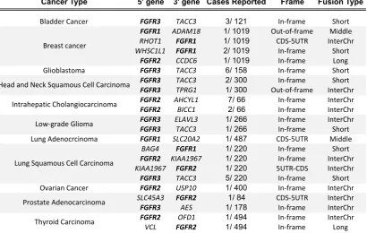
More recently, activating gene fusions in the FGFRs have been discovered in a number of cancers, typically at low incidence51,52 (Table 1). The majority of FGFR fusion partners contain dimerization domains, which induce ligand-independent receptor dimerization and oncogenic effects. FGFR3 fusions are relatively common in glioblastoma and bladder cancer, with rare reports in lung cancer52. Many FGFR3
gene fusions are with transforming acidic coiled-coil containing protein 3 (TACC3), in which the coiled coil domain is involved in protein oligomerisation and protein-protein interactions53,54. In the FGFR3-TACC3 fusion protein, the final exon at the C-terminus
of FGFR3 is replaced with TACC3, which results in oncogenic constitutive kinase activity, localisation of the fused protein to spindle poles and subsequent chromosomal segregation defects and aneuploidy51. The fused protein can activate
MAPK/ERK and JAK/STAT signalling pathways, but not PKC, due to the loss of phospholipase C (PLCγ) binding site51,55.
TACC3 is frequently FGFR3 3’ fusion partner whereas FGFR2 has several reported fusion partners. FGFR2 fusions are found in roughly 15% of intrahepatic cholangiocarcinoma56,57, and rarely in lung, thyroid and prostate cancers52. Many
fusion proteins contain protein-binding domains (citron Rho-interacting kinase (CIT), coiled-coil domain-containing protein 6 (CCDC6), cell cycle and apoptosis regulator protein 2 (CCAR2, also known as KIAA1967), oral-facial-digital syndrome 1 protein (OFD1), BicC family RNA-binding protein 1 (BICC1)) fused to the cytoplasmic tail of
fusion-receptor dimerization and ligand-independent signalling52. Interestingly,
N-terminal fusions of other proteins with FGFRs have also been reported. A fusion of the prohibitin -containing protein ER lipid raft associated 2 (ERLIN2) with FGFR1 has been described in breast cancer, and SLC45A3-FGFR2 gene fusion was identified in a patient with prostate cancer52. Although the most probable consequences of the described N-terminal fusions are increased receptor dimerization and increased kinase activation, the SLC45A3-FGFR2 gene fusion represents a unique pathogenic
mechanism, in which the entire open reading frame of FGFR2 falls under the promoter of an androgen-regulated SLC45A3, resulting in overexpression of
FGFR252.
Overexpression of FGFR2-BICC1, FGFR3-brain-specific angiogenesis inhibitor 1-associated protein 2-like protein 1 (BAIAP2L1), and FGFR3-TACC3 in 293T cells enhanced cancer cell proliferation in vitro, as well as increased susceptibility to FGFR inhibitors in vitro and in vivo52. Furthermore, stable expression of
FGFR3-BAIAP2L1, FGFR3-TACC3, and FGFR2-CCDC6 fusion proteins in a benign telomerase reverse transcriptase (hTERT) – human mammary epithelial (HME) mammary gland cell line promoted cell proliferation via increased MAPK/ERK and JAK/STAT pathway activation, highlighting a role for FGFRs in oncogenic transformation52. As more FGFR fusions emerge, their individual oncogenic potential
will need to be investigated, particularly in the case of out-of-frame fusions.
[H3] FGF ligand signalling, EMT and angiogenesis
Deregulation of FGF expression and secretion in cancer or stromal cells may also contribute to or drive carcinogenesis. Most evidence for abnormal autocrine and
progression of prostate cancer, including FGF1, FGF2, FGF6, FGF858-63 and more
recently, endocrine FGF1962,63 and FGF2364,65 (Box 1). Amplification of the 11q13 locus, including cyclin D1 (CCND1) and FGF3, FGF4 and FGF19, is frequent in many cancers. Although amplified FGFs are not expressed in many 11q13-amplified cancers66-68, in the 15% of hepatocellular carcinomas (HCC) with amplification of the 11q13 locus, FGF19 is expressed and contributes to cancer pathogenesis69. Furthermore, transgenic mice with overexpression of FGF19 at an ectopic site
(skeletal muscle) developed HCC by 10 months of age70, confirming the endocrine-like oncogenic effects of FGF19 on hepatocytes. Pre-clinical studies showed that
FGF19 stimulates tumour progression via activation of STAT371, and RNAi-mediated knockdown69 and neutralising antibodies against FGF1971,72 had a profound
anti-proliferative effect in HCC in vitro and in vivo models.
FGF2, FGF8 and FGF9 are capable of facilitating EMT in cancer by inducing mesenchymal characteristics in of epithelial cells73-77, similar to their established roles
during embryogenesis. High levels of FGF2 are expressed and secreted in triple-negative breast cancer cell lines78, specifically of mesenchymal phenotype79. In
patients, increased FGF2 levels in plasma are observed in many cancers, such as leukaemia, lung and breast cancers, particularly in metastatic disease80,81, likely
reflecting increased release of FGF2 bound to heparan sulfate proteoglycans in the extracellular matrix by invading cancer cells, although this FGF2 release is of uncertain pathogenic relevance.
Switching from an FGFR-IIIb isoform (with higher affinity for FGF1, FGF3, FGF7 and FGF10), which is enriched in epithelia, to a ‘mesenchymal’ IIIc isoform (with higher affinity for FGF1, FGF2, and FGF9)74,82,83 may facilitate EMT with enhanced FGF signalling via increased affinity for oncogenic FGFs secreted by the tumour or the
increased invasiveness of bladder and prostate84, pancreatic85 and colon cancer cell
lines 86.
FGF2 has a key role in wound healing 87,88 and angiogenesis by promoting proliferation and migration of endothelial cells 89,90 in murine models, particularly in combination with vascular endothelial growth factor (VEGF)91,92..Increased FGF2 levels were reported in patients who were resistant to anti-angiogenic agents93,
indicating a possible role for FGFs in mediating resistance to anti-VEGF therapy (Box 2). Indeed, dual inhibition of FGF and VEGF inhibited tumour growth and
angiogenesis in mouse pancreatic neuroendocrine tumours that were resistant to VEGF inhibition94.
[H3] Signal transducers
Differential expression of key signal transducing proteins may shape the signal transduction pathways activated by FGFR signalling. FRS2 amplification and protein overexpression — which may promote MAPK/ERK signalling — were reported in undifferentiated high-grade pleomorphic sarcoma and ovarian cancer, and FRS2
silencing reduced cell proliferation of liposarcoma and ovarian cancer cell lines95,96.
Growth factor receptor-bound protein 2 (GRB2) and PLCγ compete for a mutual binding site on FGFR2, and reduced GRB2 levels translate into PLCγ-mediated cancer cell migration and invasion97. A combination of increased PLCγ and low GRB2 expression levels correlate with poor clinical prognosis in ovarian98 and lung99
cancers.
[H1] Targeting FGFR in the clinic
The contribution of aberrant FGFR signalling to tumourigenesis has led to the
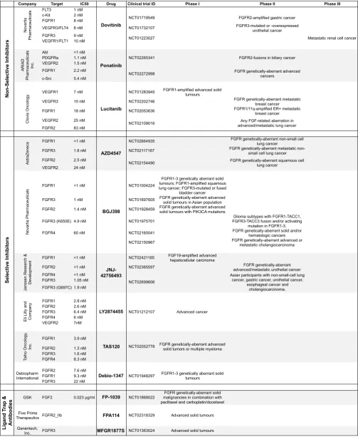
were promising in pre-clinical studies of various tumour types harbouring FGFR aberrations. Although there are no FGFR-targeted therapies approved for the treatment of cancer at present, the results of a large number of early phase therapeutic trials have revealed important information on targeting FGFR in the clinic, with therapies including small molecule tyrosine kinase inhibitors (TKIs) that target the ATP-binding cleft of the kinase domains of several growth factor receptors (multi-targeting TKIs), TKIs that selectively target the kinase domain of FGFRs (selective
TKIs), monoclonal antibodies anti-FGFR and FGF ligand traps (Table 2).
[H3] Multi-targeting TKIs
The kinase domains of FGFR, VEGFR and platelet-derived growth factor receptor (PDGFR) families are phylogenetically related, and several non-selective TKIs originally developed to inhibit the VEGFRs also inhibit FGFR. Dovitinib (TKI258) is a non-selective TKI that targets VEGFR1-3, FGFR1-3 and PDGFRb at nanomolar concentrations7. Dovitinib demonstrated prominent anti-tumour activity in a phase I
study in patients with renal cell carcinoma (RCC) 100, although reduced efficacy was
observed in a phase II study in metastatic RCC patients101. A subsequent
randomised phase III study of 570 patients for third line treatment for RCC demonstrated no difference in efficacy outcomes between dovitinib and sorafenib, another multi-targeting VEGFR inhibitor that does not appreciably inhibit FGFRs 101. Baseline levels of FGF2 did not predict for relative benefit, and were also not different between sorafenib and dovitinib when measured during treatment. These data questioned whether all efficacy of dovitinib in patients was through inhibition of VEGFR. In a separate phase II trial, treatment with dovitinib induced relatively infrequent partial responses in patients with FGFR1 or 11q13-amplified ER+ breast
potentially suggesting an oncogenic role for FGFRs in patients in whom FGFR1 or
11q13 was amplified.
Lucitanib (E3810) is another multi-TKI that targets FGFR1-2 and VEGFR1-3 among other tyrosine kinase receptors. A phase I/IIa study assessing lucitanib in solid tumours demonstrated clinical benefit in patients harbouring FGFR aberrations, with 6 out of 12 patients achieving RECIST partial response 102. Additional non-selective
TKI with anti-FGFR activity include nintedanib (BIBF1120) and ponatinib (AP24534), which so far demonstrated modest anti-tumour activities in advanced solid tumours103
and leukaemia104.
There is general uncertainty over whether these multi-targeting TKIs sufficiently inhibit FGFRs in the clinic. Dosing is limited by hypertension through VEGFR inhibition, and by non-specific toxicity102, with adverse effects specific to the selective
FGFR inhibitors frequently not observed. Stratification of patients on the basis of their FGFR expression/mutation profile identified partial responses in breast cancers with
FGFR1 (8q12) and/or 11q13 amplifications7. However it remains uncertain how much
of the activity of these TKIs is through multi-targeted inhibition of VEGFR and other non-FGFR kinases.
[H3] Selective inhibitors
In order to facilitate on-target FGFR inhibition in patients who harbour FGFR abnormalities, and also reduce toxic effects associated with multi-TKIs, selective inhibitors of the FGFRs have been developed (Table 2). The kinase domains of FGFR1-3 show high structural similarity105, and most selective inhibitors inhibit all three FGFRs to varying degrees. The FGFR4 kinase domain is structurally distinct
A retrospective analysis of the early selective inhibitor trials has revealed substantial variability in response rates between genetic aberrations. FGFR1-amplified cancers responded infrequently to selective FGFR inhibition. In a phase I study of AZD4547, an FGFR1-3 catalytic inhibitor, only one patient with FGFR1-amplified squamous NSCLC had a confirmed RECIST partial response (32% reduction in target lesions), from a total of 20 patients enrolled in the study107. In a phase I study of 132 patients with FGFR1-3 genetic aberrations, NVP-BGJ398 — a further FGFR1-3 selective
inhibitor — demonstrated partial responses in four patients with FGFR1-amplified NSCLC, and stable disease in 14 patients108. Regarding breast cancer, in a phase II
multicentre proof-of-concept study evaluating AZD4547, one out of eight patients with FGFR1-amplified breast cancer responded5 to the inhibitor. Similarly, only one
patient with FGFR1-amplified breast cancer had a tumour regression when treated with NVP-BGJ398 in the phase I study108.
The response rate in FGFR3-aberrant urothelial cancer is also uncertain. Two out of twenty patients with FGFR3-mutated bladder cancer achieved stable disease in response to AZD4547 in a phase I study107, although partial responses were also
reported in FGFR3-mutated bladder cancer in a phase I trial of NVP-BGJ398108.
Additional clinical data in patients harbouring FGFR mutations are required to reliably assess the potential of distinct individual FGFR mutations to predict response to targeted agents.
By contrast, there have been high rates of response reported for FGFR2
amplification. In a phase II trial evaluating AZD4547, three out of nine patients with
FGFR2-amplified gastric cancer had a response to AZD4547 that lasted for 27–45 weeks5. However, a separate phase II study showed no statistically-significant
paclitaxel arm)109, with evidence that response was limited by intra-tumoural
heterogeneity, as discussed later in the Review.
Tumours with FGFR fusions seem to have a high response rate to FGFR inhibition. Tumour shrinkage was observed in one cholangiocarcinoma and one HCC patient with FGFR2-BICC1 gene fusions in response to NVP-BGJ398 in a phase I study108. Consequently, this drug is now being investigated in phase II studies in
advanced-stage cholangiocarcinoma110, advanced-stage gastrointestinal stromal tumours (GIST)111 and other solid and haematologic malignancies112. Patients with urothelial
tumours harbouring either FGFR2 truncation or FGFR3-TACC3 fusion also demonstrated clinical responses in a phase I dose-escalation study of JNJ-427564936, which is now being assessed in a phase II study in unresectable
urothelial cancers with FGFR genomic aberrations113. Two more inhibitors,
LY2874455114 and TAS120112, are currently in phase I trials.
Collectively, early trials of selective TKIs proved highly successful in targeting FGFR fusions and selected patients with FGFR2 amplification, although only marginal success was seen when targeting other FGFR aberrations.
[H3] Monoclonal antibodies targeting FGF and FGFR
Although several monoclonal antibodies (mAbs) against FGFRs have been developed, limited clinical data are currently available. MGFR1877S is an anti-FGFR3 mAb that was evaluated in a phase I dose-escalation trial in patients with advanced-stage solid tumours115. Stable disease was reported to be the best response in patients with urothelial cell carcinoma (5 out of 10 patients), with
mAb against FGFR1-IIIc, named IMC-A1, was shown to induce severe anorexia in animal models117, and thus was never translated into the clinic. The FGFR2-IIIb blocking mAb FPA144 inhibited growth of FGFR2-amplified gastric cancer xenografts by 72% to 100%118 and recently entered a phase I trial119. Data from 13 patients enrolled to date in this trial showed no dose-limiting toxic effects associated with FPA144 administration, with upper respiratory infection, alopecia and fatigue reported as adverse events in more than one patient120.
FP-1039 is a FGF ligand trap; a soluble fusion protein that contains the extracellular
domain of FGFR1-IIIc splice isoform and demonstrated angiogenic and anti-proliferative properties in multiple cancer cell line models via selective sequestration of non-hormonal FGFs121. Recently, a first in-human phase I study evaluating
FP-1039 in patients with metastatic or locally advanced-stage solid tumours has been completed122. In an unselected patient population, the best response was recorded to
be stable disease (41.7%) and major adverse effects observed were diarrhoea (43.6%), fatigue (43.6%), and nausea (25.6%)122. No apparent relationship was
reported between tumour response and FGF pathway aberrations in the 39 patients enrolled.
[H1] Challenges and opportunities
[H3] Challenges of patient selection
Prospective selection of patients with specific FGFR aberrations is one of the major challenges in clinical trials. The overall infrequency of individual FGFR aberrations complicates identification of the best target population for each selective inhibitor. Further complicating early phase clinical trials have been basket trials, an approach that includes all patients with any FGFR aberration. As it has become clear that
focused on individual aberrations123. Tumour biopsy material is often limited, and
increasing evidence supports the potential to screen for FGFR aberration in plasma on circulating tumour DNA24. Non-invasive and inexpensive approaches like this could aid broader capture of genetic landscape of a tumour, and studies investigating detection of FGFR genetic aberrations in plasma are currently ongoing.
Additional challenges have emerged in selecting patients with FGFR amplification,
with ambiguity over the criteria for amplification and the importance of clonality in determining response. Early-phase trials of FGFR inhibitors selected patients on the
basis of criteria used to define HER2 amplification (gene to centromere ratio >2), yet evidence suggests only tumours with higher FGFR copy number (gene to centromere ratio >4/5) are likely to respond to FGFR inhibition24. FGFR1 protein is
frequently not overexpressed in cancers with lower levels of FGFR1 amplification 124-126, and mRNA levels of FGFR1 in those cases may be more reliable. Moreover,
intra-tumour heterogeneity presents a major selection challenge. FGFR2
amplification in gastric cancer is frequently sub-clonal109, with response observed
only in cancers with clonal amplification24. The importance of clonality in response to
mutations and fusions has yet to be explored. In general, oncogenic fusions are early truncal events in cancer, frequently occurring in genomically stable tumours, reinforcing the potential for therapeutic targeting of FGFR fusions.
[H3] Variable addiction to FGFR amplification
Increasing evidence suggests that only a fraction of cancers with FGFR aberrations are addicted to FGFR signalling. Differential activation of signal transduction pathways by different FGFRs and by distinct oncogenic events is likely critical in
of MAPK/ERK signalling is particularly important during organogenesis127,128; FGFRs
have been shown to signal primarily through ERK1/2 during development, and FGF, FGFR, and ERK1/2 loss-of-function phenotypes are very similar129. In cancer, the MAPK/ERK signalling pathway is also most strongly activated by FGFR signalling across diverse aberrations, such as mutation or overexpression of the receptor molecules.. In many cellular contexts, this dominant signalling through the MAPK/ERK pathway is insufficient to drive addiction to FGFR signalling. Although
FGFR signalling may contribute to oncogenesis and FGFR inhibition may result in reduced proliferation in cancer cell lines, this has not translated into single agent
efficacy in the clinic.
In vitro studies identified a moderate correlation between FGFR1 locus 8q12
amplification and sensitivity to FGFR inhibitors in NSCLC10 and breast cancer130. Yet
mouse models question whether FGFR1 amplification and overexpression induces oncogene addiction. Exogenous overexpression of FGFR1 in animal models does not result in malignant transformation, and induced dimerization of FGFR1 is required to trigger invasive properties in normal breast epithelial cell lines131 and
transgenic mouse models of progressive mammary gland tumourigenesis132. In FGFR1-amplified cell lines, FGFR inhibition frequently results in inhibition of MAPK/ERK signalling, but without substantially affecting other signal transduction pathways such as PI3K/AKT signalling. Co-aberrant genes in FGFR1-amplified cancers may also result in reduced addiction to the FGFR pathway, including
PIK3CA activating mutations and amplification of CCND132. Although FGFR1 may contribute to aspects of tumour progression, such as endocrine resistance in breast cancer (Box 2), FGFR1 is not a dominant oncogene.
wider control of signal transduction and mTOR activity by FGFR2 signalling24. FGFR2 amplified at very high levels results in supra-physiological FGFR2 expression, signalling and oncogene addiction, with a partial crosstalk between FGFR2 and other receptor tyrosine kinases, including ERBB3 (also known as HER3) and insulin growth factor receptor 1 (IGF1R)24.
The mechanisms through which FGFR fusion proteins mediate addiction to FGFR
signalling remain to be elucidated, and is likely to be cancer type-dependent. Overexpression of FGFR3 fusion proteins transformed 293T cells52 and Rat1A
fibroblasts51, and enhanced cell proliferation compared with overexpression of wild-type receptors52. Bladder cancer cell lines and xenograft models expressing fused
FGFR3 proteins were sensitised to the FGFR inhibition51,55, although not by
expression of FGFR3 containing hotspot mutations52,55.
HCC harbouring FGF19 amplification may also represent a subset of cancers strongly addicted to the FGFR pathway. FGF19 amplification, and consequent ligand overexpression and FGFR4 activation, contribute to HCC development70. Pre-clinical
data show that a blocking anti-FGFR4 monoclonal antibody (LD1) significantly reduced HCC xenograft growth133. A small-molecule inhibitor of FGFR4, BLU9931, with high selectivity against the other FGFR family members, suppressed tumour growth in HCC xenograft models with FGF19 amplification134. The pan-FGFR inhibitor JNJ-42756493, which inhibits FGFR4 at doses similar to those used to inhibit the other FGFR receptors, is currently being investigated in patients with advanced-stage HCC135 (Table 2).
Collectively, these data have led to a growing understanding of the importance of
demonstrated in xenograft models of NSCLC and head and neck squamous cell carcinoma (HNSCC), and in one patient136, many cancers with mutations in FGFR2
and FGFR3 display limited FGFR-dependent signalling. Select cancers with high levels of FGF19 and FGFR2 amplification and FGFR fusions present putative biomarkers of FGFR addiction and confer sensitivity to targeted agents, unlike low-level FGFR1 amplification.
Combination therapeutic approaches may overcome the limitations of single agent FGFR inhibition in FGFR1-amplified cancers. Inhibitors of the PI3K-mTOR pathway
are synergistic with FGFR inhibition, in part as mTOR activity is frequently only weakly inhibited by targeting FGFR, with synergy described both in vitro and in vivo
in HNSCC cell lines16, endometrial cancer models137, gastric adenocarcinoma24 and
HCC138. Despite these observations, combined individual toxic effects of these
inhibitors will likely become a limiting factor in implementing this combination in the clinic.
[H3] Toxicity limits pan-FGFR inhibition in the clinic
On-target toxicity from pan-FGFR1-3 inhibition — including hyperphosphatemia , skin and eye dryness, keratopathy and asymptomatic retinal pigment detachment — limits dosing 139,140. Higher specificity with antibodies, or a next generation of selective
inhibitors against a single FGFR, could minimise the appearance of adverse effects. Blockade of FGFs with the ligand binding trap FP-1039 reduced growth of FGFR1 -amplified lung cancer cell lines, xenografts121 and in a phase I study122, with no effect on serum calcium or phosphate levels. Similarly, a small-molecule ligand trap derived from long pentraxin 3 protein (NSC12) demonstrated potent anti-tumour action in FGFR-dependent xenograft models without systemic toxicity in the treated
[H3] Mechanisms of acquired resistance to FGFR inhibitors
As with the majority of targeted treatments, a growing challenge of FGFR inhibition efficacy is development of drug resistance. In vitro studies have identified
‘gatekeeper’ mutations in the FGFRs, and bypass activation of downstream signalling via alternate receptor tyrosine kinase, as frequent mechanisms of acquired or intrinsic resistance to targeted therapies (Figure 3).
‘Gatekeeper’ mutations in the ATP binding cleft that induce resistance to FGFR inhibition have been identified pre-clinically. A ‘gatekeeper’ mutation FGFR3_V555M,
along with comparable residues FGFR1_V561 and FGFR2_V564, induces resistance to multiple FGFR inhibitors in vitro142-144. Protein modelling studies
suggest that these ‘gatekeeper’ mutations in the ATP cleft strengthen the hydrophobic spine of the kinase and may create a steric conflict to hinder drug-binding efficiency143. The substitution of V561 for a ‘bulky’ Met amino acid resulted in
complete disruption of FGFR1 binding to PD173074144. Although these studies
demonstrate emergence of mutants resistant to FGFR inhibitors as mechanisms of acquired resistance pre-clinically, they are yet to be confirmed in samples of patient who have experienced clinical progression. In light of emergence of ‘gatekeeper’ mutations in FGFRs, irreversible covalent FGFR inhibitors that bind such FGFRs have been developed with the aim to overcome resistance to selective FGFR inhibitors145.
Activation of alternative receptor tyrosine kinases, in particular the ERBB receptor family has been described as an escape mechanism in FGFR-resistant tumours. FGFR3-dependent bladder cancer cell lines developed rapid resistance to the FGFR inhibitor NVP-BGJ398 via switching to signalling through either ERBB2 (also known
Furthermore, dual inhibition of FGFR3 and EGFR activity in FGFR3-mutant bladder cancer cell lines resulted in increased cell death147. In FGFR1-amplified NSCLC cell lines resistant to FGFR therapy, PDGFRA and HER2 were reported to be co-activated126. Use of novel approaches to allow detection of alternatively activated tyrosine kinase receptors or signalling pathways may augment selection of cancers for which FGFR inhibition is effective.
[H1] Conclusion
References
1 Sleeman, M. et al. Identification of a new fibroblast growth factor receptor, FGFR5. Gene 271, 171-182 (2001).
2 Itoh, N. & Ornitz, D. M. Fibroblast growth factors: from molecular evolution to roles in development, metabolism and disease. Journal of biochemistry 149, 121-130, doi:10.1093/jb/mvq121 (2011).
3 Dienstmann, R. et al. Genomic aberrations in the FGFR pathway: opportunities for targeted therapies in solid tumors. Annals of oncology : official journal of the European Society for Medical Oncology / ESMO 25, 552-563, doi:10.1093/annonc/mdt419 (2014).
4 Dieci, M. V., Arnedos, M., Andre, F. & Soria, J. C. Fibroblast growth factor receptor inhibitors as a cancer treatment: from a biologic rationale to medical perspectives. Cancer discovery 3, 264-279, doi:10.1158/2159-8290.CD-12-0362 (2013).
5 Smyth, E. C. T., N. C.; Peckitt, C.; Pearson, A.; Brown, G.; Chua, S.; Gillbanks, A.; Johnston, S. R. D.; Tarazona, N.; Cutts, R.; Kilgour, E.; Rooney, C.; Smith, N. R.; Sumpter, K. A.; Ajaz, M. A.; Thomas, A. L.; Watkins, D.; Chau, I.; Popat, S.; Cunningham D. Phase II multicenter proof of concept study of AZD4547 in FGFR amplified tumours. . ASCO Annual Meeting 33, 2508 (2015).
6 Tabernero, J. et al. Phase I Dose-Escalation Study of JNJ-42756493, an Oral Pan-Fibroblast Growth Factor Receptor Inhibitor, in Patients With Advanced Solid Tumors. Journal of clinical oncology : official journal of the American Society of Clinical Oncology 33, 3401-3408, doi:10.1200/JCO.2014.60.7341 (2015).
7 Andre, F. et al. Targeting FGFR with dovitinib (TKI258): preclinical and clinical data in breast cancer. Clinical cancer research : an official journal of
the American Association for Cancer Research 19, 3693-3702,
doi:10.1158/1078-0432.CCR-13-0190 (2013).
8 Konecny, G. E. et al. Second-line dovitinib (TKI258) in patients with FGFR2-mutated or FGFR2-non-FGFR2-mutated advanced or metastatic endometrial cancer: a non-randomised, open-label, two-group, two-stage, phase 2 study. Lancet Oncol 16, 686-694, doi:10.1016/S1470-2045(15)70159-2 (2015).
9 Yang, W. et al. Prognostic value of FGFR1 gene copy number in patients with non-small cell lung cancer: a meta-analysis. Journal of thoracic disease 6, 803-809, doi:10.3978/j.issn.2072-1439.2014.05.02 (2014).
10 Weiss, J. et al. Frequent and focal FGFR1 amplification associates with therapeutically tractable FGFR1 dependency in squamous cell lung cancer.
Science translational medicine 2, 62ra93, doi:10.1126/scitranslmed.3001451 (2010).
11 Peifer, M. et al. Integrative genome analyses identify key somatic driver mutations of small-cell lung cancer. Nature genetics 44, 1104-1110, doi:10.1038/ng.2396 (2012).
12 Cihoric, N. et al. Prognostic role of FGFR1 amplification in early-stage non-small cell lung cancer. British journal of cancer 110, 2914-2922, doi:10.1038/bjc.2014.229 (2014).
13 Reis-Filho, J. S. et al. FGFR1 emerges as a potential therapeutic target for lobular breast carcinomas. Clinical cancer research : an official journal of the
American Association for Cancer Research 12, 6652-6662,
14 Courjal, F. et al. Mapping of DNA amplifications at 15 chromosomal localizations in 1875 breast tumors: definition of phenotypic groups. Cancer research 57, 4360-4367 (1997).
15 Lee, H. J. et al. Low prognostic implication of fibroblast growth factor family activation in triple-negative breast cancer subsets. Annals of surgical oncology 21, 1561-1568, doi:10.1245/s10434-013-3456-x (2014).
16 Singleton, K. R. et al. Kinome RNAi Screens Reveal Synergistic Targeting of MTOR and FGFR1 Pathways for Treatment of Lung Cancer and HNSCC.
Cancer research 75, 4398-4406, doi:10.1158/0008-5472.CAN-15-0509 (2015).
17 Turner, N. et al. FGFR1 amplification drives endocrine therapy resistance and is a therapeutic target in breast cancer. Cancer research 70, 2085-2094, doi:10.1158/0008-5472.CAN-09-3746 (2010).
18 Mohammadi, M. et al. Crystal structure of an angiogenesis inhibitor bound to the FGF receptor tyrosine kinase domain. The EMBO journal 17, 5896-5904, doi:10.1093/emboj/17.20.5896 (1998).
19 Campbell, J. et al. Large-Scale Profiling of Kinase Dependencies in Cancer Cell Lines. Cell reports 14, 2490-2501, doi:10.1016/j.celrep.2016.02.023 (2016).
20 Guagnano, V. et al. FGFR genetic alterations predict for sensitivity to NVP-BGJ398, a selective pan-FGFR inhibitor. Cancer discovery 2, 1118-1133, doi:10.1158/2159-8290.CD-12-0210 (2012).
21 Matsumoto, K. et al. FGFR2 gene amplification and clinicopathological features in gastric cancer. British journal of cancer 106, 727-732, doi:10.1038/bjc.2011.603 (2012).
22 Turner, N. et al. Integrative molecular profiling of triple negative breast cancers identifies amplicon drivers and potential therapeutic targets.
Oncogene 29, 2013-2023, doi:onc2009489 [pii] 10.1038/onc.2009.489 (2010).
23 Ueda, T. et al. Deletion of the carboxyl-terminal exons of K-sam/FGFR2 by short homology-mediated recombination, generating preferential expression of specific messenger RNAs. Cancer research 59, 6080-6086 (1999).
24 Pearson, A. et al. High-Level Clonal FGFR Amplification and Response to FGFR Inhibition in a Translational Clinical Trial. Cancer discovery, doi:10.1158/2159-8290.CD-15-1246 (2016).
25 Verstraete, M. et al. In vitro and in vivo evaluation of the radiosensitizing effect of a selective FGFR inhibitor (JNJ-42756493) for rectal cancer. BMC cancer 15, 946, doi:10.1186/s12885-015-2000-8 (2015).
26 Reynisdottir, I. et al. High expression of ZNF703 independent of amplification indicates worse prognosis in patients with luminal B breast cancer. Cancer medicine 2, 437-446, doi:10.1002/cam4.88 (2013).
27 Baykara, O., Bakir, B., Buyru, N., Kaynak, K. & Dalay, N. Amplification of chromosome 8 genes in lung cancer. Journal of Cancer 6, 270-275, doi:10.7150/jca.10638 (2015).
28 Zhang, X. et al. Luminal breast cancer cell lines overexpressing ZNF703 are resistant to tamoxifen through activation of Akt/mTOR signaling. PloS one 8, e72053, doi:10.1371/journal.pone.0072053 (2013).
29 Fischbach, A. et al. Fibroblast growth factor receptor (FGFR) gene amplifications are rare events in bladder cancer. Histopathology 66, 639-649, doi:10.1111/his.12473 (2015).
31 Dutt, A. et al. Drug-sensitive FGFR2 mutations in endometrial carcinoma.
Proceedings of the National Academy of Sciences of the United States of America 105, 8713-8717, doi:10.1073/pnas.0803379105 (2008).
32 Helsten, T. et al. The FGFR Landscape in Cancer: Analysis of 4,853 Tumors by Next-Generation Sequencing. Clinical cancer research : an official journal of the American Association for Cancer Research, doi:10.1158/1078-0432.CCR-14-3212 (2015).
33 Greenman, C. et al. Patterns of somatic mutation in human cancer genomes.
Nature 446, 153-158, doi:10.1038/nature05610 (2007).
34 Gao, J. et al. Integrative analysis of complex cancer genomics and clinical profiles using the cBioPortal. Science signaling 6, pl1, doi:10.1126/scisignal.2004088 (2013).
35 Ibrahimi, O. A. et al. Biochemical analysis of pathogenic ligand-dependent FGFR2 mutations suggests distinct pathophysiological mechanisms for craniofacial and limb abnormalities. Human molecular genetics 13, 2313-2324, doi:10.1093/hmg/ddh235 (2004).
36 Ibrahimi, O. A. et al. Structural basis for fibroblast growth factor receptor 2 activation in Apert syndrome. Proceedings of the National Academy of
Sciences of the United States of America 98, 7182-7187,
doi:10.1073/pnas.121183798 (2001).
37 Wilkie, A. O., Patey, S. J., Kan, S. H., van den Ouweland, A. M. & Hamel, B. C. FGFs, their receptors, and human limb malformations: clinical and molecular correlations. American journal of medical genetics 112, 266-278, doi:10.1002/ajmg.10775 (2002).
38 Tanizaki, J. et al. Identification of Oncogenic and Drug-Sensitizing Mutations in the Extracellular Domain of FGFR2. Cancer research 75, 3139-3146, doi:10.1158/0008-5472.CAN-14-3771 (2015).
39 Cappellen, D. et al. Frequent activating mutations of FGFR3 in human bladder and cervix carcinomas. Nature genetics 23, 18-20, doi:10.1038/12615 (1999).
40 Rosty, C. et al. Clinical and biological characteristics of cervical neoplasias with FGFR3 mutation. Molecular cancer 4, 15, doi:10.1186/1476-4598-4-15 (2005).
41 Bernard-Pierrot, I. et al. Oncogenic properties of the mutated forms of fibroblast growth factor receptor 3b. Carcinogenesis 27, 740-747, doi:10.1093/carcin/bgi290 (2006).
42 Chen, H. et al. A molecular brake in the kinase hinge region regulates the activity of receptor tyrosine kinases. Molecular cell 27, 717-730, doi:10.1016/j.molcel.2007.06.028 (2007).
43 Hart, K. C. et al. Transformation and Stat activation by derivatives of FGFR1, FGFR3, and FGFR4. Oncogene 19, 3309-3320, doi:10.1038/sj.onc.1203650 (2000).
44 Taylor, J. G. t. et al. Identification of FGFR4-activating mutations in human rhabdomyosarcomas that promote metastasis in xenotransplanted models.
The Journal of clinical investigation 119, 3395-3407, doi:10.1172/JCI39703 (2009).
45 Andersen, S. W. et al. Breast cancer susceptibility associated with rs1219648 (fibroblast growth factor receptor 2) and postmenopausal hormone therapy use in a population-based United States study. Menopause 20, 354-358, doi:10.1097/GME.0b013e318268ca46 (2013).
46 Hunter, D. J. et al. A genome-wide association study identifies alleles in FGFR2 associated with risk of sporadic postmenopausal breast cancer.
47 Meyer, K. B. et al. Allele-specific up-regulation of FGFR2 increases susceptibility to breast cancer. PLoS biology 6, e108, doi:10.1371/journal.pbio.0060108 (2008).
48 Frullanti, E. et al. Meta and pooled analyses of FGFR4 Gly388Arg polymorphism as a cancer prognostic factor. European journal of cancer prevention : the official journal of the European Cancer Prevention Organisation 20, 340-347, doi:10.1097/CEJ.0b013e3283457274 (2011). 49 Bange, J. et al. Cancer progression and tumor cell motility are associated
with the FGFR4 Arg(388) allele. Cancer research 62, 840-847 (2002).
50 Ulaganathan, V. K., Sperl, B., Rapp, U. R. & Ullrich, A. Germline variant FGFR4 p.G388R exposes a membrane-proximal STAT3 binding site. Nature
528, 570-574, doi:10.1038/nature16449 (2015).
51 Singh, D. et al. Transforming fusions of FGFR and TACC genes in human glioblastoma. Science 337, 1231-1235, doi:10.1126/science.1220834 (2012). 52 Wu, Y. M. et al. Identification of targetable FGFR gene fusions in diverse
cancers. Cancer discovery 3, 636-647, doi:10.1158/2159-8290.CD-13-0050 (2013).
53 Ha, G. H., Kim, J. L. & Breuer, E. K. Transforming acidic coiled-coil proteins (TACCs) in human cancer. Cancer letters 336, 24-33, doi:10.1016/j.canlet.2013.04.022 (2013).
54 Hood, F. E. & Royle, S. J. Pulling it together: The mitotic function of TACC3.
Bioarchitecture 1, 105-109, doi:10.4161/bioa.1.3.16518 (2011).
55 Williams, S. V., Hurst, C. D. & Knowles, M. A. Oncogenic FGFR3 gene fusions in bladder cancer. Human molecular genetics 22, 795-803, doi:10.1093/hmg/dds486 (2013).
56 Sia, D. et al. Massive parallel sequencing uncovers actionable FGFR2-PPHLN1 fusion and ARAF mutations in intrahepatic cholangiocarcinoma.
Nature communications 6, 6087, doi:10.1038/ncomms7087 (2015).
57 Arai, Y. et al. Fibroblast growth factor receptor 2 tyrosine kinase fusions define a unique molecular subtype of cholangiocarcinoma. Hepatology 59, 1427-1434, doi:10.1002/hep.26890 (2014).
58 Giacomini, A. et al. A long pentraxin-3-derived pentapeptide for the therapy of FGF8b-driven steroid hormone-regulated cancers. Oncotarget 6, 13790-13802 (2015).
59 Wan, X. et al. Prostate cancer cell-stromal cell crosstalk via FGFR1 mediates antitumor activity of dovitinib in bone metastases. Science translational medicine 6, 252ra122, doi:10.1126/scitranslmed.3009332 (2014).
60 Tanner, Y. & Grose, R. P. Dysregulated FGF signalling in neoplastic disorders. Seminars in cell & developmental biology, doi:10.1016/j.semcdb.2015.10.012 (2015).
61 Tuomela, J. & Harkonen, P. Tumor models for prostate cancer exemplified by fibroblast growth factor 8-induced tumorigenesis and tumor progression.
Reproductive biology 14, 16-24, doi:10.1016/j.repbio.2014.01.002 (2014). 62 Feng, S., Dakhova, O., Creighton, C. J. & Ittmann, M. Endocrine fibroblast
growth factor FGF19 promotes prostate cancer progression. Cancer research
73, 2551-2562, doi:10.1158/0008-5472.CAN-12-4108 (2013).
63 Nagamatsu, H. et al. FGF19 promotes progression of prostate cancer. The Prostate 75, 1092-1101, doi:10.1002/pros.22994 (2015).
64 Feng, S., Wang, J., Zhang, Y., Creighton, C. J. & Ittmann, M. FGF23 promotes prostate cancer progression. Oncotarget 6, 17291-17301 (2015). 65 Lee, E. K. et al. FGF23: mediator of poor prognosis in a sizeable subgroup of
66 Schuuring, E. The involvement of the chromosome 11q13 region in human malignancies: cyclin D1 and EMS1 are two new candidate oncogenes--a review. Gene 159, 83-96 (1995).
67 Karlsson, E. et al. High-resolution genomic analysis of the 11q13 amplicon in breast cancers identifies synergy with 8p12 amplification, involving the mTOR targets S6K2 and 4EBP1. Genes, chromosomes & cancer 50, 775-787, doi:10.1002/gcc.20900 (2011).
68 Ormandy, C. J., Musgrove, E. A., Hui, R., Daly, R. J. & Sutherland, R. L. Cyclin D1, EMS1 and 11q13 amplification in breast cancer. Breast cancer research and treatment 78, 323-335 (2003).
69 Sawey, E. T. et al. Identification of a therapeutic strategy targeting amplified FGF19 in liver cancer by Oncogenomic screening. Cancer cell 19, 347-358, doi:10.1016/j.ccr.2011.01.040 (2011).
70 Nicholes, K. et al. A mouse model of hepatocellular carcinoma: ectopic expression of fibroblast growth factor 19 in skeletal muscle of transgenic mice. The American journal of pathology 160, 2295-2307, doi:10.1016/S0002-9440(10)61177-7 (2002).
71 Zhou, M. et al. Separating Tumorigenicity from Bile Acid Regulatory Activity for Endocrine Hormone FGF19. Cancer research 74, 3306-3316, doi:10.1158/0008-5472.CAN-14-0208 (2014).
72 Wu, A. L. et al. FGF19 regulates cell proliferation, glucose and bile acid metabolism via FGFR4-dependent and independent pathways. PloS one 6, e17868, doi:10.1371/journal.pone.0017868 (2011).
73 Chen, J. et al. TGF-beta1 and FGF2 stimulate the epithelial-mesenchymal transition of HERS cells through a MEK-dependent mechanism. Journal of cellular physiology 229, 1647-1659, doi:10.1002/jcp.24610 (2014).
74 Shirakihara, T. et al. TGF-beta regulates isoform switching of FGF receptors and epithelial-mesenchymal transition. The EMBO journal 30, 783-795, doi:10.1038/emboj.2010.351 (2011).
75 Fillmore, C. M. et al. Estrogen expands breast cancer stem-like cells through paracrine FGF/Tbx3 signaling. Proceedings of the National Academy of
Sciences of the United States of America 107, 21737-21742,
doi:10.1073/pnas.1007863107 (2010).
76 Acevedo, V. D. et al. Inducible FGFR-1 activation leads to irreversible prostate adenocarcinoma and an epithelial-to-mesenchymal transition.
Cancer cell 12, 559-571, doi:10.1016/j.ccr.2007.11.004 (2007).
77 Marek, L. et al. Fibroblast growth factor (FGF) and FGF receptor-mediated autocrine signaling in non-small-cell lung cancer cells. Molecular pharmacology 75, 196-207, doi:10.1124/mol.108.049544 (2009).
78 Sharpe, R. et al. FGFR signaling promotes the growth of triple-negative and basal-like breast cancer cell lines both in vitro and in vivo. Clinical cancer research : an official journal of the American Association for Cancer Research
17, 5275-5286, doi:10.1158/1078-0432.CCR-10-2727 (2011).
79 Abramson, V. G., Lehmann, B. D., Ballinger, T. J. & Pietenpol, J. A. Subtyping of triple-negative breast cancer: implications for therapy. Cancer
121, 8-16, doi:10.1002/cncr.28914 (2015).
80 Ruotsalainen, T., Joensuu, H., Mattson, K. & Salven, P. High pretreatment serum concentration of basic fibroblast growth factor is a predictor of poor prognosis in small cell lung cancer. Cancer epidemiology, biomarkers & prevention : a publication of the American Association for Cancer Research, cosponsored by the American Society of Preventive Oncology 11, 1492-1495 (2002).
81 Nguyen, M. et al. Elevated levels of an angiogenic peptide, basic fibroblast growth factor, in the urine of patients with a wide spectrum of cancers.
82 Yan, G., Fukabori, Y., McBride, G., Nikolaropolous, S. & McKeehan, W. L. Exon switching and activation of stromal and embryonic fibroblast growth factor (FGF)-FGF receptor genes in prostate epithelial cells accompany stromal independence and malignancy. Molecular and cellular biology 13, 4513-4522 (1993).
83 Ranieri, D., Belleudi, F., Magenta, A. & Torrisi, M. R. HPV16 E5 expression induces switching from FGFR2b to FGFR2c and epithelial-mesenchymal transition. International journal of cancer. Journal international du cancer 137, 61-72, doi:10.1002/ijc.29373 (2015).
84 Chaffer, C. L. et al. Mesenchymal-to-epithelial transition facilitates bladder cancer metastasis: role of fibroblast growth factor receptor-2. Cancer research 66, 11271-11278, doi:10.1158/0008-5472.CAN-06-2044 (2006). 85 Ishiwata, T. et al. Enhanced expression of fibroblast growth factor receptor 2
IIIc promotes human pancreatic cancer cell proliferation. The American journal of pathology 180, 1928-1941, doi:10.1016/j.ajpath.2012.01.020 (2012).
86 Matsuda, Y., Hagio, M., Seya, T. & Ishiwata, T. Fibroblast growth factor receptor 2 IIIc as a therapeutic target for colorectal cancer cells. Mol Cancer Ther 11, 2010-2020, doi:10.1158/1535-7163.MCT-12-0243 (2012).
87 Broadley, K. N. et al. Monospecific antibodies implicate basic fibroblast growth factor in normal wound repair. Laboratory investigation; a journal of technical methods and pathology 61, 571-575 (1989).
88 Ortega, S., Ittmann, M., Tsang, S. H., Ehrlich, M. & Basilico, C. Neuronal defects and delayed wound healing in mice lacking fibroblast growth factor 2.
Proceedings of the National Academy of Sciences of the United States of America 95, 5672-5677 (1998).
89 Compagni, A., Wilgenbus, P., Impagnatiello, M. A., Cotten, M. & Christofori, G. Fibroblast growth factors are required for efficient tumor angiogenesis.
Cancer research 60, 7163-7169 (2000).
90 Ronca, R., Giacomini, A., Rusnati, M. & Presta, M. The potential of fibroblast growth factor/fibroblast growth factor receptor signaling as a therapeutic target in tumor angiogenesis. Expert opinion on therapeutic targets 19, 1361-1377, doi:10.1517/14728222.2015.1062475 (2015).
91 Pepper, M. S., Mandriota, S. J., Jeltsch, M., Kumar, V. & Alitalo, K. Vascular endothelial growth factor (VEGF)-C synergizes with basic fibroblast growth factor and VEGF in the induction of angiogenesis in vitro and alters endothelial cell extracellular proteolytic activity. Journal of cellular physiology
177, 439-452, doi:10.1002/(SICI)1097-4652(199812)177:3<439::AID-JCP7>3.0.CO;2-2 (1998).
92 Yan, W., Bentley, B. & Shao, R. Distinct angiogenic mediators are required for basic fibroblast growth and vascular endothelial growth factor-induced angiogenesis: the role of cytoplasmic tyrosine kinase c-Abl in tumor angiogenesis. Molecular biology of the cell 19, 2278-2288, doi:10.1091/mbc.E07-10-1068 (2008).
93 Kopetz, S. et al. Phase II trial of infusional fluorouracil, irinotecan, and bevacizumab for metastatic colorectal cancer: efficacy and circulating angiogenic biomarkers associated with therapeutic resistance. Journal of clinical oncology : official journal of the American Society of Clinical Oncology
28, 453-459, doi:10.1200/JCO.2009.24.8252 (2010).
95 Zhang, K. et al. Amplification of FRS2 and activation of FGFR/FRS2 signaling pathway in high-grade liposarcoma. Cancer research 73, 1298-1307, doi:10.1158/0008-5472.CAN-12-2086 (2013).
96 Luo, L. Y. et al. The Tyrosine Kinase Adaptor Protein FRS2 Is Oncogenic and Amplified in High-Grade Serous Ovarian Cancer. Mol Cancer Res 13, 502-509, doi:10.1158/1541-7786.MCR-14-0407 (2015).
97 Timsah, Z. et al. Competition between Grb2 and Plcgamma1 for FGFR2 regulates basal phospholipase activity and invasion. Nat Struct Mol Biol 21, 180-188, doi:10.1038/nsmb.2752 (2014).
98 Timsah, Z. et al. Grb2 depletion under non-stimulated conditions inhibits PTEN, promotes Akt-induced tumor formation and contributes to poor prognosis in ovarian cancer. Oncogene 35, 2186-2196, doi:10.1038/onc.2015.279 (2016).
99 Timsah, Z. et al. Expression pattern of FGFR2, Grb2 and Plcgamma1 acts as a novel prognostic marker of recurrence recurrence-free survival in lung adenocarcinoma. Am J Cancer Res 5, 3135-3148 (2015).
100 Angevin, E. et al. Phase I study of dovitinib (TKI258), an oral FGFR, VEGFR, and PDGFR inhibitor, in advanced or metastatic renal cell carcinoma. Clinical cancer research : an official journal of the American Association for Cancer Research 19, 1257-1268, doi:10.1158/1078-0432.CCR-12-2885 (2013). 101 Motzer, R. J. et al. Dovitinib versus sorafenib for third-line targeted treatment
of patients with metastatic renal cell carcinoma: an open-label, randomised phase 3 trial. Lancet Oncol 15, 286-296, doi:10.1016/S1470-2045(14)70030-0 (2doi:10.1016/S1470-2045(14)70030-014).
102 Soria, J. C. et al. Phase I/IIa study evaluating the safety, efficacy, pharmacokinetics, and pharmacodynamics of lucitanib in advanced solid tumors. Annals of oncology : official journal of the European Society for Medical Oncology / ESMO 25, 2244-2251, doi:10.1093/annonc/mdu390 (2014).
103 Okamoto, I. et al. Phase I safety, pharmacokinetic, and biomarker study of BIBF 1120, an oral triple tyrosine kinase inhibitor in patients with advanced solid tumors. Mol Cancer Ther 9, 2825-2833, doi:10.1158/1535-7163.MCT-10-0379 (2010).
104 Cortes, J. E. et al. A phase 2 trial of ponatinib in Philadelphia chromosome-positive leukemias. The New England journal of medicine 369, 1783-1796, doi:10.1056/NEJMoa1306494 (2013).
105 Johnson, D. E. & Williams, L. T. Structural and functional diversity in the FGF receptor multigene family. Advances in cancer research 60, 1-41 (1993). 106 Lesca, E., Lammens, A., Huber, R. & Augustin, M. Structural analysis of the
human fibroblast growth factor receptor 4 kinase. Journal of molecular biology
426, 3744-3756, doi:10.1016/j.jmb.2014.09.004 (2014).
107 Andre F., R. M., Dean E., Varga A., van der Noll R., Stockman P. K., Ghiorghiu D., Kilgour E., Smith P. D., Macpherson M., Lawrence P., Hastie A. and Schellens J. H. M. Results of a phase I study of AZD4547, an inhibitor of fibroblast growth factor receptor (FGFR), in patients with advanced solid tumors. AACR 104th Annual Meeting (2013).
108 Nogova, L. et al. Evaluation of BGJ398, a Fibroblast Growth Factor Receptor 1-3 Kinase Inhibitor, in Patients With Advanced Solid Tumors Harboring Genetic Alterations in Fibroblast Growth Factor Receptors: Results of a Global Phase I, Dose-Escalation and Dose-Expansion Study. Journal of clinical oncology : official journal of the American Society of Clinical Oncology
35, 157-165 (2017).
Catherine Geh, Elaine Kilgour. in 2015 ASCO Annual Meeting. abstr 4014 (J Clin Oncol ).
110 US National Library of Medicine. ClinicalTrials.gov, https://clinicaltrials.gov/ct2/show/NCT02150967 (2014).
111 US National Library of Medicine. ClinicalTrials.gov, https://clinicaltrials.gov/ct2/show/NCT02257541 (2014).
112 US National Library of Medicine. ClinicalTrials.gov, https://clinicaltrials.gov/ct2/show/NCT02160041 (2014).
113 US National Library of Medicine. ClinicalTrials.gov, https://clinicaltrials.gov/ct2/show/NCT02365597 (2015).
114 US National Library of Medicine. ClinicalTrials.gov, https://clinicaltrials.gov/ct2/show/NCT01212107 (2010).
115 US National Library of Medicine. ClinicalTrials.gov, https://clinicaltrials.gov/ct2/show/NCT01363024 (2011).
116 Odonnell, P. et al. A Phase I Dose-escalation Study of MFGR1877S, a Human Monoclonal Anti-fibroblast Growth Factor Receptor 3 (FGFR3) Antibody, in Patients (pts) with Advanced Solid Tumors. European Journal of Cancer 48, 191-192, doi:10.1016/S0959-8049(12)72418-8 (2012).
117 Sun, H. D. et al. Monoclonal antibody antagonists of hypothalamic FGFR1 cause potent but reversible hypophagia and weight loss in rodents and monkeys. American journal of physiology. Endocrinology and metabolism
292, E964-976, doi:10.1152/ajpendo.00089.2006 (2007).
118 Gemo, A. T. et al. Abstract 5446: FPA144: A therapeutic antibody for treating patients with gastric cancers bearing FGFR2 gene amplification. Cancer research 74, 5446-5446, doi:10.1158/1538-7445.am2014-5446 (2014).
119 US National Library of Medicine. ClinicalTrials.gov, https://clinicaltrials.gov/ct2/show/NCT02318329 (2014).
120 Bendell, J. C. et al. FPA144-001: A first in human study of FPA 144, an ADCC-enhanced, FGFR2b isoform-selective monoclonal antibody in patients with advanced solid tumors. ASCO Meeting Abstracts 34, 140 (2016).
121 Harding, T. C. et al. Blockade of nonhormonal fibroblast growth factors by FP-1039 inhibits growth of multiple types of cancer. Science translational medicine 5, 178ra139, doi:10.1126/scitranslmed.3005414 (2013).
122 Tolcher, A. W. et al. A phase I, first in human study of FP-1039 (GSK3052230), a novel FGF ligand trap, in patients with advanced solid tumors. Annals of oncology : official journal of the European Society for Medical Oncology / ESMO 27, 526-532, doi:10.1093/annonc/mdv591 (2016). 123 Andre, F., Delaloge, S. & Soria, J. C. Biology-driven phase II trials: what is
the optimal model for molecular selection? Journal of clinical oncology : official journal of the American Society of Clinical Oncology 29, 1236-1238, doi:10.1200/JCO.2010.31.6877 (2011).
124 Malchers, F. et al. Cell-autonomous and non-cell-autonomous mechanisms of transformation by amplified FGFR1 in lung cancer. Cancer discovery 4, 246-257, doi:10.1158/2159-8290.CD-13-0323 (2014).
125 Wynes, M. W. et al. FGFR1 mRNA and protein expression, not gene copy number, predict FGFR TKI sensitivity across all lung cancer histologies.
Clinical cancer research : an official journal of the American Association for Cancer Research 20, 3299-3309, doi:10.1158/1078-0432.CCR-13-3060 (2014).
126 Kotani, H. et al. Co-active receptor tyrosine kinases mitigate the effect of FGFR inhibitors in FGFR1-amplified lung cancers with low FGFR1 protein expression. Oncogene, doi:10.1038/onc.2015.426 (2015).
128 Brewer, J. R., Mazot, P. & Soriano, P. Genetic insights into the mechanisms of Fgf signaling. Genes & development 30, 751-771, doi:10.1101/gad.277137.115 (2016).
129 Corson, L. B., Yamanaka, Y., Lai, K. M. & Rossant, J. Spatial and temporal patterns of ERK signaling during mouse embryogenesis. Development 130, 4527-4537, doi:10.1242/dev.00669 (2003).
130 Shiang, C. Y. et al. Amplification of fibroblast growth factor receptor-1 in breast cancer and the effects of brivanib alaninate. Breast cancer research and treatment 123, 747-755, doi:10.1007/s10549-009-0677-6 (2010).
131 Xian, W., Schwertfeger, K. L., Vargo-Gogola, T. & Rosen, J. M. Pleiotropic effects of FGFR1 on cell proliferation, survival, and migration in a 3D mammary epithelial cell model. The Journal of cell biology 171, 663-673, doi:10.1083/jcb.200505098 (2005).
132 Welm, B. E. et al. Inducible dimerization of FGFR1: development of a mouse model to analyze progressive transformation of the mammary gland. The Journal of cell biology 157, 703-714, doi:10.1083/jcb.200107119 (2002). 133 French, D. M. et al. Targeting FGFR4 inhibits hepatocellular carcinoma in
preclinical mouse models. PloS one 7, e36713, doi:10.1371/journal.pone.0036713 (2012).
134 Hagel, M. et al. First Selective Small Molecule Inhibitor of FGFR4 for the Treatment of Hepatocellular Carcinomas with an Activated FGFR4 Signaling Pathway. Cancer discovery 5, 424-437, doi:10.1158/2159-8290.CD-14-1029 (2015).
135 US National Library of Medicine. ClinicalTrials.gov, https://clinicaltrials.gov/ct2/show/NCT02421185 (2015).
136 Liao, R. G. et al. Inhibitor-sensitive FGFR2 and FGFR3 mutations in lung squamous cell carcinoma. Cancer research 73, 5195-5205, doi:10.1158/0008-5472.CAN-12-3950 (2013).
137 Gozgit, J. M. et al. Combined targeting of FGFR2 and mTOR by ponatinib and ridaforolimus results in synergistic antitumor activity in FGFR2 mutant endometrial cancer models. Cancer chemotherapy and pharmacology 71, 1315-1323, doi:10.1007/s00280-013-2131-z (2013).
138 Scheller, T. et al. mTOR inhibition improves fibroblast growth factor receptor targeting in hepatocellular carcinoma. British journal of cancer 112, 841-850, doi:10.1038/bjc.2014.638 (2015).
139 Gavine, P. R. et al. AZD4547: an orally bioavailable, potent, and selective inhibitor of the fibroblast growth factor receptor tyrosine kinase family. Cancer research 72, 2045-2056, doi:10.1158/0008-5472.CAN-11-3034 (2012).
140 Zhao, G. et al. A novel, selective inhibitor of fibroblast growth factor receptors that shows a potent broad spectrum of antitumor activity in several tumor xenograft models. Mol Cancer Ther 10, 2200-2210, doi:10.1158/1535-7163.MCT-11-0306 (2011).
141 Ronca, R. et al. Long-Pentraxin 3 Derivative as a Small-Molecule FGF Trap for Cancer Therapy. Cancer cell 28, 225-239, doi:10.1016/j.ccell.2015.07.002 (2015).
142 Blencke, S. et al. Characterization of a conserved structural determinant controlling protein kinase sensitivity to selective inhibitors. Chemistry & biology 11, 691-701, doi:10.1016/j.chembiol.2004.02.029 (2004).
143 Byron, S. A. et al. The N550K/H mutations in FGFR2 confer differential resistance to PD173074, dovitinib, and ponatinib ATP-competitive inhibitors.
Neoplasia 15, 975-988 (2013).