INTRODUCTION
Breast cancer is a life-threatening disease that predominantly affects women. The Global Cancer Statistics 2018 report esti-mated 2,089,000 new cases of breast cancer in 2018 globally [1]. In females, breast cancer was the leading cause of mortality (15.0%) among the ten most common cancers [1]. Breast can-cer is also on the rise in China. Moreover, the age of onset of breast cancer is dropping in China. In early 2019, the National Cancer Center of China reported that there were 304,000 breast cancer cases in 2015 and that breast cancer was the first cause of morbidity and the fifth cause of mortality among all cancers in females [2].
Traditional Chinese Medicine (TCM) has been widely used as an alternative therapy for breast cancer. Some TCM treatments can significantly improve the quality of life and overall survival of breast cancer patients [3-5]. TCM is
related to the unity and integrity of the human body and its relationship with nature. As the basic substance of which the human body is made, Qi is involved in promoting, warming, defending, and consolidating and governing action in the body. Qi stems from the congenital essence of parents before birth and is converted from daily diet as well as natural air that we breathe in through the lung. Qi deficiency is common in clinical practice and typically presents as pale complex-ion, shortness of breath, limb weakness, dizziness, sweating, and low voice [6-7]. Zhao Jingfang, a well-known oncologist and practitioner of traditional Chinese medicine, proposed a therapy called “Balance via Minor Manipulation”, charac-terized by accurate pinpointing, simple prescriptions, small doses, and regulation of spleen and stomach. Zhao believes that the treatment of breast cancer should rely on regulating the circulation of Qi, blood, and fluid in the middle energizer (spleen and stomach) of the body. Only through the har-mony between the three the immune system of the body can strengthen and pathogenic factors can be eliminated. Number 3 Prescription (WD-3) is a prescription developed according to the Zhao’s philosophy of “Balance via Minor Manipulation” in 1997. Several previous studies showed that the treatment with WD-3 could improve the quality of life of patients with advanced colon and gastric cancer. Namely, the achieved dis-ease control rate (88.16%) and the 3-year overall survival rate (61.18%) among patients with advanced gastric cancer receiv-ing treatment with WD-3 were significantly improved in com-parison with untreated patients [8,9]. WD-3 has been used in the treatment of breast cancer for a long time in our hospital,
WD-3 inhibits the proliferation of breast cancer cells by
regulating the glycolytic pathway
Xiaodan Zhu#, Lu Zhao#, Jianliang You, Yiqun Ni, Zhipeng Wei, Qing Xue, Chunhui Jin*
ABSTRACT
Number 3 Prescription (WD-3) is an herbal remedy used in traditional Chinese medicine that has been shown to improve the outcomes of patients with advanced colon and gastric cancers. This study aimed to investigate the effect of WD-3 on proliferation, glycolysis, and hexoki-nase 2 expression in breast cancer cells. Four breast cancer cell lines (MDA-MB-231, BT-549, MCF-7, and MCF-7/ADR-RES) were treated with different concentrations of WD-3 compared with blank control (phosphate-buffered saline). Each of the breast cancer cell lines was also divided into WD-3, paclitaxel, and blank control group. Cell proliferation and morphology were assessed by MTT assay, nuclear Hoechst 33258 staining, or immunofluorescence. Apoptosis was analyzed by flow cytometry. High performance liquid chromatography was used for mea-surement of ATP, ADP, and AMP. Hexokinase 2 expression was analyzed by Western blot and quantitative reverse transcription PCR. WD-3 inhibited proliferation and increased apoptosis in all four breast cancer cell lines, in a dose-dependent manner. ATP and EC (energy charge) were significantly decreased in WD-3-treated BT-549 and MDA-MB-231 cells. WD-3 significantly downregulated the protein and mRNA expression of hexokinase II in BT-549 cells, however, not in the other three breast cancer cell lines. Our findings indicate that WD-3 targets the glycolytic pathway in breast cancer cells to exert its antitumor activity.
KEYWORDS: Breast cancer; WD-3; glycolysis; hexokinase 2; apoptosis; ATP; antitumor activity
Department of Oncology, Wuxi Hospital affiliated to Nanjing University of Chinese Medicine, Wuxi, China
*Corresponding author: Chunhui Jin, Department of Oncology, Wuxi Hospital affiliated to Nanjing University of Chinese Medicine, 8 Zhongnan West Road, Wuxi, Jiangsu 214071, China. E-mail: [email protected]
#These authors equally contributed
DOI: http://dx.doi.org/10.17305/bjbms.2019.4530 Submitted: 22 November 2019/Accepted: 10 January 2020 Conflict of interest statement: The authors declare no conflict of interests
showing good therapeutic efficacy. However, the specific therapeutic mechanism of WD-3 in breast cancer needs to be further clarified.
The energy metabolism of breast cancer cells largely dif-fers from that of normal cells, which is mainly manifested in the increase of glycolysis. As breast cancer progresses, the cells become increasingly dependent on adenosine triphos-phate (ATP) produced by the glycolytic pathway [10-13]. Thus, inhibition of the glycolytic pathway by targeting key enzymes and metabolic factors has become a viable strategy for developing antitumor drugs, which has been achieved in some in vitro and in vivo experiments [14]. Hexokinase is the first rate-limiting enzyme in the glycolytic pathway and is highly expressed in many types of tumors [15]. It is gener-ally believed that hexokinase 2, the most common subtype of hexokinases in tumor cells, not only regulates glycolysis, but also inhibits apoptosis by binding to voltage-dependent anion channel (VDAC) on the mitochondrial outer membrane [16]. This study aimed to investigate the effect of WD-3 on pro-liferation, glycolysis, and hexokinase 2 expression in breast cancer cells.
MATERIALS AND METHODS
Drug preparation
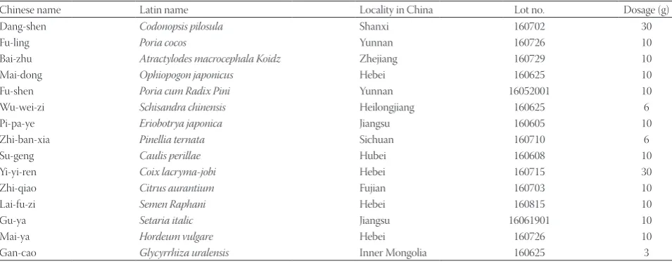
WD-3 prescription (Table 1), which is mainly composed of Codonopsis pilosula,Poria cocos,Atractylodes macrocephala Koidz,Ophiopogon japonicus,Poria cum Radix Pini,Schisandra chinensis, Eriobotrya japonica, Pinellia ternata,Caulis perillae, Coix lacryma-jobi,Citrus aurantium,Semen Raphani,Setaria italic,Hordeum vulgare, and Glycyrrhiza uralensis, was pre-pared by the Reagent Preparation Room, Wuxi Hospital Affiliated to Nanjing University of Chinese Medicine (Jiangsu Food and Drug Administration [2005] No. 401, Jiangsu med-icine license 204002111). The herbs comprising WD-3 were
identified and decocted together. In the first step, herbs were placed in a gallipot, soaked in water eight times for 1 h, heated, and kept boiling for 1 h, resulting in distilled Liquid 1. In the second step, the herbs were immersed in five times the volume of water in a pan for 30 min and cooked for 1 h, and Liquid 2 was filtered. In the third step, Liquid 1 and Liquid 2 were com-bined and left for over 12 h. The resulting Liquid 3 was filtered and concentrated to one tenth of the original solution [17]. White granulated sugar (20 g), sodium benzoate (0.4 g), and ethylparaben (0.05 g) were added to the Liquid 3. Finally, the mixture were boiled for 15 min, sterilized at 110°C, and stored in the refrigerator at 4°C. Paclitaxel injection (5 ml: 30 mg × 1 bottle/box, lot number: 17060411), provided by Yangtze River Pharmaceutical Group Co., Ltd was used as positive control. Phosphate buffered saline (PBS) was used in blank control group.
Cell lines
Breast cancer cell lines MDA-MB-231, BT-549, MCF-7, and MCF-7/ADR-RES and fluorescently labeled MDA-MB-231/ RFP (red fluorescent protein)), BT-549/RFP, MCF-7/RFP, and MCF-7/ADR-RES/RFP were provided by Anticancer Biotechnology (Beijing) Co., Ltd, Beijing, China.
Reagents
Rabbit monoclonal anti-Hexokinase II (Cat. No. 2867) was purchased from Cell Signaling Technology, Co. Ltd, USA; anti-glyceraldehyde 3-phosphate dehydrogenase (GAPDH, Cat. No. YM3029) from Immunoway, USA; ATP (Cat. No. A7699), adenosine diphosphate (ADP, Cat. No. A2754), ade-nosine monophosphate (AMP, Cat. No. A6885), and MTT (Cat. No. M2128) from Sigma-Aldrich, USA; perchloric acid (Cat. No. 10019318), potassium dihydrogen phosphate (Cat. No. 10017618), sodium hydroxide (Cat. No. 10019762), and potassium hydroxide (Cat. No. 10017018) from Sinopharm
TABLE 1. Herbs included in WD-3
Chinese name Latin name Locality in China Lot no. Dosage (g)
Dang-shen Codonopsis pilosula Shanxi 160702 30
Fu-ling Poria cocos Yunnan 160726 10
Bai-zhu Atractylodes macrocephala Koidz Zhejiang 160729 10
Mai-dong Ophiopogon japonicus Hebei 160625 10
Fu-shen Poria cum Radix Pini Yunnan 16052001 10
Wu-wei-zi Schisandra chinensis Heilongjiang 160625 6
Pi-pa-ye Eriobotrya japonica Jiangsu 160605 10
Zhi-ban-xia Pinellia ternata Sichuan 160710 6
Su-geng Caulis perillae Hubei 160608 10
Yi-yi-ren Coix lacryma-jobi Hebei 160715 30
Zhi-qiao Citrus aurantium Fujian 160703 10
Lai-fu-zi Semen Raphani Hebei 160815 10
Gu-ya Setaria italic Jiangsu 16061901 10
Mai-ya Hordeum vulgare Hebei 160726 10
Chemical Reagent Co., Ltd, China; RPMI 1640 culture medium (SH30809.01) from Hyclone, USA; fetal bovine serum (FBS, 040011ACS) from Biological Industry, Israel; Penicillin-Streptomycin (Pen-Strep, Cat.No. 15140122) from Gibco, USA; Hoechst 33258 (Cat. No. H21491) and Dead Cell Apoptosis Kit with Annexin V FITC/PI (Cat. No. V13242) from Invitrogen, USA; 2 × T5 Fast qPCR Mix (Cat. No. TSE202) and Golden star RT6 cDNA Synthesis kit (Cat. No. TSK301S) from TSINGKE, China; Trizol reagent (Cat. No. 15596018) from Life Technologies, USA; M-MLV Reverse Transcriptase (Cat. No. M5313) from Promega, USA; PowerUp SYBR Green
Master Mix (Cat. No.A25742) from ABI and 5 × RNA Loading Buffer (Cat. No. CW0611A) from CWBio Co.Ltd, China.
Instruments
The instruments used were OLYMPUS IMT-2 fluores-cence microscope (OLYMPUS, Japan), laser confocal scan-ning microscopy (LCSM, Thermo scientific, USA), Agilent 1200 high performance liquid chromatography (HPLC, Agilent, USA), ELx800 microplate reader (BioTek, USA), flow cytometry (BD Biosciences, USA), Roche LightCycler 480II real-time PCR (Roche, Switzerland), PowerPac Basic electro-phoresis instrument (BIO-RAD, USA).
MTT assay
The breast cancer cells in the logarithmic phase were seeded in a 96-well plate (4 × 10³ cells per well) for cell
adhe-sion. After 48 h treatment with WD-3 (paclitaxel positive control and blank control were also set), 0.5 m/mL MTT solution was added. Two hours later, the supernatant was dis-carded and 150 μL of dimethyl sulfoxide (DMSO) was added. The samples were then oscillated at low speed on the shaking table for 10 min to fully dissolve the crystals. OD450 was mea-sured and the half maximal inhibitory concentration (IC50)
was calculated for each sample as follows: IC50 = (OD450 of
positive control group – OD450 of samples)/OD450 of pos-itive control group × 100 %. Each experiment was performed in triplicate.
Nuclear Hoechst 33258 staining
Dual-color fluorescent cells in the logarithmic phase were prepared as a suspension at 4 × 10⁵ cells/mL. Cell suspension
was seeded in a 96-well plate (100 μL per well) for cell adhe-sion. WD-3 treatment group, paclitaxel treatment group, and blank control group were set. After a treatment for 24 h, the cells were incubated with 0.5 μg/mL Hoechst 33258 solution for 20 min at room temperature, followed by PBS wash 3 times with 5 min interval. Finally, cell morphology was observed
Transfection and screening of dual-color
fluorescent cells and immunofluorescence
Dual-color fluorescent cells expressing RFP in the cytoplasm and green fluorescent protein (GFP) in the nucleus were used. GFP was stably transfected into RFP-positive cells. Briefly, PT-67 H2B/GFP cells were packaged and grown to the loga-rithmic phase. The supernatant was collected and filtered (pore size: 0.22 μm). MDA-MB-231/RFP, BT-549/RFP, MCF-7/RFP, MCF-7/ADR-RES/RFP cells were transfected with a filtration repeated 2–4 times. At 48 h, GFP-positive nuclei were partially observed under a fluorescence microscope. RFP-positive cells were screened by treatment with 200, 400, and 800 μg/mL G418. After 1–2 weeks, the cells in a good condition and with rel-atively high ratio of dual-color fluorescent proteins were selected for monoclonal screening. These cells were inoculated into a 96-well plate with one cell per well. One week later, the cells with monoclonal formation were subjected to amplification culture. The clones with the brightest fluorescence were preserved.
Dual-color fluorescent cells in the logarithmic phase were prepared as a suspension at 4 × 10⁵ cells/mL. Cell suspension
was injected into a 96-well plate (100 μL per well) for cell adhesion overnight. After a treatment with WD-3 or pacli-taxel for 24 h and 48 h, cell morphology was observed under a fluorescence microscope.
Apoptosis determination
The cells were seeded in a 6-well plate (1 × 10⁵ cells per
well). After culture for 18–24 h, different concentrations of WD-3 were added, and paclitaxel positive control and blank control groups were set. Then the cells were collected and centrifuged at 1500 r/min for 10 min. The precipitant was fixed in 1 ml of 75% ethanol at 4°C overnight. After 24 h treatment, fixed cells were centrifuged at 1000 r/min for 10 min, and the precipitant was incubated with 200 μL of binding buffer, 10 μL of Annexin V/FITC (1 μg/mL), and 5 μL of propidium iodide [PI] (2 μg/mL) at 4°C, in dark for 15 min. Cell apoptosis was determined using a flow cytometry.
HPLC
Cells in the logarithmic phase were seeded into 100-mm culture dishes and treated with different doses of WD-3. The same amount of cells was collected from each sample, the cells were quantified for protein concentration and sub-jected to HPLC. The following equations were used: Total adenine nucleotides (TAN) = ATP + ADP + AMP; Energy charge (EC) = (ATP + 0.5 ADP)/(ATP + ADP + AMP).
Western blot
plate. After specific treatment, the cells were lysed for total protein extraction using radioimmunoprecipitation assay (RIPA) buffer and quantified by bicinchoninic acid (BCA) method. The protein sample was loaded for electrophoresis and transferred on a polyvinylidene fluoride (PVDF) mem-brane. Membranes were blocked in 5% skim milk for 2 h and subjected to incubation with primary (anti-hexokinase 2 [HK2] antibody) and secondary antibodies (mouse monoclonal anti-GAPDH). Bands were detected by enhanced chemilumines-cence (ECL) and analyzed by ImageJ Software, NIH, USA.
Quantitative reverse transcription PCR
(qRT-PCR)
Breast cancer cells (2 mL of suspension, 5 × 10⁴ cells/mL) in the logarithmic phase were seeded into a 96-well plate. WD-3 group and blank control group were established. After 24 h of culture, TRIzol kit was used to extract total RNA, and M-MLV kit was used for reverse transcription, according to the manufacturer’s protocols. The RT-PCR steps were 16°C for 30 min, 42°C for 30 min, and 70°C for 15 min, total of 30 cycles. The samples were stored in a refrigerator at -20°C. For each sample, 2 μl of cDNA was amplified with target gene prim-ers and reference gene primprim-ers (Table 2) for 40 cycles (95°C, 1 min; 95°C, 15 sec; and 60°C, 50 sec). The relative expression was quantified with the formula 2-△△CT [18].
Statistical analysis
Data analysis was performed using IBM SPSS Statistics for Windows, Version 19.0. (IBM Corp. Armonk, NY). Qualitative data were analyzed by chi-square and rank sum test. Quantitative data were analyzed by the Student’s t-test and analysis of variance (ANOVA). A value of p < 0.05 was considered statistically significant.
RESULTS
WD-3 treatment inhibited the proliferation of
breast cancer cells
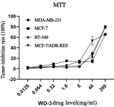
Breast cancer cells MDA-MB-231, BT-549, MCF-7, and MCF-7/ADR-RES were treated with different concentrations of WD-3 (0, 0.0128, 0.064, 0.32, 1.6, 8, 40, and 200 mg/mL). Proliferation inhibition rate was determined by MTT assay. WD-3 treatment markedly inhibited the proliferation of the four breast cancer cell lines (Figure 1). The inhibition rate grad-ually increased in a dose-dependent manner. IC50 values of the four breast cancer cell lines were calculated and shown in Table 3. The inhibitory effect of WD-3 on the proliferation rate was much more pronounced in MCF-7/ADR-RES cells, the lowest inhibition rate was observed in the hormone-depen-dent MCF-7 cell line.
Cell morphology changes in breast cancer cells
after WD-3 treatment
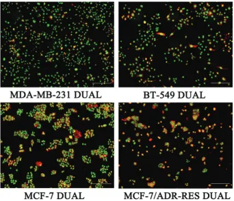
Cell morphology changes following WD-3 treatment were observed by laser confocal imaging. Breast cancer cells were divided into WD-3 group (80 mg/mL), paclitaxel group (3 μg/ mL), and blank control group. Cells were treated with 80 mg/mL WD-3 or 3 μg/mL paclitaxel for 24 h. As shown in Figure 2, chro-matin condensation, aggregation, marginalization, and fragmen-tation were observed in both WD-3 group and paclitaxel group. Four dual-color fluorescent breast cancer cell lines MDA-MB-231 DUAL, BT-549 DUAL, MCF-7 DUAL, and MCF-7/ADR-RES DUAL were successfully established (Figure 3). These dual-color fluorescent cells were treated with different concentrations of WD-3 (20, 40, and 80 mg/mL) for
FIGURE 1. Proliferation inhibition rate of WD-3 in breast can-cer cells by MTT assay. Breast cancan-cer cell lines MDA-MB-231, BT-549, MCF-7, and MCF-7/ADR-RES were treated with differ-ent concdiffer-entrations of WD-3 (0, 0.0128, 0.064, 0.32, 1.6, 8, 40, and 200 mg/mL). WD-3 treatment markedly inhibited the proliferation of the four breast cancer cell lines. The inhibition rate gradually increased in a dose-dependent manner.
TABLE 2. Primer sequences used in quantitative reverse transcription polymerase chain reaction (qRT-PCR)
Gene name Sequence (5’ to 3’) Product size (bp)
HK2 F:TGCCACCAGACTAAACTAGACGR:CCCGTGCCCACAATGAGAC 227
GAPDH F:AGGTCGGTGTGAACGGATTTG 95 R:GGGGTCGTTGATGGCAACA
HK2: Hexokinase 2; GAPDH: Glyceraldehyde 3-phosphate dehydrogenase
TABLE 3. IC50 values of WD-3 (mg/mL) for four breast cancer cell lines
Cell line MDA-MB-231 BT-549 MCF-7 MCF-7/ADR-RES IC50 78.79±1.34* 90.00±9.16* 146.89±6.69 62.00±14.23*
24 h and 48 h. Cell morphology changes were observed under the OLYMPUS IMT-2 fluorescence microscope (Figure 4). The cells in blank control group were normal in morphology.
RFP-positive cytoplasm and GFP-positive nucleus were clear (nuclei were yellow-green due to RFP overlap). Membrane folds were clearly distinguishable.
FIGURE 2. Laser confocal imaging of four breast cancer cell lines treated with WD-3. Breast cancer cell lines MDA-MB-231,
BT‑549, MCF‑7, and MCF‑7/ADR‑RES were divided into WD‑3 (80 mg/mL), paclitaxel (TAX, 3 μg/mL), and blank control (phos -phate-buffered saline) group. Cells were treated for 24 h. Chromatin condensation, aggregation, marginalization, and
fragmenta-tion were observed in both WD‑3 group and paclitaxel group. Scale bar, 50 μm.
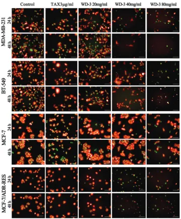
The following apoptotic morphology changes were observed in the four breast cancer cell lines after WD-3 treat-ment: cell surface protrusion and fold disappearance, rounded and smaller cells, excessive intracellular vacuoles, chromatin condensation (fluorescence enhancement), and nuclear decay (rippled or creased). Apoptotic bodies were observed at high-dose WD-3. Apoptosis increased in a high-dose- and time-depen-dent manner in all four breast cancer cell lines. Obvious apop-tosis was observed in MCF-7 DUAL after 80 mg/mL WD-3 treatment. In addition to MCF-7 DUAL, a decreased number of living cells and an increased number of apoptotic cells were observed in the remaining three breast cancer cell lines fol-lowing 40 mg/mL and 80 mg/mL WD-3 treatment. Under
the same-dose of WD-3 (80 mg/mL), the lowest number of living cells was observed in MCF-7/ADR-RES DUAL and the highest number in MCF-7 DUAL cell line, which was consis-tent with the MTT results.
WD-3-induced apoptosis in breast cancer cells as
determined by flow cytometry
Cells were treated with 40, 80, and 160 mg/mL of WD-3. Paclitaxel (5 μg/mL) and blank control group were also set. After treatment for 24 h, the cells were stained with Annexin V/FITC and PI and subjected to flow cytometry (Figure 5A). Apoptotic rate increased in a dose-dependent manner and
FIGURE 4. Fluorescence imaging of four breast cancer cell lines treated withWD-3 at different time points and concentrations. MDA-MB-231 DUAL, BT-549 DUAL, MCF-7 DUAL, and MCF-7/ADR-RES DUAL were treated with different concentrations of
WD‑3 (20, 40, and 80 mg/mL) or paclitaxel (TAX, 3 μg/mL) for 24 h and 48 h. Blank control was treated with phosphate‑buffered
saline. WD-3 treatment caused cell surface protrusion and fold disappearance, rounded and smaller cells, excessive intracellular
vacuoles, chromatin condensation (fluorescence enhancement), and nuclear decomposition (rippled or creased). Apoptotic bod -ies were observed at high-dose WD-3. Apoptosis increased in a dose- and time-dependent manner. In particular, WD-3 treatment
living cell ratio decreased (Figure 5B) in all four breast cancer cell lines, suggesting that WD-3 induced apoptosis in breast cancer cells.
WD-3 affected ATP, ADP, and AMP levels in
breast cancer cells
To investigate the effect of WD-3 on intracellular levels of ATP, ADP, and AMP breast cancer cells were treated with 0 (control), 20 (low-dose), and 60 mg/mL (high-dose) WD-3
for 24 h. Compared with control, ATP levels and EC dose-de-pendently decreased in BT-549 and MDA-MB-231 cells. In MCF-7 and MCF-7/ADR-RES cells, the changes in ATP, ADP, and AMP levels were not significant (Figure 5 C and D).
WD-3 downregulated hexokinase 2 protein level in
BT-549 cells
Hexokinase 2 protein levels were quantified in the four breast cancer cell lines. WD-3 treatment (60 mg/mL,
FIGURE 5. WD-3 inhibited the growth of human breast cancer cells. (A and B) Flow cytometry was used to analyze cell apopto-sis. Breast cancer cell lines MDA-MB-231, BT-549, MCF-7, and MCF-7/ADR-RES were treated with 40, 80, and 160 mg/mL of
WD‑3 or 5 μg/mL of paclitaxel for 24 h. Blank control was treated with phosphate‑buffered saline. Apoptotic rate increased in
a dose-dependent manner and living cell ratio decreased in all four breast cancer cell lines. (C and D) Changes in ATP and EC after WD-3 treatment in the four breast cancer cell lines. Breast cancer cells were treated with 0 (control), 20 (low-dose), and 60 mg/mL (high-dose) WD-3 for 24 h. Compared with control, ATP levels and EC dose-dependently decreased in BT-549 and MDA-MB-231 cells. (E) The protein levels of hexokinase 2 were determined by Western blot. GAPDH was used an internal control.
WD‑3 treatment (60 mg/mL, high‑dose) significantly downregulated hexokinase 2 protein level in BT‑549 cells. (F) The mRNA lev
-els of hexokinase 2 were analyzed by qRT‑PCR. WD‑3 treatment (60 mg/mL, high‑dose) significantly decreased the mRNA level of
hexokinase 2 in BT-549 cells. *p < 0.05 and **p < 0.01 vs. 0 mg/mL; #p < 0.05 vs. control group. ATP: Adenosine triphosphate;
EC: Energy charge; GAPDH: Glyceraldehyde 3-phosphate dehydrogenase; qRT-PCR: Quantitative reverse transcription polymerase chain reaction.
B
C A
high-dose) significantly downregulated hexokinase 2 protein level in BT-549 cells. Downregulation of hexokinase 2 was not significant in the other three breast cancer cell lines (Figure 5E).
WD-3 downregulated hexokinase 2 mRNA level in
BT-549 cells
Hexokinase 2 mRNA levels were quantified in the four breast cancer cell lines. WD-3 treatment (60 mg/mL, high-dose) significantly decreased the mRNA level of hexokinase 2 in BT-549 cell line (Figure 5F), but not in the other three cell lines.
DISCUSSION
There are various TCM theories on the pathogenesis of breast cancer. Accordingly, an increasing number of in vitro studies have been conducted to verify the anti-breast-can-cer effects of Compound Chinese Traditional Medicines (CCTMs) that function through the dissipation of blood stasis, detoxification, warming Yang, relief of liver symp-toms, and enhancement of body resistance [19]. It was reported that Xiaojindan, a CCTM for blood stasis dissipa-tion and detoxificadissipa-tion, remarkably inhibited cell invasion and epithelial–mesenchymal transition (EMT) in breast cancer by regulating p38 mitogen-activated protein kinase (MAPK) and c-Jun N-terminal kinase (JNK) MAPK path-ways [20]. The traditional formula Tongluosanjie Pill could suppress cell growth and estrogen synthesis in breast can-cer cells MCF-7 [21]. Wang et al. [22] demonstrated that a yang-warming CCTM could significantly arrest MCF-7 cells in S phase and inhibit metastasis by mediating matrix metalloproteinase-2 (MMP-2)/tissue inhibitor of MMP-2 (TIMP-2) balance. Another liver-soothing and spleen-en-hancing detoxification prescription could also suppress the proliferation of MCF-7 cells by inducing cell apoptosis [23]. In this study we investigated the effect of WD-3 on breast cancer, a disease that we considered to be a result of spleen deficiency. As suggested by Zhao, breast cancer treatment should act on the spleen and stomach to regulate Qi, elim-inate dampness, and resolve phlegm. WD-3 is mainly com-posed of Codonopsis pilosula, Poria cocos, Atractylodes mac-rocephala Koidz,Ophiopogon japonicus, Poria cum Radix Pini, Schisandra chinensis, Eriobotrya japonica Thunb, Pinellia ternata, Caulis perillae, Coix lacryma-jobi, Citrus auran-tium, Semen Raphani, Setaria italic, Hordeum vulgare, and Glycyrrhiza uralensis. All the ingredients are mild and non-toxic and suitable for long-term regulation of the spleen and stomach [24]. We found that WD-3 treatment inhibited the proliferation, improved the morphology, and increased the apoptosis of breast cancer cells.
According to TCM, different physical constitutions lead to differences in breast cancer pathogenesis. Multiple pathological subtypes of breast cancer show obvious het-erogeneity [25]. In this study, we selected four human breast cancer cell lines with different biological properties. MDA-MB-231 cell line is derived from human breast inva-sive ductal carcinoma, a highly metastatic cell line charac-terized as estrogen receptor negative [ER(-)], progesterone receptor negative [PR(-)], and human epidermal growth factor receptor 2 [HER2(-)]. BT-549 cell line is derived from human papillary invasive ductal carcinoma that is ER(-), PR(-), and HER2(-). MCF-7 is derived from human breast invasive ductal carcinoma with ER(+), PR(+), and HER2(-). MCF-7/ADR-RES is a doxorubicin-induced multidrug-re-sistant breast cancer cell line [26,27]. Although WD-3 treat-ment inhibited the proliferation of all four breast cancer cell lines in our study, the IC50 values were significantly differ-ent, with the highest in MCF-7/ADR-RES and the lowest in MCF-7 cell line (the IC50 values were in a descending order for 7/ADR-RES, MDA-MB-231, BT-549, and MCF-7). Obvious apoptosis was observed in MCF-7 DUAL after 80 mg/mL WD-3 treatment. In addition to MCF-7 DUAL, a decreased number of living cells and an increased num-ber of apoptotic cells were observed in the remaining three breast cancer cells following 40 mg/mL and 80 mg/mL WD-3 treatment. Under the same-dose of WD-3, the low-est number of living cells was observed in MCF-7/ADR-RES DUAL and the highest number in MCF-7 DUAL. WD-3 treatment dose-dependently decreased ATP levels and EC in BT-549 and MDA-MB-231 cells, but did not cause significant changes in MCF-7 and MCF-7/ADR-RES cells. Moreover, WD-3 treatment significantly downregulated hexokinase 2 level in BT-549 cells, but not in the other three breast cancer cell lines. Consistent with previous studies [28], breast can-cer cell lines with different biological characteristics may use different pathways for proliferation and metabolism.
hand, in tumor cells, only 2 moles of ATP and lactic acid are produced through glycolysis. Compared with normal cells, the glycolysis in tumor cells consumes more nutrients and produces less energy. The spleen functions to nourish the whole body with cereal essence; once diseased, the spleen fails to transform nutrients and produce energy, which in terms of energy production efficiency can be compared to glycolysis. Xu et al. [30] indicated that spleen deficiency and respiratory dysfunction lead to hypoxia and upregu-lation of hypoxia-inducible factor 1-alpha (HIF-1α). They suggested that, in obese people, chronic inflammation and cytokine release caused by spleen deficiency is a major fac-tor triggering glycolysis in colorectal cancer. The proposed hypothesis “spleen deficiency-mitochondria-aerobic gly-colysis-tumor” suggests that replenishing Qi to invigorate the spleen could improve mitochondrial metabolism and inhibit aerobic glycolysis in tumor cells, thus suppressing tumor growth [31]. Our study found that WD-3 treatment significantly decreased ATP levels and EC in BT-549 and MDA-MB-231 breast cancer cells, indicating that WD-3 treatment can reduce the energy production for tumor cell proliferation. Hexokinase 2, the first rate-limiting enzyme in the glycolytic pathway of breast cancer, can initiate and maintain the glycolytic rate during proliferation of cancer cells. Therefore, hexokinase 2 is a viable target to inhibit ATP production in breast cancer cells and induce apopto-sis. We showed that WD-3 treatment markedly downregu-lated hexokinase 2 level in BT-549 breast cancer cells. WD-3 may inhibit tumor cell proliferation by intervening with the glycolytic pathway. To some extent, our findings support the hypothesis of “spleen deficiency-mitochondria-aerobic glycolysis-tumor”. However, the regulatory effects of WD-3 on glycolysis-related genes and its pharmacological mecha-nism need to be further investigated.
CONCLUSION
Our preliminary results showed that WD-3 affects apop-tosis, glycolysis, and hexokinase 2 expression in breast cancer cells. WD-3 inhibited the proliferation and increased apop-tosis of breast cancer cells. In BT-549 cell line, WD-3 both decreased the energy production and downregulated hexoki-nase 2. Our findings indicate that WD-3 targets the glycolytic pathway in breast cancer cells to exert its antitumor activity.
ACKNOWLEDGMENTS
This work study supported by the Youth program of Wuxi municipal Health Committee (grant no.Q201602 and Q201704).We would like to thanks for the technical support from Beijing AnTaiKang biotechnology co., LTD
REFERENCES
[1] Bray F, Ferlay J, Soerjomataram I, Siegel RL, Torre LA, Jemal A. Global cancer statistics 2018: GLOBOCAN estimates of incidence and mortality worldwide for 36 cancers in 185 countries. CA Cancer J Clin 2018;68:394-424.
https://doi.org/10.3322/caac.21492.
[2] Zheng RS, Sun KX, Zhang SW, Zeng HM, Zou XN, Chen R, et al. Report of cancer epidemiology in China, 2015. Chin J Onc 2019;41:19-28.
https://doi.org/10.3760/cma.j.issn.0253-3766.2019.01.005. [3] Fei S, Wan DG, Liu F, Jia LQ, Zhang YH, Lou YN. Advances in TCM
research on the prevention and treatment of triple-negative breast cancer. Chinese Journal of Basic Medicine in Traditional Chinese Medicine 2018;24:1649-51.
[4] Kapinova A, Stefanicka P, Kubatka P, Zubor P, Uramova S, Kello M, et al. Are plant-based functional foods better choice against cancer than single phytochemicals? A critical review of current breast can-cer research. Biomed Pharmacother 2017;96:1465-77.
https://doi.org/10.1016/j.biopha.2017.11.134.
[5] Zhu L, Li L, Li Y, Wang J, Wang Q. Chinese herbal medicine as an adjunctive therapy for breast cancer: a systematic review and meta-analysis. Evid Based Complement Alternat Med 2016;2016:9469276.
https://doi.org/10.1155/2016/9469276.
[6] Zhao Q, Gao X, Guangli Y, Zhang A, Sun H, Han Y, et al. Chinmedomics facilitated quality-marker discovery of Sijunzi decoc-tion to treat spleen qi deficiency syndrome. Front Med 2019;1-22. https://doi.org/10.1007/s11684-019-0705-9.
[7] Shu Q, Sun D, Wang H, Liang F, Gerhard L, Daniela L, et al. Differences of acupuncture and moxibustion on heart rate variabil-ity in qi-deficiency syndrome: a randomized controlled trial. Chin Acup Moxib (Zhongguo Zhen Jiu) 2017;37:25-30.
https://doi.org/10.13703/j.0255-2930.2017.01.006.
[8] You JL, Zhou LY, Xu M. Clinical research of the treatment of advanced gastric cancer using Chinese herbal medicine WD-3. Hubei J Tradi Chin Med 2004;26:8-9.
[9] Zhou LY, Shan ZZ, You JL. Clinical observation on treatment of colonic cancer with combined treatment of chemotherapy and Chinese herbal medicine. Chin J Integr Med 2009 15:107-11. https://doi.org/10.1007/s11655-009-0107-y.
[10] Warburg O. On the origin of cancer cells. Science 1956;123:309-14. https://doi.org/10.1126/science.123.3191.309.
[11] Salamon S, Podbregar E, Kubatka P, Büsselberg D, Caprnda M, Opatrilova R, et al. Glucose metabolism in cancer and ischemia: possible therapeutic consequences of the Warburg effect. Nutr Cancer 2017;69:177-83.
https://doi.org/10.1080/01635581.2017.1263751.
[12] Lebelo MT, Joubert AM, Visagie MH. Warburg effect and its role in tumourigenesis. Arch Pharm Res 2019;42:833-47.
https://doi.org/10.1007/s12272-019-01185-2.
[13] Sun L, Suo C, Li ST, Zhang H, Gao P. Metabolic reprogramming for cancer cells and their microenvironment: beyond the Warburg effect. Biochim Biophys Acta Rev Cancer 2018;1870:51-66. https://doi.org/10.1016/j.bbcan.2018.06.005.
[14] Li Y, Zhang J. Advances in glycolysis-based therapy for breast can-cer. Chin J Clin Oncol 2015;42:1063-6.
[15] Zhang Q, Zhang Y, Zhang P, Chao Z, Xia F, Jiang C, et al. Hexokinase II inhibitor, 3-BrPA induced autophagy by stimulating ROS forma-tion in human breast cancer cells. Genes Cancer 2014;5:100-12. https://doi.org/10.18632/genesandcancer.9.
[16] Shoshan-Barmatz V, Mizrachi D. VDAC1: from structure to cancer therapy. Front Oncol 2012;2:164.
https://doi.org/10.3389/fonc.2012.00164.
[17] Chen S, Ye ZQ, Li ZW, Zhao CX, Chen GJ, Zhou JZ, et al. Wenyang Huoxue Jiedu formula inhibits thin-cap fibroatheroma plaque for-mation via the VEGF/VEGFR signaling pathway. J Ethnopharmacol 2018;219:213-21.
[18] Livak KJ, Schmittgen TD. Analysis of relative gene expression data using real-time quantitative PCR and the 2(-Delta Delta C(T)) method. Methods 2001;25:402-8.
https://doi.org/10.1006/meth.2001.1262.
[19] Que GY, You JL. LianliangYou’s experience in the treatment of breast cancer. New TCM 2018;50:224-6.
[20] Peng B, He R, Xu QH, Sun LH, Han JY, Li JR. Inhibitory effect of Xiaojintan on metastasis of breast cancer cells and its mechanism. Chin J TCM 2018;33:4916-9.
[21] Zhang SY, Wang T, Wang YY. Inhibition of serum Tongluo Sanjie Pill on growth of breast cancer cells MCF-7. Shanghai Univ TCM 2012;26:90-4.
[22] Wang Z, Zhang WX. Effect of CCTM Warming Yang on cell cycle arrest, anti-invasion and anti-metastasis of breast cancer cells MCF-7. Chin Archi TCM 2018;36:1569-72.
[23] Li LP, Yang X, Pan B, Tan XN, Luo J, Jiang YL. Effect of detoxification prescription of soothing liver and strengthening spleen on prolif-eration and apoptosis of breast cancer cells MCF-7. J. Hunan Univ TCM 2018;38:645-9.
[24] Zhou LY, Shan ZZ, You JL. Clinical observation on treatment of colonic cancer with combined treatment of chemotherapy and Chinese herbal medicine. Chin J Integr Med 2009;15:107-11. https://doi.org/10.1007/s11655-009-0107-y.
[25] Lacroix M, Leclercq G. Relevance of breast cancer cell lines as models for breast tumours: an update. Breast Cancer Res Treat 2004;83:249-89.
https://doi.org/10.1023/B: BREA.0000014042.54925.cc.
[26] Xiaodan Z. Anti-tumor effect of the couplet medicinals rhizoma bolbostemmae-Fritillaria thunbergii on breast cancer. Beijing University of Chinese Medicine; 2015.
[27] Gao Y, Tollefsbol TO. Combinational proanthocyanidins and res-veratrol synergistically inhibit human breast cancer cells and impact epigenetic-mediating machinery. Int J Mol Sci 2018;19. pii: E2204. https://doi.org/10.3390/ijms19082204.
[28] Xiao-dan Z, Chao A, Kai-wen H, Meng Y, Chong-yi L. Study on the combined function of Bolbostemma Paniculatum Saponins and Peimine in vitro of human breast cancer cell. China Journal of Traditional Chinese Medicine and Pharmacy 2015;30:1508-13. [29] Yang Y, Liu Y, Zhang F, Sun YH, Wang Y, Zhang Z, et al. Relevance
between spleen deficiency and viscera diseases based on mitochon-drial research. J TCM 2018;59:1742-6.
[30] Xu WJ, Sun XG. Spleen deficiency is the key pathogenesis of energy metabolism disorder and epithelial-mesenchymal transition in col-orectal cancer. Chin J Basi Med 2015;21:500-20.
[31] Rong ZB, Luo AM, Yao NL. A new hypothesis of etiology and patho-genesis: spleen deficiency-mitochondria-aerobic glycolysis-tumor. Journal of The Fourth Military Medical University 2016;7:19-22.
Related articles published in BJBMS
1. Effect of CCT137690 on long non-coding RNA expression profiles in MCF-7 and MDA-MB-231 cell lines
Tuğçe Balcı Okcanoğlu et al., BJBMS, 2020
2. Dichloromethane fractions of Scrophularia oxysepala extract induce apoptosis in MCF-7 human breast cancer cells

![Crystal structure of 3,14 diethyl 2,13 diaza 6,17 diazoniatricyclo[16 4 0 07,12]docosane dinitrate dihydrate from synchrotron X ray data](data:image/gif;base64,R0lGODlhAQABAIAAAP///wAAACH5BAEAAAAALAAAAAABAAEAAAICRAEAOw==)