BOSNIAN JOURNAL OF BASIC MEDICAL SCIENCES 2008; 8 (4): 350-355&
Abstract
Th e aim of the study was to detect HIV RNA in seropositive patients using RT-PCR method and thus, to
establish PCR methodology in the routine laboratory works.
Th e total of examined persons were divided in two groups: ) persons seropositive for HIV; and )
healthy persons - randomly selected blood donors that made the case control group. Th e subjects age
was between and years (average ,).
ELFA test for combined detection of HIV p antigen and anti HIV-+ IgG and ELISA test for detec-tion of antibodies against HIV- and HIV-, were performed for each examined person. RNA from the whole blood was extracted using a commercial kit based on salt precipitation. Detection of HIV RNA was performed using RT-PCR kit. Following nested PCR, the product was separated by electrophoresis
in , agarose gel. Th e result was scored positive if the band of bp was visible regardless of intensity.
Measures of precaution were taken during all the steps of the work and HIV infected materials were disposed of accordingly.
In the group of blood donors ELFA, ELISA and RT-PCR were negative. Assuming that prevalence of HIV
infection is zero, the clinical specifi city of RT-PCR is . Th e analytical specifi city of RT-PCR method
was tested against Hepatitis C and B, Human Papiloma Virus, Cytomegalovirus, Herpes Simplex Virus,
Rubella Virus, Mycobacterium tuberculosis, Chlamydia trachomatis. None of these templates yielded am-plicon. In the group of seropositive persons, samples were analyzed. HIV RNA was detected in samples. ELISA and ELFA test were positive in all samples. Diff erent aliquots of the samples were tested independently and showed the same results. After diff erent periods of storing the RNA samples at –ºC,
RT-PCR reaction was identical to the one performed initially. Th e obtained amplicons were maintained
frozen at –ºC for a week and the subsequently performed electrophoresis was identical to the previous
one. Th e reaction is fast, simple for manipulation; with low detection level of IU/ml. RT-PCR needs a
small amount of RNA, as well as a small volume of sample. HIV RNA was detected in diff erent periods of time with diff erent clinical presentations in patients, with or without antiretroviral therapy.
RT-PCR method gives the opportunity for reliable determination of HIV- RNA with border of detection
of IU/ml. Th e test is reproducible and has high analytical and clinical specifi city.
KEY WORDS: HIV, RT-PCR
RT-PCR DETECTION OF
HIV IN REPUBLIC OF
MACEDONIA
Golubinka Bosevska¹*, Nikola Panovski², Eleni Dokić², Violeta Grunevska³
¹ Republic Institute for Health Protection, Divizija No , Skopje, Macedonia
² Institute for Microbiology, Faculty of Medicine, Vodnjanska bb, Skopje, Macedonia
³ Clinic for Infectious Diseases, Faculty of Medicine, Vodnjanska bb, Skopje, Macedonia
BOSNIAN JOURNAL OF BASIC MEDICAL SCIENCES 2008; 8 (4): 351-355
Introduction
Th ree crucial questions regarding HIV diagnostics have been addressed in medicine worldwide over the last years: whether the subject was infected, how active HIV replication was, and how sensitive to retroviral therapy the virus was? Detection of virus genome made by in situ hybridization or Polymerase Chain Reaction (PCR) is an attempt to provide answers to all of the three ques-tions. Two methods for detection of HIV RNA are avail-able: Target amplifi cation with modalities of Reverse transcriptase (RT) PCR and Nucleic acid sequencebased amplifi cation assay (NASBA), and Signal amplifi -cation, as branch DNA test (-). Clinical relevance of those assays dependens on their reproducibility and am-plifi cation dynamics for diff erent genotypes. Some stud-ies addressed comparisons of the results obtained from diff erent assay systems (-). Th e aims of the study are to introduce PCR methodology in HIV diagnostics in our country and to detect HIV RNA with nested RT-PCR in seropositive subjects in order to evaluate analytical specifi city and reproducibility of this method.
Patients and Methods
Th e case control study involved subjects, divided in two groups:
a) HIV seropositive subjects ( patients), and b) Sedentary healthy subjects ( blood donors). Seropositive patients, according to the HIV trans-mission, were divided in three groups: heterosexuals
BOSNIAN JOURNAL OF BASIC MEDICAL SCIENCES 2008; 8 (4): 352-355) PCR mix for nested PCR of: four dNTPs, X reac-tion buff er, primers for nested amplifi careac-tion, cresol red, glycerol and deionizated water; ) PCR Enzyme – with polymerase activity; ) Positive control; ) Mineral oil. Deoxyuracil triphosphate (dUTP) and uracil-N-glycosylase are added to the reaction (-). Th e method is based on HIV RNA transcription into complementary DNA copy (cDNA). Amplifi cation was conducted under following conditions: for reverse tran-scription min at °C, min at °C, min. at °C- one cycle; and for amplifi cation sec at °C, sec at °C, sec at °C – cycles and one cycle at °C for min. Th e conditions of the nested PCR were: min at °C one cycle; sec at °C, sec at °C, sec at °C for cycles; and one cycle at °C for min (-). PTC-™ Th ermal Cycler (MJ Research, inc.) was used. PCR products were separated using , agarose gel electrophoresis. Ethidium bromide was added (). UV transiluminator was used for visualiza-tion of the bands. The result was scored positive if band of bp was present regardless of intensity. Th e procedures were performed in Virology Laboratory
in Republic Institute for Health Protection, Skopje, Mace-donia, with biological protection class II. One-way pro-cess (pre-amplifi cation to post-amplifi cation) was used.
Results
HIV RNA was not detected in any of the case control patients: examples were not reactive to RT PCR kit. Sup-posing that prevalence of HIV- infection in seronegative blood donors is zero, we obtained test specifi city . To estimate analytical specificity of the test, we at-tempted amplifi cation of nucleic acids pf Hepatitis C
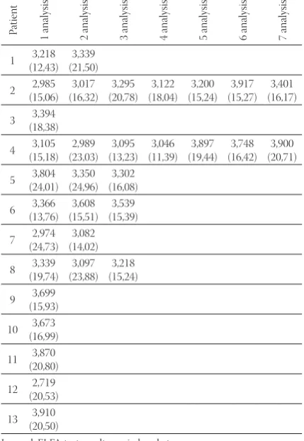
and B, Human Papiloma Virus, Cytomegalovirus, Herpes Simplex, Rubella, Mycobacterium tuberculosis, Chla-mydia trachomatis. All the test reactions were negative. HIV RNA was detected in samples (Table ). In patients, ELFA HIV duo was also positive for anti-HIV IgG and p antigen. ELISA test was positive which indicated presence of HIV antibodies (Table ). In patients HIV RNA was detected during the first visit. Six patients came to see the doctor only once. HIV RNA was detected in three of them. Patient No. showed undetectable level of HIV RNA in two tests. In patient No. HIV RNA was detected during the first visit, but it was undetectable during the second one. One patient was identifi ed as HIV infected based on the presence of HIV antibodies. Infection was con-firmed by HIV RNA detection in three consecutive tests. In patients No. and fl uctuations in HIV RNA test results were detected following seven series of tests. In order to estimate reproducibility, diff erent aliquots of the samples were tested independently. All of them showed the same results. After diff erent periods of stor-age at -°C, samples were resubmitted to reverse transcription and amplifi cation. In all samples the results were identical to the fi rst round of test. Also, amplicons
Patient
1 analysis 2 analysis 3 analysis 4 analysis 5 analysis 6 analysis 7 analysis
1 +
-2 + - + - - +
-3 +
4 + - - + - - +
5 + + +
6 - +
-7 -
-8 + -
-9 -10 + 11 -12 -13 + P atien t
1 analysis 2 analysis 3 analysis 4 analysis 5 analysis 6 analysis 7 analysis
1 3,218 (12,43) 3,339 (21,50) 2 2,985 (15,06) 3,017 (16,32) 3,295 (20,78) 3,122 (18,04) 3,200 (15,24) 3,917 (15,27) 3,401 (16,17) 3 3,394 (18,38) 4 3,105 (15,18) 2,989 (23,03) 3,095 (13,23) 3,046 (11,39) 3,897 (19,44) 3,748 (16,42) 3,900 (20,71) 5 3,804 (24,01) 3,350 (24,96) 3,302 (16,08) 6 3,366 (13,76) 3,608 (15,51) 3,539 (15,39) 7 2,974 (24,73) 3,082 (14,02) 8 3,339 (19,74) 3,097 (23,88) 3,218 (15,24) 9 3,699 (15,93) 10 3,673 (16,99) 11 3,870 (20,80) 12 2,719 (20,53) 13 3,910 (20,50) Legend: (+) = present, (-) = absent.
TABLE 1. Presence of HIV RNA in patients
Legend: ELFA test results are in brackets
BOSNIAN JOURNAL OF BASIC MEDICAL SCIENCES 2008; 8 (4): 353-355
were frozen at –°C, and reanalyzed by electrophore-sis. Th e results did not diff er from the fi rst electrophore-sis. Th e results of electrophoresis are shown in Figure .
Discussion
We tested a commercial test, applicable for extraction, reverse transcription and amplifi cation of HIV RNA. In routine work time consumed is a very important thing. Period of hours that elapsed during our analysis (from samples received to the fi nal results) is relatively short. We isolated total RNA from whole blood, with ready for use kit based on salt precipitation ac-cording Nelson and Kim recomendations (). Low amount of required material is of ut-most importance. Our method required μl of whole blood to yield μl of isolated RNA. Cunningham et al. compared RT PCR and DNA PCR tests for the detection of HIV and con-cluded that the former is more sensitive ( to ). Sensitivity of qualitative RNA test is more pronounced than of the quantitative one (). The test that we used did have performance of RNA detection of IU/ml as the lowest limit. Using the primers located for pol gene sequence
aga-
BOSNIAN JOURNAL OF BASIC MEDICAL SCIENCES 2008; 8 (4): 354-355rose gel, the results were identical to the previous run. Nelson and Kim consider that material may evaporate when maintained at room temperature, which results in little and invisible gel product (). Incorporation of positive and negative controls is neces-sary for correct interpretation of the test. Positive control consists of RNA molecule with high level of transcription. We used deionized sterile water as negative control (). False positive or negative results may result from prim-ers mispriming or non-optimized temperature of an-nealing. We used hot start technique with thermostable polymerase to increase the sensitivity of PCR reaction. With aim of reducing the risk of false positive results
the kits incorporated uracil–N–glykosilase (,). Nested PCR is a procedure that increased specific-ity of the test and has the potential to detect very little amounts of DNA/RNA. Amplifi cation of the grade is gained after the fi rst round of PCR consisting of - cycles. Nested PCR results in amplifi cation of the grade (, ). Nested PCR was important in the detec-tion of very small amounts of HIV RNA in the serum of asymptomatic HIV positive patients that were
nega-tive for p antigen in the study of Munoz et al. (). Optimization of PCR reaction is very impor-tant. Electrophoretic analysis should reveal only one band. Undetectable HIV RNA means either that RNA is bellow the low border of detection or that the virus subtype is not detectable with this test. In order to improve the reliability of those mo-lecular assays better standardization is important. Universal precaution and elimination of RNAase activity is provided in the lab, during the process (,). One way (preamplifi cation to post-amplifi cation) method is used. Safety action is maintained in order to prevent contami-nation and ensure reliable results. We used class II bio-safety with HEPA-high-effi ciency particulate air (,). Th is qualitative test, introduced in our country for the first time, can gain results when serology cannot be-cause of the ELISA limits of detection, as in cases of possible HIV exposure, person with clinical symptoms of acute retroviral syndrome, or newborns of HIV in-fected mothers. Since quantitative tests for measuring of viral load are still not introduced, this test can also be used in monitoring the effi cacy of antiretroviral therapy.
Conclusion
According to our data we can conclude that RT-PCR method enables reliable determination of HIV- RNA with limit of detection at IU/ml. Th e test is reproducible and has high analytical and clinical specifi city. Qualitative RT-PCR meth-od for the detection of HIV RNA is recommended for use in routine laboratory work for documentation of HIV infec-tion, as well as for the clinical management of HIV infected patients and monitoring of antiretroviral therapy effi cacy.
References
() Bootman J., Heath A.B., Hughes H.H. An international collabora-tive study on the detection of an HIV- genotype B fi eld isolate by nucleic acid and amplifi cation techniques. J. Virol. Methods. ;(-): -.
() Efremov D.G. Molecular detection and typisation of pathogenic microbes. Prilozi : XVIII, –: –
() Segondy M., Izopet J., Pellegrin I., Montes B., Dumon B., Pasqui-erc M., et al. Comparasion of the Quantiplex HIV- RNA . As-say with the Amplicor HIV- Monitor . AsAs-say for the Quan-tifi cation of the levels of Human Immunodefi ciency virus type RNA in plasma of patients receiving Didonosien combination therapy. J. Clin. Microbiol. ; (): -.
() Kirstein L.M., Mellors J.W., Rinaldo C.R., Margolick J.B., Giorgi J.V., Phair J.P., Dietz E., Gupta P., Sherlock C.H., Hogg R., Mon-taner J.S.G., Muñoz A. Eff ects of anticoagulant, processing delay, and assay method (Branched DNA versus Reverse Transcriptase PCR) on measurement of Human Immunodefi ciency Virus Type RNA levels in plasma. J. Clin. Microbiol. ; (): – .
() Nolte F.S., Boysza J., Thurmond C., Clark W.S., Lennox J.L. Clinical comparison of an enhanced–sensitivity branched–DNA assay and reverse transcription – PCR for quantitation of Hu-man Immunodefi ciency Virus Type RNA in plasma. J. Clin. Microbiol.;; : –.
() Pachl C., Todd J.A., Kern D.G. Rapid and precise quantifi cation of HIV-RNA in plasma using a branched DNA (bDNA) signal amplifi cation assay. J AIDS & Hum Retrovirol.;:-. () Kern D., Collins M., Fultz T. An enhanced sensitivity branched
DNA assay for quantifi cation of human immunodefi ciency virus type RNA in plasma. J. Clin. Microbiol. ; (): -.
() Erice A., Brambilla D., Bremer J., Brooks J., Kokka R., Yen-Lieber-man B., et al. PerforYen-Lieber-mance Characteristics of the QUANTIPLEX HIV- RNA . Assay for Detection and Quantitation of Human Immunodefi ciency Virus Type RNA in Plasma. J. Clin. Micro-biol. ;: –.
() Dewar R.L., Highbarger H.C., Sarmiento M.B. Application of branched DNA signal amplifi cation to monitor human immu-nodefi ciency virus type burden in human plasma. J. Infec. Dis. ;: –.
() Mulder J., McKinney N., Christopherson C. Rapid and simple PCR assay for quantifi cation of human immunodefi ciency virus type RNA in plasma: Application to acute retroviral infection. J. Clin. Microbiol. ; ( ): -.
BOSNIAN JOURNAL OF BASIC MEDICAL SCIENCES 2008; 8 (4): 355-355
Acknowledgment
I would like to thank Academician Professor Georgi Efremov, from Macedonian Academy of Science and Arts for initial idea for this thesis.
() Fischer M., Huber W., Kallivroussis A., Ott P., Opravil M., Luthy R., Weber R., Cone R.W. Higly sensitive methods for quantitation of human immunodefi ciency virus type RNA from plasma, cells and tissues. J. Clin. Microbiol. ;(): -.
() Revets H., Marissens D, De Wit S, Lacor P, Clumeck N, Lauw-ers S, et al. Comparative evaluation of NASBA HIV- RNA QT, AMPLICOR-HIV MONITIR, and QUANTIPLEX HIV RNA Assay, three methods for quantifi cation of Human Immunode-fi ciency Virus type RNA in plasma. J. Clin. Microbiol. ;: -.
() Michael N.L., Herman S.A., Kwok S., Dreyer K., Wang J., et al. Development of calibrated viral load standards for Group M Subtypes of Human Immunodefi ciency Virus Type and perfor-mance of an improved AMPLICOR HIV- MONITOR test with isolates of diverse subtypes. J. Clin. Microbiol. ; (): -.
() Nolte F.S., Boysza J., Th urmond C., Scott C.W., Lennox J.L. Clini-cal comparison of an enhanced–sensitivity branched–DNA assay and reverse transcription – PCR for quantitation of Human Im-munodefi ciency Virus Type RNA in plasma. J. Clin. Microbiol. ;: –.
() Myers T.W., Gelfand D.H. Reverse transcription and DNA am-plification by a Thermus thermophilus DNA polymerase. Bio-chemistry ;: –.
() Gerard G.F., Alessio D. Reverse transcriptase: Th e use of cloned Moloney leukemia virus reverse transcriptase to synthesize DNA from RNA. In: Enzymes of Molecular Biology, Methods in Mo-lecular Biolog., Humana Totowa,. New York. ;(): – . () Innis M.A., Gelfl and D.H., Snisky J.J., White T.J. PCR Protocols: A
guide to Methods and Applications. Academic, San Diego, . () Mullis K.B., Falcona F.A. Specifi c synthesis of DNA in vitro via
a polymerase–catalyzed chain reaction. Meth. Enzym., ;: –.
() Innis M.A., Myambo K.B., Gelfl and D.H., Brow M.A. DNA se-quencing with Th ermus aquaticus DNA polymerase and direct sequencing of polymerase chain reaction–amplifi ed DNA. Proc. Nat. Acad. Sci. ;: –.
() Jia-Horng K., Heptonstall J., Ding-Shinn C. Molecular methods of measurement of hepatitis B virus, hepatitis C virus, and human immunodefi ciency virus infection: implications for occupational health practice. Occup. Environ. Med. ;:-. () Sambrook J., Fritsch E.F., Maniatis T. Molecular Cloning: A
Labo-ratory Manual, Cold Spring Harbor LaboLabo-ratory. :().
() Nelson M., Kim J. Methods in molecular medicine. HIV proto-cols. Humana Press, New Jersey, .
() Cunningham C.K., Charbonneau T.T., Song K., Patterson D., Sullivan T., Cummins T.,et al. Comparison of human immuno-defi ciency virus DNA polymerase chain reaction and qualita-tive and quantitaqualita-tive RNA polymerase chain reaction in human immunodefi ciency virus -exposed infants. Pediatr. Infect. Dis. J., ;(): -.
() Fischer A., Lejczak C., Lambert C., Servais J., Makombe N., Rusine J., et al. Simple DNA extraction method for dried blood spots and comparison of two PCR assays for diagnosis of vertical Human Immunodefi ciency Virus Type transmission in Rwanda. J. Clin. Microb., ; :-.
() Coleman W.B., Tsongalis G.J. Molecular diagnostics for the Clini-cal Laboratorian. Humana Press. NJ; :-.
() Longo M.C., Beringer M.S., Hartley J.L. Use of uracil DNA glyco-sylase to control carry – over contamination in polymerase chain reactions. Gen. ;–.
() Kwok S. Procedures to minimize PCR – product carry – over, in
PCR protocols: A Guide to Methods and Application, Academic Press, San Diego.:pp.–.
() Pantaleo G., Cohen O.J., Schacker T., Vaccarezza M., Graziosi C., Rizzardi G.P., et al. Evolutionary pattern of human immunode-fi ciency virus (HIV) replication and distribution in limph nodes following primary infection: implications for antiviral therapy. Nat. Med. ; (): -.
() World Health Organization. Laboratory biosafety manual. WHO. Geneva, .
() Van Bueren J., Larkin D.P., Simpson R.A. Inactivation of hu-man immunodefi ciency virus type by alcohols. J. Hosp. Infect. .():-.
() Kwok S., Higuchi R. Avoiding false positives with PCR. Nature ;: -.
() Sarkar G., Sommer S.S. Sheding light on PCR contamination. Na-ture ;: -.