University of Pennsylvania
ScholarlyCommons
Publicly Accessible Penn Dissertations
1-1-2012
Engineering Patterns to Study Vascular Biology
Jan BaranskiUniversity of Pennsylvania, [email protected]
Follow this and additional works at:http://repository.upenn.edu/edissertations Part of theBiomedical Commons
This paper is posted at ScholarlyCommons.http://repository.upenn.edu/edissertations/610 Recommended Citation
Baranski, Jan, "Engineering Patterns to Study Vascular Biology" (2012).Publicly Accessible Penn Dissertations. 610.
Engineering Patterns to Study Vascular Biology
Abstract
Proper growth of blood vessels is critical for development, wound healing and homeostasis. This process is regulated by a variety of microenvironmental cues including growth factor signaling, cell-cell contacts and mechanical and biochemical signals from the extracellular matrix. The work presented in this dissertation encompasses the application of engineering principles to the study of angiogenesis and vascular biology within the contexts of tissue engineering and vascular disease.
In Chapter 2, we present a novel strategy for generating a spatially patterned vascular network in vivo. Future development of clinically viable engineered tissues hinges on the ability to generate functional vasculature capable of delivering blood to parenchymal cells deep within the tissue. The vascularization strategy described here utilizes tissue constructs that contain patterned ‘cords’ of endothelial cells. Implantation of these
constructs into mice leads to the formation of stable capillaries in a spatially controlled geometry. The capillaries become perfused with host blood as early as 3 days post implantation, remain stable for at least 28 days in vivo, are largely comprised of implanted endothelial cells, and are invested by α-SMA positive pericytes. We further demonstrate that spatial patterning of vascular architecture improves the function of engineered hepatic tissues. Specifically, co-implantation of patterned endothelial cell cords with primary hepatocyte aggregates suggested that organized vascular architecture significantly improved albumin promoter activity within the tissues.
In Chapter 3, we describe the development of an organotypic vascular wall model and show that pulmonary arterial smooth muscle cells (PASMCs) isolated from patients with idiopathic pulmonary arterial
hypertension (IPAH) exhibit a hyperproliferative phenotype in culture. While normal control PASMCs display Rac1-mediated growth control, the higher proliferation in IPAH PASMCs is dependent on increased RhoA activity. We observed that focal adhesion assembly and focal adhesion kinase signaling are abnormally increased in IPAH PASMCs and show that antagonizing adhesion signaling by direct inhibition of FAK abrogates IPAH PASMC hyperproliferation in vitro.
In summary, our strategy for rapidly inducing the formation of spatially controlled capillaries comprises a novel technique for spatial control of vessel growth in vivo. Functional studies with engineered hepatic tissues also demonstrate the potential of this technique to be used in vascularizing engineered solid organs. Findings from our investigation into aberrant IPAH SMC proliferation suggest that a mechanosensitive proliferative control mechanism underlies IPAH etiology.
Degree Type
Dissertation
Degree Name
Doctor of Philosophy (PhD)
Graduate Group
First Advisor
Christopher S. Chen
Keywords
angiogenesis, regenerative medicine, tissue engineering, vascular architecture, vascularization
Subject Categories
ENGINEERING PATTERNS TO STUDY VASCULAR BIOLOGY
Jan D. Baranski A DISSERTATION
in Bioengineering
Presented to the Faculties of the University of Pennsylvania in
Partial Fulfillment of the Requirements for the Degree of Doctor of Philosophy
2012
Supervisor of Dissertation: ___________________________
Christopher S. Chen, Professor, Bioengineering
Graduate Group Chairperson: ___________________________
Beth A. Winkelstein, Professor, Bioengineering
Dissertation Committee:
Peter F. Davies, Professor, Pathology and Laboratory Medicine Sandra W. Ryeom, Assistant Professor, Cancer Biology
ACKNOWLEDGMENTS
I owe a wealth of gratitude to the many people who have supported me during this long endeavor. I would like to first thank my advisor, Chris Chen for his mentorship and support. His enthusiasm and passion for science have provided constant inspiration, and his lab provided a rich and stimulating environment that allowed me to freely pursue my ideas. I owe thanks to my committee for taking time to evaluate my work and for pushing me to always strive for a higher standard.
Next, I would like to express my gratitude to past and present lab members who have provided much insightful feedback, support, and camaraderie. Ravi, Sami, Colette, Wes, Colin, Jeroen, Jordan, Esteban, and Michele helped guide me through the daily toils of graduate school. I owe immense gratitude to my collaborators, especially Ritu, Mike, Dana, Kelly, and Sri, who have directly contributed to the work presented in this
dissertation.
ABSTRACT
ENGINEERING PATTERNS TO STUDY VASCULAR BIOLOGY
Jan D. Baranski
Christopher S. Chen
Proper growth of blood vessels is critical for development, wound healing and
homeostasis. This process is regulated by a variety of microenvironmental cues including
growth factor signaling, cell-cell contacts and mechanical and biochemical signals from
the extracellular matrix. The work presented in this dissertation encompasses the
application of engineering principles to the study of angiogenesis and vascular biology
within the contexts of tissue engineering and vascular disease.
In Chapter 2, we present a novel strategy for generating a spatially patterned
vascular network in vivo. Future development of clinically viable engineered tissues
hinges on the ability to generate functional vasculature capable of delivering blood to
parenchymal cells deep within the tissue. The vascularization strategy described here
utilizes tissue constructs that contain patterned ‘cords’ of endothelial cells. Implantation
of these constructs into mice leads to the formation of stable capillaries in a spatially
controlled geometry. The capillaries become perfused with host blood as early as 3 days
post implantation, remain stable for at least 28 days in vivo, are largely comprised of
implanted endothelial cells, and are invested by α-SMA positive pericytes. We further
demonstrate that spatial patterning of vascular architecture improves the function of
cords with primary hepatocyte aggregates suggested that organized vascular architecture significantly improved albumin promoter activity within the tissues.
In Chapter 3, we describe the development of an organotypic vascular wall model and show that pulmonary arterial smooth muscle cells (PASMCs) isolated from patients with idiopathic pulmonary arterial hypertension (IPAH) exhibit a hyperproliferative phenotype in culture. While normal control PASMCs display Rac1-mediated growth control, the higher proliferation in IPAH PASMCs is dependent on increased RhoA activity. We observed that focal adhesion assembly and focal adhesion kinase signaling are abnormally increased in IPAH PASMCs and show that antagonizing adhesion signaling by direct inhibition of FAK abrogates IPAH PASMC hyperproliferation in vitro.
In summary, our strategy for rapidly inducing the formation of spatially controlled capillaries comprises a novel technique for spatial control of vessel growth in vivo.
TABLE OF CONTENTS
ACKNOWLEDGMENTS ... ii
ABSTRACT ... iii
LIST OF FIGURES ... ix
CHAPTER 1: BACKGROUND AND SIGNIFICANCE ... 1
1.1 Scope of dissertation ... 1
1.2 Introduction to tissue engineering ... 1
1.2.1 General strategies in tissue engineering ... 2
1.2.2 Current challenges in engineering solid tissues ... 4
1.2.3 Organotypic models of vascular smooth muscle ... 4
1.3 Angiogenesis in physiology and disease ... 6
1.3.1 Steps of angiogenesis ... 6
1.3.2 Regulation of angiogenesis by growth factors ... 8
1.3.3 Regulation of angiogenesis by the extracellular matrix ... 11
1.3.4 Regulation of angiogenesis by cell-cell contacts ... 13
1.3.5 Therapeutic angiogenesis and its role in the clinic ... 15
1.4 Tissue engineering approaches to promote angiogenesis ... 17
1.4.1 Biofunctional materials for inducing angiogenesis ... 18
1.4.2 Cell-based strategies for generating microvasculature ... 19
1.5 The vasculature in a disease context: pulmonary arterial hypertension ... 22
1.5.1 Pathology and current treatments ... 23
1.5.3 Regulation of smooth muscle cell proliferation by the microenvironment ... 28
1.6 Summary ... 30
CHAPTER 2: Patterned vascular networks improve function of engineered tissues in vivo
... 31
2.1 Introduction ... 31
2.2 Generation of patterned endothelial cell ‘cords’ in vitro ... 32
2.2.1 Design, fabrication and optimization of a second-generation device for
generating patterned cell cords ... 32
2.2.2 Screening cell types and coculture ratios for use in cords ... 41
2.2.3 In vitro characterization of endothelial cell cords ... 46
2.3 Vascularizing engineered tissues in vivo via implantation of endothelial cell cords54
2.3.1 Development of in vivo implantation model ... 54
2.2.2 Implantation of endothelial cell cords drives formation of mature capillaries in vivo ... 62
2.2.3 Implantation of patterned endothelial cell constructs yields a perfused in vivo
vasculature with conserved geometry ... 71
2.2.4 Patterned vasculature within engineered hepatic tissues maintains hepatocyte function ... 74
2.4 Materials and Methods ... 79
2.5 Conclusions ... 86
CHAPTER 3: Proliferative regulation by adhesion and RhoGTPases in smooth muscle cells is altered in idiopathic pulmonary arterial hypertension ... 88
3.2 Development of an organotypic arterial wall model ... 92
3.3 Contractility measurements of IPAH PASMCs ... 98
3.4 Further characterization of IPAH PASMCs ... 103
3.5 Proliferative regulation by adhesion and Rho GTPases is altered in IPAH PASMCs ... 109
3.5.1 Proliferation in IPAH SMCs is Rho-dependent ... 109
3.5.2 Adhesion signaling is altered in IPAH PASMCs ... 111
3.5.3 High proliferation in IPAH PASMCs is mediated by altered adhesion signaling ... 114
3.6 Materials and Methods ... 115
3.7 Conclusions ... 120
CHAPTER 4: Discussion and Future Directions ... 122
4.1 Discussion of patterned endothelial cell cords for vascularizing engineered tissues ... 122
4.1.1. In vitro generation of endothelial cell cords ... 122
4.1.2 The vascularization response to implanted endothelial cell cords ... 124
4.1.3 Role of cell type in the vascularization response upon implantation of endothelial cell cords ... 126
4.1.4 Role of growth factor signaling in the vascularization response upon implantation of endothelial cell cords ... 128
4.1.6 Role of cell-cell contacts in the vascularization response upon implantation of
endothelial cell cords ... 132
4.1.7 Anastomosis with the host vasculature ... 134
4.1.8 Patterning of vasculature via implantation of endothelial cell cords ... 136
4.1.9 Remaining challenges for vascularization of engineered tissues via implantation of endothelial cell cords ... 138
4.2 Discussion of studies on proliferative regulation in IPAH SMCs ... 140
4.2.1 Generation of an in vitro arterial wall model ... 140
4.2.2 Mechanisms underlying abnormal proliferation in IPAH smooth muscle cells ... 143
4.2.2 Assessing the phenotype of IPAH smooth muscle cells ... 145
4.2.3 Clinical translation of findings ... 146
4.3 Future directions ... 147
4.3.1 Applications of vascularization via patterned EC cords ... 147
4.3.2 Future understanding of proliferative regulation in IPAH ... 149
LIST OF FIGURES
Figure 2.1 Early technique for generating patterned cell cords ... 34
Figure 2.2 Removal of cords from PDMS template ... 35
Figure 2.3 First- and second-generation devices for generating cords. ... 37
Figure 2.4 Removal of EC cords into fibrin ... 40
Figure 2.5 Schematic of process used to generate EC cords using second-generation PDMS devices ... 41
Figure 2.6 Networking behavior of e.End2 cells in collagen gels ... 43
Figure 2.7 Networking behavior of HUVECs in collagen gels ... 44
Figure 2.8 Networking behavior of HUVECs + C3H10T1/2 cells collagen gels ... 46
Figure 2.9 Relative distribution of HUVECs, 10T1/2s within collagen in EC cords ... 47
Figure 2.10 Cytoskeletal tension is required for cord contraction ... 49
Figure 2.11 Contribution of C3H10T1/2 cells to cord contractility ... 51
Figure 2.12 Generation of EC cords with varying diameters and geometries ... 53
Figure 2.13 Gross anatomy of tissue constructs resected 5 days post-implantation ... 55
Figure 2.14 Early evidence of blood in implanted tissue constructs containing EC cords ... 56
Figure 2.15 Development and implantation of a backing structure implantable tissue constructs ... 58
Figure 2.16 Preservation of geometry and presence of blood in EC cords resected 7 days post-implantation ... 60
Figure 2.17 Frozen sectioning of tissue constructs ... 61
Figure 2.18 Implanted EC cords drive formation of mature capillaries ... 63
Figure 2.20 Composition of vessels at graft periphery. ... 67
Figure 2.21 Cells are required for vascularization ... 68
Figure 2.22 Effect of varying 10T1/2 to HUVEC ratios on in vivo capillary formation . 70 Figure 2.23 Effects of cord diameter on vascularization in vivo ... 71
Figure 2.24 Perfusion with host blood and patterning of in vivo vasculature ... 73
Figure 2.25 Capillary sprouting between adjacent cords ... 74
Figure 2.26 EC cords within engineered rat hepatic tissues improve function ... 76
Figure 2.27 EC cords within engineered human hepatic tissues improve function ... 78
Figure 3.1 Ascorbic acid treatment of bPASMCs ... 93
Figure 3.2 Culture of bPAECs atop a layer of bPASMCs (medium density) ... 95
Figure 3.3 Culture of bPAECs atop a layer of bPASMCs (high density) ... 96
Figure 3.4 ‘Reverse’ arterial wall model. ... 97
Figure 3.5 ppMLC expression levels in IPAH PASMCs... 99
Figure 3.6 IPAH SMC contractility in collagen gels. ... 100
Figure 3.7 Measurements of traction forces generated by IPAH PASMCs ... 102
Figure 3.8 Adhesion of control and IPAH PASMCs to fibronectin-coated substrates .. 104
Figure 3.9 Effects of cell density and N-cadherin expression on IPAH PASMC proliferation ... 106
Figure 3.10 SMC marker expression levels of control versus IPAH PASMCs ... 108
Figure 3.11 Perlecan deposition patterns in IPAH PASMC cultures ... 109
Figure 3.12 Proliferation rates of control and IPAH SMCs in culture ... 111
Figure 3.13 Focal adhesion size, structure, and FAK phosphorylation status in IPAH PASMCs ... 113
CHAPTER 1: BACKGROUND AND SIGNIFICANCE
1.1 Scope of dissertation
Two studies are presented in this dissertation. In Chapter 2, we describe the development and application of a novel strategy for vascularizing engineered tissues in vivo. In
Chapter 3, while we begin with a description of work aimed at developing an organotypic arterial wall model, our main focus shifts to a study into proliferative control mechanisms that might underlie smooth muscle cells in pulmonary arterial hypertension. A common introduction to these two studies is presented here in Chapter 1 and is organized as follows: 1) brief definition and overview of tissue engineering, 2) introduction to
angiogenesis and vascular biology, 3) overview of strategies for promoting angiogenesis via tissue engineering approaches, and 4) introduction to the pathology of vascular disease and pulmonary arterial hypertension. Topics including remaining challenges and future directions are discussed in Chapter 4.
1.2 Introduction to tissue engineering
As medical technology improves and life expectancy throughout the world increases, the demand for organs grows. Currently, approximately 500,000 patients benefit from organ transplants in the US each year, and millions of additional surgical procedures are
continues to severely outpace demand by an ever-greater margin. Indeed, the median waiting period for a kidney in the US in 2005 was 1,269 days (Network & Recipients, 2011). Apart from whole organ transplants, therapies utilizing tissue regeneration or replacement hold much promise for treating a variety of conditions. Considering diabetes alone, the potential is enormous – 17.5 million patients were affected in the US in 2007, and the disease represents a $116 billion annual burden in excess medical expenditures (American Diabetes Association, 2007).
Since first being described in the late 1980s, the field of tissue engineering is slowly beginning to meet some of the demands for organs and organ replacement therapies. Its goal is to develop biological substitutes that maintain, improve or restore normal tissue function (Heineken & Skalak, 1991; Langer & Vacanti, 1993; Vacanti, 2012). Tissue engineering embodies efforts to create new therapies that rely on a fundamental understanding and control of the structure-function relationship in native tissues. It is an inherently multidisciplinary field that marries the application of engineering principles with a fundamental understanding of the underlying biological processes (Orlando, Wood, et al., 2011b). Though clinical trials with tissue-engineered therapies have only recently begun, preliminary results from these and other in vitro and preclinical animal studies strongly suggest that many novel therapies will emerge over the next several decades.
1.2.1 General strategies in tissue engineering
understood, a growing number of studies utilizing these materials and their interactions with cells as a means of repairing or replacing injured tissues quickly coalesced into its own field (Vacanti, 2012). Today, tissue engineering strategies can be divided into two general categories: 1) ones that rely solely on growth-inducing substances and scaffolds and 2) ones that rely on cells placed on or within synthetic scaffolds or natural matrices (Langer & Vacanti, 1993). Accordingly with the field’s origins, a substantial amount of research has focused on efforts to generate materials with bioactive properties (R. R. Chen & Mooney, 2003; Lutolf & Hubbell, 2005). Indeed, within the context of vascular tissue engineering, much work has focused on using biomaterials to help drive the body’s natural angiogenic processes (Cleary et al., 2012; Lovett, Lee, Edwards, & Kaplan, 2009; Phelps & García, 2010). Significant contributions in this area include the controlled release of growth factors for inducing vessel growth (Peters, Polverini, & Mooney, 2002), insights into of the role of matrix mechanics on capillary morphogenesis (Ghajar, Chen, Harris, Suresh, et al., 2008a), and the development of a variety of synthetic materials with broad applications in tissue engineering (Lin & Anseth, 2008; X. Liu, Holzwarth, & Ma, 2012).
interest (Rustad et al., 2010). While significant challenges remain, advances in the study of materials and cell biology are continuing to drive the development of novel and increasingly effective tissue-engineered therapies.
1.2.2 Current challenges in engineering solid tissues
Therapies for replacing organs such as engineered skin, bladder, airways, urethra and large blood vessels have recently showed promise in early stage clinical trials and have already become available to small numbers of patients (Hibino et al., 2011; Macchiarini et al., 2008; Orlando, Wood, et al., 2011b; H. Yanaga, Imai, Fujimoto, & Yanaga, 2009). The ability to engineer large solid organs with high metabolic demands such as kidney, liver and pancreas, however, remains elusive. These cell dense and highly vascularized tissues are complex and remain difficult to construct in the laboratory (Vacanti, 2012). A key obstacle to their design is the limitation of diffusion of nutrients and waste products to and from cells within the tissue. Since cells must be located within 100 - 200 µm of blood vessels to ensure survival, our ability to create a functional vasculature remains paramount to overcoming the challenge of creating large organs for use in humans (Rouwkema, Rivron, & van Blitterswijk, 2008). The development of a strategy to
effectively vascularize engineered tissue is a major goal of the work described in Chapter 2 of this dissertation.
1.2.3 Organotypic models of vascular smooth muscle
2011; Paquet et al., 2010). Such models allow a much wider variety of manipulations and are used for the in vitro study of phenomena that might otherwise be prohibitively
expensive, time consuming, or invasive for in vivo experimentation. In the context of vascular biology, current organotypic models include microfluidic devices and similar systems that mimic one or more physiological processes within angiogenesis, vascular injury, inflammation, and tumor extravasation (Chrobak, Potter, & Tien, 2006; Kamat et al., 2011; Zervantonakis et al., 2012; Zheng et al., 2012). While the adoption of
endothelial-only angiogenic models has been growing significantly, the development of more complete arterial wall models has been largely unexplored. Indeed, the few reported uses of an in vitro EC-SMC bilayer focus on the direct interactions between ECs and SMCs, and we found no reports of its use to study disease (Fillinger, Sampson, Cronenwett, Powell, & Wagner, 1997; Niwa, Sakai, Watanabe, Ohyama, & Karino, 2007; Ziegler, Alexander, & Nerem, 1995). In a study presented in Chapter 3 of this dissertation, we sought to create an organotypic model of vascular smooth muscle for use in the study of pulmonary arterial hypertension.
1.3 Angiogenesis in physiology and disease
Angiogenesis, or the sprouting of capillaries from preexisting blood vessels, is a complex process that relies on a number of spatiotemporally regulated biochemical and
mechanical cues . When tissues within multicellular organisms grow to sizes beyond the diffusion limit, they rely on angiogenesis to recruit new vessels and gain access to the blood supply (Carmeliet & Jain, 2000). Furthermore, formation of a functional
vasculature is critical for tissues’ ability to receive nutrients and remove metabolic waste, and angiogenesis plays a central role in development, wound healing and a number of disease states with each step regulated by a balance between pro- and anti-angiogenic cues .
1.3.1 Steps of angiogenesis
neural guidance cues such as semaphorins and plexins (Torres-Vázquez et al., 2004). The intricate sprouting process is further regulated notch and delta-like-4 (Dll4) interactions in the emerging sprout (Phng & Gerhardt, 2009). While individual endothelial cells first initiate the sprouting process, the emerging sprout consists of numerous stalk cells following the guidance of a leader, or tip cell. The sprouting cells actively compete for the tip cell position in a process that involves the up- and down-regulation of VEGF receptors and is mediated by Notch and Dll4 signaling (Jakobsson et al., 2010). As sprouting continues, additional gradients of molecules including platelet-derived growth factor B (PDGFB) and angiopoietin help recruit supporting pericytes to the nascent endothelial sprouts (Hellberg, Ostman, & Heldin, 2010; Jain, 2003). At this stage of angiogenesis, the newly formed vessels begin to form lumens capable of carrying blood via a process of vacuole fusion (Kamei et al., 2006; Strilic et al., 2009). The maturing vessels are further stabilized by the presence of blood flow, establishment of tight junctions via cell-cell interactions between claudins and occludins, and adherens junctions via VE-cadherin interactions (Dejana & Giampietro, 2012; Dejana, Tournier-Lasserve, & Weinstein, 2009; Jain, 2003). Ultimately, all of these tightly coordinated steps lead to the development of functional, blood-carrying vessels that may be further pruned in order to generate an optimal network of vessels capable of effectively delivering blood to growing or injured tissues (Hlushchuk et al., 2011). Lastly, the dysregulation of vessel growth contributes to the pathogenesis of many disorders
1.3.2 Regulation of angiogenesis by growth factors
The process of angiogenesis is regulated by a diversity of cues from growth factors, the ECM, and cell-cell interactions. Of the soluble factors that regulate angiogenesis, the VEGF signaling pathway has been the most widely studied. The VEGF gene family consists of VEGF-A, VEGF-B, VEGF-C, VEGF-D and placental growth factor (PlGF) (Ferrara et al., 2003). By far, the most studied isoform is VEGF-A, which signals primarily through VEGFR2 (Adams & Alitalo, 2007). It is essential for embryonic angiogenesis and deletion of a single allele results in embryonic lethality (Carmeliet et al., 1996). In general, VEGF-A has been found to be critical for all tissues and
developmental stages in which vessel growth is essential. The VEGF isoforms are
Platelet-derived growth factor (PDGF) is critical to the later stages of the angiogenic process (Jain, 2003). Once sprouting is underway, endothelial cells release PDGF-B to recruit pericytes, which express PDGF receptor-beta (Gaengel, Genové, Armulik, & Betsholtz, 2009; Hellberg et al., 2010). In vivo studies have shown that knockout of PDGF-B leads to pericyte deficiencies, which result in fragile and leaky vessels (Quaegebeur, Segura, & Carmeliet, 2010). PlGF, first thought to be a
proangiogenic factor critical during development, is dispensable during development and is relevant only in disease(Carmeliet et al., 2001). Similar to VEGF, basic fibroblast growth factor was one of the first angiogenic factors to be discovered (Carmeliet & Jain, 2011). bFGF and the related family of FGFs bind to FGF receptors (FGFRs) on
endothelial cells or can stimulate angiogenesis indirectly by inducing the release of angiogenic factors from other cell types (Beenken & Mohammadi, 2009). Most notably, the inhibition of FGFR signaling in quiescent endothelial cells wash shown to cause vessel disintegration, suggesting that basal levels of FGF are required for the
maintenance of vascular integrity (Murakami et al., 2008).
al., 2009). In quiescent endothelial cell layers such as those present in mature vessels, Ang-1 helps further stimulate quiescence by causing Tie2 clustering at cell-cell junctions, recruiting additional mural cells, and stimulating basement membrane deposition
(Saharinen et al., 2008). In an angiogenic context, however, endothelial tip cells release Ang-2, which antagonizes Ang-1/Tie2 signaling and causes increased angiogenic activity through enhanced mural cell detachment, increased vascular permeability through
loosening of cell-cell junctions, and eventually increased endothelial cell sprouting (Augustin et al., 2009).
The most recently implicated signaling system in angiogenesis is the Notch signaling pathway. During sprouting, activation of VEGFR-2 in tip cells upregulates the expression of Dll4, a soluble ligand for the Notch receptor (Hellström et al., 2007; Phng & Gerhardt, 2009). In turn, the Notch activation in neighboring endothelial cells
downregulates VEGFR-2 and upregulates VEGFR-1, making the stalk cells less responsive to the sprouting activity of VEGF. This interplay between VEGFR-1 and VEGFR-2 expression in neighboring cells coordinates the cells’ migration toward the tip cell position within growing sprouts (Gaengel et al., 2009; Jakobsson et al., 2010).
topics of investigation (Arima et al., 2011; Greenberg et al., 2008; Jakobsson et al., 2010).
1.3.3 Regulation of angiogenesis by the extracellular matrix
The ECM provides cells with a physical anchorage to their surrounding environment and is often a dynamic component of the cellular microenvironment. Vascular cells adhere to the surrounding ECM through integrins and actively degrade and secrete new matrix during periods of angiogenic activity (Carmeliet, 2003; D'Amore & Thompson, 1987). Due to the heterodimeric nature of integrins and their ability to signal bidirectionally, integrin signaling during angiogenesis is both more complex and less well understood than growth factor signaling. While not all integrins are expressed in quiescent
endothelium, integrins α1β1, α2β1, α4β1, α5β1, α6β1, α9β1, α6β4, αvβ3 and αvβ5 have all been implicated in angiogenesis (Avraamides, Garmy-Susini, & Varner, 2008; Desgrosellier & Cheresh, 2010; Hodivala-Dilke, 2008). Animal and in vitro studies and have shown that blocking integrin αvβ3, αvβ5, α5β1, α1β1, or α2β1 inhibits angiogenesis
in vivo or in ex vivo angiogenic models (Brooks, Silletti, Schalscha, Friedlander, & Cheresh, 1998; Friedlander et al., 1995; Kim, Bell, Mousa, & Varner, 2000; Senger et al., 1997). Integrins αvβ3 and αvβ5 are particularly well studied and appear to be upregulated in response to proangiogenic factors in both healthy tissues and in tumors (Avraamides et al., 2008; R. Silva, D'Amico, Hodivala-Dilke, & Reynolds, 2008). Further complexity, however, is suggested by integrin knockout studies, which suggest that integrins αv, β3,
Given integrins’ ability to transmit signals bidirectionally across the cell membrane and to interact with a variety of extracellular molecules, they are said to function as hubs that coordinate endothelial cell behavior during angiogenesis (Contois, Akalu, & Brooks, 2009). Specifically, integrins possess the ability to bind to growth factors such as VEGF, bFGF, and Ang-1 as well as receptors including VEGFR-2 and FGFR (Avraamides et al., 2008). These interactions have been shown to stimulate vessel growth by upregulating and activating proteases secreted by invading tip cells and
promoting vessel maturation by enhancing the adhesion of endothelial cells and pericytes to their shared basement membrane (Desgrosellier & Cheresh, 2010; Hodivala-Dilke, 2008).
Finally, it is important to note the role of the ECM components in regulating angiogenesis. Endothelial cells and the surrounding pericytes share a basement
membrane that is primarily composed of laminin and type IV collagen (Carmeliet, 2003; Simon-Assmann, Orend, Mammadova-Bach, Spenlé, & Lefebvre, 2011). During
state (Ghajar, George, & Putnam, 2008b). A specific example includes the cleavage of type IV collagen, which exposes cryptic binding sites that bind upregulated αvβ3
integrins during angiogenesis (J. Xu et al., 2001). During quiescence, the presence of the surrounding ECM components helps maintain endothelial cells in an antiproliferative state (Carmeliet & Jain, 2011). Lastly, MMPs help regulate the angiogenic process by liberating matrix-bound growth factors such as VEGF and FGF (Bergers et al., 2000). While matrix bound isoforms of VEGF have been shown so support vessel branching, soluble VEGF that has been cleaved by MMPs has been shown to enlarge vessels (Iruela-Arispe & Davis, 2009). An additional layer of complexity is introduced into the
regulation of angiogenesis by proteases due to their ability to release anti-angiogenic signals such as angiostatin (Folkman, 2007).
1.3.4 Regulation of angiogenesis by cell-cell contacts
involvement of different connexins in endothelial-endothelial, smooth muscle-smooth muscle and endothelial-smooth muscle gap junctions. Nevertheless, studies have shown that endothelial specific knockout of Cx40 and Cx37 in mice results in severe vascular abnormalities (Simon & McWhorter, 2002). In conjunction with cell culture models, these studies have suggested the importance connexins in the angiogenic process (Bazzoni & Dejana, 2004; Hill, Rummery, Hickey, & Sandow, 2002). The underlying mechanisms, however, remain an area of ongoing research.
In addition to gap junctions, quiescent endothelial cells normally maintain cell-cell junctions via tight junctions (claudins, occludins) and adherens junctions (cadherins) (Jain, 2003). During angiogenesis, however, the junctions between endothelial cells are temporarily dissociated, thus limiting the role of cell-cell junctional proteins in sprouting endothelial cells to steps in which the cells are acting as a synchronized unit (Wallez & Huber, 2008). Within this context, studies have shown that VE-cadherin is required for proper lumen formation and localization of CD34 (Strilic et al., 2009). During the latter stages of angiogenesis, N-cadherin stabilizes contacts between endothelial cells and pericytes, which in turn enhance the production of basement membrane proteins
including laminins and collagen IV (Stratman, Malotte, Mahan, Davis, & Davis, 2009). Ultimately, it is a complex interplay of pro- and antiangiogenic growth factors,
complexity, however, that also makes angiogenic pathways susceptible to a variety of misregulated signals during disease (Ferrara & Kerbel, 2005).
1.3.5 Therapeutic angiogenesis and its role in the clinic
Blood vessels nourish virtually every organ in the body and are critical for development, wound healing and maintenance of homeostasis. In disease, however, insufficient vessel growth can lead to stroke, myocardial infarction, ulcerative disorders and
neurodegeneration (Carmeliet & Jain, 2000). Abnormal vessel growth helps drive cancer, inflammatory disorders and pulmonary hypertension among other diseases (Carmeliet, 2003; Folkman, 2007). While anti-angiogenic therapies are by far more widely studied, therapeutic vascularization of ischemic tissues remains a central goal in the study of angiogenesis. Treatment of ischemic heart disease via angiogenic therapies, for example, is receiving significant interest after several clinical studies have demonstrated
preliminary success in providing additional blood flow to poorly vascularized areas in patients with myocardial ischemia (Laham et al., 1999; Rosengart et al., 1999;
Schumacher, Pecher, Specht, & Stegmann, 1998). In such physiologic contexts where revascularization of injured tissues is delayed or pathological due to insufficient vessel growth, VEGF-related therapies remain the most widely studied (Carmeliet, 2003). Other angiogenic therapies, however, that attempt to introduce angiogenic cues from sources such as platelets and monocytes are also being considered (Bottomley et al., 1999; Griga, Werner, Köller, Tromm, & May, 1999).
attempts at promoting new vessel growth that hinge upon the delivery of single growth factors such as VEGF or bFGF have led to the formation of leaky vessels and failed in clinical trials (Henry et al., 2001; Stewart et al., 2009). As additional angiogenic pathways are being elucidated, new clinical targets are continually emerging. For
example, angiopoietin has now been shown to help repair or prevent damaged and leaky vessels in diabetic retinopathy, acute macular degeneration, and ischemia/reperfusion injury (Thurston et al., 2000; 1999). Overall, molecules from the VEGF family,
Angiopoietin-1, β-Estradiol, the FGF family, IL-8, Leptin, MCP-1, MMPs, NOS, PDGF-BB, TNF-α, Angiogenin, TGF have all been studied in various animal models with promising preclinical results (Pandya, Dhalla, & Santani, 2006).
of patients suffering form peripheral arterial disease resulted in restored limb function (Tateishi-Yuyama et al., 2002; Tomita et al., 1999). Larger placebo-controlled studies, however, will again be required to validate these findings and assess long-term benefits.
The failures of growth factor therapies and emergence of cell-based strategies highlights the need for a more integrated approach to angiogenic therapies (Yoshida et al., 2010). In an attempt to help restore the appropriate microenvironmental cues necessary for proper vascularization of tissue, many tissue-engineered vascularization strategies are quickly emerging as potentially viable methods for properly revascularizing injured tissues (Thwaites, Reebye, Mintz, Levicar, & Habib, 2012).
1.4 Tissue engineering approaches to promote angiogenesis
Shortly after the inception of tissue engineering as a field, vascularization quickly
emerged as a central topic of research (Kaully, Kaufman-Francis, Lesman, & Levenberg, 2009; Lovett et al., 2009). The need to vascularize engineered tissues combined with unmet clinical needs for therapeutic angiogenesis have sparked significant research efforts. Novel integrated approaches that utilize growth factors, synthetic matrices and cells often in combination hold great promise. It should be noted, however, that this area of research is in a much more nascent state than the angiogenic therapies discussed in the previous section, and none of the approaches discussed here have yet been thoroughly tested in human clinical trials. As these efforts begin to enter human trials over the
categorized into two major categories: 1) approaches that utilize biofunctional materials and 2) approaches that utilize cells to induce vascularization (Lovett et al., 2009).
1.4.1 Biofunctional materials for inducing angiogenesis
vivo (Tengood, Kovach, Vescovi, Russell, & Little, 2010; Tengood, Ridenour, Brodsky, Russell, & Little, 2011).
Recently, PEG hydrogels have emerged as an alternative synthetic scaffold for inducing angiogenesis (Zisch et al., 2003). Since natural ECMs are hydrogels, the structure of PEG provides a close synthetic mimic for host ECM (Moon et al., 2011). PEG hydrogels also possess highly tunable mechanical properties and can be
functionalized with various proteins or peptides to render the material adhesive and degradable (Lutolf & Hubbell, 2003). Angiogenic studies with PEG hydrogels have demonstrated enhanced angiogenesis using the materials in in vitro, ex vivo and in vivo
models of angiogenesis (Leslie-Barbick, Moon, & West, 2009; Moon, Lee, & West, 2007).
While promising, the application of synthetic or natural matrices alone has yet to demonstrate the ability to rapidly vascularize tissues. These approaches, however, are helping to provide insights into the design of more complicated strategies involving the use of exogenous cells. For example, Putnam and colleagues have focused on the interplay between ECM mechanics and vascularization potential (Ghajar, Chen, Harris, Suresh, et al., 2008a; Kniazeva & Putnam, 2009). This work is paving the road for understanding critical design parameters in developing the next generation of vascularization strategies.
1.4.2 Cell-based strategies for generating microvasculature
tissue (X. Chen et al., 2010; Jain, Au, Tam, Duda, & Fukumura, 2005). Specifically, large engineered tissues must become quickly vascularized (1-2 days) upon implantation in order to prevent parenchymal cell death (Orlando, Baptista, et al., 2011a). In a disease setting, ischemic tissues need to be revascularized as quickly as possible to minimize necrosis and tissue damage. Accordingly, attempts to more rapidly vascularize tissues have attempted to leverage the use of cells embedded within natural or synthetic scaffolds to help speed up the vascularization process (Kaully et al., 2009; Lovett et al., 2009).
The driving principle behind cell-based vascularization strategies is to implant a rudimentary vascular network or a population of vascular cells capable of rapidly connecting with the host vasculature (X. Chen et al., 2009). Most current studies in this area remain limited to natural matrices. Though synthetic polymer chemistry is advancing rapidly, natural matrices inherently posses the proper adhesive, mechanical and
degradable properties known to be necessary for proper vascularization (Kaully et al., 2009). It is likely that a switch to synthetic materials will occur over time as they become better understood and more tunable to these applications.
A seminal study in the area of cell-based vascularization approaches was first performed by Jain and colleagues in which they combined endothelial cells and
material or in vitro culture conditions in an attempt to find a set of optimal conditions for generating functional vasculature in vivo (Lovett et al., 2009). In another study Jain and colleagues used endothelial progenitor cells to demonstrate the ability of their engineered vessels to remain stable up to one year in vivo upon implantation (Au, Tam, Fukumura, & Jain, 2008b). Most recently, work by George and colleagues has focused on rapidly vascularizing the engineered tissues once implanted. Using endothelial cells and a variety of supporting cell types embedded in a fibrin matrix, they demonstrated the formation of perfused vessels within one day of implantation (X. Chen et al., 2009; 2010). Additional biological insights have shown that the type of cell used within these engineered
constructs can come from an autologous source (Melero-Martin et al., 2008), and that the presence of myeloid cells, an immune component, is necessary for proper vessel
formation (Fantin et al., 2010). Evidence also suggests that implanted vascular cell networks anastomose with the host vasculature via a novel “wrapping-and-tapping” mechanism (Cheng et al., 2011).
that by preculturing cells within fibrin constructs, which allows them to organize into rudimentary networks, a more rapid formation of perfused vasculature can be achieved in vivo upon implantation (X. Chen et al., 2009). More recent work has shown that
organized vessel networks improve the function of skeletal muscle grafts upon implantation (Koffler et al., 2011). Together these studies suggest that patterning of engineered vasculature is an area worthy of further investigation. Indeed, vasculature generated via developmental processes is guided by a number of patterning cues and, through a sequence of regulated spatiotemporal signals, reaches a geometry that is optimal for the perfusion of the surrounding tissue (LaRue et al., 2003; Melani &
Weinstein, 2010; Rivron et al., 2012; Ruhrberg et al., 2002). In the engineered context, an ability to pattern the vasculature de novo would usher in the possibility of studying optimal vessel patterns and subsequently using them to develop larger functional engineered tissues for use in the clinic (Orlando, Baptista, et al., 2011a; Rustad et al., 2010).
1.5 The vasculature in a disease context: pulmonary arterial
hypertension
elevated pulmonary arterial pressure (Lourenço, Fontoura, Henriques-Coelho, & Leite-Moreira, 2011). In patients with PAH, the increased vascular resistance in the pulmonary circulation places a greater workload on the heart and ultimately leads to right ventricular failure and premature death. PAH can be hereditary, associated with other conditions (including congenital heart disease, HIV and others) or idiopathic. Idiopathic pulmonary arterial hypertension (IPAH), which occurs in the absence of identifiable risks, is
particularly difficult to diagnose (Gaine & Rubin, 1998; Rubin, 1997). Patients present with general symptoms and diagnosis is only made after the exclusion of other heart and lung diseases known to cause elevations in pulmonary arterial pressure (Chin & Rubin, 2008). In patients with IPAH, the estimated survival rates for patients receiving treatment at 1, 2, and 3 years are 83, 67, and 58%, respectively (Humbert et al., 2010). Despite advances in hypertension research, no cure currently exists for PAH (Agarwal & Gomberg-Maitland, 2011).
1.5.1 Pathology and current treatments
Pulmonary arterial hypertension is typified by hyperproliferation and remodeling within the small pulmonary arteries, which results in increased blood pressure within the lung vasculature. Characteristic abnormalities in the pulmonary arteries of PAH patients include thickening of the intimal, medial and adventitial compartments (Chin & Rubin, 2008; Gaine & Rubin, 1998; Rubin, 1997). Detailed histological examination of
pulmonary arterioles in patients with IPAH shows neointimal thickening, pulmonary arteriolar occlusion, and plexiform lesions, suggesting that IPAH is a primarily
de novo synthesis of components of the extracellular matrix (ECM) (Chin & Rubin, 2008; Farber & Loscalzo, 2004; Humbert et al., 2004; Jones & Rabinovitch, 1996; Jones,
Cowan, & Rabinovitch, 1997; Pietra et al., 1989).
Three classes of drugs are currently used to treat PAH: prostacyclin analogs, endothelin receptor antagonists and phosphodiesterase type 5 inhibitors (Agarwal & Gomberg-Maitland, 2011). The vasoconstriction and progressive vascular cell
proliferation seen in PAH patients may stem from low levels of endogenous prostacyclin (Rubin et al., 1982). Thus, prostacyclin analogs were introduced to target the
prostaglandin I receptor, which is capable of inducing potent vasodilation through the activation of cyclic adenosine monophosphate (Agarwal & Gomberg-Maitland, 2011). The most notable drug in this category and the current first-line preferred drug for PAH treatment is epoprostenol. Its long-term benefits have been reported by two large centers and it remains as the only therapy for advanced PAH that has proven to enhance survival (Barst et al., 1996). While demonstrating very favorable outcomes and improving
Endothelin-A and endothelin-B are targets of the second class of drugs used in the treatment of PAH . Endothelin-1 is a potent vasoconstrictor and has been shown to
increase smooth muscle cell proliferation (Boniface & Reynaud-Gaubert, 2011). Endothelin-1 levels in the circulating plasma of PAH patients are increased, further suggesting that its receptors may be a good target for treating PAH (Rubens et al., 2001). Bosentan and ambrisentan are two endothelin receptor antagonists currently in use today. While administration of these two drugs must be closely monitored due to potential liver toxicity issues, they remain widely prescribed to PAH patients (Rubin et al., 2002; Vatter et al., 2002).
Phosphodiesterase inhibitors act by inhibiting phosphodiesterase 5 in the nitric oxide-cGMP pathway. Briefly, nitric oxide elicits vasodilatory and antiproliferative effects via the release of cyclic guanosine monophosphate (cGMP). cGMP, however, is typically rapidly broken down by phosphodiesterase 5 (Agarwal & Gomberg-Maitland, 2011). Thus, this category of PAH drugs aims to induce pulmonary vasorelaxation by preventing this rapid degradation of cGMP (Agarwal & Gomberg-Maitland, 2011). Sildenafil and tadafil comprise this category of drugs and are both administered orally (Galie et al., 2009; Ghofrani et al., 2002).
treatments outside the realm of lung transplantation currently exist (Agarwal &
Gomberg-Maitland, 2011; Lourenço et al., 2011). Lastly, it should be noted that most of the currently administered drugs have been approved based on short-term studies. Few long-term well-controlled studies have been done to assess clinical outcomes (Barst et al., 2009). In summary, while significant advances in the treatment of PAH have been made over the past two decades, no cure currently exists and little is understood about the underlying etiology of the disease.
1.5.2 The role of vascular smooth muscle cells in pulmonary hypertension
The majority of drugs first used in treating PAH were chosen for their vasodilatory effects. However, several have now demonstrated anti-proliferative effects in vitro and in animal models (Jiang, Zhou, & Liu, 2012; Wolf, Sauter, Risler, & Brehm, 2005).
Abnormal SMC proliferation within pulmonary arteries of PAH patients is one of the hallmark characteristics of the disease (Humbert et al., 2004; Lourenço et al., 2011). Hyperproliferating SMCs are found in the plexiform lesions that are found occluding the intraluminal space in patients with sever PAH (Pietra et al., 1989).
It was shown more than thirty years ago that injured SMCs undergo a shift from a contractile phenotype to one that is synthetic and characterized by increases in
proliferation, migration and ECM synthesis (Chamley, Campbell, McConnell, &
Corsini et al., 1999; Porter et al., 2002)). It has been reported that a possible mechanism of action in this context is through Rho GTPase prenylation (Laufs, Marra, Node, & Liao, 1999). More recent studies have specifically implicated aberrant PDGF signaling in SMC hyperproliferation (Humbert et al., 2004). Ito and colleagues showed that PDGF
stimulation of SMCs isolated from lungs of PAH patients resulted in higher proliferation rates than those in control SMCs (Ogawa et al., 2005). In a follow-up study, they further demonstrated that simvastatin inhibits this PDGF-induced hyperproliferation through an upregulation of the cell cycle inhibitor p27 (Ikeda et al., 2010). Interestingly, they found that simvastatin treatment correlated with the disorganization of actin fibers and the inhibition of Rho translocation from the cytoplasm to the membrane, again suggesting that Rho GTPase signaling may play an important role. Their basic findings are also supported by studies that demonstrate the reversal of experimentally induced PAH in rodent models via the administration of PDGF inhibitors (Schermuly et al., 2005).
having direct involvement in the progression of PAH. Combined, these studies give hints at the types of molecular targets that might be at the focus of future PAH therapies.
1.5.3 Regulation of smooth muscle cell proliferation by the microenvironment
Fully differentiated smooth muscle cells in the healthy adult typically exhibit very low rates of proliferation (S. Schwartz, Campbell, & Campbell, 1986). During times of injury or vascular disease, however, these cells undergo a phenotypic switch and begin
proliferating rapidly (A. W. Clowes, Reidy, & Clowes, 1983). While SMC proliferation is largely controlled by the same mechanisms as in other cell types, there are several microenvironmental cues that are of additional relevance when considering smooth muscle cell biology in the disease context. First, growth factors and other compounds that are involved in angiogenesis and vascular disease such as nitric oxide, EGF and PDGF have been shown to regulate SMC proliferation in vivo (Ferns et al., 1991; Lindner & Reidy, 1991; Nabel et al., 1993).
Overall, essentially all potent SMC mitogens tend to activate receptors with intrinsic tyrosine kinase activity (Cadena & Gill, 1992). In turn, these receptors activate
corresponding intracellular pathways including MAP kinase, protein kinase B and protein kinase C (Berra et al., 1993; Coffer, Jin, & Woodgett, 1998; Seger & Krebs, 1995). Ultimately, these growth factors share activation of the cyclin-dependent kinases in the cell cycle pathway as a final common signaling pathway (Newby & Zaltsman, 2000).
In addition to growth factors, vascular SMC proliferation is regulated by other extracellular cues derived from the surrounding ECM(Assoian & Marcantonio, 1997). Most notably, heparan sulfate proteoglycans inhibit proliferation directly by inhibiting signaling by growth factors via the protein kinase C pathway (Y. Wang & Kovanen, 1999). Other components of the basement membrane have also been shown to help maintain a non-proliferative phenotype in normal SMCs. Specifically, Ross and
signals and mechanics may play a role in the disease progression. This area of research, however, remains largely unexplored in the context of PAH.
1.6 Summary
CHAPTER 2: Patterned vascular networks improve function of
engineered tissues
in vivo
2.1 Introduction
Engineered organ tissues are emerging as a new class of therapies to combat the scarce supply of heterologous donor organs available for transplant (Vacanti & Langer, 1999). While highly promising, a critical limitation in the field is the successful vascularization of large tissue constructs (Orlando, Baptista, et al., 2011a; Rustad et al., 2010). The future utility of engineered tissues relies on the development of vascular architectures that effectively deliver blood to these large and geometrically complex tissues (Kaully et al., 2009).
random vascular self-assembly and tubulogenesis within natural and engineered ECMs as described above in section 1.3.2. While promising, such strategies are currently unable to provide the spatial and geometric control of vessel architecture that is necessary for parenchymal cell survival within solid tissues (Vacanti, 2012).
In this chapter, we describe the development and application of a novel approach for creating functional, spatially organized vascular architectures within engineered tissues. The underlying hypothesis of our work is that patterned constructs of endothelial cells can be used to induce vascularization in vivo. Our approach utilizes micropatterning techniques to organize endothelial cells into geometrically defined ‘cords’ that drive the formation of fully functional, patterned capillaries upon implantation into mice. We also demonstrate the ability of our vascularization strategy to maintain the viability and proper function of engineered hepatic tissue.
2.2 Generation of patterned endothelial cell ‘cords’
in vitro
2.2.1 Design, fabrication and optimization of a second-generation device for
generating patterned cell cords
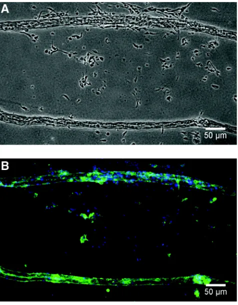
Figure 2.2. Removal of cords from PDMS template. Phase contrast (A) and fluorescence (B) images of HUVEC cords removed after 24 h in culture into 2.4 mg/ml collagen gel. Cells are labeled for actin (green) and nuclei (blue). Data reprinted with permission (Raghavan, Nelson, Baranski, Lim, & Chen, 2010a).
Laschke et al., 2010). We predicted that the established cell-cell contacts within cords comprised of endothelial cells would help enhance their angiogenic potential and that patterned cord geometry would be preserved upon implantation and subsequent vessel formation in vivo. More broadly, we hypothesized that the implantation of patterned EC cords could be used to control the architecture of engineered vasculature in vivo.
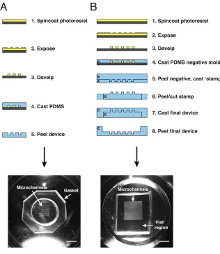
While effective in demonstrating a proof of concept, use of the first-generation device for creating EC cords in extensive studies of vascularization was prohibitively time consuming, required large numbers of cells (> 6 million per device), and yielded cords that were limited in length (< 1 cm). To overcome these limitations, we designed, fabricated and optimized a second-generation system for producing and removing EC cords (Fig. 2.3). Fabrication of the new devices utilized a double-casting technique to generate microchannels with surrounding flat regions within a PDMS well rather than relying on a separate gasket (Fig. 2.3A, B). The second generation device was designed to enable generation of larger numbers of cords using fewer cells (2 million per device). The new device configuration allowed for cords to be easily removed from the
Figure 2.3. First- and second-generation PDMS devices for generating cords. (A) Process flow describing technique for fabricating first-generation device for generating cords. (B) Process flow describing technique for fabricating second-generation devices for generating cords. Macroscopic images of final devices are shown in bottom panels. (bars: 10 mm)
The complete redesign of the device required several optimizations to techniques for generating cords. First, treatment of the PDMS with Pluronic F-127 required
PDMS hydrophilic, making it largely non-adhesive to proteins and cells (M.-H. Wu, 2009). Here, a Pluronic treatment that is too mild will prevent proper cord formation by allowing cells to adhere and migrate on the PDMS surfaces. Conversely, a Pluronic treatment that is too harsh will render the PDMS surfaces almost entirely non-adhesive causing the cords to float out of the microchannels upon addition of growth medium. Through empirical tests, we determined that proper Pluronic treatment that achieves the correct balance of adhesive cues is obtained by exposing the PDMS templates to a 0.02% (w/v) Pluronic F-127 solution for 10 minutes. While we found that decreasing the
concentration to below 2x10⁻⁵ % was required to observe any effects of decreased concentration, the length of Pluronic treatment had a significant impact on its effect. Additionally, it should be noted that the negative PDMS masters from which the final devices are cast could be reused no more that three times. After three casts, we observed that the resulting devices were more adhesive to collagen and cells despite treatment with Pluronic. Due to the double casting process, devices cast more than three times also began to exhibit defects in the walls of the microchannels.
step. In addition, the time required for the polymerization of the collagen required a balance between ensuring that the gel has fully formed and preventing too much drying of the sample, which led to cell death. Through a series of optimizations, we found that inverting the sample for 5 minutes over a bath of water during polymerization achieved the correct balance of gelation time and humidity and resulted in viable cords. Additional complexity is introduced by the technique used when adding growth medium to the polymerized cords immediately prior to culture. Upon methodically employing a variety of techniques, we determined that allowing large drops of medium to fall onto the microchannels yielded the best results. Briefly, five drops of growth medium are first dispensed into the center and four corners of the microchannels from a height of several millimeters. Subsequent drops are dispensed in a similar manner into the regions between those already placed onto the substrate. As the second set of drops is introduced, the growth medium accumulates into a larger pool that covers the entire area of
microchannels containing cords. The final volume of medium is brought up to 750 µl by dispensing slowly into one of the corners of the device. Importantly, extreme care must be taken during this process not to dislodge the cords from the microchannels during these final steps.
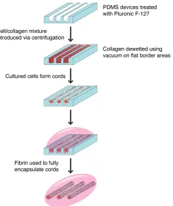
microchannels containing cords onto a pool of pre-polymerized fibrin yielded the best results. Use of a PDMS gasket helps to ensure that constructs with a controlled thickness are generated. After 15 minutes of polymerization, the sample can then be flooded with PBS, which allows the positive buoyancy of the PDMS to help lift it off of the underlying fibrin matrix. Several layers of cords can be stacked on top of each other to generate more complex geometries. In the final step, an additional layer of fibrin is added to encapsulate the exposed surface of the cords. The large area of the microchannels allows for 6 mm discs to be generated, which are suitable for implantation into mice (Fig. 2.4). A schematic of the process for generating cords using the second-generation of devices is shown in Figure 2.5.
Figure 2.5 Schematic of process used to generate EC cords using second-generation PDMS devices. Design modifications including increased microchannel area and incorporation of flat border regions allow for the removal and generation of larger cords, which are amenable to implantation into mice.
2.2.2 Screening cell types and coculture ratios for use in cords
Melero-Martin et al., 2008; Traktuev et al., 2009). Here, we screened the ability of HUVECs and eEnd.2 cells to organize into networks when cultured within collagen gels in vitro. Given our hypothesis that established cell-cell contacts might help enhance the angiogenic effect of implanted cords, we attempted to choose a cell mixture capable of extensive cell-cell interactions and networking behavior. We first tested the eEnd.2 murine embryonic endothelial cell line by culturing the cells in a 2.5 mg/ml collagen gel for 4 days (Williams, Courtneidge, & Wagner, 1988). While these cells exhibited localized networking behavior, significant clumping was observed and large increases in cell number indicated high rates of proliferation (Fig. 2.6). This high rate of proliferation in 3D led us to conclude that the eEnd.2 cell type was a poor candidate for use in a
vascularization strategy whose goal it is to generate stable vessels in vivo. Next, we tested human umbilical vein endothelial cells (HUVECs), a primary endothelial cell type
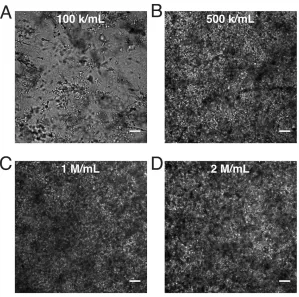
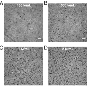
Figure 2.7 Networking behavior of HUVECs in collagen gels. Brightfield imaging of HUVECs cultured in 2.5 mg/ml collagen for 4 days at concentrations of 1x10⁵ cells/ml (A), 5x10⁵ cells/ml (B), 1x10⁶ cells/ml (C) and 2x10⁶ cells/ml (D) shows limited networking or cell-cell interactions. (bars: 100 µm)
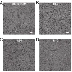
C3H10T1/2 is a murine cell line of mesenchymal origin with demonstrated potential to differentiate into pericytes and promote vessel maturation (Hirschi,
Figure 2.8 Networking behavior of HUVECs + C3H10T1/2 cells collagen gels. Brightfield imaging of 1x10⁶ HUVECs cultured in 2.5 mg/ml collagen for 4 days at with no 10T1/2s (A), 1:50 (B), 1:30 (C) or 1:10 (D) ratios of 10T1/2s to HUVECs. (bars: 100 µm)
2.2.3 In vitro characterization of endothelial cell cords
Having created a robust platform for generating cords, we performed several
characterizations to better understand the composition of cords and the mechanics and cellular dynamics of the cord formation process. First, time-lapse imaging of cells
(Fig. 2.9A,B). Hematoxylin and eosin (H&E) and sirius red staining of fully contracted, paraffin embedded cords suggested that some clustering and wrapping of cells around a core of compacted collagen might also occur (Fig. 2.9C). These occasional clusters, however, were randomly distributed along the length of the cords.
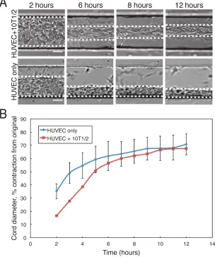
Once in culture, cords contracted to approximately 65% of their original diameter over a period of 10-12 hours (Fig. 2.10). To verify that this contraction is driven by cells exerting tractional forces against the surrounding ECM we treated cords with the myosin IIa inhibitor blebbistatin or the RhoA inhibitor Y27632. When treated with these
inhibitors immediately after seeding, the cords remained in their uncontracted state (Fig. 2.10). Wash our of the inhibitors after 6.5 or 7.5 hours led to a rapid resumption of the contraction process. Within 6 hours of wash out, the cords had contracted to
To determine whether the presence of 10T1/2 cells affected the dynamics of cord contraction, we generated HUVEC only cords and compared their contraction rate to that of HUVEC + 10T/1/2 cords (Fig. 2.11). Given the contractile nature of mural cells in vivo, we predicted that cords containing 10T1/2s would contract more rapidly or to a higher degree than those containing HUVECs alone. Interestingly, both cord types
To demonstrate the potential of using EC cords to pattern vasculature into a variety of geometries, we attempted to generate cords with a range of diameters and shapes. While in our previous work we demonstrated that varying collagen concentration could be used to alter final cord diameter, this approach is limited to a small range of diameters (Raghavan, Nelson, Baranski, Lim, & Chen, 2010a). Here, we chose to maintain a consistent collagen concentration while varying the PDMS microchannel width. To maintain a 1:1 aspect ratio for the various microchannel widths, separate wafers with matching heights were fabricated for each width. For cords with varying geometries, 150 µm wide and 150 µm deep microchannels were used. Several minor optimizations were necessary when generating cords of varying diameters. Cords smaller than 100 µm yielded a much-reduced volume of collagen within the PDMS
of the cords and surrounding flat regions. These modifications allowed us to successfully generate cords with diameters that ranged from 25 µm to 500 µm (Fig. 2.12A) and a variety of architectures (Fig. 2.12B).
2.3 Vascularizing engineered tissues
in vivo
via implantation of
endothelial cell cords
2.3.1 Development of in vivo implantation model
parametrial fat pad strongly suggested that blood was present within or near the implanted cords (Fig. 2.14).
Figure 2.14 Early evidence of blood in implanted tissue constructs containing EC cords. H&E staining suggesting presence of blood in tissue constructs implanted adjacent to the parametrial fat pad. Fibrin gels containing EC cords directly sutured to the parametrial fat pad and resected 5 days PI. Arrowheads denote cords with evidence of red blood cells in transverse (left panel) or longitudinal cross section. (bars: 100 µm)
shape upon implantation. First, we created our tissue constructs atop a thin (1 mm) layer of PDMS. The combined PDMS and fibrin gel with cords were cut into 6 mm discs using a biopsy punch. Small (0.75 mm) holes were punched near the periphery of the PDMS to allow for sutures to be passed through the structure for attaching to host tissue (Fig 2.15A). Without these holes near the discs’ periphery, passing a suture needle through the PDMS discs led to tearing. This approach successfully maintained the shape/geometry of the constructs upon implantation. However, we found that the PDMS discs engrafted to the host adipose tissue making their removal without disrupting the implanted constructs extremely difficult. Because PDMS cannot be sectioned resecting the entire fat pad and implanted construct with the PDMS still attached would prohibit any subsequent histological analysis. Given these complications with PDMS, we next attempted to use a polypropylene mesh as a backing material for the tissue constructs. We cut small squares of mesh material (~1 cm2) and embedded them in fibrin. This procedure was performed atop a Pluronic-treated PDMS-coated petri dish to aid in the detachment of the fibrin prior to
implantation. We next inverted cords onto the mesh/fibrin layer and removed them as before. The resulting assembly consisted of a layer of cords positioned immediately atop the polypropylene mesh with the entire assembly encased in fibrin (Fig. 2.15B). While it was possible to section the polypropylene mesh, we found that the mesh filaments prevented any imaging of the embedded cords. To enable potential intravital imaging of our tissue constructs, we utilized a ‘donut’-shaped
heated cork borers. The smooth outer edge facilitated their manipulation during surgery, while the inner hole would allow for direct imaging of the cords in the
future. Use of a mesh material allowed for the needle and sutures to be passed directly through the periphery of the mesh without any further modifications during
implantation (Fig. 2.15D).
atop ‘donut-shaped’ mesh embedded in fibrin. Inner hole facilitated imaging of cords while smooth outer edge aided in implantation. (bar: 1 mm) (D) Mesh ring sutured sutured atop the parametrial fat pad in preparation for implantation. (bar: 4 mm)
The polypropylene mesh incorporated into our design could be readily sectioned and successfully preserved the geometry of the implanted tissue constructs. These