MINIMAL INVASIVE DENTISTRY
Swadheena Patro
Institute of Dental Sciences, Campus
A R T I C L E I N F O
INTRODUCTION
The aim of MID is to halt the disease first and then to restore the lost tooth structure and to keep teeth healthy and functional for life. A most important element is achieved through implementing the important strategies for keeping teeth free from carious lesions. These strategies are considered to be:
1. Early caries detection and risk assessment
2. Remineralization of demineralized enamel and dentin 3. Optimal caries preventive measures
4. Minimally invasive operative interventions 5. Repair rather than replacement of the restorations.
The four core principles of MID can be summarized as follows:
1. Recognition- To identify and assess any potential caries risk factors early, through lifestyle analysis, saliva testing and using plaque diagnostic tests.
2. Reduction- To eliminate or minimize caries risk factors through altered fluid balance reducing the intake of dietary cariogenic foods addressing lifestyle habits. 3. Regeneration- To arrest and reverse incipient lesions
using appropriate topical agents including fluoride Casein Phosphopeptides-Amorphous Calcium phosphate (CPP-ACP).
International Journal of Current Advanced Research
ISSN: O: 2319-6475, ISSN: P: 2319-6505,
Available Online at www.journalijcar.org
Volume 7; Issue 5(G); May 2018; Page No.
DOI: http://dx.doi.org/10.24327/ijcar.2018
Copyright©2018 Swadheena Patro., Rini Behera and Sumita Mishra
Attribution License, which permits unrestricted use, distribution, and reproduction in any medium, provided the original w
*Corresponding author: Sumita Mishra
Institute of Dental Sciences, Campus-3, SOA University, Kalinga Nagar
Article History:
Received 20th February, 2018
Received in revised form 20th March, 2018 Accepted 8th April, 2018 Published online 28th May, 2018
Key words:
Demineralization, Remineralization, Dental caries, CPPACP, Composites, Atraumatic Restorative treatment
MINIMAL INVASIVE DENTISTRY
Swadheena Patro., Rini Behera and Sumita Mishra*
Institute of Dental Sciences, Campus-3, SOA University, Kalinga NagarA B S T R A C T
Objective: Minimally Invasive Dentistry (MID) emphasizes
management strategies resulting in less destruction of tooth structure. The primary focus in the medical model of caries management is identifying and eliminating the causative factors for caries, along with repairing damage caused by c
the traditional surgical model because the disease is viewed as an infection rather than as a lesion and its treatment objective is to reduce or eliminate pathogens. The medical model synthesizes knowledge of the disease process into a simple conceptual model using new technologies. Overview: This section should include a brief summary of the findings of the review. Conclusions: The dentist role in MID is recognizing, intercepting and prevention of demineralized sites. Furthermore, assessing the caries risk and maintaining a state of oral health followed by monitoring over extended period of time. Thus, with the philosophy of extension for prevention would lead to extension for ruining teeth. With old challenges at hand to which applying minimally invasive techniques leads to maximum preservation with minimal removal of tooth structure. Technological innovations in materials and techniques gives better results and higher patient acceptance and an easier practice.
The aim of MID is to halt the disease first and then to restore the lost tooth structure and to keep teeth healthy and functional for life. A most important element is achieved through implementing the important strategies for keeping teeth free
us lesions. These strategies are considered to be:
Early caries detection and risk assessment
Remineralization of demineralized enamel and dentin
Minimally invasive operative interventions of the restorations. 1
The four core principles of MID can be summarized as
To identify and assess any potential caries risk factors early, through lifestyle analysis, saliva testing and using plaque diagnostic tests.
eliminate or minimize caries risk factors through altered fluid balance reducing the intake of dietary cariogenic foods addressing lifestyle habits.
To arrest and reverse incipient lesions using appropriate topical agents including fluorides and
Amorphous Calcium
4. Repair- When cavitation is present and surgical intervention is required. Bioactive materials are used to restore the tooth and promote internal healing of the dentine particularly in cases of deep dentine caries where the risk of iatrogenic pulpal injury is high
An accurate diagnosis of the disease is mandatory.
Early Caries Detection
In order to conserve tooth structure and perform minimally invasive dentistry, carious lesions must be detected at the earliest possible time.
Various Methods of Diagnosis
Visual-tactile method
Conventional methods
1. Tactile examination 2. Visual examination
Advances in visual method
Illumination
1. Ultrasonic illumination 2. Ultrasonic imaging
3. Fiber optic transillumination (FOTI) 4. Wavelength dependent FOTI 5. Digital imaging FOTI (DIFOTI)
a. Dyes
b. Endoscopy filtered fluoroscence (EFF) method.
International Journal of Current Advanced Research
6505, Impact Factor: 6.614
www.journalijcar.org
; Page No. 12697-12702
//dx.doi.org/10.24327/ijcar.2018.12702.2241
Swadheena Patro., Rini Behera and Sumita Mishra. This is an open access article distributed under the Creative Commons Attribution License, which permits unrestricted use, distribution, and reproduction in any medium, provided the original w
3, SOA University,
Nagar
Minimally Invasive Dentistry (MID) emphasizes conservative caries management strategies resulting in less destruction of tooth structure. The primary focus in the medical model of caries management is identifying and eliminating the causative factors for caries, along with repairing damage caused by caries. This is a departure from the traditional surgical model because the disease is viewed as an infection rather than as a lesion and its treatment objective is to reduce or eliminate pathogens. The medical model cess into a simple conceptual model using new : This section should include a brief summary of the findings of the The dentist role in MID is recognizing, intercepting and prevention rmore, assessing the caries risk and maintaining a state of oral health followed by monitoring over extended period of time. Thus, with the philosophy of extension for prevention would lead to extension for ruining teeth. With old challenges at ch applying minimally invasive techniques leads to maximum preservation with minimal removal of tooth structure. Technological innovations in materials and techniques gives better results and higher patient acceptance and an easier practice.
When cavitation is present and surgical intervention is required. Bioactive materials are used to restore the tooth and promote internal healing of the dentine particularly in cases of deep dentine caries where the risk of iatrogenic pulpal injury is high.2, 3
An accurate diagnosis of the disease is mandatory.
In order to conserve tooth structure and perform minimally invasive dentistry, carious lesions must be detected at the
Various Methods of Diagnosis of Dental Caries
Ultrasonic illumination
Fiber optic transillumination (FOTI) Wavelength dependent FOTI Digital imaging FOTI (DIFOTI)
Endoscopy filtered fluoroscence (EFF) method.
Research Article
Radiographic methods
Conventional methods
a. Intraoral periapical x-rays (IOPA) b. Bitewing radiographs
c. Panorex radiography d. Xeroradiography
Recent advances in radiographic techniques
a. Digital imaging
b. Computerized image analysis c. Subtraction radiography
d. Tuned aperture computerized tomography e. Magnetic resonance micro imaging (MRMI)
Electrical conductance measurement
Lasers
1. Argon laser 2. Diode lasers
3. Qualitative laser fluorescence
4. Diagnodent (Quantitative laser fluorescence) 5. Optical coherence tomography
6. Polarization sensitive optical coherence tomography (PSOCT)
7. Frequency-domain laser-induced infrared photo thermal radiometry & modulated luminescence (PTR/LUM) 8. Dye enhanced laser fluorescence.
The accuracy of any diagnostic test is measured according to its “sensitivity” and “ specificity”.
Caries Risk Assessment
Caries-risk is “the probability of future caries disease development.”
Disease development includes both primary disease (new carious lesions) and secondary disease (lesion progression or re-activated carious lesions).
The three levels of prevention linked to the steps are:-
Primary prevention (step 1)- to prevent the intra family transmission of mutans streptococci and delay the establishment in infants, toddlers, and young children.
Secondary prevention (step 2)- to prevent, arrest, or reverse the microbial shift before any clinical manifestations of the disease occur.
Tertiary prevention (step 3)- focuses on limiting (stopping) the progression of caries process by initiating remineralization therapy of the existing lesions.
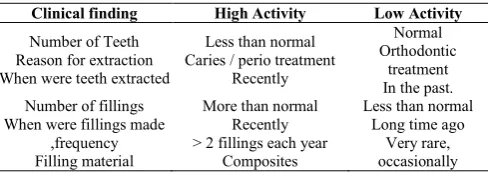
Clinical examination for evaluation of caries activity
A few important points for estimating caries activity are presented in table below:
Table 1 Estimation of Carious Activity
Clinical finding High Activity Low Activity
Number of Teeth Reason for extraction When were teeth extracted
Less than normal Caries / perio treatment
Recently
Normal Orthodontic
treatment In the past. Number of fillings
When were fillings made ,frequency Filling material
More than normal Recently > 2 fillings each year
Composites
Less than normal Long time ago
Very rare, occasionally
Gold glass ionomers
Number of enamel lesions and cavities Localization Surface colour
X-rays
More than expected Appeared recently. Smooth or lingual surfaces,
lower incisors Soft, light Progression into dentin
None old , neglected Predilection sites
only Fissures. Hard, Dark Regression, still
within enamel.
Oral hygiene
Aggravating factors
Poor Crowded teeth Deep Fissures Marginal overchanges Orthodontic appliances
Acceptable Spaced arches shallow fissures Perfect adaptation
Removable appliances
Caries-Activity Tests for the Dental Office
Caries activity tests should reflect the three overlapping circles presented by Keyes in 1962: (1) the bacterial challenge, (2) the sugar content of the diet, and (3) tooth and host resistance (susceptibility) with remineralization potential. In the light of these requirements following tests should be suggested.
Bacterial challenge- Determination of mutans streptococci as an indicator of relative risk.
Diet- determination of lactobacilli as an indicator of sugar content in the diet.
Remineralization potential - salivary flow rate and buffer capacity as an indicator of potential biological repair.
Host susceptibility - caries experience as an indicator of past activity.
Remineralization
After the disease control, the loss of minerals from tooth hard tissues needs to be addressed and the oral balance between de and remineralization processes on the tooth surface should be regained. This may be done through "external remineralization" (on the tooth surface) and in cavity walls through '"internal remineralization".4
Reduction in Cariogenic Bacteria
Casein Phosphopeptide Amorphous Calcium Phosphate Complexes (CPP-ACP)
These complexes of tryptic casein phosphopeptides have the ability to stabilize calcium phosphate in solution as amorphous calcium phosphate (ACP).5,6 This complex is a nano cluster of ACP with four multiphosphorylated peptides that prevent its growth to the critical size required for nucleation, phase transformation and precipitation. Remineralization of enamel is correlated with the degree of saturation for dicalcium phosphate dihydrate (CaHPO4.2 H2O).7
Flowable composites
Flowable composites were developed mainly for special handling properties for composite resins. Because of low viscosity the composites are indicated for preventive resin restoration as pit and fissure sealants and for repair of porcelain or gold alloy restoration.8
Indirect Composite Resin
Nanocomposites
Nanotechnology may provide composite resins with a dramatically smaller filler particle size that can be dissolved in higher concentrations and polymerized into the resin system. The molecules in these materials can be designed to be compatible when coupled with a polymer and provide unique characteristics (i.e., physical, mechanical, optical). 10,11
Antimicrobial Composite
Antimicrobial properties of composites may be accomplished by introducing agents such as silver or one or more antibiotics into the material. Microbes are subsequently killed on contact with the materials or through leaching of the antimicrobial agents into the body environment.12 Silver and titanium particles were introduced into dental composites, respectively, to introduce antimicrobial properties and enhance the biocompatibility of the composites.13
Several reports have described the incorporation of a methacryloyloxy-dodecyl-pyridinium-bromide monomer in composite resins that showed no release of the incorporated monomer but still exhibited antibacterial properties.14
Stimuli Responsive Composite
Stimuli-responsive materials possess properties that may be considerably changed in a controlled fashion by external stimuli like changes of temperature, mechanical stress, pH, moisture, or electric or magnetic fields.
Fiber Reinforced Composite
Fiber reinforced composites have numerous industrial and aerospace applications because they are light, strong and non flammable.15 ,16
Self-healing Composite
One of the first self-repairing or self-healing synthetic materials was an epoxy system which contained resin filled microcapsules. If a crack occurs in the epoxy composite material, that microcapsules are destroyed near the crack and release the resin. The resin subsequently fills the crack and reacts with a Grubbs catalyst dispersed in the epoxy composite, resulting in a polymerization of the resin and a repair of the crack.17
Atraumatic Restorative Treatment
Ultraconservative treatment approaches are recommended for treating cavitated dentin lesions, which includes preservation of as much sound tooth structure as possible.18 ART is an example of MID.19
It consists of two components: sealing of caries prone pits and fissures with a sealant, and use of a sealant in combination with restoring cavitated dentin lesions.20 The main difference between the ART approach and other minimally invasive operative interventions is that ART uses hand instruments only in conjunction with adhesive materials or systems.
The instruments used in ART are; mouth mirror, a probe, tweezers, a dental hatchet, excavators, and an applier / carver. The consumable materials required are cotton wool rolls, cotton wool pellets, petroleum jelly, tumbler/cup, wooden wedges, and matrix bands or plastic strips.
In practice, Glass-Ionomer Cement (GIC) has become the most predominantly used material mainly because of its delayed
setting reaction that allows handling of the material before it is completely set.
Step-by-Step procedure of ART for use with glass-ionomer restorative material is
1. Isolation of the tooth.
2. Cleaning of the tooth to permit examination of the carious lesion.
3. Access to the caries is achieved using a hatchet or other suitable instrument.
4. Caries is removed with excavators.
5. The cavity and adjacent pits and fissures are further cleaned with a conditioner.
6. The glass-ionomer is mixed according to the manufacturer's instructions.
7. The cavity and pits and fissures are slightly overfilled with the mixed glass-ionomer.
8. A gloved index finger is placed over the top of the filling material and pressure is applied to press the filling material into the cavity and the pits and fissures. 9. The finger is removed carefully.
10. Excess filling material is removed.
11. The occlusion is checked and the filling adjusted.
The term "modified ART' refers to ART which is performed in places where traditional dental equipment is available instead of in field situations. This modification uses rotary equipment, the drill, to open the tooth cavity, followed by the normal ART procedure in cleaning and restoring the cavity. The use of rotary equipment makes the total procedure quicker and easier.
The management of ART failures follows principles of restoration repair instead of replacement
Table 2 Principles of Restoration Repair
ART Failure Management
Material wear >0.5mm
Partial material loss
Complete material loss
Caries related to restoration margin
Cleaning of GIC surface, Application of dentine condition.
Placement of new GIC Layer Cleaning fracture surface, Application of
dentin conditioner Placement of new GIC layer Cleaning cavity surface, Application of
dentin condition Placement of new GIC layer Caries removal using hand excavator
Cleaning GIC surface Application of dentin conditioner
Placement of new GIC layer
Preventive Resin Restoration
Preventive sealant restoration is indicated primarily on the occlusal surfaces of permanent molars and premolars when occlusal caries has involved a minimal amount of dentin, and may also be indicated for primary molars.21 They are used especially when the cavity in a pit or fissure is small and discrete. Larger cavities should be restored with amalgam or a posterior composite while smaller cavities may be restored with sealant alone.22, 23
Sealants
Figure 1 Diagrammatic depiction of the morphological types of fissure system u, u shaped, v, v shaped, Y1 and Y2, deep narrow fissure systems.
Deep and narrow fissures (Y1 and Y2) are associated with a higher local caries risk, and should be sealed independent of general caries risk.
Indications for fissure sealing24
Local risk General risk Sealing
Low (shallow fissure) low no
High (deep fissure) low yes
Low (shallow fissure) high yes
High (deep fissure) high yes
The "Tunnel" Restoration
A tunnel preparation is made for removal of proximal caries by making an access through occlusal surface while leaving the marginal ridge intact. This technique was first used in primary molars by Jinks in 1963 and later on Hunt and Knight used this procedure for restoration of small proximal lesions.Most clinical studies report, silver-cermet glass ionomer as the preferred restorative material. This technique has fallen from favor because of the difficulty of completely removing the
caries, subsequent frequent collapse of the marginal ridge, and occlusal wear of the restorative material. Hasselrot (1998) reported a 50% survival rate of tunnel restorations in permanent teeth over a 6-year period.25
Figure 2 The Tunnel Preparation
The Proximal "Slot" Preparation
In the "slot" preparation, access to the caries is gained through the marginal ridge, but preserving this structure wherever possible (Mount and Ngo 2000).
The occlusal fissure is maintained intact and when the cavity is restored with resin composite, the fissure can be protected with sealant. The cavity design allows better visualization of the caries than the tunnel design, allowing the removal of unsupported enamel. The cavity can be restored with glass ionomer and a bonded surface laminate of composite resin to resist heavy occlusal contact. Slot preparations to be restored with amalgam require mechanical retention. Grooves 0.5-mm wide must be cut into the facial and lingual walls of the proximal box using the full diameter of a No. l/4 round bur. They are placed 0.25mm from the enamel-dentin junction in the opposing facial and lingual walls, and parallel to the external surface of the tooth. The grooves should be quite distinct, extending from the gingival floor of the slot preparation to the occlusal surface
Figure 3 Proximal Slot Preparation
Chemomechanical Caries Removal (CMCR)
Chemo mechanical caries removal is a method for minimally-invasive, gentle dentine caries removal based on biological principles (Zinc et al., 1988).26 CMCR involves the use of a gel (carisolv) that is applied to the caries affected area of the dentine, which selectively reacts with denatured collagen, thereby making the carious dentine softer. Specially designed instruments are used to remove the softened diseased portion of the tooth, while healthy tissue is preserved. The treatment is quiet and effective. Many patients and dentists call it "a silent revolution".
Table 3 Indications for the use of each of the four types of sealant restoration
Type of sealant
restoration Indications
Type-1
Fissure sealant alone
Stained and decalcified fissure
No radiographic sign of dentine involvement
Less than two other carious lesions in the mouth.
Type-2
Composite plus sealant
Stained and decalcified fissure
More than two other carious lesions in the mouth
Enamel biopsy shows lesions confined to enamel
Type-3
Glass ionomer cement plus cement
Enamel biopsy indicated
Cavity in dentine with minimal lateral spread
Margins not in occlusal contact
Type-4
Laminate restoration
Enamel biopsy indicated
Lesions in dentine with lateral spread along DEJ
Cavity margins in occlusal contact
Amalgam restoration
Enamel biopsy indicated
Large radioluscency in dentin.
Significant lateral spread along DEJ
It has the following advantages over traditional drilling
1. Less perception of pain and more comfortable for patient.
2. Less fear and anxiety to method, leads to less discomfort to patients especially in children.
3. Removes only infected layer and leads to more tissue preservation.
4. No pulpal irritation.
5. Well suited to the treatment of deciduous teeth, dental phobic's and medically compromised patients.
6. Better removal of caries in uncooperative patients. 7. Useful in physically handicapped patients.
8. Useful in patients with T.B like infectious diseases (prevent droplet infection).
Papacarie and Carisolv can be successfully used in special health care needs (SHCN) patients and phobic adults in pediatric dentistry and public health sectors.27
Papacarie
In Brazil 2003, Formula ea cao by Sao Paulo, first time introduced papain gel as Papacarie for chemomechanical caries removal agent (Bussadori et al., 2005).28 Papacarie is a product which is patented, registered and approved by ANVISA in Brazil. Its main components are papain, chloramine and toluidine blue.
Chloramine
A compound comprised by chlorine and ammonia has bactericidal and disinfectant properties widely used as an irrigating solution of radicular canals in order to chemically soften the carious dentine.
Toluidine Blue
Initially, the malachite green was used as coloring agent, however, after a few studies toluidine blue was found highly effective against Streptococcus mutans that fixes into the bacterial membrane.
Ozone Therapy
Ozone therapy can be defined as a versatile bio-oxidative therapy in which oxygen/ozone is administered via gas or dissolved in water or oil base to obtain therapeutic benefits.29 Ozone therapy has a wide range of applications in treating various diseases owing to its unique properties including antimicrobial, immune stimulant, analgesic, anti hypnotic, detoxicating, bioenergetic and biosynthetic actions. Currently ozone finds its application in the management of inflammatory bowel disorder, specifically ulcerative colitis, Crohn's disease and chronic bacterial diarrhea, cancer, stroke and AIDS.
Lasers
The term LASER is an acronym for ‘Light Amplification by the Stimulated Emission of Radiation’. The most commonly used lasers in dentistry at present are Carbon Dioxide (C02), Erbium: Yttrium, Aluminum, Garnet (Er:YAG), Erbium, Chromium: Yttrium, Scandium, Gallium Garnet (Er, Cr:YSGG), Neodymium: YAG and diode.
CONCLUSIONS
G.V.Black introduced the concept of "Extension for prevention" which has gradually evolved in modern times into "prevention of extension" resulting in conservation of tooth
structure in which lies the basis of MID, either surgically or byremineralization procedures. Thus MID is directed at detecting, diagnosing, intercepting and treating dental caries on the microscopic level. Minimally intervention philosophy relies on identifying and assessing the risk factors early, preventing disease by eliminating the risk factors, and restoring the health of the oral environment.
References
1. Burk FJT. Minimal intervention isn’t just small cavities. Dent Update 2008; 35:509.
2. Walsh LJ. A system for total environmental management (STEM) of the oral cavity, and its application to dental caries control. Internat Dent SA
Australas Edn 2008;3:34-48.
3. Brostek AM, Bochnek AJ, Walsh LJ. Minimally invasive dentistry: A review and update. Shanghai J Stomatol 2006; 15:225- 249.
4. Yassin OM. In vitro studies of the effect of a dental explorer on the formation of an artificial carious lesion.
ASDC J Dent Child 1995; 62:111-117.
5. Reynolds EC, Cain CJ, Webber FL. Anticariogenicity of calcium phosphate complexes of trypticcasein phosphopeptides in the rat. J Dent Res 1995;74:1272-1279.
6. Reynolds EC. Anticariogenic complexes of amorphous calcium phosphate stabilized by casein phosphopeptides: a review. Spec Care Dentist 1998; 18:8-16.
7. Huq NL, Cross KJ, Reynolds EC. Molecular modeling of the multi phosphorylate casein phosphopeptide alphaSl-casein based on NMR constraints. J Dairy Res
2004;71:28-32.
8. Cohen R. The expanded use of improved flowable composite. Dent Town 2008;64:25-35.
9. Nandini S. Indirect resin composites. J Conserv Dent
2010;13:184-94.
10. Terry DA. Applications of nanotechnology. Ed Comment 2004;16:417-22.
11. Loguercio AD, Alessandra R, Mazzocco KC, Dias AL, Busato AL, Singer Jda M, et al. Microleakage in class II composite resin restorations: Total bonding and open sandwich technique. J Adhes Dent 2002;4:137-44. 12. Joyee JL, Cook CN. Packable resin composites. Ann
Essences Dent Clin Update 2003;25:19-21.
13. Yeli M, Kidiyoor KH, Nain B, Kumar P. Recent advances in composite resins - A review. J Oral Res Rev 2010;2:8-14.
14. Beyth N, Yudovin-Farber I, Bahir R, Domb AJ, Weiss EI. Antibacterial activity of dental composites containing quaternary ammonium poly ethylenimine nano particles against Streptococcus mutans.
Biomaterials 2006;27:3995-4002.
15. Rosensteil SF, Land MF, Fujimoto J. Contemporary Fixed Prosthodontics. 3rd ed. St. Louis:Mosby; 2001. p. 697-706.
16. Joyee JL, Cook CN. Packable resin composites. Ann
Essences Dent Clin Update 2003;25:19-21.
17. Yeli M, Kidiyoor KH, Nain B, Kumar P. Recent advances in composite resins - A review. J Oral Res Rev 2010; 2:8-14.
containing quaternary ammonium poly ethylenimine nano particles against Streptococcus mutans.
Biomaterials 2006; 27:3995-4002.
19. Grossman ES, Mickenautsch S. Microscope observations of ART excavated cavities and restorations. SADJ. Sep 2002; 57:359-63.
20. Frencken JE, Holmgren CJ. Atraumatic restorative treatment for dental caries. Nijmegen, STI Book b.v.; 1999. ISBN 906759024X.
21. Gwinnett A J. A comparison of proximal carious lesions as seen by clinical radiography, contact microradiography and light microscopy. J Am Dent
Assoc 1971; 83:1078-1080.
22. Millman C K. Fluoride syndrome. Br Dent J 1985; 154:341.
23. Gray GB, Shellis P. Infiltration of resin into white spot caries like lesions of enamel: an in vitro study. Eur J
Prosthodont Restor Dent 2002; 10:27-32.
24. Staehle HJ. Fissurenversiegelungen and preventive Adhasivfullungen. In: Einwag J, Pieper K. Kinderzahnheil-kunde.Urban&Schwarzenberg
2002:210-212.
25. Hasselrot L. Tunnel restorations in permanent teeth: a 7-year follow-up study. Swed Dent J 1998; 22; 1-7. 26. Zinc JH, Mclnnes-Ledoux P, Capdeboscq C, Weinberg
R. Chemomechanical caries removal a clinical evaluation. J. Oral Rehab 1988; 15:23-33.
27. Carrillo C, Tanaka M, Cesar M, Use of papain gel in disabled patients. J Dent Child 2005; 75:222-228. 28. Bussadori SK, Castro LC, Galvao AC, Papain gel: A
new chemomechanical caries removal agent. J Clin Pediatr Dent 2005; 30:115-119.
29. Bocci V. Ozone as Janus: this controversial gas can be either toxic or medically useful. Mediators Inflamm 2004; 13:3–11.
How to cite this article:
Swadheena Patro., Rini Behera and Sumita Mishra (2018) 'Minimal Invasive Dentistry', International Journal of Current
Advanced Research, 07(5), pp. 12697-12702. DOI: http://dx.doi.org/10.24327/ijcar.2018.12702.2241