University of Pennsylvania
ScholarlyCommons
Publicly Accessible Penn Dissertations
Spring 5-17-2010
High-Throughput Engineering and Analysis of
Class II Mhc/Peptide Binding by Yeast Co-Display
Wei JiangUnversity of Pennsylvania, [email protected]
Follow this and additional works at:http://repository.upenn.edu/edissertations
Part of theBiochemical and Biomolecular Engineering Commons
This paper is posted at ScholarlyCommons.http://repository.upenn.edu/edissertations/419
Recommended Citation
Jiang, Wei, "High-Throughput Engineering and Analysis of Class II Mhc/Peptide Binding by Yeast Co-Display" (2010).Publicly Accessible Penn Dissertations. 419.
High-Throughput Engineering and Analysis of Class II Mhc/Peptide
Binding by Yeast Co-Display
Abstract
Polymorphisms of major histocompatibility complex (MHC) and molecular mechanisms of their antigen-presenting specificity and promiscuity have great impact on T cell-mediated immune responses and related diseases. Challenges in elucidating the characteristics of antigenic peptide binding by MHC motivate the development of high throughput experimental tools to quantitatively analyze interactions between hundreds of MHC allelic proteins and various peptide sequences. We demonstrated such a method by co-displaying target peptides and class II MHC (MHC-II) on the yeast surface in an intracellular association-dependent manner. The optimized yeast co-display system enabled quantitative mapping of side-chain preferences and general motifs for peptides binding to MHC-II by site-directed mutagenesis or peptide library screening, and also allowed rapid tailoring of MHC-II peptide binding specificity by directed evolution approaches, which derived MHC-II allelic mutants with altered peptide binding specificity or hyper-promiscuity. Comparison of these experimentally engineered mutants with naturally discovered MHC-II proteins recovered valuable information about structure-function relationship in the evolutionary mechanisms for polymorphic MHC-II molecules, which could direct future immunotherapeutic innovation.
Degree Type Dissertation
Degree Name
Doctor of Philosophy (PhD)
Graduate Group
Chemical and Biomolecular Engineering
First Advisor Wei Jiang
Keywords
yeast surface display, MHC-II, directed evolution, peptide binding, yeast co-display
Subject Categories
HIGH-THROUGHPUT ENGINEERING AND ANALYSIS OF CLASS II
MHC/PEPTIDE BINDING BY YEAST CO-DISPLAY
Wei Jiang
A DISSERTATION
in
Chemical and Biomolecular Engineering
Presented to the Faculties of the University of Pennsylvania
in
Partial Fulfillment of the Requirements for the
Degree of Doctor of Philosophy
2010
Supervisor of Dissertation Signature________________ Eric T. Boder, Associate Professor
Chemical and Biomolecular Engineering, University of Tennessee
Graduate Group Chairperson Signature________________
Matthew J. Lazzara, Assistant Professor of Chemical and Biomolecular Engineering
Dissertation Committee
David J. Graves, Associate Professor of Chemical and Biomolecular Engineering
Dimitri S. Monos, Professor of Pathology and Laboratory Medicine, Children’s Hospital of Philadelphia
ACKNOWLEDGEMENTS
First of all, I would like to thank my advisor Dr. Eric Boder for his help and
guidance on my Ph.D. project for the past five and a half years. Eric is such a nice
person that I really felt comfortable and confident to do research abroad with him.
During countless friendly communications with him, both of my academic background
and English presentation skills have been improved dramatically. Most importantly, the
freedom Eric gave me upon designing projects and performing experiments helped me
developing the potential to think and work independently. I would also like to thank Dr.
Dimitrios Monos, Dr. Scott Diamond, and Dr. David Graves, for being in my thesis
committee and proposing helpful suggestions to my project. As an immunologist, Dr.
Monos offered a lot of great opinions and valuable immunological background support
on my research. I would like to give special thanks to Dr. Lawrence Stern (University of
Massachusetts Medical School) for sharing the plasmid pDLM1-drb1s, which provides
the necessary MHC-II gene to start my project.
In the Boder Lab, many people deserve my acknowledgements. Andrew Nields was
the first one tutoring me through most of experimental techniques for months and
assisting me on the MHC-II engineering project, which he had been working on for
many years. Even though we did not have too long overlap periods in the lab, Andrew
did help me a lot to cross both research and language barriers. I would like to thank
Ranganath Parthasarathy, an experienced chemist and bioengineer with whom I spent
the most years in lab. Ranganath provided both technical expertise and experimental
easier and more bearable after lab relocation to Tennessee. Jeong Lee, Lauren Pepper,
and Kyudam Oh not only shared their interesting ideas with me in research but also
befriend to me in my life. Sheldon Park, Shyam Subramanian, Choi Fong Lu and the
undergraduate student Nick Dang also assisted me a lot during my first years of Ph.D.
study at Penn.
There were many people at University of Tennessee who have provided assistance to
my research. Nancy Neilsen (College of Veterinary Medicine) spent a lot of time and
patience on showing me technical details on how to set up a cell sorter for better
performing one of my key experiments, which indeed greatly helped me to understand
the theoretical background beyond the experiment itself and to perform troubleshooting
thereafter. Dianne Trent (Department of Comparative Medicine) and Shige Eda
(Department of Forestry, Wildlife, and Fisheries) also kindly operated their equipments
for me to complete many flow cytometric analyses. Joe May (Division of Biology) gave
me a lot of suggestions on how to generate better DNA sequencing results and offered
couples of reruns for my samples with unsuccessful results. Other members joining the
Boder lab after relocation: Vince Price, Ellen Messenger, Maryam Raeeszadeh
Sarmazdeh, and Sathish Dharmalingam, together created an enjoyable lab atmosphere
for my research in Tennessee.
Finally, I would like to thank my parents – Zuozhao Jiang and Deling Zheng in
China for their continuous understanding, encouragement and support both on my life
and oversea study, especially during the time when I was writing this dissertation.
This thesis work was funded by the U.S. National Science Foundation and the U.S.
ABSTRACT
HIGH-THROUGHPUT ENGINEERING AND ANALYSIS OF CLASS II
MHC/PEPTIDE BINDING BY YEAST CO-DISPLAY
Wei Jiang
Eric T. Boder
Polymorphisms of major histocompatibility complex (MHC) and molecular mechanisms
of their antigen-presenting specificity and promiscuity have great impact on T
cell-mediated immune responses and related diseases. Challenges in elucidating the
characteristics of antigenic peptide binding by MHC motivate the development of high
throughput experimental tools to quantitatively analyze interactions between hundreds of
MHC allelic proteins and various peptide sequences. We demonstrated such a method
by co-displaying target peptides and class II MHC (MHC-II) on the yeast surface in an
intracellular association-dependent manner. The optimized yeast co-display system
enabled quantitative mapping of side-chain preferences and general motifs for peptides
binding to MHC-II by site-directed mutagenesis or peptide library screening, and also
allowed rapid tailoring of MHC-II peptide binding specificity by directed evolution
approaches, which derived MHC-II allelic mutants with altered peptide binding
specificity or hyper-promiscuity. Comparison of these experimentally engineered
mutants with naturally discovered MHC-II proteins recovered valuable information
about structure-function relationship in the evolutionary mechanisms for polymorphic
TABLE OF CONTENTS
CHAPTER 1. BACKGROUND AND SIGNIFICANCE 1
1.1. Overview 1
1.2. Central role of CD4+ T cells in adaptive immune responses 3
1.2.1. Brief introduction for Immune system and lymphocytes 3
1.2.2. Differentiation and Function of CD4+ T cells 4
1.2.3. Activation of CD4+ T cells by APCs 7
1.2.4. Signaling pathways for CD4+ T cell activation 8
1.3. MHC-II and antigenic peptide binding 10
1.3.1. Classification and evolution of polymorphic MHC 10
1.3.2. Structure of MHC-II proteins 12
1.3.3. MHC-II pathway of antigen processing in APC 13
1.3.4. Peptide binding characteristics of MHC-II 15
1.4. Protein engineering technologies in this thesis work 17
1.4.1. Cell-surface display for target proteins 17
1.4.2. Analyzing immunofluorescently labeled cells by flow cytometry 20
CHAPTER 2. DEVELOPMENT OF YEAST CO-DISPLAY – A NOVEL
METHODOLOGY FOR CHARACTERIZING MHC-II/PEPTIDE BINDING 24
2.1. Introduction 24
2.2. Materials and Methods 27
2.2.1. Construction of plasmids 27
2.2.2. Buffers and media for yeast cultivation and treatment 31
2.2.3. Antibodies and labeling reagents 33
2.2.4. Generation and cultivation of yeast strains 33
2.2.5. Peptide binding assay for classical surface displayed HLA-DR1 34 2.2.6. Surface-stripping assay by DTT or Factor Xa treatment 35
2.2.7. Immunofluorescent labeling of yeast 35
2.2.8. Flow cytometric analysis of labeled cells 36
2.3. Results 36
2.3.1. Classical yeast display of functional HLA-DR1 heterodimer 36 2.3.2. Surface display of FLU peptide and their capability for anchoring soluble HLA-DR1 41 2.3.3. Yeast co-display of FLU and FLU-anchored soluble HLA-DR1 46 2.3.4. Stripping of FLU/HLA-DR1 complex off the surface of yeast 48 2.3.5. Verification of genotype-phenotype linkage between plasmids and co-displayed proteins 53
2.4. Discussion 55
CHAPTER 3. OPTIMIZATION OF YEAST CO-DISPLAY FOR QUANTITATIVE
ANALYSIS 59
3.1. Introduction 59
3.2. Materials and Methods 62
3.2.1. Media for yeast growth and induction 62
3.2.2. Primary and secondary labeling reagents 63
3.2.3. General culturing procedure for yeast co-display 64
3.2.4. General fluorescent labeling procedure 64
3.2.5. Quantitative analysis of flow cytometric data 65
3.3. Results 67
3.3.1. Evaluation of medium, temperature and time for induction 67
3.3.2. Optimization of cell density for induction 70
3.3.3. Optimization of immunofluorescent labeling 75
3.4. Discussion 78
3.4.1. Optimal working condition for yeast co-display 78 3.4.2. Quantitative analysis of co-displayed FLU and HLA-DR1 80
CHAPTER 4. IDENTIFICATION OF P1 POCKET PROFILE AND PREDICTION OF HLA-DR1-SPECIFIC LIGANDS 84
4.1. Introduction 84
4.2. Materials and Methods 88
4.2.1. P1 variant-expressing plasmid construction and yeast transformation 88 4.2.2. P1 anchor preference assay via yeast co-display 89
4.2.3. Construction of peptide library 89
4.2.4. Library screening and FACS sorting 91
4.2.5. Positive clone isolation and plasmid recovery 91
4.3. Results 92
4.3.1. Quantitative mapping of P1 pocket profile by saturation mutagenesis 92 4.3.2. Determination of HLA-DR1 specific ligands by screening peptide library 98
4.4. Discussion 104
4.4.1. Yeast co-display can quantitatively determine pocket profiles 104 4.4.2. Potential of yeast co-display for determining binding motif of DR-specific ligands in a high
throughput manner 105
CHAPTER 5. ENGINEERING HLA-DR1 BY DIRECTED EVOLUTION FOR ALTERED PEPTIDE BINDING SPECIFICITY 108
5.1. Introduction 108
5.2. Materials and Methods 110
5.2.4. Positive clone isolation and characterization 114
5.3. Results 115
5.3.1. Construction and screening of HLA-DR1 mutant library 115 5.3.2. Characterization of positive clones containing target HLA-DR1 mutants 119 5.3.3. Backcrossing and new library construction and screening 127 5.3.4. Characterization of positive clones of backcrossed libraries 128 5.3.5. Evaluation for false positive sorted from yeast library 133
5.4. Discussion 136
5.4.1. Appropriate strategy for engineering peptide binding specificity of MHC-II 136 5.4.2. Common features of essential mutations in HLA-DR1 138 5.4.3. Residues in P1 pocket defining peptide binding specificity 142 5.4.4. Significance of essential mutations occurred outside P1 pocket 145 5.4.5. Evolutionary hint for MHC-II in peptide binding specificity 149 5.4.6. Summary of engineering HLA-DR1 for altering peptide binding specificity 151
CHAPTER 6. CONCLUSIONS OF THIS THESIS WORK AND THE FUTURE 152
6.1. Development of a quantitative, high throughput engineering platform for studying
protein-protein interactions 152
6.2. Generation of peptide binding spectrum of MHC-II for various applications 153
6.3. Potential applications of yeast co-display 155
LIST OF TABLES
TABLE 1-1 HLA ALLELES NUMBERS (ADAPTED FROM IMGT/HLA DATABASE,
ASSIGNED OCTOBER 2009) 11
TABLE 2-1 MFI FOR HA OR DR SIGNAL ON THE SURFACE OF YEAST BEFORE OR
AFTER DTT TREATMENT. 50
TABLE 2-2 MFI FOR HA OR DR SIGNAL ON THE SURFACE OF YEAST BEFORE OR
AFTER FACTOR XA TREATMENT. 52
TABLE 2-3 MFI FOR HA OR DR SIGNAL ON THE SURFACE OF YEAST BEFORE OR
AFTER LONGER FACTOR XA TREATMENT. 53
TABLE 3-1 DIFFERENT BATCHES OF YPG MEDIA USED IN THIS CHAPTER. 63
TABLE 3-2 ANTIBODIES WITH WORKING DILUTION RECOMMENDED BY
CORRESPONDING SUPPLIERS. 76
TABLE 5-1 RATIOS OF SORTED CELLS TO TOTAL CELLS AFTER EACH ROUND OF
SORTING. 119
TABLE 5-2 MUTATIONS IN SELECTED HLA-DR1 MUTANTS. 124
TABLE 5-3 MUTATIONS IN HLA-DR1 MUTANTS AFTER BEING BACKCROSSED WITH
LIST OF FIGURES
FIGURE 1-1 PROLIFERATION AND FUNCTION OF CD4+ T CELLS. 5
FIGURE 1-2 SIGNALING PATHWAYS BETWEEN CD4+ T CELL AND APCS. 7
FIGURE 1-3 THREE-DIMENSIONAL STRUCTURE OF HUMAN LEUKOCYTE
ANTIGEN-DR1 IN COMPLEX WITH FLU PEPTIDE. 13
FIGURE 1-4 PEPTIDE PROCESSING IN APC VIA MHC-II MATURATION PATHWAY. 14
FIGURE 1-5 CLASSICAL YEAST SURFACE DISPLAY OF TARGET PROTEIN. 20
FIGURE 2-1 DESIGN OF THE YEAST CO-DISPLAY SYSTEM FOR HLA-DR1. 27
FIGURE 2-2 EXPRESSION CASSETTES FOR CLASSICAL YEAST DISPLAY OF HLA-DR1
HETERODIMER. 37
FIGURE 2-3 CLASSICAL YEAST DISPLAY AND IMMUNOFLUORESCENT DETECTION OF
HLA-DR1 HETERODIMER. 38
FIGURE 2-4 FLUORESCENCE INTENSITY FROM LABELED YEAST DISPLAYING
FLU-LINKED OR “EMPTY” HLA-DR1. 39
FIGURE 2-5 PEPTIDE BINDING CAPABILITY OF CLASSICAL YEAST DISPLAYED
HLA-DR1 HETERODIMER. 40
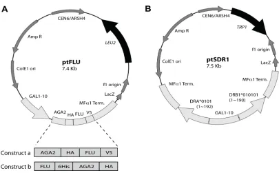
FIGURE 2-6 MAP OF YEAST SHUTTLE VECTORS FOR YEAST CO-DISPLAY. 42
FIGURE 2-7 SURFACE LEVELS OF TWO FLU FUSION CONSTRUCTS AND ANCHORED
SOLUBLE HLA-DR1. 43
FIGURE 2-8 SIMULTANEOUS DETECTION OF BOTH HA AND V5 EPITOPE TAGS
FLANKING FLU PEPTIDE. 45
FIGURE 2-9 YEAST CO-DISPLAY AND SURFACE DETECTION OF FLU PEPTIDE AND
FLU-BOUND SOLUBLE HLA-DR1. 47
FIGURE 2-10 DTT AND FACTOR XA REACTING SITES IN TWO FLU/HLA-DR1
DISPLAYING SCHEMES. 48
FIGURE 2-11 EFFECT OF DTT ON SURFACE DISPLAY OF AGA2P OR FLU/HLA-DR1
COMPLEXES. 49
FIGURE 2-12 EFFECT OF FACTOR XA PROTEASE ON SURFACE DISPLAY OF
FLU/HLA-DR1. 51
FIGURE 2-13 EFFECT OF FACTOR XA ON FLU/HLA-DR1 DISPLAY LEVEL AFTER
FIGURE 2-14 CONTROL EXPERIMENTS FOR EXAMINING POSSIBLE SWITCHING OF SOLUBLE HLA-DR1 MOLECULES AMONG DIFFERENT YEAST CELLS IN THE SAME
CULTURE. 54
FIGURE 3-1 QUANTITATIVE ANALYSIS OF RELATIVE HLA-DR1 AMOUNT ON YEAST SURFACE USING FLOW CYTOMETRIC DATA GENERATED FROM
ANTI-DR-LABELED YEAST. 67
FIGURE 3-2 EFFECT OF MEDIUM, TEMPERATURE AND TIME LENGTH USED FOR YEAST INDUCTION ON HLA-DR1 SURFACE DISPLAY LEVEL. 70
FIGURE 3-3 EFFECT OF DENSITIES OF CELLS COLLECTED AND INITIATED FOR
INDUCTION ON HLA-DR1 SURFACE DISPLAY LEVEL. 72
FIGURE 3-4 CELL GROWTH CURVE FOR CO-DISPLAYING YEAST CULTURED IN
SD-SCAA. 73
FIGURE 3-5 FURTHER EVALUATION FOR THE EFFECT OF CELL AGE AND INITIAL
CELL DENSITY. 74
FIGURE 3-6 TITRATION OF ANTIBODIES USED FOR IMMUNOFLUORESCENT LABELING OF HLA-DR1 AND HA-TAG ON THE SURFACE OF YEAST. 77
FIGURE 3-7 QUANTITATION FOR RELATIVE BINDING OF MHC-II TO PEPTIDES ON THE
SURFACE OF YEAST. 83
FIGURE 4-1 ANCHOR-POCKET REGIONS IN THE STRUCTURE OF HLA-DR1 ASSOCIATED
WITH FLU PEPTIDE. 88
FIGURE 4-2 FLUORESCENT DETECTION OF HA-TAG AND V5-TAG SIMULTANEOUSLY ON THE SURFACE OF YEAST DISPLAYING P1 VARIANT OF FLU PEPTIDE. 94
FIGURE 4-3 FLUORESCENT DETECTION OF HA-TAG AND V5-TAG SIMULTANEOUSLY ON THE SURFACE OF YEAST CO-EXPRESSING AGA2P-P1-VARIANT AND HLA-DR1.
95
FIGURE 4-4 SIMULTANEOUS DETECTION OF P1-VARIANT AND HLA-DR1 ON THE
SURFACE OF CO-EXPRESSING YEAST. 97
FIGURE 4-5 P1 POCKET PROFILE OF HLA-DR1 ANALYZED BY YEAST CO-DISPLAY. 98
FIGURE 4-6 FLUORESCENT-ACTIVATED CELL SORTING OF A LIBRARY OF RANDOMIZED PEPTIDES FOR HLA-DR1 SPECIFIC LIGANDS BY YEAST
CO-DISPLAY. 100
FIGURE 4-7 RELATIVE HLA-DR1 AMOUNT ON THE SURFACE OF SELECTED CLONES FROM YEAST LIBRARY CO-EXPRESSING RANDOMIZED PEPTIDE SEQUENCES
AND HLA-DR1. 101
FIGURE 4-8 SEQUENCES OF PEPTIDE FUSIONS DISPLAYED BY SORTED DR-POSITIVE
FIGURE 5-1 P1 POCKET IN PEPTIDE BINDING SITE OF HLA-DR1. 117
FIGURE 5-2 RELATIVE HLA-DR1 AMOUNT ON THE SURFACE OF SELECTED POSITIVE CLONES CO-DISPLAYING HLA-DR1 MUTANTS AND TARGET P1 VARIANT: A.
P1-VAL, B. P1-ALA, OR C. P1-GLU. 121
FIGURE 5-3 P1 ANCHOR PREFERENCE FOR HLA-DR1 AND ITS VARIANT-BINDING
MUTANTS. 126
FIGURE 5-4 RELATIVE HLA-DR1 AMOUNT ON THE SURFACE OF CLONES SORTED OUT OF WILD TYPE-BACKCROSSED LIBRARIES FOR CO-DISPLAYING OF HLA-DR1
MUTANTS AND TARGET P1 VARIANT. 129
FIGURE 5-5 P1 ANCHOR PREFERENCE FOR SELECTED HLA-DR1 MUTANTS FROM
BACKCROSSED LIBRARIES. 133
FIGURE 5-6 FLOW CYTOMETRIC ANALYSIS FOR EXAMINATION OF FALSE POSITIVE. 135
FIGURE 5-7 ESSENTIAL MUTATION SITES IMPLIED BY DIRECTED EVOLUTION VIA YEAST CO-DISPLAY FOR ALTERING PEPTIDE BINDING PROPERTIES. 140
FIGURE 5-8 POLYMORPHIC SITES OF PROTEIN ENCODED BY DRB ALLELES. 141
FIGURE 5-9 SELECTED EXAMPLES OF SUBSTITUTIONS AT ESSENTIAL MUTATION SITES WITHIN P1 ANCHOR-POCKET REGION DOMINATING OR AFFECTING P1
POCKET PROFILE. 144
FIGURE 5-10 SUBSTITUTION AT ESSENTIAL MUTATION SITES OUTSIDE P1 ANCHOR-POCKET REGION FAVORING FOR ACCOMMODATION OF P1 VARIANTS. 147
FIGURE 6-1 AN EXAMPLE OF PEPTIDE BINDING SPECTRUM OF DR PROTEINS
Chapter 1. Background and significance
1.1. Overview
Biological evolution makes creatures on the earth more and more sophisticated as
well as better and better developed. For example, to protect against diseases caused by
various agents in the surrounding, organisms have developed a defending bio-system
called immune system1-3 by gradually accumulating and coordinating subtle helpful
immunological features supplied by specifically differentiated cells and naturally
evolved molecules over thousands or millions of generations. Natural evolution is slow
and results are determined by complex fitness constraints existing in nature.
Nonetheless, evolution remains a powerful paradigm for optimizing biological systems.
Thus, in vitro methods for directed evolution4, 5, wherein researchers can control fitness
drivers guiding change, have garnered substantial interest in the last two decades. These
methods are commonly and most simply applied to proteins6, 7, the basic unit of
biological function.
Most highly developed vertebrates, such as mammals and humans, have a relatively
complicated immune system consisting of many types of organs, tissues, cells, and
proteins, which connect with each other in an elaborate and dynamic process. Within
this network, CD4+ T cell, a sub-group of lymphocytes, serves as a director to control
and optimize the function of other immune effector cells8 and a booster to maximize the
efficacy of immunity. The role of these lymphocytes is so important that dysfunction or
deficiency syndrome (AIDS) caused by a notorious virus, HIV10. It is of great
importance to understand the mechanism for stimulation of CD4+ T cell responses and
find a possible way to modulate their activation and function. Studies of CD4+ T cells at
the molecular level indicate that two signals are involved in the process of their epitope
specific activation11, 12. The primary pathway for transmitting signal 1 is predominantly
determined by the molecular interaction between T cell receptor (TCR) and class II
major histocompatibility complex (MHC-II) associated with antigenic peptide processed
and presented by professional antigen-presenting cells (APCs)11, 13. Therefore,
peptide-binding specificity of MHC-II proteins plays an important role in defining the epitope
specificity of T cell activation and evolution of MHC-II for altered specificity implicates
potentials in regulation of helper T cell-mediated immune responses.
As one of the most polymorphic membrane proteins14, 15, MHC has already been
selected for millennia by nature16, 17 so that each MHC-II allele is poly-specific for a
large set of antigenic peptides derived from both endocytosed pathogens and cytosolic
proteins18-22 and only a few alleles are required and expressed by each individual for
presentation of peptides and initiation of T cell responses to overwhelm a verity of
foreign invaders. However, environmental changes enforce improvement of MHC’s
peptide selectivity such that CD4+ T cell mediated immune responses could function
against mutagenic pathogens. Laboratorial protein engineering techniques enable an
initial evaluation of these evolutionary processes experimentally, which not only help to
understand structure-function relationship for naturally evolved MHC-II molecules but
This thesis work is focused on 1) development of a quantitative, high throughput in
vitro protein engineering methodology – yeast co-display, for studying the interaction
between various peptides and diverse MHC-II molecules (Chapter 2 and 3); 2)
characterization at the molecular level of peptide binding specificity for selected MHC-II
allele (HLA-DR1), determination of side chain preference for some pocket-like region in
the binding site of DR1 (or pocket profile23), and prediction of DR1 specific ligands
using the novel technology (Chapter 4); 3) modification of pockets within the peptide
binding groove of DR1 for improved and/or altered peptide binding specificity by
directed evolution, which has a potential to regulate T cell activation (Chapter 5).
1.2. Central role of CD4+ T cells in adaptive immune responses
1.2.1. Brief introduction for Immune system and lymphocytes
The immune system that most vertebrates have not only inherits the oldest innate
immunity, but also develops a more evolved adaptive immunity, first appeared in jawed
vertebrates2 and became increasingly specialized with further evolution. Adaptive
immune system consists of macrophages and other phagocytic cells, lymphocytes and
their derivatives such as antibodies1, 3, 24. Lymphocytes are the only cell types in the
body capable of specifically recognizing and distinguishing different antigenic
determinants and are responsible for the adaptive immune response, specificity and
memory25. A lot of congenital and acquired immunodeficiencies are related to
molecular defects in the development and function of lymphocytes or their products9.
There are three major subsets of lymphocytes25: 1) Natural killer (NK) cells, a part of
innate immune system, can recognize and kill infected cells directly without a need for
additional activation; 2) B cells, main components of adaptive immune system, are
responsible for secreting antibodies, which stimulate humoral immune responses by
directly capturing antigens; 3) T cells, another group of main components in adaptive
immune system, only respond to antigens processed and presented by APCs via a
MHC-invoked secretory pathway and stimulate cell-mediated immune responses. Two
important effector cells differentiated from T lymphocytes involved in MHC-restricted
activations are T helper cells (Th cells, expressing CD4 coreceptors for MHC-II binding)
and CD8+ cytotoxic T lymphocytes (CTLs, expressing CD8 coreceptors for MHC-I
binding).
1.2.2. Differentiation and Function of CD4+ T cells
Not like NK cells, antibodies or CTLs, Th cells do not have the ability to eliminate
infected pathogens by themselves, however, taking up almost half of the lymphocyte
population in human body, they do play an important role in adaptive immunity or even
innate immunity. Because of their regulatory function, these effector cells of CD4+ T
cells are usually designated as helper cells. Of particular interest is that CD4+ T cells
may differentiate into two different subsets of effector cells, Th1 and Th2, which produce
distinct cytokines and perform distinct effector functions. Once being activated, Th1 cells
are essential in stimulating antibody secretion and determining B cell antibody class
switch, in promoting the activation and proliferation of CTLs8, and in regulating activity
cell-mediated immunity (Figure 1-1). Th2 cells on the hand mainly function as
suppressers for these effector cell-mediate responses. Both cell subsets are involved in
formation of tissue injury and inflammation as well as generation of memory T cells,
which may persist for years after acute immune responses are down-regulated and
disappeear25.
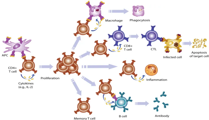
Figure 1-1 Proliferation and function of CD4+ T cells. CD4+ T cells are activated by antigenic peptide presented by professional APCs and driven to the clonal expansion by cytokines (e.g. interleukin-2). The activated and differentiated Th cells can further activate and differentiate macrophages, CD8+ T cells, and
B cells, or introduce inflammation. The main function of activated macrophages, CD8+ T cells, and B cells are to lyse phagocytosed antigens, to kill infected cells or tumor cells, and to secrete antigen-specific antibodies, respectively. Once antigens are eliminated, Th cells will be down regulated and
entering a memory phase. (Adapted from Abbas, A.K., Lichtman, A.H. & Pober, J.S. Cellular and molecular Immunology, 4/e (Saunders, philadelphia, 2000)).
Both B cell and macrophages are professional APCs for Th cells29, so immature B
cells or macrophages can process and present antigen fragments to specifically activate
CD4+ cells, which in turn will fully activate B cells and macrophages by cytokines (e.g.,
In contrast, CD8+ T cells do not serve as professional APCs to Th cells and their
development and function do not necessarily require presence of Th cells. In a strong
innate immune response, the primary activation of CD8+ T cells can be elicited by
dendritic cells (DCs), if microbe directly infects DCs35, or if cross-presentation36, 37 of
microbial antigens is sufficient. However, the participation of Th cells is usually
required for CD8+ T cell responses to tumor cells38, viral infections39, 40 and for
expansion of memory CD8+ T cells41, 42. Communication between these two types of T
cells is largely mediated by professional APCs, such as DCs43, via interleukin-2 (IL-2)
signaling41 (Figure 1-1) or costimulatory signaling mediated by CD40:CD40 ligand39, 44,
45 or CD28:B7-1/B7-2 pathway42 (Figure 1-2).
Thus, CD4+ helper T cell actually plays a central role in adaptive immunity. The
possibility of controlling or adjusting CD4+ T cell development and function at
molecular level would present intriguing potential for immunotherapy of numerous
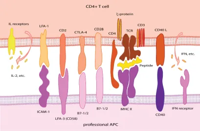
Figure 1-2 Signaling pathways between CD4+ T cell and APCs.
1.2.3. Activation of CD4+ T cells by APCs
In order to enter the functional stage, naïve T cells need to be activated and
differentiated into effector T cells by specialized APCs, called “professional APCs”
(Figure 1-1). As mentioned earlier, DCs, B cells and macrophages can all serve as
professional APCs to present antigenic peptides for CD4+ T cell recognition and
activation29, 35. However, the amount of MHC-II and cotimulators they can express, the
phase of adaptive immune responses at which they can activate CD4+ T cells, the
locations where they meet with T cells, and the functional feedback received from Th
cells are all different25.
B cells and macrophages with lower levels of MHC-II mainly present antigen to
responses in tissues, which might cause delayed type hypersensitivity. The main
purpose of this antigen presenting process is to further activate B cells and macrophages
themselves and fully accomplish their function. DCs on the contrary, resident in
epithelia and tissues, can capture antigens and transport them to secondary lymphoid
organs, including lymph nodes and spleen and present them to naïve CD4+ T cells.
They can express large amount of MHC-II and costimulators on the surface and are the
most effective APCs for initiating primary T cell responses.
Adoptive transplantation of autologous APCs such as DCs has been used in
immunotherapy to generate more effective T cell-mediated immune responses26, 49.
However, the labor intensity and high expense on producing APCs and the unreliable
quality and quantity of the available APCs that can be used in adoptive immunotherapy
all limit their usage in clinical applications. Therefore, investigators have started to
develop various artificial APCs (aAPCs) to enhance the generation of antigen specific T
cells27. No matter which method is going to be used, first of all, it is critical to clarify
the molecular mechanism that leads to the activation of CD4+ T cells by APCs.
1.2.4. Signaling pathways for CD4+ T cell activation
T cell activation needs both antigen recognition and costimulation. After antigens
are specifically processed and presented by professional APCs to CD4+ T cells, T cells
will receive both “signal 1” through TCRs and “signal 2” via costimulator receptors
from professional APCs11, 12.
The primary signaling pathway, which determines the specificity of CD4+ T
ζprotein-associated TCRs (TCR complexes) and APC-processed antigenic peptide-
coupled MHC-II molecules (peptide/MHC-II complexes) along with the CD4
coreceptor12, 13 (Figure 1-2). The antigen specificity of T cell response is greatly
dependent on antigenic peptides processing and presentation by APCs and peptide-MHC
complex recognition of TCRs on the T cell surface.
Other than the first signaling pathway, there are several intermolecular
ligand-receptor interactions that function to deliver the second signal which is necessary for
optimization of T cell activation and differentiation: CD28:B7-1/B7-2, the principal
pathway for delivering second signals for T cell activation12, 13; CD2: CD58, signal
transducer as well as intercellular adhesion molecules. Some other ligand-receptor pairs
might only serve as adhesion molecules, such as integrin LFA-150: ICAM-1/ICAM-2,
however, they are necessary for keeping T cells and APCs in close contacting with each
other for signaling transduction25 (Figure 1-2).
Although the appearance of costimulatory molecules is substantial, specialized
MHC-II proteins on the surface of APCs perform the main task of displaying
cell-associated antigens for recognition by CD4+ T cells. Therefore, intensive studies using
in vivo and in vitro methods have been carried out and will keep focusing on elucidating
the cellular and molecular basis of antigen processing inside APCs as well as interaction
between antigenic peptide and MHC molecules, which is also a key to the construction
of aAPCs and to the design of vaccines for modulating T cell responses. One purpose of
this thesis work is trying to develop a quantitative, high throughput method for better
1.3. MHC-II and antigenic peptide binding
1.3.1. Classification and evolution of polymorphic MHC
The MHC locus is a large gene cluster found in the genome of most vertebrates. The
main products encoded by these diverse genes are expressed as trans-membrane
glycoproteins on the surface of APCs, which have the ability to degrade and process
antigens into short fragments (peptides) and transport them using MHC proteins to the
surface for recognition by antigen-specific T cells. Two well-known subgroups of MHC
proteins are 1) class I (MHC-I), expressed on the surface of all nucleated cells and
responsible for presenting intracellular peptides to CD8+ T cells, and 2) class II
(MHC-II), expressed on the surface of professional APCs, such as DCs, macrophages and B
cells and responsible for presenting extracellular peptides to CD4+ T cells51. In humans,
MHC proteins are referred to as human leukocyte antigen (HLA), and most intensely
studied HLA alleles are HLA-A, HLA-B, HLA-C (for class I) and HLA-DR, HLA-DP,
HLA-DQ (fcor class II).
MHC is the most polymorphic gene in the human genome. Up till today, multiple
alleles have been found for all the nine classical genes (the heavy chain for each of class
I alleles and two chains for each of class II alleles), among which the most
conspicuously diverse loci, HLA-A, HLA-B, HLA-C and HLA-DRB, have 893, 1431,
569 and 814 known gene alleles that can encoding 681, 1165, 431 and 637 proteins
Table 1-1 HLA alleles numbers (adapted From IMGT/HLA Database, assigned October 2009)
HLA class I HLA class II
Gene
A B C DRA DRB DQA1 DQB1 DPA1 DPB1
Alleles 893 1,431 569 3 814 35 106 28 136
Proteins 681 1,165 431 2* 637 26 77 16 118
HLA-DRB Alleles
Gene
DRB1 DRB2 DRB3 DRB4 DRB5 DRB6 DRB7 DRB8 DRB9
Alleles 722 1 52 13 19 3 2 1 1
Proteins 572 0 42 7 16 0 0 0 0
* The two DRA proteins have only one difference at the cytoplasmic tail.
The evolutionary force that has created and maintained such astounding allelic
diversity is postulated to be balancing selection52, 53
, a broad term that identifies any kind
of natural selection in which no single allele is absolutely most fit. This striking feature
of MHC genes as well as proteins not only creates fertile grounds for evolutionary
biologists but also provides resourceful platforms for protein engineers. Although a high
degree of MHC polymorphism is found in human population, an individual can only
express approximately 18 class I or class II proteins, which are usually individually
specific, so transplantation rejection and autoimmune diseases might occur when trying
to transplant organs or tissues between individuals. Because of these limitations,
immunologists and bioengineers would always like to investigate the common and
distinct molecular properties of MHC proteins and develop methods for monitoring the
in vitro methodology we will talk about in this thesis allows discovery of the molecular
relationship among MHC-II alleles in a functionality-dependent manner.
1.3.2. Structure of MHC-II proteins
An MHC-II protein is a non-covalently associated heterodimer consist of an αchain
(32-34kDa) and a β chain (29-32kDa). Each subunit contains two extracellular
domains54-59 (Figure 1-3A), a trans-membrane spanning domain and a cytoplasmic tail.
The N-terminal α1 and β1 domains interact to form the peptide-binding groove,
composed of two α-helical walls and a floor formed by eight strands of anti-parallel β
-sheets (Figure 1-3B). Polymorphisms of MHC-II are mainly due to the existence of
various possible residues at the same position within the peptide binding groove15. In
contrast with class I, the class II molecules leave both ends of the peptide-binding
groove open for accommodating longer peptides up to 30 residues59, 60. This peptide
binding domain is responsible for loading peptides and presenting them to T cells61. The
α2 and β2 domains fold into immunoglobulin (Ig) domains, responsible for CD4
coreceptor binding and are relatively conserved among various alleles of a particular
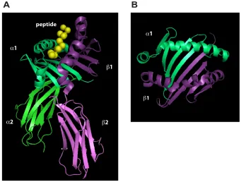
Figure 1-3 Three-dimensional structure of human leukocyte antigen-DR1 in complex with FLU peptide54. A. Extra-cellular domains of DR1 associated with FLU peptide (derived from influenza hemagglutinin residues 306-318: PKYVKQNTLKLAT, also known as HA306-318 elsewhere54; and we use “FLU” henceforward in order to distinguish it from another HA epitope tag derived from the same protein). B. Top view of the peptide-binding site formed by α1 and β1 domains. Images were made using PyMOL software (Delano, W.L., The PyMOL Molecular Graphics System (2002) on world wide web http://www.pymol.org) by rendering molecular coordinates in the code file 1DHL.
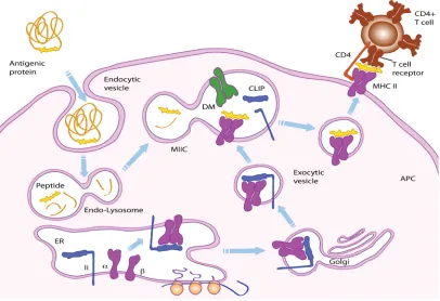
1.3.3. MHC-II pathway of antigen processing in APC
The maturation of MHC-II and the peptide binding and transport by MHC-II take
place inside professional APCs upon the exposure to foreign antigens29, 63 (Figure 1-4).
After capturing antigens, these specialized APCs will internalize the extracellular
proteins into vesicular compartments, such as endosomes and lysosomes, where
proteolytic enzymes degrade antigens into short fragments (peptides) under the acidic
condition. Newly assembled MHC-II in endoplasmic reticulum (ER) are transported
through Golgi complex to late endosomes and lysosomes with the help of invariant chain
membrane fusion of different endosomes and lysosomes, MHC-II and antigenic peptides
enter the same vesicle, called the MHC class II compartment, or MIIC64, which contains
all components required for peptide/MHC-II association such as enzymes, MHC-II, the
Ii (or invariant chain-derived peptides), and HLA-DM (product of a non-polymorphic
gene located at the locus of MHC-II gene cluster) with the ability to catalyze peptide
binding65. To remove Ii from the peptide-binding site, proteolytic enzymes first cleave Ii
into class II-asociated invariant chain peptide (CLIP), and then DM helps to catalyze the
conformational change66 of MHC-II for peptide exchange between CLIP and high
affinity antigenic peptides. Once mature MHC-II molecules are associated and
stabilized by antigenic peptides66-69, the peptide/MHC-II complex can be delivered by
exocytic vesicles to the surface of APC for recognition by CD4+ T cells.
1.3.4. Peptide binding characteristics of MHC-II
There are several important features of the interaction between MHC-II proteins and
specific peptides21, 70. First of all, even though each MHC-II molecule contains a single
peptide binding groove capable of accommodating one peptide at a time, it has the
ability to specifically recognize and present a large set of peptides derived from
enormous numbers of antigens18-20. This explains why each individual expressing only a
few different MHC-II alleles (normally less than 12) could defend against infections of
various foreign agents.
Secondly, peptides bound to MHC-II share common structural features that favor
their interaction. The optimal length of specific peptides is between 12 to 16 residues,
with 9 consecutive residues directly contacting with the binding groove59. Some have
their side chains pointing down to pocket-like region within the groove serving as
anchors for MHC-II binding, whereas the rest face up with their side chains away from
the groove for recognition by TCRs (Figure 4-1). This feature reflects the antigen
specificity of both MHC-II and TCRs.
Thirdly, the association is relatively stable with a very low off-rate and the peptide
binding affinity of MHC-II is higher than the peptide/MHC-II complex binding affinity
of TCR25. This allows peptide/MHC complexes to persist long enough on the surface of
APCs for T cell recognition.
Fourthly, MHC-II shows a broader specificity (promiscuity) for peptide binding than
the specificity of antigen recognition by TCRs25. They can bind different sources of
cell stimulation. This actually increases the chance of autoimmunity and the tolerance of
self-like foreign antigens.
All the information about peptide/MHC-II interactions may be used in vaccine
design for enhancing CD4+ T cell-mediated adaptive immunity or in immunotherapy for
treating T cell-related diseases26-28, 38, 46-49, 71, 72. However, most of these strategies based
on applying known T cell specific epitopes presented by self MHC-II molecules for
stimulating CD4+ T cell activation, which are limited by the restricted set of epitopes
recognized by the few types of self expressed MHC-II alleles found in each individual.
Therefore, there will always be some nonspecific epitope or mutated epitope bearing
antigens with the capability of escaping the recognition of MHC-II as well as CD4+ T
cells and causing sever immune diseases to certain MHC-II alleles expressing
individuals. In these cases, immunotherapeutic methods relying on MHC-II-specific
antigens will become less effective, and alternative strategies such as improving the
range of peptides presentable by MHC-II or changing the specificity of certain alleles
could be useful.
Even though each individual could only bear a few MHC alleles and will inevitably
come across with the problem of transplantation rejection when a foreign allele showing
up in their bodies, hundreds or even thousands of alleles do exist in nature among
different individuals and their protein products share similar structures and functions.
This large freedom of the balancing selection for MHC-II in nature suggests that any
self-MHC allele has the potential of being mutated or modified subtly without affecting
most of its characteristics, possibly including self-tolerance. Furthermore, if a selection
affinity of an MHC-II molecule should be able to be “directedly” edited or evolved by
introducing subtle changes such as single mutations in the protein sequence leaving most
of the other properties and functions unchanged. Creating and applying this kind of
self-derived or self-evolved tolerant MHC-II molecules in the development of vaccines and
aAPCs would become a breakthrough in vaccination and immunotherapy.
In this thesis work, we will take the first step of this big project on evaluating the
possibility of developing novel MHC-II molecules with minimum residue substitutions,
which can specifically recognize and present targeted peptides for T cell activation.
1.4. Protein engineering technologies in this thesis work
1.4.1. Cell-surface display for target proteins
Display of recombinant proteins on the surface of cells or viral particles not only
serves as an expression system for correct assembly of proteins but also provides a
platform for directly exploring and improving protein functions such as immunogenicity,
ligand binding specificity and enzymatic activities, etc. by directed evolution73-89. Up till
today, various cellular systems such as bacteriophages, bacteria, yeast,
baculovirus-insect cells and baculovirus/retrovirus-mammalian cells have been developed and widely
used for displaying and engineering proteins of interest. There is no expression system
perfectly suitable for all kinds of in vitro engineering applications, so advantages and
disadvantages usually need to be considered and compromised for acquiring a specific
accomplishment.
environments and less modifications, because these prokaryotic organisms are normally
lack of the post-translational machinery that helps properly assembling eukaryotic
especially human proteins. However, the simplicity of culturing and cloning these lower
hierarchy single-cell systems make them priorities for engineering a great number of
well studied eukaryotic polypeptides by construction and screening of libraries of
variants with a size of 108~1010. Investigation of large pool of variants and screening for
better fitness has become more and more useful via directed evolution for understanding
function of various proteins nowadays, thus, other than cellular platform based
displaying system, ribosome display93 or even mRNA display94-96 have been derived for
expression of even larger libraries for protein engineering in a cell-free environment
without limitation of transfection and/or transformation efficiency. The latter two
systems are very useful for screening a large variety of short peptides in some cases, but
less preferred for polypeptides, whose function relies on tertiary structure.
On the other hand, insect cell display76, 97 and mammalian cell display75, 82, 98, 99
inherit the advantage of eukaryotic expression systems, which provide more accurate
protein folding regulatory and auxiliary mechanism and better similarity to in vivo
conditions. As a result, large membrane proteins with sophisticated structures or
membrane protein complexes consist of multiple non-covalently associated monomers
are better to be studied within these higher-level organisms. For the cost, one must
sacrifice diversity and the number of protein variants that can be tested, or, labor
intensity as well as time and expense cost has to be increased dramatically to obtain
similar library of protein mutants for screening novel functionality, which sometimes is
biotechnology. Additionally, even with the help of baculoviral100 or retroviral89
technology, the difficulty of applying mammalian cells to engineer heterologous proteins
make them less commonly used than other displaying systems. The ability of producing
viral particles (e.g. baculovirus or retrovirus) for displaying interest polypeptides though
provides more flexibility to extend the application of these two kinds of virus infected
eukaryotic displaying systems84, 89.
In a lot of cases, diversity and complexity of certain eukaryotic protein are both
desired when performing specific structure-function analysis, such as the one in this
thesis work; hence, yeast display73, 74, 101 represents an ideal in vitro research platform,
which has the ability to translate and modify proteins under control of eukaryotic
expression though a little different from mammalian cells, while lose little molecular
cloning advantage as a single cellular expression system. Theoretically, one can
generate a combinatorial library with up to 109 individual clones using yeast, which is
able to cover combinations of 20 standard amino acids in 7 different sites within a
relatively complex protein. It is for sure that this single-cell eukaryotic displaying
system has its potential on occupying more and more research fields in protein
engineering.
In this thesis, we are going to display proteins of interest on the surface of yeast and
extend the display technology to fully utilize yeast to characterize interaction between
two or more proteins, e.g. peptides and DR1 heterodimer.
To display a protein of interest on the surface of yeast, the gene coding for protein of
strain EBY10074 (a GAL1-AGA1::URA3 ura3-52 trp1 leu2Δ1 his3Δ200 pep4::HIS2
prb1Δ1.6R can1 GAL), heterologous genes in the vector will be translated to protein of
interest fused by Aga2 protein (Aga2p), which is the subunit of a native yeast surface
protein, a-agglutinin103, 104. Directed by a signal peptide, the protein fusion will enter
protein secretory pathway, where Aga2p will associate with the other subunit of a
-agglutinin (Aga1p) by forming two disulfide bonds. Once transported outside yeast, the
assembled a-agglutinin will anchor protein of interest on the surface by the designed
covalent linkage between Aga2p and protein of interest (Figure 1-5).
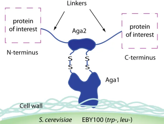
Figure 1-5 Classical yeast surface display of target protein. Protein of interest can be covalently linked to either N-terminus or C-terminus of Aga2p, which serves as the anchor for surface-displaying protein of interest.
1.4.2. Analyzing immunofluorescently labeled cells by flow
cytometry
Detection of protein on yeast surface is mainly carried out by applying
proteins on the surface of yeast, cells are usually enriched in growth media and then
switched to induction media for a certain time for optimal protein expression.
Consequently, cells are collected by centrifugation and labeled with a primary labeling
reagent specific for protein of interest displayed on yeast surface. In some cases, the
primary reagent is conjugated with a fluorophore for direct fluorescent labeling. In other
cases, a secondary reagent conjugated with a fluorescent dye is used for indirect
fluorescent labeling. These conjugated fluorescent dyes can be detected via flow
cytometry. All labeling work performed in this thesis work used indirect labeling, which
normally gives the stronger signal.
Other than proteins of interest themselves, small epitope tags such as HA, V5, 6His,
c-Myc, etc., are wildly used and fused to nonfunctional ends of target protein enabling
indirect checking of expression and presentation of protein of interest on the surface of
yeast. Due to the small size (5-20 residues), epitope tags will not affect the tagged
protein’s biochemical properties, but can be recognized by epitope-specific antibodies,
which also allows immunofluorescent labeling. Fluorescent labeling can be performed
for one epitope or more at the same time as long as primary and secondary reagents have
lowest cross-reactivity against each other. Double labeling by using an antibody specific
for protein of interest and another one specific for a fused epitope tag was used
frequently in this thesis work.
Flow cytometer allows suspension of labeled yeast cells flowing dropwise through a
detecting area at a high speed, where fluorophores coupled on yeast surface can be
mirror. At the same time, forward light scattering (FSC) and 90° side light scattering
(SSC) can also be observed, which correlate with volume of yeast cells and their
intracellular complexity, respectively. The dot-plot of these two parameters always
shows a certain pattern (e.g. Figure 3-1 (i)), which is significantly different from that of
bacteria or mammalian cells, allowing a first validation for properly cultivation of yeast
and an easy checking for contamination.
Sometimes, yeast displaying target protein is desired to be recovered from a large
pool of yeast cells, a more developed flow cytometer, called cell sorter, will make this
possible. Cell sorter can isolate individual cells via an electronic field and direct them
into the corresponding collection tubes. This process is usually called
fluorescent-activated cell sorting (FACS).
After flow cytometry or cell sorting, flow cytometric data can be analyzed in
different plots. The simplest is using a one-dimensional histogram by cell events (or cell
numbers) vs. fluorescence intensity of the fluorophore used for labeling protein of
interest (e.g. Figure 2-4 (i) and (ii)). This histogram will normally exhibit a distribution
of cell numbers at different intensities, the mean of which statistically represents how
well the labeled protein is displayed on the surface of yeast. Usually, yeast cells
themselves will give an auto-fluorescent background in most flow data, which can be
determined by preparing a few unlabeled or irrelevant yeast controls in parallel. If
double labeling of two proteins on yeast surface is performed, other than histograms, a
two-dimensional dot-plot can be generated by plotting the fluorescence intensity of one
fluorophore against the other (e.g. Figure 2-4 (iii)). The dot-pot will normally display
proteins or epitopes displayed on yeast surface can be examined simultaneously. It is
critical to set up appropriate compensation when running a double-labeled sample,
because a lot of times, the emission from one fluorophore can overflow into channels
used to detect other fluorophores, especially for some strong signals. In those cases,
single labeled controls need to be used to adjust the compensation so that no signal
interfering occurs.
Most recent flow cytometers are equipped with at least two lasers, which can excite
fluorophores with excitation wavelengths in a fairly large range, thus, if properly
selected, two fluorophores with excitation wavelengths far from each other can be used
for double labeling and excited by two lasers respectively on flow cytometer without any
compensation. This will greatly increase the efficacy and accuracy of flow detection. In
this thesis work, we are going to take advantage of this feature so that no compensation
Chapter 2. Development of yeast co-display – a novel
methodology for characterizing MHC-II/peptide binding
2.1. Introduction
Antigen specificity of CD4+ T cell-mediated immune response is primarily
determined by peptide presented by MHC-II molecules on the surface of APCs. In depth
characterization of peptide binding by MHC-II is critical to understanding issues in
vaccine design, autoimmune disease, infectious disease progression, and transplantation
rejection28, 46, 72.
MHC-II is a transmembrane protein, whose extracellular portion consists of two
heterologous chains, α and β, each of which containing two domains54. Mature MHC-II
proteins function to capture antigenic peptides processed inside professional
antigen-presenting cells (APCs) and present them on the surface of APCs for recognition by
CD4+ T cells to mediate adaptive immunity. The peptide-binding site of MHC-II
formed by α1 and β1 domains contains several pocket-like regions, which prefer to
accommodate specific side chains of anchor residues on peptides58, 59. The engaged
peptides are always heterogeneous in size with both termini extended beyond the
binding groove60. Even though a lot of common features for peptide binding have
already been discovered, the majority of peptide binding motif and anchor preference
information for distinct alleles of MHC-II still needs to be further characterized so that
specific target peptide/MHC-II pairs can be applied for immunotherapeutic application
MHCs are the most polymorphic glycoproteins known in nature, out of which each
individual only expresses a few alleles, so it is challenging to investigate peptide-binding
properties of all these trans-membrane proteins in vivo or ex vivo. Therefore a variety of
in vitro methods have been developed for studying the interaction between peptide and
MHC-II. A routinely used in vitro approach entails purifying soluble recombinant
MHC-II molecules from different expression systems such as B cell lines105, insect
cells106, yeast107, or Escherichia coli108-112 and then characterizing binding of these
molecules to different peptides generated either chemically by solid-phase synthesis113,
114 or genetically by cell or non-cell display technologies. The former one using
synthesized peptides enables coupling of all kinds of quantitative assays for acquiring
binding affinity and kinetics18, 112, 115-123, whereas the latter allows construction of big
libraries of peptide sequences for a high throughput screening and investigation90.
However, the labor-intensive preparation of soluble MHC-II proteins and lengthy
binding assays limit the efficacy and throughput of these methods for mapping MHC-II
binding specificities across the large number of existing alleles. Other attempts focused
on assaying binding of soluble peptide by MHC-II expressed natively on mammalian
cell surface124-128, which still did not completely get away from the difficulty of
manipulating MHC-II proteins.
Alternatively, engineered cell-surface display systems such as phage129-131, yeast102,
132-134, baculovirus-insect cells76, and mammalian cells98, 135 have been tried for
displaying recombinant extracellular domain of MHC molecules, suggesting a potential
antigenic peptide was always genetically introduced by covalent linkage, which not only
brings in unknown artificial effects but also restrain the flexibility of using these
methods to quantify peptide binding. Sometimes, acid/base half of leucine zipper was
attached to C-terminus of either chain of MHC-II to facilitate protein association and
surface anchorage76, which was not necessary in other display system but could alter
MHC-II native folding. These limitations make current cell-surface display system less
proper for characterizing the interaction between peptides and MHCs. The lack of a
rapid, efficient, robust, and quantitative methodology for characterizing the peptide
binding specificity and promiscuity of MHC-II alleles remains a bottleneck. Several
two-hybrid expression systems76, 81, 88, 102, 136-141 suggest a way to manipulate
multi-proteins in their native forms within the same cell and a possibility to improve current
cell-surface display technology to study interaction between peptide and MHC-II.
Herein, we report a novel, quantitative, high throughput methodology, yeast
co-display (Figure 2-1), for characterizing and engineering peptide-binding specificity of
MHC-II. As a model system, the extracellular domains of the MHC-II human leukocyte
antigen HLA-DR1 were expressed as a secreted heterodimer in Saccharomyces
cerevisiae. Simultaneously, an antigenic peptide known to bind to HLA-DR1 was
surface-displayed. Intracellular binding between the MHC-II and the peptide antigen
thus anchored the soluble MHC-II to the cell surface upon secretion, allowing detection
by immunofluorescence. The relative abundance of MHC-II compared to peptide on the
cell surface depended on the strength of binding between these species. Accordingly,
genetically manipulating either partner, enabling the application of directed evolution
approaches for high throughput characterization or engineering.
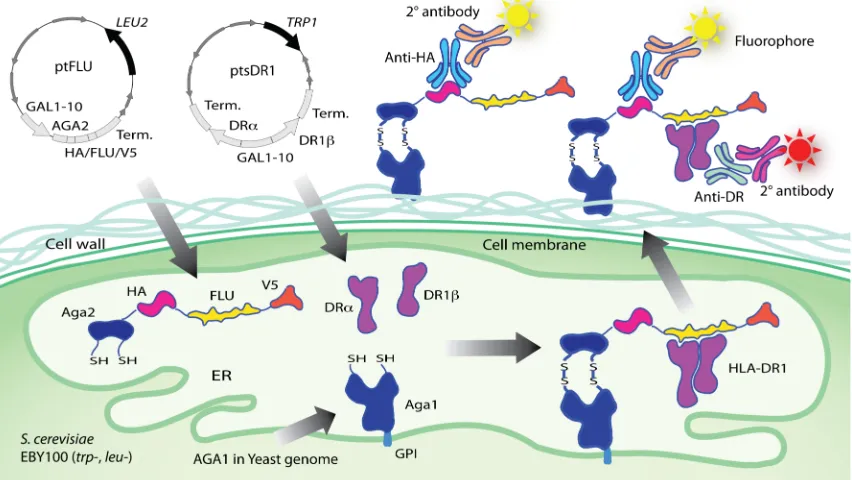
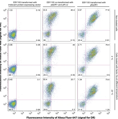
Figure 2-1 Design of the yeast co-display system for HLA-DR1. Two plasmids direct the expression of MHC-II ectodomain heterodimer and a peptide respectively in Saccharomyces cerevisiae yeast stain EBY100 (URA+, trp-, leu-). Aga2p-fused FLU peptide (derived from influenza hemagglutinin residues 306-318: PKYVKQNTLKLAT) is expressed flanked by HA tag (influenza hemagglutinin 98-106: YPYDVPDYA, different from FLU) and V5 tag (short peptide: GKPIPNPLLGLDST), enabling detection of peptide levels by immunofluorescent staining with antibodies specific for either tag. The α and β chain of HLA-DR1 ectodomain are expressed from separate cassettes and assembled in endoplasmic reticulum (ER) as a soluble non-covalently associated heterodimer. FLU peptide is anchored to the cell surface via native processing and secretion of the assembled a-agglutinin protein (composed of the Aga1p and Aga2p subunits) as described74, and the HLA-DR1 heterodimer is anchored by noncovalent binding to FLU. Relative fluorescence levels of different fluorophores coupled to anti-tag and anti-DR reagents indicate the level of saturation of available peptides by bound HLA-DR1. GPI: glycosyl phosphatidylinositol, an anchor helping a-agglutinin to transport through the secretory pathway104.
2.2. Materials and Methods
2.2.1. Construction of plasmids
Primers or oligonucleotides were synthesized commercially either by Invitrogen
stated, most enzymes were purchased from New England Biolabs (NEB, Ipswich, MA).
Taq (Qiagen, Germantown, MD or NEB) or Vent DNA polymerase catalyzed
amplification of polymerase chain reaction (PCR) products. Enzymatic digested PCR
products and plasmids were all confirmed by gel electrophoresis and purified before
ligation, which was catalyzed by T4 DNA Ligase (NEB or Invitrogen). Either QIAquick
Gel Extraction Kit (Qiagen) or Wizard SV Gel and PCR Cleanup System (Promega,
Madison, WI) was used for purification of DNA or PCR products without significant
differences. Either chemical- or electro- transformation in Escherichia coli strain DH5α
(Invitrogen) was performed for cloning and amplifying plasmids, which can be isolated
by using QIAprep Spin Miniprep Kit (Qiagen) and confirmed by DNA sequencing
(DNA Sequencing Facility, Department of Genetics, University of Pennsylvania, PA or
Molecular Biology Resource Facility, Division of Biology, University of Tennessee,
TN)
Plasmids for classical surface display of HLA-DR1
The gene expressing the extracellular domain of HLA-DR1 β chain allele
DRB1*010101 (1-190) was cloned from plasmid pDLM1-drb1s (kindly provided by Dr.
L. Sten, Umass) by PCR using N-terminal primer W3 (5’–TAATCCCGGGGACCTAA
GTATGTGAAGCAGAATACACTGAAGCTGGCAACCGGAGGTGGTTCACTAGT
GCCACGGGGCTCTGGAGGAAAGCTTGGAGACACCCGACCACGTTTCTT) and
C-terminal primer W4 (5’–TACTGGATCCAGAACCACCACCACCAGAACCACCAC
CACCAGAACCACCACCACCGCTAGCTGCTCTCCATTCCACTGTGAG), such that
sequence encoding the 13-residue FLU peptide (derived from influenza hemagglutinin
the N-terminus. The PCR product was ligated into plasmid Z47102 in place of the
original expression cassette coding for allele DRB1*0401 via annealing of sticky ends
obtained by XmaI/BamHI double digest, to create pfluDR1 for surface-displaying
FLU-fused HLA-DR1 (Figure 2-2). Another vector pDR1 expressing “empty” HLA-DR1
was constructed likewise but without adding FLU peptide by using another N-terminal
primer W1 (5’–AGCTCCCGGGGAGACACCCGACCACGTTTCTT) instead of W3.
Plasmids for classical surface display of FLU peptide
The entire expression cassette (GAL1-10//AGA2//HA//scFv 4-4-20//MFα Term.)
was excised from pCT30274 by double digest using KpnI and SacI and cloned into
KpnI/SacI partially digested yeast shuttle vector pRS315142 to create a plasmid with a
LEU2 selectable marker. Expression cassette (scFv 4-4-20 //MFαTerm.)within NheI
and SacI sites in this plasmid was substituted by a PCR product encoding
(FLU//SphI//MFα Term.), obtained by using N-terminal primer W20 (5’–
TCTAGCTAGCCCTAAGTATGTGAAGCAGAATACACTGAAGCTGGCAACCTAA
GCATGCCAACAGTGTAGATGTAACAA) and C-terminal primer W21 (5’–
CTGTGAGCTCAATTCTCTTAGGATTCG), to construct pFLU. Similarly, W34 (5’–
TCTAGCTAGCCCTAAGGCGGTGAAGCAGAATACACTGAAGCTGGCAACCTA
AGCATGCCAACAGTGTAGATGTAACAA) was used instead of W20 for
constructing pFLUA, which directs expression of a FLU analogue with Tyr308
substituted by Ala (encoded by underlined GCG) at the putative anchor position P1
(refer to Figure 4-1).