Birth Size and Brain Function 75 Years Later
WHAT’S KNOWN ON THIS SUBJECT: The fetal origins of adult disease hypothesis proposes that suboptimal fetal development may condition the later risk of disease, particularly
cardiovascular disease. However, this hypothesis has never been tested for diseases of the aging brain.
WHAT THIS STUDY ADDS: Thisfirst study of its kind provides clinical measures suggesting that small birth size, as an indicator of an adverse intrauterine environment, has lifelong
consequences for brain tissue volume and cognitive function. In addition, it shows that the effects of a suboptimal intrauterine environment on late-life cognitive function were particularly present in those with lower educational levels.
abstract
BACKGROUND:There are several lines of evidence pointing to fetal and other early origins of diseases of the aging brain, but there are no data directly addressing the hypotheses in an older population. We inves-tigated the association of fetal size to late-age measures of brain structure and function in a large cohort of older men and women and explored the modifying effect of education on these associations.
METHODS: Within the AGES (Age Gene/Environment Susceptibility)-Reykjavik population-based cohort (born between 1907 and 1935), archived birth records were abstracted for 1254 men and women who∼75 years later underwent an examination that included brain MRI and extensive cognitive assessment.
RESULTS:Adjustment for intracranial volume, demographic and medical history characteristics, and lower Ponderal index at birth (per kg/m3), an indicator of third-trimester fetal wasting, was significantly associ-ated with smaller volumes of total brain and white matter;bs (95% confidence intervals) were 21.0 (21.9 to 20.0) and 20.5 (21.0 to 20.0) mL. Furthermore, lower Ponderal index was associated with slower processing speed and reduced executive functioning but only in those with low education (b [95% confidence interval]: 20.136 [20.235 to 20.036] and20.077 [20.153 to20.001]).
CONCLUSIONS:Thisfirst study of its kind provides clinical measures suggesting that smaller birth size, as an indicator of a suboptimal in-trauterine environment, is associated with late-life alterations in brain tissue volume and function. In addition, it shows that the effects of a suboptimal intrauterine environment on late-life cognitive function were present only in those with lower educational levels.Pediatrics
2014;134:761–770
AUTHORS:Majon Muller, MD,a,bSigurdur Sigurdsson,
MSc,cOlafur Kjartansson, MD,c,dPalmi V. Jonsson, MD,c,e
Melissa Garcia, MPH,aMikaela B. von Bonsdorff, PhD,a,f
Ingibjorg Gunnarsdottir, PhD,gInga Thorsdottir, PhD,g
Tamara B. Harris, MD,aMark van Buchem, MD, PhD,h
Vilmundur Gudnason, MD, PhD,cand Lenore J. Launer,
PhDa
aLaboratory of Epidemiology and Population Sciences, Intramural
Research Program, National Institute on Aging, Bethesda, Maryland;bDepartments of Gerontology and Geriatrics, and hRadiology, Leiden University Medical Center, Leiden, Netherlands; cThe Icelandic Heart Association, Kopavogur, Iceland;
dDepartments of Neurology and Radiology, andeDepartment of
Geriatrics, andgUnit for Nutrition Research, Landspitali
University Hospital, Reykjavik, Iceland;fDepartment of Health
Sciences, Gerontology Research Centre, University of Jyväskylä, Jyväskylä, Finland
KEY WORDS
birth size, education, brain atrophy, cognition, aging
ABBREVIATIONS
AGES—Age Gene/Environment Susceptibility CI—confidence interval
CSF—cerebral spinalfluid GM—gray matter ICV—intracranial volume PI—Ponderal index RS—Reykjavik Study TB—total brain WM—white matter WML—white matter lesion
Dr Muller conceptualized and designed the study, drafted the initial manuscript, and carried out the initial analyses; Drs Sigurdsson, Kjartansson, Jonsson, and Garcia designed the data collection instruments of the Age Gene/Environment Susceptibility (AGES)–Reykjavik Study and critically reviewed the manuscript; Dr von Bonsdorff carried out the initial analyses and critically reviewed the manuscript; Drs Gunnarsdodottir and Thorsdottir designed part of the data collection instruments of the Reykjavik Study and critically reviewed the manuscript; Drs Harris, van Buchem, and Gudnason designed the data collection instruments of the AGES-Reykjavik Study, coordinated and supervised data collection, and critically reviewed the manuscript; Dr Launer conceptualized and designed the study, designed the data collection instruments of the AGES-Reykjavik Study, coordinated and supervised data collection, and critically reviewed the manuscript; and all authors approved thefinal manuscript as submitted.
www.pediatrics.org/cgi/doi/10.1542/peds.2014-1108
doi:10.1542/peds.2014-1108
Accepted for publication Jun 26, 2014
Address correspondence to Lenore J. Launer, PhD, National Institute on Aging, Laboratory of Epidemiology and Population Sciences, Suite 3C309, Gateway Building, 7201 Wisconsin Ave, Bethesda, MD 20892-9205. E-mail: [email protected]
There are several lines of evidence sug-gesting that early-life influences may be important to consider when identifying risk factors for pathologic brain aging later in life.1To date, these early infl u-ences have been captured by head size, as an indirect measure of fetal experi-ence,2,3and by education, which may not only reflect positive neurotropic effects but also risk factors for dementia such as socioeconomic factors and the load of cardiovascular disease.4,5
The fetal origins of adult disease hypothesis proposes that abnormal fetal development, deduced from small birth size, may induce permanent changes in the structure, me-tabolism, and physiology of fetal organs, for example, through epigenetic mecha-nisms.6,7 These changes in combination with effects of environmental exposures during childhood, such as education, may condition the later risk of disease.8Although these lines of evidence suggest early-life influences on cardiovascular disease, this hypothesis has never been tested for brain disease, which is highly prevalent and de-bilitating in older populations. Furthermore, several lines of evidence have suggested that higher educational achievement in early life is related to a reduced dementia risk later in life,9which is consistent with the cognitive reserve hypothesis.10However, it is unknown if these early-life influences (birth size and education) interact with respect to late-life disease risk.
In this first study of its kind to our knowledge, we investigated the associ-ation of birth size to late-age measures of brain structure and cognitive function and explored the modifying effect of education in these associations in a large cohort of older men and women (mean age: 75 years) who participated in the Age Gene/Environment Susceptibility (AGES)– Reykjavik Study (RS).
METHODS
AGES-RS
This analysis is based on the cohort of men and women who participated in
the RS and its follow-up, the AGES-RS. The RS (1967–1996) was initiated by the Icelandic Heart Association to study cardiovascular disease and risk fac-tors in middle age. The cohort included men and women born between 1907 and 1935 and living in Reykjavik in 1967. The midlife data reported here were obtained from the study examination closest to the time that the participants were 50 years of age. The AGES-RS was initiated in 2002 to examine the con-tribution of genetic susceptibility and gene-environment interaction to con-ditions common in old age.11From 2002 to 2006, a random sample of survivors of the original RS cohort was invited to the AGES-RS, and 5764 subjects (72% response rate) participated in the study. All participants in the AGES-RS under-went extensive evaluation, including a standard clinical evaluation, brain MRI, and neuropsychological testing. The pro-tocol was approved by the Icelandic Na-tional Bioethics Committee (VSN 00-063), the Icelandic Data Protection Authority, and by the institutional review board of the National Institute of Aging; all partic-ipants provided written informed consent.
Birth Size
Original midwifes’birth records on co-hort members born in the greater Reykjavik area were stored at the Na-tional Archives of Iceland, Reykjavik. Birth records were found and matched for 4828 individuals who participated in the RS. Data on birth size were abstracted by 2 researchers and included birth weight and length, as well as information on whether the birth was preterm (defined at that time as birth length ,48 cm), singleton, or multiple12; data on gesta-tional age were not recorded. Birth weight was recorded to the nearest 0.05 kg and length in centimeters from crown to heel. Ponderal index (PI) was calcu-lated as birth weight (kg) divided by cubic length (m3). Low PI indicates having soft tissue mass below normal for the stage of skeletal development.
This measure generally reflects impaired fetal growth in late gestation, leading to disproportionate fetal growth, and is an indicator of fetal wasting.6,13
Late-Life Brain Structure
Magnetic resonance images were ac-quired on a study-dedicated 1.5-T Sign Twinspeed EXCITE system (General Electric Medical Systems, Waukesha, WI) and were acquired during the late-life examination as part of the AGES-RS. The structural image protocol and the processing of the images have been fully described elsewhere.13Briefly, brain tis-sue volumes (mL), including gray matter (GM), white matter (WM), cerebral spinal fluid (CSF), and white matter lesions (WMLs), were generated by using the multispectral magnetic resonance im-ages and a high-throughput automatic image analysis pipeline. The pipeline, described previously, was based on the Montreal Neurologic Institute pipeline and optimized for use in the AGES-RS.14,15 Tissue classification was achieved with an artificial neural network classifier in the 4-dimensional intensity space de-fined by the 4 sequences (fluid attenua-tion inversion recovery and T1-, proton density-, and T2-weighted). Quality con-trol was based on visual inspection of a verification image for each subject in-cluding 14 a priori selected slice loca-tions from each of the pulse sequences (T1, proton density, T2, andfluid attenu-ation inversion recovery), evenly dis-tributed across the entire brain in the axial, coronal, and sagittal planes. Total brain (TB) volume was computed as the sum of GM, WM, and WML. Total in-tracranial volume (ICV) was computed as the sum of TB and CSF.
Late-Life Cognitive Function
these tests, 3 cognitive domain com-posite scores were calculated to form reliable and valid cognitive domain measures and to increase precision and were based on a theoretical grouping of tests, similar to other population-based studies17,18: (1) the memory composite score includes the immediate and delayed recall sections of the modified version of the California Verbal Learning Test; (2) the processing speed compos-ite includes the Figure Comparison Test, Digit Symbol Substitution Test, and the Stroop Test 1 (word reading) and 2 (color naming); and (3) the executive function composite includes a short version of the Cambridge Neuropsychological Test Au-tomated Battery Spatial Working Mem-ory Test, the Digits Backward Test, and Stroop Test 3 (word-color interference). A confirmatory factor analysis was used to check thefit of the composites to the data; results indicated that the 3-factor structurefit the data reasonably well.16 Composite measures were computed by converting raw scores to stan-dardizedzscores and averaging them across the tests in each composite. Interrater reliability for all tests was excellent (Spearman correlations range: 0.96–0.99).
Covariates
Body weight and height were measured at midlife and late-life. As part of the AGES-RS, participants were administered a standardized questionnaire that in-cluded questions about education, child-hood environment, and their lifetime education. Education was categorized into low (primary school) versus high (secondary school/college/university). Occupation was categorized into “homemaker/miscellaneous,” “manual,” “service workers,”and“professional.”19 Lifestyle factors and cardiovascular risk/disease developed during adult-hood may explain any association we find between birth size and brain.6–8 Therefore, we considered in our analyses
the following factors: smoking and alco-hol consumption categorized as current/ former/none; hypertension defined as antihypertensive treatment and/or sys-tolic blood pressure.140 mm Hg and/ or diastolic blood pressure.90 mm Hg; diabetes defined as having a history of diabetes, using glucose-modifying medi-cation, or with a fasting glucose of$126 mg/dL; dyslipidemia defined as total cholesterol$240 mg/dL or use of statins; and prevalent coronary artery disease based on self-reported history, angina pectoris on the Rose questionnaire, or evidence on electrocardiogram of possi-ble or probapossi-ble myocardial infarction.
Analytical Sample
Of the 4828 original Reykjavik cohort participants with available birth records, 1682 (35%) participated in the AGES-RS, including 1254 individuals with brain volume and cognition data. Compared with data published for the whole Reykjavik population with birth record data (N= 4828), the individuals in the analytical sample of this study had sim-ilar birth size and midlife body size.12 Compared with the total AGES-RS sample with brain MRI (N= 4614), the individuals in the analytical sample were younger (75 vs 76 years), had more years of ed-ucation (% with low eded-ucation: 18% vs 24%), and had somewhat larger ICV (1511 vs 1501 mL) and TB volume (1087 vs 1079 mL).
Data Analysis
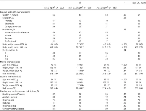
Subject characteristics at birth, midlife, and late life were calculated for the total population (N= 1254) and for descriptive purposes according to categories of PI (#23.0, 23.1–27.0, and.27.0 kg/m3) to create low (,25%), normal (25%–75%), and high (.75%) PI. Statistical differ-ences across PI categories were calcu-lated by using analysis of covariance for continuous measurements and x2 for categorical variables. Linear regression adjusted for age and gender was used to
investigate the association of continu-ous birth size measures (weight, length, and PI) to late-life ICV, absolute brain volumes (TB, GM, WM, WML, and CSF), and cognitive function (memory, pro-cessing speed, and executive functioning) (model 1). All outcome variables were normally distributed except for WML vol-ume, which was log (ln) transformed. For ICV and brain volumes, additional ad-justments were made for education (model 2) and for midlife weight and height, because birth size and head size are both strongly related to adult body size (model 3). Analyses for brain tissue were adjusted for ICV (model 4), because absolute brain tissue is strongly related to head size and to obtain relative brain volumes as indicators of brain atrophy. In addition, to examine if the effect of edu-cation on the “birth size–brain aging” relation was explained by socioeconomic status, additional adjustments were made for occupation. Finally, to examine whether relations between birth size measures and brain structure and function were explained by lifestyle and cardiovascular risk factors, additional adjustments were made for history of smoking, alcohol, hy-pertension, diabetes, hyperlipidemia, and coronary artery disease.
these categories was calculated. Analy-ses were carried out with IBM SPSS Statistics 18.0 (SPSS, Inc, Chicago, IL).
RESULTS
The 1254 participants had a mean age of 75 years (10th–90th percentile: 69–82 years) at the late-life examination and 50 years (10th–90th percentile: 42–58 years) at the midlife examination; 57% were women. The mean (SD) birth weight was 3.7 (0.5) kg, birth length was 52 (2.5) cm, and PI was 25.9 (3.4) kg/m3. Compared with individuals with a higher PI at birth, those with a lower PI were more oftenfirst-borns, younger at midlife and late-life examinations, and were more often men and smok-ers. Education was not different across the PI groups (Table 1).
At late life, mean (SD) ICV was 1510 (145) mL and mean (SD) TB volume was 1087 (101) mL, which is 72% of ICV. Mean (SD) WM, GM, WML, and CSF volumes were 388 (48), 679 (63), 20 (19), and 425 (83) mL, respectively.
Birth Size and Late-Life Brain Structure
Lower birth weight, birth length, and PI were associated with smaller ICV, smaller absolute brain volumes, and larger absolute CSF volume later in life (Table 2, model 1). After adjusting for ICV, the associations of lower birth weight and lower birth length to late-life brain volumes (model 3) were not significant. However, lower PI remained significantly associated with smaller relative TB (and larger CSF) and WM
volumes, but not with GM and WML vol-umes (model 3). Per each kg/m3 de-crease in PI, mean (95% CI) relative TB and WM volumes decreased by 21.0 (21.8 to20.1) and20.5 (20.9 to20.1) mL, respectively. Further adjustment for lifestyle and cardiovascular risk factors (model 4) did not change these effect estimates:21.0 (21.9 to20.0) and20.5 (21.0 to20.0) mL, respec-tively.
The relation between PI and brain vol-umes was linear (Fig 1;P-trend = .03). Adjusted mean differences between low PI (#23.0 kg/m3) and high PI (.27.0 kg/m3) were significant for volumes of ICV-adjusted TB, WM, and CSF (b[95% CI]:29.4 [218.4 to20.3],23.4 [26.9 to 20.7], and 9.4 [0.3 to 18.4] mL, re-spectively. Education did not modify the TABLE 1 Characteristics of the Total Population and Those in the Lowest, Middle, and Highest Quartiles of PI at Birth
PI P Total (N= 1254)
#23.0 kg/m3(n= 202) 23.1–27.0 kg/m3(n= 663) .27.0 kg/m3(n= 389) General and birth characteristics
Gender, % female 53 59 58 .09 57
Education, % .25
Primary 17 19 19 18
Secondary 54 53 47 52
College/university 29 28 34 30
Occupation, % .27
Homemaker/miscellaneous 40 45 45 44
Manual 12 16 15 15
Services 26 23 23 24
Professional 22 16 17 17
Birth weight, mean (SD), kg 3.4 (0.6) 3.7 (0.5) 4.0 (0.5) ,.001 3.7 (0.5) Birth length, mean (SD), cm 54.2 (3.1) 52.7 (2.1) 51.3 (2.2) ,.001 52.5 (2.5)
Parity, mother, % .04
0 39 30 23 30
1–2 36 41 41 40
$3 25 29 36 30
Midlife characteristics
Age, mean (SD), y 49 (6) 50 (6) 51 (6) ,.001 50 (6) Height, mean (SD), cm 173 (9) 171 (9) 171 (9) .04 171 (9) Weight, mean (SD), kg 73 (13) 74 (13) 74 (13) .87 74 (13) BMI, mean (SD) 24.6 (3.4) 25.2 (3.5) 25.3 (3.3) .03 25.1 (3.4) Late-life characteristics
Age, mean (SD), y 74 (5) 75 (5) 76 (5) ,.001 75 (5) Height, mean (SD), cm 170 (10) 168 (9) 167 (9) .02 168 (9) Weight, mean (SD), kg 78 (15) 77 (14) 77 (15) .83 77 (15) BMI, mean (SD) 26.8 (4.4) 27.4 (4.3) 27.4 (4.5) .26 27.3 (4.4) Lifestyle and cardiovascular risk factors, %
Smoking: current/former 62 62 55 .07 59
Alcohol : current 69 71 72 .74 71
Hypertension 74 81 79 .25 79
Diabetes 11 15 10 .18 13
Hyperlipidemia 52 47 50 .42 48
association of birth size measures with brain volumes (P-interaction..10).
Birth Size and Late-Life Brain Function
Lower birth weight, birth length, and PI were not directly associated with late-life memory performance, processing speed, or executive functioning after controlling for age, gender, and edu-cation (Supplemental Table 1). How-ever, the interaction between PI and education was significant for process-ing speed and executive function but not for memory (P-interaction = .04 and .007, respectively).
Stratified analysis showed that lower PI (per SD) was significantly associated with slower processing speed and poorer executive functioning in partic-ipants with low education (b=20.136 [95% CI:20.235 to20.036],P= .008, and b=20.077 [95% CI:20.153 to20.001],
P= .04) but not in those with higher education (b= 0.007 [95% CI:20.033 to 0.047] andb= 0.038 [95% CI:20.004 to 0.077]). To better visualize the inter-action, we present the low- and high-education effects by tertiles of PI (Fig 2). Further adjustment for occupa-tion and lifestyle and cardiovascular risk factors yielded similar results as pre-sented (data not shown). TB and WM volumes were correlated with process-ing speed and executive functionprocess-ing but not with memory performance; corre-lation coefficients for TB volume were 0.21 (P,.001), 0.21 (P,.001), and 0.03 (P..05) and for WM volume were 0.22 (P,.001), 0.23 (P,.001), and 0.03 (P.
.05). When including WM volume and ICV in the model, the associations between PI and processing speed and executive functioning in the low-education group were attenuated (b= 20.032 [95% CI: 20.060 to20.004] andb=20.020 [95% CI: 20.045 to 0.004]). Education did not modify the association of birth weight or length with cognitive func-tion (P-interaction..10).
There was no change in the associa-tions after excluding preterm births (n = 24), excluding individuals with prevalent dementia (n= 34), or after adjusting for birth year. Also, to in-vestigate whether being small for ges-tational age explained our findings, analyses were repeated relating birth weight,10th percentile20(#3.0 kg) to the outcome measures, and we found no relation.
DISCUSSION
In this study we provide unique data linking 2 current hypotheses about risk factors for pathologic changes in brain characteristics during life: (1) there are early origins of late-life brain aging (alterations in brain structure and func-tion) and (2) education reduces risk of cognitive impairment in multiple path-ways. We found that small birth size, in-dicated by lower birth weight, shorter birth length, and lower PI were all strongly associated with smaller head size (ICV) and smaller absolute brain tissue volumes in late life. When taking into account head size, lower PI, as an indicator of disproportionate growth retardation, was associated with smaller
late-life relative brain volumes, specifi -cally with smaller relative TB and WM volumes. This finding was reflected in cognitive performance; lower PI was associated with slower late-life pro-cessing speed and reduced executive functioning but only in those with lower educational levels.
This population-based study with a large sample has several unique strengths related to the availability of recorded data across the life span. Birth size data were abstracted from midwife records, which were not available in previous studies on early-life risk factors for late-age evidence of brain pathology.1 We used an extensive range of cognitive tests, quantitatively assessed total and tissue-specific brain volumes, and were able to control for ICV, midlife body size, and cardiovascular risk to estimate the independent effect of small birth size on late-life brain volumes. Furthermore, the subset of participants included in these analyses did not differ in birth size from those who were not in the AGES follow-up. A limitation of this study is that we were not able to specifically study the effect of preterm birth and being small for gestational age, which
resulted in proportional growth retar-dation, as opposed to disproportional retardation reflected by PI. Furthermore, although we adjusted for both adult body and head size to account for the strong correlation between body and brain size present at birth, we cannot rule out residual confounding. Another limitation is that no data were available on neonatal head circumference, which could have helped, at least in part, to unravel the mechanisms involved in the birth size–brain pathology connection. Finally, education usually is highly cor-related with socioeconomic status and therefore difficult to disentangle from one another. However, additional ad-justment for occupation, as a proxy for socioeconomic status, showed that the effect of low PI on reduced cognitive performance in the low-education group was not explained by occupation.
Although we found that all measures of birth size were strongly associated with late-life head size, only PI, as an in-dicator of fetal wasting, revealed an association with brain volumes, par-ticularly WM volume. Clinical observa-tions have shown that growth inhibition in early pregnancy produces a propor-tionally undersized fetus that affects both length and weight, so PI is normal.6 Growth inhibition in late pregnancy has less effect on fetal length but could result in disproportionate thinness at birth (low PI).6Our findings therefore suggest that the critical period of im-paired fetal growth for our outcomes occurred in late pregnancy, which is when the organs and tissues undergo critical periods of development.21 Nor-mal brain development in this critical period of late pregnancy involves both development of GM and WM. However, the formation of WM and myelination starts in the second half of pregnancy and oligodendrocyte differentiation oc-curs during the last gestational weeks of prenatal development and therefore is severely compromised in several
FIGURE 1
conditions affecting fetal nutrition in this critical period.22,23 That the last gestational weeks are critical for brain development is confirmed by human MRI studies showing that preterm-born individuals have abnor-mally altered brain structures, par-ticularly the WM,24–29 and that lower placental weight is associated with altered late-life WM integrity in older individuals.30
Our secondfinding is that small birth size contributed to reduced late-life cognitive performance only in partic-ipants with lower educational achieve-ment during early life. The association
was clearest in the processing speed and executive functioning tests that rely on WM integrity to transmit signals throughout the brain. Our findings are in line with a study showing that late-middle-aged persons, affected by exposure to famine before birth, had poorer executive functioning than those not affected.31 Better than ex-pected cognitive performance in the presence of brain pathology has been observed in the context of the cogni-tive reserve hypothesis.10The concept of cognitive reserve provides an ex-planation for differences between individuals in susceptibility to
age-related brain changes or pathology, whereby some people, for example, those with higher educational achieve-ment, can tolerate more of these changes than others and maintain function.
Thus, suboptimal pre- and postnatal environments could result in a delay in myelination affecting axonal conduc-tion velocity and could conceivably underlie long-term cognitive perfor-mance.23We know that in an environ-ment of impaired fetal growth, the brain is preferentially perfused to maintain oxygen supply.32Of particular interest is that the observed variations in birth anthropometric measures are likely to be physiologic in this sample. Also, the mean birth weight in the North Atlantic, which is among the highest in the world12and the high level of edu-cation in Iceland, with a widespread literacy rate since the end of the 18th century, suggest that even subtle normal variations in intrauterine en-vironment and educational levels may lead to differences in postnatal brain structure and function. Because our study sample had more years of edu-cation than the total AGES-RS cohort, it is expected that effect sizes would increase when studying this relation in populations with proportionately smaller birth sizes and lower educa-tional levels.
Possible pathways exist to link these suboptimal pre- and postnatal envi-ronments to smaller brain volumes and reduced cognitive functioning in late life (Fig 3). A chain of events triggered by abnormal or suboptimal fetal develop-ment, through, for example, maternal malnutrition or placental insufficiency, leading to small birth size, lower so-cioeconomic status, an unhealthy life style, and increased cardiovascular risk could explain our findings. Our study revealed that individuals with lower birth PI were more likely to smoke dur-ing adult life. Because there is a strong
FIGURE 2
relation between smoking behavior of parents and children, thisfinding could reflect that the mothers of in-dividuals with lower PI smoked more during their pregnancy. However, ad-justing for smoking and other mea-sures of socioeconomic status and lifestyle factors did not change our results. Another possibility is that fetal undernutrition directly affects neurodevelopment or accelerated brain aging.1 In our study, small birth size was not only related to reduced life head size but also to smaller late-life relative brain volumes. And, although brain volumes and cognitive function were measured at 1 point in time, this finding could suggest that suboptimal
fetal development is involved in the disruption of optimal brain growth as well as in neurodegenerative processes and loss of brain tissue. Although this is a genetically homogenous population, genetic influences could explain, at least in part, the relation between birth size and brain development.33,34 Epi-genetic changes may also play an im-portant role in propagating these trajectories. Recent investigations give insight into how epigenetic changes in the fetal environment, such as alterations in methylation of genes involved in regu-lating cortical development, may have lasting effects on developmental and aging processes, through influencing the expression of key regulatory genes
or progenitor cells.7,35,36 For instance, it has been hypothesized that fetal un-dernutrition affects the differentiation of progenitors into myelin-forming oli-godendrocytes, which occurs during the last gestational weeks of human de-velopment.22 In addition, it has been shown that early-life socioeconomic status, including education, contributes to DNA methylation variation.37
CONCLUSIONS
This study is thefirst to our knowledge that shows that small birth size is associated with smaller late-life brain volumes and that higher levels of ed-ucation, as an indicator of cognitive
FIGURE 3
reserve, may minimize the effects of a suboptimal intrauterine environ-ment on late-life cognitive function (Fig 3). These study results provide a framework for linking several ac-tively pursued areas of research on
the origins of chronic disease, including dementia, and studies investigating the contribution to dementia risk of edu-cation, small head size, and cognitive reserve. A clinical implication of our findings is that interventions to boost
cognitive reserve throughout childhood and adult life, for example, by increasing educational levels, hold promise to prevent late-life cognitive impairment, particularly in those with small birth size.
REFERENCES
1. Whalley LJ, Dick FD, McNeill G. A life-course approach to the aetiology of late-onset dementias.Lancet Neurol. 2006;5(1):87–96
2. Mortimer JA, Snowdon DA, Markesbery WR. Head circumference, education and risk of dementia:findings from the Nun Study.J Clin Exp Neuropsychol. 2003;25(5):671–679
3. Perneczky R, Wagenpfeil S, Lunetta KL, et al; MIRAGE Study Group. Head circumference, atrophy, and cognition: implications for brain reserve in Alzheimer disease. Neu-rology. 2010;75(2):137–142
4. Snowdon DA, Kemper SJ, Mortimer JA, Greiner LH, Wekstein DR, Markesbery WR. Linguistic ability in early life and cognitive function and Alzheimer’s disease in late life: findings from the Nun Study. JAMA. 1996;275(7):528–532
5. Bennett DA, Wilson RS, Schneider JA, et al. Education modifies the relation of AD pa-thology to level of cognitive function in older persons.Neurology. 2003;60(12):1909–1915
6. Barker DJ, Gluckman PD, Godfrey KM, Harding JE, Owens JA, Robinson JS. Fetal nutrition and cardiovascular disease in adult life.Lancet. 1993;341(8850):938–941
7. Gluckman PD, Hanson MA. Living with the past: evolution, development, and patterns of disease.Science. 2004;305(5691):1733–1736
8. Gluckman PD, Hanson MA, Cooper C, Thornburg KL. Effect of in utero and early-life conditions on adult health and disease.
N Engl J Med. 2008;359(1):61–73
9. Meng X, D’Arcy C. Education and dementia in the context of the cognitive reserve hy-pothesis: a systematic review with meta-analyses and qualitative meta-analyses. PLoS ONE. 2012;7(6):e38268
10. Tucker AM, Stern Y. Cognitive reserve in ag-ing.Curr Alzheimer Res. 2011;8(4):354–360
11. Harris TB, Launer LJ, Eiriksdottir G, et al. Age, Gene/Environment Susceptibility-Reykjavik Study: multidisciplinary applied phenomics.
Am J Epidemiol. 2007;165(9):1076–1087
12. Gunnarsdottir I, Birgisdottir BE, Benediktsson R, Gudnason V, Thorsdottir I. Relationship between size at birth and hypertension in
a genetically homogeneous population of high birth weight.J Hypertens. 2002;20(4): 623–628
13. Cole TJ, Henson GL, Tremble JM, Colley NV. Birthweight for length: Ponderal index, body mass index or Benn index?Ann Hum Biol. 1997;24(4):289–298
14. Zijdenbos AP, Forghani R, Evans AC. Auto-matic“pipeline” analysis of 3-D MRI data for clinical trials: application to multiple sclerosis.IEEE Trans Med Imaging. 2002;21 (10):1280–1291
15. Sigurdsson S, Aspelund T, Forsberg L, et al. Brain tissue volumes in the general pop-ulation of the elderly: the AGES-Reykjavik Study.Neuroimage. 2012;59(4):3862–3870
16. Saczynski JS, Jonsdottir MK, Sigurdsson S, et al. White matter lesions and cognitive performance: the role of cognitively com-plex leisure activity.J Gerontol A Biol Sci Med Sci. 2008;63(8):848–854
17. de Groot JC, de Leeuw FE, Oudkerk M, et al. Cerebral white matter lesions and cogni-tive function: the Rotterdam Scan Study.
Ann Neurol. 2000;47(2):145–151
18. Wilson RS, Mendes De Leon CF, Barnes LL, et al. Participation in cognitively stimulat-ing activities and risk of incident Alzheimer disease.JAMA. 2002;287(6):742–748
19. Groffen DA, Koster A, Bosma H, et al; Age, Gene/Environment Susceptibility-Reykjavik Study Investigators. Socioeconomic fac-tors from midlife predict mobility limitation and depressed mood three decades later:
findings from the AGES-Reykjavik Study.BMC Public Health. 2013;13:101: doi: 10.1186/1471-2458-13-101
20. Katz J, Lee AC, Kozuki N, et al; CHERG Small-for-Gestational-Age-Preterm Birth Working Group. Mortality risk in preterm and small-for-gestational-age infants in low-income and middle-income countries: a pooled country analysis. Lancet. 2013;382(9890): 417–425
21. Rees S, Inder T. Fetal and neonatal origins of altered brain development. Early Hum Dev. 2005;81(9):753–761
22. Liu J, Casaccia P. Epigenetic regulation of oligodendrocyte identity. Trends Neurosci. 2010;33(4):193–201
23. Tolcos M, Bateman E, O’Dowd R, et al. In-trauterine growth restriction affects the maturation of myelin.Exp Neurol. 2011;232 (1):53–65
24. Stewart AL, Rifkin L, Amess PN, et al. Brain structure and neurocognitive and behav-ioural function in adolescents who were born very preterm.Lancet. 1999;353(9165): 1653–1657
25. De Bie HM, Oostrom KJ, Boersma M, et al. Global and regional differences in brain anatomy of young children born small for gestational age.PLoS ONE. 2011;6(9):e24116
26. Allin MP, Kontis D, Walshe M, et al. White matter and cognition in adults who were born preterm.PLoS ONE. 2011;6(10):e24525
27. Northam GB, Liégeois F, Chong WK, Wyatt JS, Baldeweg T. Total brain white matter is a major determinant of IQ in adolescents born preterm.Ann Neurol. 2011;69(4):702–711
28. Walhovd KB, Fjell AM, Brown TT, et al; Pe-diatric Imaging, Neurocognition, and Ge-netics Study. Long-term influence of normal variation in neonatal characteristics on human brain development.Proc Natl Acad Sci USA. 2012;109(49):20089–20094
29. Raznahan A, Greenstein D, Lee NR, Clasen LS, Giedd JN. Prenatal growth in humans and postnatal brain maturation into late adolescence.Proc Natl Acad Sci USA. 2012; 109(28):11366–11371
30. Shenkin SD, Bastin ME, Macgillivray TJ, Deary IJ, Starr JM, Wardlaw JM. Birth parameters are associated with late-life white matter integrity in community-dwelling older peo-ple.Stroke. 2009;40(4):1225–1228
31. de Rooij SR, Wouters H, Yonker JE, Painter RC, Roseboom TJ. Prenatal undernutrition and cognitive function in late adulthood.
Proc Natl Acad Sci USA. 2010;107(39): 16881–16886
problems in the general population.Am J Epidemiol. 2008;168(10):1145–1152
33. Ikram MA, Fornage M, Smith AV, et al; Early Growth Genetics Consortium; Cohorts for Heart and Aging Research in Genomic Epide-miology Consortium. Common variants at 6q22 and 17q21 are associated with intracranial volume.Nat Genet. 2012;44(5):539–544
34. Stein JL, Medland SE, Vasquez AA, et al; Alzheimer’s Disease Neuroimaging Initiative;
EPIGEN Consortium; IMAGEN Consortium; Saguenay Youth Study Group; Cohorts for Heart and Aging Research in Genomic Epide-miology Consortium; Enhancing Neuro Imaging Genetics through Meta-Analysis Consortium. Identification of common variants associated with human hippocampal and intracranial volumes.Nat Genet. 2012;44(5):552–561
35. Petanjek Z, Kostovic I. Epigenetic regulation of fetal brain development and
neuro-cognitive outcome.Proc Natl Acad Sci USA. 2012;109(28):11062–11063
36. Chouliaras L, Rutten BP, Kenis G, et al. Epi-genetic regulation in the pathophysiology of Alzheimer’s disease. Prog Neurobiol. 2010;90(4):498–510
37. Borghol N, Suderman M, McArdle W, et al. Associations with early-life socio-economic position in adult DNA methylation. Int J Epidemiol. 2012;41(1):62–74
(Continued fromfirst page)
PEDIATRICS (ISSN Numbers: Print, 0031-4005; Online, 1098-4275).
Copyright © 2014 by the American Academy of Pediatrics
FINANCIAL DISCLOSURE:The authors have indicated they have nofinancial relationships relevant to this article to disclose.
FUNDING:This work was supported by a grant from the National Institutes of Health (N01-AG-1-2100), with contributions from NINDS, NEI, NIDCD, NHLBI, NIA Intramural Research Program, the Icelandic Heart Association (Hjartavernd), and the Icelandic Parliament (the Althingi). Funded by the National Institutes of Health (NIH).
DOI: 10.1542/peds.2014-1108 originally published online September 1, 2014;
2014;134;761
Pediatrics
B. Harris, Mark van Buchem, Vilmundur Gudnason and Lenore J. Launer
Garcia, Mikaela B. von Bonsdorff, Ingibjorg Gunnarsdottir, Inga Thorsdottir, Tamara
Services
Updated Information &
http://pediatrics.aappublications.org/content/134/4/761 including high resolution figures, can be found at:
References
http://pediatrics.aappublications.org/content/134/4/761#BIBL This article cites 37 articles, 7 of which you can access for free at:
Subspecialty Collections
http://www.aappublications.org/cgi/collection/neurology_sub Neurology
sub
http://www.aappublications.org/cgi/collection/fetus:newborn_infant_ Fetus/Newborn Infant
following collection(s):
This article, along with others on similar topics, appears in the
Permissions & Licensing
http://www.aappublications.org/site/misc/Permissions.xhtml in its entirety can be found online at:
Information about reproducing this article in parts (figures, tables) or
Reprints
DOI: 10.1542/peds.2014-1108 originally published online September 1, 2014;
2014;134;761
Pediatrics
B. Harris, Mark van Buchem, Vilmundur Gudnason and Lenore J. Launer
Garcia, Mikaela B. von Bonsdorff, Ingibjorg Gunnarsdottir, Inga Thorsdottir, Tamara
Majon Muller, Sigurdur Sigurdsson, Olafur Kjartansson, Palmi V. Jonsson, Melissa
Birth Size and Brain Function 75 Years Later
http://pediatrics.aappublications.org/content/134/4/761
located on the World Wide Web at:
The online version of this article, along with updated information and services, is
http://pediatrics.aappublications.org/content/suppl/2014/08/26/peds.2014-1108.DCSupplemental Data Supplement at:
by the American Academy of Pediatrics. All rights reserved. Print ISSN: 1073-0397.