University of New Hampshire Scholars' Repository
Molecular, Cellular and Biomedical Sciences
Scholarship
Molecular, Cellular and Biomedical Sciences
4-26-2017
Pb2+ tolerance by Frankia sp. strain EAN1pec
involves surface-binding
Teal Furnholm
University of New Hampshire, Durham
Medhat Rehan
University of New Hampshire, Durham
Jessica Wishart
University of New Hampshire, Durham
Louis S. Tisa
University of New Hampshire, Durham, [email protected]
Follow this and additional works at:
https://scholars.unh.edu/mcbs_facpub
This Article is brought to you for free and open access by the Molecular, Cellular and Biomedical Sciences at University of New Hampshire Scholars' Repository. It has been accepted for inclusion in Molecular, Cellular and Biomedical Sciences Scholarship by an authorized administrator of University of New Hampshire Scholars' Repository. For more information, please [email protected].
Recommended Citation
Pb
2+
tolerance by
Frankia
sp. strain EAN1pec involves
surface-binding
Teal Furnholm,1Medhat Rehan,1,2,3Jessica Wishart1,4and Louis S. Tisa1,
*
Abstract
Several Frankia strains have been shown to be lead-resistant. The mechanism of lead resistance was investigated for
Frankia sp. strain EAN1pec. Analysis of the cultures by scanning electron microscopy (SEM), energy dispersive X-ray
spectroscopy (EDAX) and Fourier transforming infrared spectroscopy (FTIR) demonstrated thatFrankia sp. strain EAN1pec
undergoes surface modifications and binds high quantities of Pb+2. Both labelled and unlabelled shotgun proteomics
approaches were used to determine changes inFrankiasp. strain EAN1pec protein expression in response to lead and zinc.
Pb2+specifically induced changes in exopolysaccharides, the stringent response, and the phosphate (pho) regulon. Two metal
transporters (a Cu2+-ATPase and cation diffusion facilitator), as well as several hypothetical transporters, were also
upregulated and may be involved in metal export. The exported Pb2+ may be precipitated at the cell surface by an
upregulated polyphosphate kinase, undecaprenyl diphosphate synthase and inorganic diphosphatase. A variety of metal
chaperones for ensuring correct cofactor placement were also upregulated with both Pb+2and Zn+2stress. Thus, this Pb+2
resistance mechanism is similar to other characterized systems. The cumulative interplay of these many mechanisms may
explain the extraordinary resilience ofFrankia sp. strain EAN1pec to Pb+2. A potential transcription factor (DUF156) binding
site was identified in association with several proteins identified as upregulated with heavy metals. This site was also discovered, for the first time, in thousands of other organisms across two kingdoms.
INTRODUCTION
The actinorhizal symbiosis involves a mutualism between the nitrogen-fixing actinobacteria,Frankia, and a variety of woody dicotyledonous plants, termed actinorhizal plants, that results in the development of a root nodule structure [1]. The symbiosis is responsible for the ability of the acti-norhizal plants to inhibit harsh environments. Actiacti-norhizal plants are able to colonize mine tailings and lands highly contaminated with industrial waste with the help ofFrankia
[2–4]. Besides their symbiont lifestyle,Frankiais also found as a member of the soil environment although less informa-tion is known about this lifestyle [5].
Elucidation of three Frankia genomes has revealed new potential in respect to metabolic diversity, natural product biosynthesis, and stress tolerance, which may aid the cos-mopolitan nature of the actinorhizal symbiosis [6–8]. In recent years, several more Frankia strains have been sequenced [9, 10]. These databases are providing a wealth of
information and have been used in genome mining [8, 11, 12], comparative genomics [6, 8], transcriptomics [13–17] and proteomics approaches [18], and have provided a base-line for this study.
Most studies on the effects of metals on Frankia physiol-ogy have centred on essential metals: Fe+3 acquisition via siderophores [19], Ni+2 and hydrogenase activity [20, 21], Ca+2in vesicle development and function [22]. Metals and
Frankia–plant interactions have also focused on nodulation
effects with B, Zn+2, Ca+2 or acidic soils [23–25]. A few studies have centred on toxic metals that have no known essential functions [26–30]. Previously, we investigated the metal tolerance levels of 12 Frankia strains using plate assays and found thatFrankiacultures exhibit elevated lev-els of tolerance to various heavy metals including Pb+2, SeO2, Cu+2 and AsO4 [26]. A binding or sequestration
mechanism for Pb+2-resistance was proposed in that study. A bioinformatics analysis of the Frankia genomes
Received 18 November 2016; Accepted 23 January 2017
Author affiliations:1Department of Cellular, Molecular, and Biomedical Sciences, University of New Hampshire, Durham, NH, USA;2Department of
Genetics, College of Agriculture, Kafrelsheikh University, Egypt; 3Department of Plant Production and Protection, College of Agriculture and
Veterinary Medicine, Qassim University, Saudi Arabia;4Department of Microbiology, Oregon State University, Corvallis, OR, USA.
*Correspondence:Louis S. Tisa, [email protected]
Keywords:Actinorhizal symbiosis; nitrogen fixation; metal resistance; soil microbe; Bioremediation; proteomics.
Abbreviations:EDAX, energy dispersive X-ray spectroscopy; EPS, exopolysaccharide; FTIR, Fourier transforming infrared spectroscopy; MTC, maxi-mum tolerance concentration; PPX, exopolyphosphatase; SEM, scanning electron microscopy; TFBS, transcription factor binding site; TMS, trans-membrane segment.
predicted several metal tolerance mechanisms that need experimental validation [31]. The Cu+2 resistance mecha-nism in Frankia sp. strain EuI1c has been shown to involve surface binding and transport proteins [32]. The goal of this study was to further investigate the mecha-nisms of Pb+2-resistance and to identify genes potentially involved in these mechanism.
METHODS
Frankiastrains and growth conditions
Frankia sp. strain EAN1pec [33] was used in this study. Stock cultures were grown and maintained in basal MP growth medium with NH4Cl as the nitrogen source and
20 mM fructose as a carbon and energy source, as described previously [34]. Basal MP growth medium consisted of 50 mM morpholinopropanesulfonic acid (MOPS) and 10 mM K2H-KH2PO4 buffer pH 6.8. To this was added
separately sterilized (final concentrations) 2.0 mM MgSO4,
20 µM FeCl3-100 µM nitrilotriacetic acid (NTA), 1.0 mM
Na2MoO4. 2H2O, 1.0 ml l 1of a trace salts mixture, 20 mM
fructose and 5.0 mM NH4Cl. The concentrated trace salts
solution contained (g l 1) 2.5 Fe2(SO4)3. 7H2O, 5.0 MnCl2
. 2H2O, 1.0 ZnSO7. 7H2O, 0.2 Co(NO3)2. 6H2O and 10.0
CaCl2. H2O.
Lead sensitivity assay
A 24-well growth assay was used to determine lead toler-ance levels ofFrankiaas described previously [35]. Briefly, cells were grown for 15 days in MP medium containing dif-ferent concentrations of Pb(NO3)2or Pb-acetate and growth
was measured by total cellular protein content as described below. The effect of phosphate on lead toxicity was tested in MP medium containing ultra-low, low and medium phos-phate levels (0.1, 1.0 and 10.0 mM, respectively). Cultures were tested under nitrogen-sufficient (5 mM NH4Cl as a
nitrogen source) and nitrogen-deficient (N2 as a nitrogen
source) conditions. Growth yield was determined by sub-tracting the protein content of the inoculum. The effect of ZnCl2on growth was also determined.
Total cellular protein determination
Protein content was measured by the BCA (bicinchoninic acid) method [36].
Scanning electron microscopy (SEM)
For these experiments, Frankia cultures were grown for 2 weeks in medium supplemented with or without 0, 3 or 5 mM Pb(NO3)2. Cells were harvested by centrifugation at
10 000gfor 10 min. Harvested samples were processed via a critical point drying method [37]. Briefly, after the cells were washed three times with phosphate-buffered saline (PBS, pH 7.4), the samples were fixed by incubation in mod-ified Karnovsky¢s fixative solution (2 % paraformaldehyde and 2.5 % glutaraldehyde in 0.1 M phosphate buffer, pH 7.4) for 4 h. The samples were washed with PBS, followed by a distilled water wash. The washed fixed cells were dehy-drated by critical point drying through a series of alcohol
dehydration steps (30, 50, 70, 90 and 100 %). The dehy-drated samples were layered with t-butyl alcohol for freeze-drying and sputter coated with gold. The samples were viewed at 1000 to 20 000 magnification on a scanning electron microscope (AMRAY 3300FE).
SEM-energy dispersive X-ray spectroscopy (EDAX)
For these experiments, samples were coated with carbon and subjected to elemental composition analysis by the use of an EDAX microanalysis system (PGT/Brucker/imix-PC) coupled to the scanning electron microscope according to the method described by Kessiet al. [38]. Frankiacultures were exposed to 0, 3 or 5 mM Pb(NO3)2for 15 days.
Precip-itate formed in cell-free culture media with 8 mM Pb was also analysed by EDAX. Intensities of each element were averaged from four fields (10 000 magnification) for each condition.
Fourier transforming infrared spectroscopy (FTIR)
Cells were grown in medium containing different concen-trations (0, 3, 5 or 8 mM) of Pb(NO3)2.Five day-old cultures
were harvested by centrifugation at 13 000gfor 10 min and washed 3 in sterile distilled H2O. The washed cells were
frozen at –80
C and lyophilized for 48 h in a Freezezone 6Lplus freeze dryer (LABConco). FTIR analysis of the lyophilized samples were performed as described previously [35] on a diamond attenuated total reflectance (ATR) Nico-let iS10 (Thermo Scientific). The average FTIR spectrum for three replicates and replicate spectrum variation were deter-mined by the use of the Omnic software package (Thermo Scientific).
Protein preparation for proteomics studies
For these studies, cultures were incubated for 5 days under metal stress conditions [3 mM Pb(NO3)2 or 3 mM ZnSO4]
or under no metal stress (control). The cells were har-vested by centrifugation at 1000 g and washed with cold TE buffer (20 mM Tris, 10 mM EDTA, pH 7.5) to remove non-specifically bound metals. The washed cells were col-lected by centrifugation at 1000 g, the pellet was resus-pended in 10 ml protein extraction solution [4 % CHAPS, 20 mM Tris-HCl, 8M urea, 1 mM MgCl2, dithiothreitol
(DTT), phenylmethylsulfonyl fluoride (PMSF), and a pro-tein inhibitor cocktail (Sigma)], and passed through a French pressure cell press at 20 000 psi and 4
C. The crude cell lysate was centrifuged 10 000gand 4C for 15 min to remove unbroken cells and the supernatant fluid was transferred to a new 50 ml plastic conical tube. Proteins were precipitated with 40 ml cold acetone for 1 h and cen-trifuged at 10 000gand 4C for 15 min. After the acetone was discarded, the protein pellet was dried and stored at
60
C until digestion and multidimensional protein iden-tification technology (MudPIT) analysis.
Isobaric tagging for relative and absolute quantitation (iTRAQ) protein analysis
urea, 0.3 M triethylammonium bicarbonate (TEAB). An amino acid analysis was performed on 2 µl of each sample after acid hydrolysis on a Hitachi L-8900 A Amino Acid Analyzer. Based on these results, 25 µg of each sample was used for enzymatic digestion and iTRAQ labelling. Prior to digestion, the samples were reduced using 1 µl of 45 mM dithiothreitol (#20290; Pierce Thermo Scientific), and incu-bated at 37
C for 20 min. The reaction mixture was alky-lated with the addition of 2 µl of 100 mM iodoacetamide (#I1149; Sigma-Aldrich) and incubated in the dark at room temperature for 20 min. The urea concentration was diluted to 2 M by adding 12 µl water. Samples were enzymatically digested with the addition of 1.25 µg lysyl endopeptidase (#125-02543; Wako) and incubated at 37
C for 5 h. This treatment was followed by the addition of 1.25 µg trypsin (Promega Seq. Grade, 25 µl of 1 mg ml 1 in 50 mM acetic acid) and incubation at 37
C for 16 h.
Each dried 8-plex iTRAQ label vial was dissolved in 50 µl of 100 % 2-propanol. After vortexing, this mixture was trans-ferred to the appropriate vial (C21 113; C 22 114; Pb 7-21 115; Pb 7-22 116; Zn 7-7-21 117; Zn 7-22 118) and incubated at room temperature for 2 h. For technical repli-cates, two of the samples were labelled with two different tags (C-7-21 119; Pb 7-22 121). After labelling, the tagged samples were combined and acidified (2 µl of 1 M phospho-ric acid). Cation exchange chromatography of the combined sample was performed on an Applied Biosystems Vision Workstation. This system uses a 2.1200 mm PolySul-foethyl A column (PolyLC) with a linear 118 min gradient (Buffer A: 10 mM potassium phosphate, 25 % acetonitrile pH 3.0; Buffer B: 10 mM potassium phosphate, 25 % aceto-nitrile pH 3.0, plus 1 M potassium chloride).
Twenty cation-exchange fractions were collected, Speedvac dried, and resuspended in 5 µl of 70 % formic acid. The frac-tions were diluted to 15 µl with 0.1 % trifluoroacetic acid (TFA) prior to loading 30 % of each fraction onto an Applied Biosystems QSTAR Elite [hybrid Liquid chroma-tography–mass spectrometry (LC-MS/MS)] interfaced with a Waters nanoACQUITY UPLC system. Trapping was done on a Waters Symmetry C18 180 µm20 mm trap column at 15 µl min 1, 99 % Buffer A (100 % water, 0.1 % formic acid) for 1 min. Peptide separation was performed on a 1.7 µm, 75 µm250 mm nanoACQUITY UPLC column (35
C) at 500 nl min 1with Buffer A (100 % water, 0.1 % formic acid) and Buffer B (100 % acetonitrile, 0.1 % formic acid). The gradient was 99 % Buffer A at initial conditions to 2 min with a linear gradient to 50 % Buffer B at 60 min, and 85 % Buffer B at 61 min. AB SCIEX ProteinPilot version 3.0 soft-ware was used for protein identification and iTRAQ quanti-tation. All iTRAQ results were uploaded into the Yale Protein Expression database (YPED; http://yped.med.yale. edu/repository/) [39]. All reported iTRAQ data was at an FDR of1 %.
Label-free protein quantitation
A separate 1.4 µg aliquot of each sample was digested as above, but using 0.07 µg of lysyl endopeptidase. A total of
0.25 µg was analysed using LC-MS/MS on a Thermo Scien-tific LTQ Orbitrap which was also equipped with a Waters nanoAQCUITY UPLC system, and a Waters Symmetry C18 180 µm 20 mm trap column and a 1.7 µm, 75 µm250 mm nanoACQUITY UPLC column (35
C) for peptide separation. Analysis was performed in triplicate after randomization and with two blanks runs inbetween samples. Trapping was done at 7 µl min 1, 99 % Buffer A (100 % water, 0.1 % formic acid) for 3 min with peptide sep-aration at 300 nl min 1 with Buffer A (100 % water, 0.1 % formic acid) and Buffer B (100 % acetonitrile, 0.075 % for-mic acid). A linear gradient (91 min) was run with 5 % Buffer B at initial conditions, 40 % Buffer B at 90 min, and 85 % Buffer B at 91 min.
The LC-MS/MS data were processed with the use of Progen-esis LCMS software [Nonlinear Dynamics. (www.nonlinear. com)]. Protein identification was performed using the Mas-cot search algorithm. Search parameters were 15 p.p.m. pep-tide tolerance and 0.6 Da MS/MS tolerance, up to +7 charge state, and variable carbamidomethylated cysteine and oxi-dized methionine. Data were standaroxi-dized to a single sam-ple to minimize retention time (RT) variability between runs. Mass accuracy was <3 p.p.m. Features within RT ranges of 0–25 min were filtered out, as were features with charge +6 and +1. A normalization factor was calculated for each run to account for differences in sample load between injections. The experimental design was set up to group multiple injections from each run.
Bioinformatics analysis of the proteome data
For each feature in the dataset, the normalized abundances, maximum fold change, and ANOVA values were calculated. These data were imported into Mascot Distiller for ion iden-tification using the NCBIFrankiasp. strain EAN1pec geno-mic database (www.ncbi.nlm.nih.gov/nuccore/NC_009921), and proteins with aP<0.05 and peptide ions scores of25 were included in the final analysis. Proteins identified using MASCOT were imported into the Progenesis LCMS soft-ware, where search hits are assigned to corresponding fea-tures (Intensities). The exponentially modified protein abundance index (emPAI) scores were also used for com-parative quantitation from the resulting MASCOT file [40]. The ion score cut-off was set to 25, and those data with
Identification of a transcription factor binding site
An in-house PERL program was used to analyse intergenic regions ofFrankiasp. strain EAN1pec potential metal resis-tance genes for potential transcription factor binding sites. A self-complementary palindromic motif was discovered upstream of Cu2+ P-type ATPase genes. To determine whether or not this motif is specific to Frankia sp. strain EAN1pec, the search was expanded to upstream regions (300 bp) of a total of 35 784 genes including: 1. CopA- or ZntA-like (COG2217) P-type ATPase transporters; 2. CopZ-like (COG2608) metal chaperones; and 3. orthologues to the Pb2+ upregulated CopA-associated secreted protein (Fra-nean1_5746). The intergenic regions were downloaded from the IMG database (http://img.jgi.doe.gov/). The PERL pro-gram used the regular expression‘ATACCC (2,8) GGGTAT’
(with one allowed bp substitution) and identified 8126 genes with intergenic regions containing the motif. The list was reduced to one species per genera and sequences were sorted by distance of motif to start codon.
Genes potentially containing the motif were analysed using Phylogibbs software (www.phylogibbs.unibas.ch/cgi-bin/ phylogibbs.pl) [41] and a conserved motif sequence logo was created. A search was conducted in the Frankia sp. strain EAN1pec genome to determine whether other genes are potentially co-regulated by this motif.Frankiasp. strain EAN1pec genes containing a minimum of five complemen-tary palindromic basepairs from the conserved motif with a maximum of two base substitutions, and between 13–17 bp in length were identified using the PERL program. Genes were searched manually to identify those with proteins upregulated with Pb2+or Zn2+, or those in potential operons with upregulated proteins.
Gene expression analyses
For these experiments, all solutions and material were DEPC-treated to prevent RNA degradation. For the RNA experiments, replicateFrankiasp. strain EAN1pec cultures (n=3) were grown under Pb2+ challenge for 3 days. RNA extractions were performed by the Triton X-100 method as previously described [11]. To remove contaminating DNA, the RNA was subjected to treatment with DNAse I (New England Biolabs) according to manufacturer’s specifica-tions. The RNA concentration for each sample was deter-mined by a Nanodrop 1000 spectrophotometer (Thermo Scientific). RNA from each replicate was pooled and con-verted to cDNA using random hexamer primers, 500 ng RNA and SuperScript III reverse transcriptase according to the manufacturer’s instructions (Invitrogen). The cDNA was diluted to 10 ng µl 1in RNAse-free H2O and stored at
20
C until use.
Frankiagene expression analyses were performed by qRT-PCR using specific primers (Table S1, available in the online Supplementary Material) and SYBR Green PCR Master Mix (Applied Biosystems) as described previously [12]. Briefly, each 25 µl reaction contained 100 ng template cDNA, 300 nM of the forward and reverse primer mix, and SYBR
Green PCR Master Mix. Parameters for the Agilent MP3000 were as follows: (1) 95
C 15 min, (2) 40 cycles of 95 C for 15 s and 60
C for 30 s, and (3) finally, a thermal disassocia-tion cycle of 95
C for 60 s, 55
C for 30 s, and incremental increases temperature to 95
C for 30 s. Reactions were per-formed in triplicate and the comparative threshold-cycle method was used to quantify gene expression. The results were standardized with gyrB expression levels. Relative expression (fold changes) was determined by the DDCt
method [42], with the control (no metal stress) as the calibrator.
Statistical analysis
Statistical tests were performed using JMP software utilizing Fit Model analysis with standard least squares coupled with restricted maximum-likelihood (REML) to estimate random variance components. Significance threshold was set at aP-value of 0.05. A two-way ANOVA was conducted on growth and gene expression experiments and levels of signifi-cance were determined by the Tukey-Kramer HSD test.
RESULTS
Effect of phosphate levels on Pb2+resistance by
Frankiasp. strain EAN1pec
Previous studies with a plate assay indicate that many
Frankiastrains exhibited high levels of lead tolerance, while the other strains tested were sensitive [26]. A 24-well plate assay (Fig. S1), showed that Frankia growth was signifi-cantly stimulated by low levels of Pb2+for both Pb(NO3)2
and Pb(C2H3O2)2 exposure. The nitrogen or phosphorous
status of the media had no effect on this increased growth at low levels of Pb(NO3)2 or Pb(C2H3O2)2. The minimum
inhibitory concentration (MIC) and maximum tolerance concentration (MTC) values for Pb2+ under nitrogen-sufficient conditions were similar to those observed with
Frankia growth on solid medium [26]. At 0.1, 1.0 and 10 mM phosphate levels, the MTC values were 1, 2 and 3, respectively. At 0.1 mM phosphate, the MIC value was 5 mM and it was greater than 9 mM for the other two phos-phate levels.
Pb2+induced morphological changes in theFrankia cell surface
When observed under phase-contrast microscopy, Pb2+ -resistant Frankia sp. strain EAN1pec formed a precipitate that was associated with its hyphae. Fig. S2 shows
Frankia sp. strain EAN1pec grown in medium with 8.0 mM Pb(NO3)2or 6.0 mM ZnSO4. The Pb2+precipitate
content as expected. Precipitates formed in the Frankia
medium resembled pyromorphite [Pb5(PO4)3Cl], a stable
precipitate formed under typical surface geochemical con-ditions [44]. Unlike the medium-formed precipitate, there is a complete absence of detectable chloride ions in the
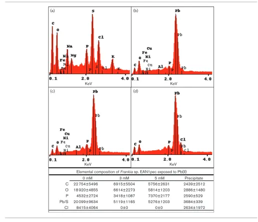
Frankia-bound Pb2+ precipitate (Fig. 2). Treatment with EDTA also failed to remove this membrane-bound precipi-tate (data not shown). These results indicate that the
Frankia Pb2+ precipitate did not simply form from the medium as pyromorphite, but was generated from some cellular process. The EDAX results also indicated that the proportion of phosphate was much higher forFrankia cul-tures exposed to higher Pb2+concentrations which suggest that different Pbx(PO4)x compounds form and bind to the
Frankiacell surface depending on Pb2+concentration. To examine surface property changes, FTIR analysis was used to characterize the general types of molecules present based on specific wave number areas [45]. FTIR spectra were collected forFrankiasp. strain EAN1pec cells exposed to Pb2+-stress and no Pb2+control cells (Fig. S3). Exposures of 3 to 8 mM Pb(NO3)2caused several changes in the
spec-tral pattern forFrankia. Specific changes were observed in the wavelength regions of 742, 776, 974, 1028, 1386, 1454, 1543 and 1719 cm 1.
Pb+2-induced proteins
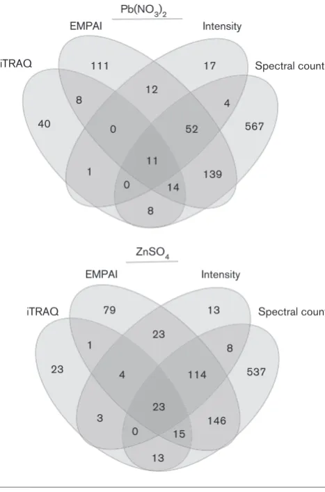
Relative protein abundance was determined by 2-D HPLC MS/MS in both labelled (iTRAQ) and unlabelled samples for Frankia sp. strain EAN1pec cells exposed to either 3 mM Pb2+ or Zn2+. For comparative purposes, Zn2+ was included as a divalent, beneficial metal that is toxic at ele-vated levels. The MTC and MIC values for Zn2+were 3 and 8 mM, respectively (data not shown). From these experi-ments, 1430 statistically significant proteins (those exhibit-ing at least a twofold change in expression) were identified, and represent 19.9 % of theFrankiasp. EAN1pec predicted proteins. Only 395 of the identified proteins were deter-mined in the labelled (iTRAQ) samples, likely due to high abundance proteins overwhelming other signals in the pooled samples (Fig. 3).
Of the 1430 positively regulated proteins, 428 proteins responded only to Pb2+, and 437 responded only to Zn2+. Table 1 shows the results for upregulated proteins that may significantly contribute to metal resistance in Frankia sp. strain EAN1pec. Pb2+induced three copper-related proteins including a fused CopC/D domain transporter (Fra-nean1_7241), a Cu2+P-type ATPase (Franean1_1679) and a negatively charged secreted protein (Franean1_5746) that is found almost exclusively in association with Cu2+ P-type ATPases (Table 1a). A Kup-type potassium transporter (Fra-nean1_4647) was also upregulated with Pb2+. Under osmotic and acid stress conditions, Kup permeases import K+ and may provide the high K+ levels needed for exopolyphospha-tase (PPX) activity [46]. Zn2+stress did not exclusively induce any metal transporters, but caused upregulation of the Zn2+ -and Ni2+-binding hydrogenase maturation factor (HypA, Fra-nean1_2483). In Frankia sp. strain EAN1pec, other metal-binding proteins including bacterioferritins (Franean1_2250, Franean1_2437), cobaltochelatase (CobN, Franean1_4862), ferric-binding protein (FbpA; Franean1_4453), and iron-sulfur assembly proteins (Franean1_2086, Franean1_2088) were upregulated in response to either Zn+2 alone or both metals (Table 1a). Exposure to both metals also induced the upregulation of a cation diffusion facilitator (CDF, Fra-nean1_2205), which functions as a general divalent metal exporter [47].
Among the Pb2+- and Zn2+-upregulated proteins, 321 (22 %) were hypothetical proteins and 143 of these proteins have no significantly homologous functional domains. There were 92 hypothetical proteins containing transmembrane segments (TMS), and based on individual inspection of predicted mem-brane orientation, 35 contained large extracellular regions or were potential transporters (Table S2a). Some of the hypothet-ical proteins demonstrated a limited (<30 %) homology to other known proteins, including: 1. cell wall modification enzymes (Franean1_5160, Franean1_6801); 2. surface lipid modification enzymes (Franean1_0195, Franean1_2417, Fra-nean1_7260); and 3. some very large membrane-anchored proteins that were possibly involved in divalent cation- or polysaccharide-binding (Franean1_0453, Franean1_5995). Small proteins with high percentage cysteine (metallothio-neins) or histidine (metallohistins) are known mechanisms of metal resistance, and have previously been predicted in
Frankia[48]. Using an in-house PERL algorithm, the amino acid composition for all theFrankiasp. strain EAN1pec pro-teins was determined (data not shown). Among the 137 hypo-theticalFrankiasp. strain EAN1pec proteins that score in the top 1 % for cysteine (>3 %) or histidine (>5 %) content, 14 have increased levels of expression with metals in this study (Table 1b).
Heavy metals are known to affect sulfur homeostasis by oxi-dizing disulfide and iron-sulfur bonds and binding to thiol amino acids and other cellular thiol molecules (e.g. glutathi-one, mycothiol) [48]. Pb2+induced alkanesulfonate mono-oxygenase (Franean1_3757) expression (Table 1c), which indicates that the cell was undergoing sulfur starvation [49].
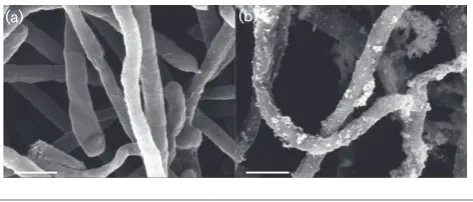
(a) (b)
Fig. 1.SEM ofFrankiasp. strain EAN1pec grown underPb2+-stress.
Frankiasp. strain EAN1pec was grown for 15 days in basal growth medium with or without 3.0 mM Pb(NO3)2as described in Methods. (a)
Several sulfur assimilation proteins including a sulfate trans-porter (SulP, Franean1_0735), sulfite reductase (Fra-nean1_6105), and cobalamin-independent methionine synthase (Franean1_1081) were upregulated with Pb2+ induction (Table 1c). Zn2+also upregulated two proteins from the sulfur assimilation pathway including a sulfate adenylyltransferase (Franean1_4225), and a cobalamin-dependent methionine synthase (Franean1_1643). Disulfide bond oxidoreductase (DsbA, Franean1_3881), which is responsible for correct formation of disulfide bonds in nascent proteins, was upregulated with Pb2+ inductionPb2+ induction (Table 1c). DsbA has been linked to Zn2+ resis-tance and alkaline phosphatase production [50]. InFrankia
sp. strain EAN1pec, one of the two phosphate metabolism-related proteins upregulated with Zn+2 was alkaline phos-phatase (Franean1_1306; the other upregulated protein was a guanosine pentaphosphate [(p)ppGpp] synthase (Fra-nean1_5139) (Table 1c).
Pb+2 also induced (p)ppGpp synthase (Franean1_5138) and other proteins involved in phosphorous metabolism, includ-ing a phosphate starvation protein (PhoH, Franean1_0834), a polyphosphate kinase (Ppk, Franean1_1116), and a polyphos-phate phosphatase (Ppx, Franean1_6144) (Table 1c). Phos-phate starvation leads to (p)ppGpp increases, which positively regulates the geneppk, leading to a temporary polyphosphate
(a) (b)
(c) (d)
KeV KeV
KeV KeV
C 22 754±5496 6915±5504 5756±2631 2439±2512
O 18 920±4855 6614±2273 5814±1203 2886±1480
P 4532±2724 3418±1087 7370±2177 2590±529
Pb/S 20 099±9634 5119±1165 5276±1203 3684±339
Cl 8415±4064 0±0 0±0 2634±1972
Elemental composition of Frankia sp. EAN1pec exposed to Pb(II)
0 mM 3 mM 5 mM Precipitate
Fig. 2.SEM-EDAX ofFrankiasp. strain EAN1pec grown under Pb2+-stress.Frankiasp. strain EAN1pec was grown for 15 days in basal
growth medium with 0, 3 or 5 mM Pb(NO3)2as described in Methods. For comparative purposes, lead precipitate formed in the
absence ofFrankia was also included. Panels (a) (d), show EDAX spectra for control cells and corresponding element analysis: (a) 0 mM Pb(NO3)2; (b) 3 mM Pb(NO3)2; (c) 5 mM Pb(NO3)2; (d) Pb
+2
accumulation [51]. High levels of polyphosphate are known to chelate metals (e.g. Zn) in the cytoplasm. When expressed with Ppx, polyphosphate increases Cu2+and Cd2+resistance [52, 53]. Both Pb2+- and Zn2+-induction caused an upregula-tion of PhoU (Franean1_6147). This Zn2+-binding, self-regu-lating global regulator is known to help repress phosphate uptake and secondary metabolism under normal conditions [54]. Therefore, PhoU upregulation indicates these processes have been de-repressed.
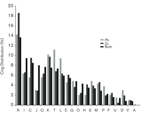
COG distribution of upregulated proteins
The distribution of the upregulated proteins into COG catego-ries was determined and the results are shown in Fig. 4. Based on COG distribution of upregulated proteins, Pb2+induced: (1) gene regulation proteins (COG T) such as histidine or ser-ine/threonine kinases; (2) DNA replication and repair mecha-nisms (COG L); and (3) a variety of inorganic metabolism proteins (COG P). Zn2+ primarily upregulated general
function (COG R) and energy metabolism (COG C) enzymes, including a variety of hydrolases, oxidoreductases and dehy-drogenases. Those proteins that were upregulated by both metals were primarily involved in lipid metabolism (COG I), protein translation (COG J) machinery (ribosomes), and protein modification (COG O) mechanisms involved in pro-tein folding (e.g. GroES/EL chaperonins). Secondary metabo-lism proteins (COG Q) were also upregulated with both metals, many falling into two predicted biosynthesis clusters. The first were from a polyketide biosynthesis cluster (Fra-nean1_2389, Franean1_2392, Franean1_2396), which is pre-dicted to produce a spore pigment [8]. The second included members of an erythronolide biosynthesis cluster (Fra-nean1_3887–Franean1_3896), whose predicted product was a specialized lipid [8].
Surface modification proteins
Several other important differences in surface and lipid pro-tein expression patterns for Pb2+and Zn2+inductions were also observed. Pb2+ exposure upregulated nearly twice as many (32 vs 18) surface sugar- and lipid-related proteins compared to Zn2+(Table S2b). These proteins included sev-eral families of glycosyltransferases, extracellular polysac-charide synthesis (EPS)-related, sugar phosphate permeases, and mycolic acid lipid exporters.
Zn+2-induction caused an upregulation of squalene-phy-toene synthase cluster of proteins (Franean1_3971, Fra-nean1_5716+17, Franean1_5128+32) (Table 1d), which are predicted to be involved in carotenoid and hopanoid lipid production [8]. Pb2+and Zn2+induction resulted in upregu-lation of several proteins involved in phytoene precursor biosynthesis proteins as well (Franean1_0162, Fra-nean1_0845, Franean1_0798 and Franean1_1170). Polyke-tide-based secondary metabolite genes are also upregulated with Pb2+and Zn2+, including polyketide cyclase and several erythronolide synthetase (Table 1d). Polyketides and other secondary metabolites have been shown to be significantly increased in microbial communities exposed to heavy met-als [55, 56].
Recognition of a metal-induced regulatory binding site
An in-house PERL algorithm was used to find potential intergenic regulatory sites of metal-related genes in the
Frankiasp. EAN1pec genome. A potential transcription fac-tor binding site (TFBS) was discovered upstream of each of the three operons containing Cu2+P-type ATPases (CopA1: Franean1_1679, CopA2: Franean1_5747, CopA3: Fra-nean1_6538). This TFBS was also identified upstream of a metal-sensing repressor (DUF156) orthologue (Fra-nean1_3360) and a potential Pb2+-precipitation protein undecaprenyl diphosphatase (UppP, Franean1_2469). When the whole Frankiasp. strain EAN1pec genome was analysed, 32 genes were identified as potential regulated by this TFBS, 13 of which had Pb2+- or Zn2+-upregulated pro-teins (Table S3). Orthologues ofFrankiasp. strain EAN1pec heavy-metal-resistance genes that have this motif upstream
139
Spectral count Intensity
EMPAI
iTRAQ
1 0
4 12
8
11
8 0
52
14 111
40 567
17
146
Spectral count Intensity
EMPAI
iTRAQ
3 4
8 23
1
23
13 0
114
15 79
23 537
13
Pb(NO3)2
ZnSO4
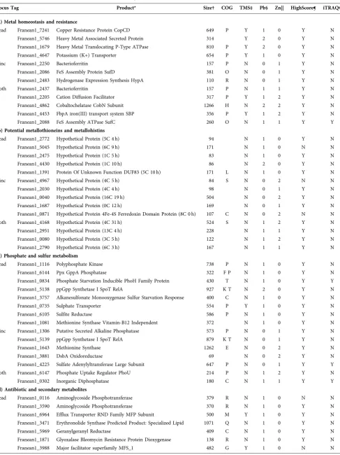
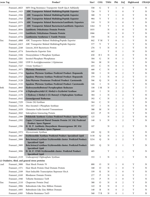
Table 1.SelectedFrankiasp. EAN1pec proteins positively regulated with Pb2+or Zn2+potentially involved in metal resistance
Locus Tag Product* Size† COG TMS‡ Pb§ Zn|| HighScore¶ iTRAQ#
(a) Metal homeostasis and resistance
Lead Franean1_7241 Copper Resistance Protein CopCD 649 P Y 1 0 Y N Franean1_5746 Heavy Metal Associated Secreted Protein 314 Y 2 0 Y N Franean1_1679 Heavy Metal Translocating P-Type ATPase 810 P Y 2 0 Y N Franean1_4647 Potassium (K+) Transporter 654 P Y 1 0 Y N Zinc Franean1_2250 Bacterioferritin 157 P N 0 1 Y N Franean1_2086 FeS Assembly Protein SufD 381 O N 0 1 Y N Franean1_2483 Hydrogenase Expression Synthesis HypA 110 R N 0 1 Y N Both Franean1_2437 Bacterioferritin 157 P N 1 1 Y N Franean1_2205 Cation Diffusion Facilitator 317 P Y 1 2 Y N Franean1_4862 Cobaltochelatase CobN Subunit 1266 H N 2 2 Y N Franean1_4453 FbpA iron(III) transport system SBP 356 P Y 1 2 Y N Franean1_2088 FeS Assembly ATPase SufC 260 O N 1 1 Y Y
(b) Potential metallothioneins and metallohistins
Lead Franean1_2772 Hypothetical Protein (5C 4 h) 94 N 1 0 Y N Franean1_5045 Hypothetical Protein (6C 9 h) 171 N 1 0 N N Franean1_2475 Hypothetical Protein (1C 5 h) 83 N 1 0 Y N Franean1_4430 Hypothetical Protein (1C 10 h) 86 N 2 0 Y N Franean1_1391 Protein Of Unknown Function DUF83 (5C 10 h) 171 L N 1 0 Y N Zinc Franean1_4967 Hypothetical Protein (4C 5 h) 84 S N 0 2 N N Franean1_2030 Hypothetical Protein (4C 4 h) 98 N 0 1 Y N Franean1_0040 Hypothetical Protein (16C 19 h) 504 N 0 2 Y N Franean1_1687 Hypothetical Protein (0C 12 h) 169 N 0 1 Y N Franean1_0871 Hypothetical Protein 4Fe-4S Ferredoxin Domain Protein (8C 0 h) 107 C N 0 2 N N Both Franean1_4168 Hypothetical Protein (4C 31 h) 524 S N 1 2 Y N Franean1_2951 Hypothetical Protein (13C 4 h) 228 N 1 1 Y N Franean1_0080 Hypothetical Protein (3C 5 h) 122 N 1 2 Y N Franean1_2790 Hypothetical Protein (6C 3 h) 167 N 1 1 Y N
(c) Phosphate and sulfur metabolism
Lead Franean1_1116 Polyphosphate Kinase 738 P N 1 0 Y N Franean1_6144 Ppx GppA Phosphatase 322 F P N 1 0 Y N Franean1_0834 Phosphate Starvation Inducible PhoH Family Protein 430 T N 1 0 Y Y Franean1_5138 ppGpp Synthetase I SpoT RelA 927 K T N 2 0 Y N Franean1_3757 Alkanesulfonate Monooxygenase Sulfur Starvation Response 400 C N 1 0 Y N Franean1_0735 Sulphate Transporter 554 P Y 1 0 Y N Franean1_6105 Sulfite Reductase 586 P N 1 0 Y N Franean1_1081 Methionine Synthase Vitamin-B12 Independent 372 N 1 0 Y N Zinc Franean1_1306 Putative Secreted Alkaline Phosphatase 573 P N 0 1 Y N Franean1_5139 ppGpp Synthetase I SpoT RelA 879 K T N 0 1 Y N Franean1_1643 Methionine Synthase 1262 E N 0 2 Y N Franean1_3881 DsbA Oxidoreductase 69 N 0 2 Y N Franean1_4225 Sulfate Adenylyltransferase Large Subunit 647 P N 0 1 Y Y Both Franean1_6147 Phosphate Uptake Regulator PhoU 214 P N 1 2 Y N Franean1_0302 Inorganic Diphosphatase 180 C N 1 1 Y Y
(d) Antibiotic and secondary metabolites
Table 1.cont.
Locus Tag Product* Size† COG TMS‡ Pb§ Zn|| HighScore¶ iTRAQ#
Franean1_6823 MFS Drug Resistance Transporter EmrB QacA Subfamily 558 G Y 1 0 Y N Franean1_1925 ABC Transporter Related Multidrug/Peptide Exporter 726 V Y 1 0 Y N Franean1_1926 ABC Transporter Related Multidrug/Peptide Exporter 588 V Y 1 0 Y N Franean1_3763 ABC Transporter Related Multidrug/Peptide Exporter 984 V Y 1 0 Y N Franean1_0376 ABC Transporter Related Bacteriocin/Lantibiotic Exporters 318 V N 1 0 Y N Franean1_0377 ABC Transporter Related Bacteriocin/Lantibiotic Exporters 1264 V Y 1 0 Y N Franean1_0056 Lantibiotic Dehydratase Domain Protein 1035 N 1 0 Y N Franean1_1332 Lantibiotic Dehydratase Domain Protein 1066 N 2 0 Y N Franean1_6754 Lanthionine Synthetase C Family Protein 358 N 1 0 Y N Zinc Franean1_4888 ABC Transporter Related Multidrug/Peptide Exporter 842 V M Y 0 1 Y N Franean1_6413 ABC Transporter Related Multidrug/Peptide Exporter 577 V Y 0 1 Y N Franean1_2248 Linocin_M18 Bacteriocin Protein 276 S N 0 1 Y N Franean1_4774 Enterobactin Exporter Ents 443 Y 0 1 N N Franean1_5184 Deoxyxylulose-5-Phosphate Synthase 650 H I N 0 2 Y N Franean1_5201 Inositol-Phosphate Phosphatase 304 G N 0 1 Y N Franean1_5202 UDP-N-Acetylglucosamine 2-Epimerase 584 M N 0 2 Y N Franean1_7227 Citrate Synthase I 455 C N 0 1 Y N Franean1_3971 Phytoene Desaturase 468 C N 0 1 Y N Franean1_5716 Squalene Phytoene Synthase Predicted Product: Hopanoids 300 I N 0 2 Y N Franean1_5717 Squalene Phytoene Synthase Predicted Product: Hopanoids 379 I N 0 2 Y Y Franean1_5128 Zeta-Phytoene Desaturase Predicted Product: Carotenoids 506 Q N 0 1 Y N Franean1_5132 Squalene Phytoene Synthase Predicted Product: Carotenoids 326 I N 0 1 Y N Both Franean1_0845 Hydroxymethylbutenyl Pyrophosphate Reductase 338 I M N 2 2 Y Y Franean1_0798 4-Diphosphocytidyl-2C-Methyl-D-Erythritol Synthase 249 I N 2 2 Y Y Franean1_1170 1-Hydroxy-2-Methyl-2-(E)-Butenyl 4-Diphosphate Synthase 384 I N 1 2 Y N Franean1_0162 Geranylgeranyl Reductase 392 C N 1 1 Y N Franean1_7229 Citrate (Si)-Synthase 364 C N 3 2 Y N Franean1_7318 Myo-Inositol-1-Phosphate Synthase 357 I N 2 2 Y N Franean1_1659 Glutamine-Scyllo-Inositol Transaminase 388 M N 2 2 Y N Franean1_3043 Siderophore-Interacting Protein 623 P N 1 1 Y N Franean1_2389 Polyketide Synthesis Cyclase Predicted Product: Spore Pigment 125 N 1 2 Y N Franean1_2392 Cupin 2 Conserved Barrel Domain Protein (1C 8 h) Predicted
Product: Spore Pigment
140 S N 1 2 Y N
Franean1_2396 H. M. P. Antibiotic Biosynthesis Monooxygenase (0C 8 h) Predicted Product: Spore Pigment
107 N 1 2 Y N
Franean1_5372 Mycocerosate Synthase 438 Q N 1 1 Y N Franean1_3887 Erythronolide Synthase Predicted Product: Specialized Lipid 3158 Q N 1 2 Y N Franean1_3888 Beta-ketoacyl synthase Erythronolide cluster. Predicted Product:
Specialized Lipid
3254 Q N 1 2 Y Y
Franean1_3889 Beta-ketoacyl synthase Erythronolide cluster. Predicted Product: Specialized Lipid
3493 Q N 2 1 Y N
Franean1_3896 H. M. P. 1TMS Erythronolide cluster. Predicted Product: Specialized Lipid
405 Y 1 1 Y N
Franean1_2119 Undecaprenyl Diphosphate Synthase 333 I N 1 1 Y N
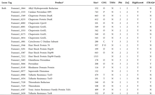
(e) Oxidative, thiol, and general stress proteins
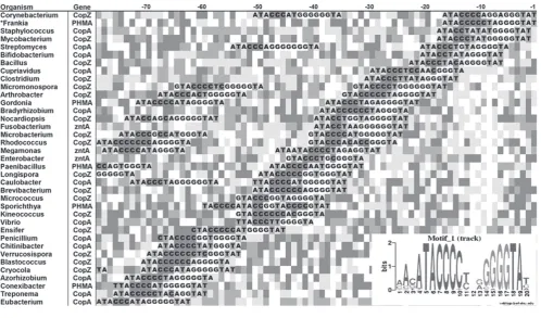
were analysed for the presence of the TFBS. Thousands of Cu2+-type ATPase (CopA) and Zn-type ATPase (ZntA) transporters, CopZ-like metal chaperones, and potential heavy-metal-associated (PHMA) secreted and lipoproteins were found with this TFBS upstream. The motif was highly conserved among many different types of bacteria and even some eukaryotes (Fig. 5). DUF156 orthologues (Pfam02583) with this TFBS motif have been found in other bacteria, including the Ni2+/Co2+-responsive regulator NcrB in Lep-tospirillium ferriphilum[57], the Cd2+/Zn2+/Ni2+-responsive regulator MreA inPseudomonas putida[58], and the Cu+2 -responsive regulator CsoR in Streptomyces lividans [59]. From these analyses, it was concluded that the identified palindrome was a CsoR/DUF156 repressor binding site.
Gene expression in response to Pb2+-stress
Transcript levels of several metal-upregulated proteins were detected by qRT-PCR (Fig. 6). One protein, glutathi-one peroxidase (GPx, Franean1_5485), was upregulated with both metals and in both labelled and unlabelled
experiments, it was selected as a control. The gpx tran-script exhibited 5.0-, 7.4- and 6.9- fold increase with expose to 3, 5 and 8 mM Pb2+ stress, respectively. Under 3 mM Pb2+ stress, the mRNA levels of both the
csoR and uppSgenes showed at 2.5- and 2.1-fold increase, respectively, which was followed by a negative dose-depen-dent response to Pb2+. None of the tested metal exporters were significantly upregulated under 3 mM Pb+2stress, but the mRNA levels of copA2 (Franean1_5747), copA3 (Fra-nean1_6538), and cdf1 (Franean1_2205) significantly increased under exposure to 8 mM Pb2+.
DISCUSSION
Frankiaimproves the survival of actinorhizal plants in the presence of high concentrations of Pb+2and other contami-nants [2, 3, 60–62]. There also appears to be a general increase in growth on both Pb(C2H3O2)2and Pb(NO3)2for up to 2 mM Pb+2.Frankiastrains were shown to have a pos-itive growth response to other metals including 2.5 mM Ni+2 and 10 mM Al+3 [20, 27, 28]. This increased growth
Table 1.cont.
Locus Tag Product* Size† COG TMS‡ Pb§ Zn|| HighScore¶ iTRAQ#
Both Franean1_1864 Alkyl Hydroperoxide Reductase 152 O N 1 2 Y N Franean1_1533 Catalase Peroxidase HPI 743 P N 1 2 Y Y Franean1_1569 Chaperone Protein DnaK 663 O N 1 1 Y N Franean1_0233 Chaperone Protein DnaK 612 O N 2 2 Y Y Franean1_6002 Chaperonin Cpn10 101 O N 2 2 Y Y Franean1_6001 Chaperonin GroEL 549 O N 1 2 Y N Franean1_3353 Chaperonin GroEL 542 O N 2 2 Y N Franean1_0175 Chaperonin GroEL 540 O N 3 2 Y N Franean1_3854 Chaperonin GroEL 545 O N 3 2 Y Y Franean1_1801 Cytochrome C Oxidase Subunit 288 C Y 3 2 Y Y Franean1_1946 Heat Shock Protein 70 957 P O N 1 2 Y N Franean1_5291 Heat Shock Protein Hsp20 199 O N 1 2 Y N Franean1_3387 Heat Shock Protein Hsp90 643 O N 2 2 Y Y Franean1_7212 Heat Shock Protein Hsp90-Family 1315 N 2 1 Y N Franean1_5485 Glutathione Peroxidase 178 O N 3 2 Y Y Franean1_3666 Peroxidase 208 O N 3 2 Y Y Franean1_0149 Rhodanese Domain Protein 116 P N 1 2 Y N Franean1_0977 Superoxide Dismutase 132 N 2 1 Y Y Franean1_0900 Tellurite Resistance TerD 479 T N 1 2 Y N Franean1_1054 Tellurite Resistance TerD 191 T N 1 1 N N Franean1_7328 Thioredoxin Reductase 348 O N 2 1 Y Y Franean1_7329 Thioredoxin 108 O N 1 2 Y N Franean1_6387 Toxic Anion Resistance Family Protein TelA 409 P N 1 2 Y N Franean1_2430 Tellurite Resistance TerB 332 P N 1 2 Y N
*Protein name and relevant manual annotations. Bold and highlighted (grey) data represent a potential functional group.
†Amino acid length of protein.
‡Transmembrane segments (TMS) present (Y) or absent (N) in protein.
§Number of samples from Pb2+-exposed cells containing indicated protein (max. 3).
||Number of samples from Zn2+-exposed cells containing indicated protein (max. 2).
with toxic metals could be due to metal stress-induced changes in sugar and phosphate metabolism observed by FTIR, EDAX, and MudPIT inFrankiasp. strain EAN1pec (discussed below). The levels of Pb+2resistance are signifi-cant. In the soil, the EPA has set a 0.96 µM limit and many Pb+2-contaminated sites are in the 5–250 µM range.
In this study, Frankia sp. strain EAN1pec was shown to absorb significant amounts of Pb2+ from its surrounding environment, and precipitate Pb+2 on the cell surface (Fig. 1). Our results also show that the bound precipitate was stable through several washings of water, acid, EDTA, or alcohol. This result indicates the presence of a specific metal-binding mechanism rather than transient sorption of Pb+2 to surface macromolecules. Based on the EDAX results, there was nearly half as much phosphate per quan-tity of Pb+2at 3 mM Pb+2than at 5 mM Pb+2. FTIR analy-sis also showed a smaller phosphate peak at 3 mM Pb+2. These observed Pb+2 precipitation and concomitant changes in surface polysaccharides, phosphates, and sulfur-ligands also occurs with several other bacterial and fungal species [43, 63].
Frankiatranscript and proteome response to Pb+2
In the absence of genetic tools, qRT-PCR and shotgun pro-teomics approaches were used to determine whether
significant changes in mRNA levels and protein expression were observed inFrankiasp. strain EAN1pec. Fig. 7 shows the interplay of the Pb+2-upregulated proteins in Frankia
sp. strain EAN1pec that contribute to its high level of Pb2+ resistance.
Pb2+export and precipitation mechanisms
Frankia sp. strain EAN1pec does not contain Zn/Cd/Pb-type ATPases, it does contain the three Cu-ATPase (CopA) paralogues. Based on the qRT-PCR results, none of the
copAgenes were upregulated at 3 mM Pb2+(Fig. 6), but the CopA1 protein was identified as upregulated with Pb2+in the proteome (Table 1a). At higher Pb2+ concentrations, both copA2 and copA3 showed a significant increase in mRNA levels (Fig. 6). Therefore, expression of heavy metal exporters and phosphate precipitation may be more import-ant at higher Pb2+levels, while the surface and stress-related changes may be sufficient to provide resistance at lower Pb2+levels.
CsoR/DUF156 is known to detect a variety of metals [64]. The CsoR/DUF156 orthologue could be involved in Pb2+ -sensing inFrankia.This gene had elevated mRNA levels at 3 mM Pb2+ exposure, but was downregulated at higher metal concentrations (Fig. 6). Furthermore, a CsoR-like metal regulatory site was found upstream of all three CuAT-Pase-containing operons and UppP (Table S3). The inverse correlation of CsoR and CopA/CDF1 expression, and the presence of potential CsoR regulatory sites near metal trans-port (CopA) and precipitation mechanisms (UppP) suptrans-port the possibility that metal export and precipitation are related inFrankiaunder high levels of metal exposure.
Other lead-response orthologues of transport proteins includ-ing CopCD, and the sugar-phosphate major facilitator super-family (MFS) transporters were upregulated with Pb2+. Pb2+ upregulated seven hypothetical proteins that are potential transporters based on the presence of multiple predicted transmembrane segments (TMS), including one (Fra-nean1_5744) that has significant metal-binding potential. Sev-eral Gram-positive and Gram-negative bacteria exploit the high affinity of Pb2+for phosphate to detoxify Pb2+as intra-and extracellular precipitates [65]. In a similar mechanism to
Ralstonia metallidurans [66], one mechanism for Frankiais that Pb2+ export is followed by precipitation by the metal-induced inorganic phosphatase (Franean1_0302), or the undecaprenyl phosphatase (UppP). Another potential mecha-nism is that undecaprenyl diphosphate itself could play an additional role in Pb2+resistance. An undecaprenyl diphos-phate synthase (UppS) protein was upregulated with both metals (Table 1d). The mRNA level of theuppSgene was also upregulated 2.1-fold as well (Fig. 6).
Metal-induced polyphosphate degradation and resultant metal-phosphate efflux has also been linked to extracellular metal precipitation [51]. In Frankia, Pb2+ upregulated the stringent response regulator (p)ppGpp synthetase (Table 1), which in turn may have upregulated a variety cellular responses including polyphosphate production. 0
2 4 6 8 10 12 14 16 18 20
Pb Zn Both
Cog
Distribution (%)
R I C J Q K T L E G O H S M P F U D V A
Fig. 4.COG categories distribution of proteins upregulated by Pb2+or
Zn2+. COG categories for proteins upregulated with Pb2+, Zn2+, or both
Cytoplasmic Pb2+could be detoxified either directly by bind-ing polyphosphate or by the formation Pb2+-phosphate
precipitates by the Pb2+-upregulated polyphosphate phospha-tase (Ppx). The strong FTIR peak at ~974 cm 1 with Pb2+, likely a P–O bond, was also seen with extracellular metal poly-phosphates [67].
Exopolysaccharides and metal adsorption
There were nearly twice as many surface sugar, cell-wall and lipid-related proteins upregulated with Pb2+than with Zn2+. A mixture of surface glycoproteins and glycolipids (collec-tively exopolysaccharides) were shown to contribute signifi-cantly to Pb2+resistance in other bacteria [68].
Both the production of an extracellular milieu (Fig. S2) and sugar–phosphate peaks identified by FTIR (Fig. S3) impli-cate a role for exopolysaccharides (EPS) in surface Pb2+ binding. Pb2+ binding to EPS is known to occur in other microbes [69, 70]. Potential EPS-related hypothetical sur-face proteins were also expressed with metals, and include cell wall (Franean1_5160, Franean1_6801), glycoprotein (Franean1_0453), or lipid modification enzymes (Fra-nean1_0195, Franean1_2417, Franean1_7260) (Table S2a). Collectively, these extracellular proteins could contribute to observed metal-sorbing extracellular matrix, either acting in a protective capacity or as a site for EPS attachment.
Fig. 5.Regulatory motif of heavy metal P-type ATPase transporters and their associated proteins. Intergenic regions up to 200 bp upstream of CopA- or ZntA-like (COG2217) P-type ATPase transporters, CopZ-like (COG2608) metal chaperones, and orthologues to the Pb2+-upregulated CopA-associated secreted protein (Franean1_5746) [indicated with asterisk] from various genera of eukaryotes and prokaryotes were searched for palindromic sequences as potential transcription factor binding sites. The identified self-complemen-tary palindromic motif was found at a variable distance from the start codon, only those found within 80 bp from the start codon are displayed. A sequence logo [lower right] of the conserved motif created from Phylogibbs software is also included.
0 mM 10
8
6
F
old c
hange
4
2
0
copA1 copA2 copA3 cdf1 gpx uppS csoR 3 mM 5 mM 8 mM
Fig. 6.Transcriptional changes of selectedFrankiagenes under Pb+2
challenge. Values represent fold changes in gene transcript levels compared to the control forFrankiasp. EAN1pec cells exposed to Pb+2
(0, 3, 5 and 8 mM). Gene transcript levels were normalized to the housekeeping genegyrBfor each Pb+2concentration. Error bars
Fig. 7.Proposed system forFrankiasp. strain EAN1pec Pb2+-resistance. Low phosphate conditions due to Pb-bound phosphate in the
media stimulate thephoregulon which upregulates both 1) the high affinity phosphate transporter PstABC (Franean1_ 0353–55) that imports phosphate, and 2) the polyphosphate kinase (PPK, Franean1_1116) which converts the phosphate to 3) polyphosphate. Poly-phosphate binds Pb2+and, using 4) exopolyphosphatase (PPX, Franean1_6144), is converted to either 5) ATP or to 6) metal phosphates
that are exported via 7) Pho84 (Franean1_3988, Franean1_6944). PPX need high levels of K+ that is provided by 8) the high affinity potassium transporter Kup (Franean1_4647). Pb2+can also be exported by heavy metal transporters 9) CDF (Franean1_2205) or 10)
CopA (Franean1_1679, Franean1_5747, Franean1_6538). 11) The classic model for extracellular Pb2+precipitation is that exported Pb2 +
Metal-related secondary metabolite production
There were specific secondary metabolite responses to Pb2+ and to Zn2+, including changes in lipids that may be due to the stringent response (Table 1d). InStreptomyces, phytoene synthesis is under the control of SigR, an oxidative-thiol stress regulator [71]. Mycocerosate is another large lipid that acts as a protective mechanism by reducing cell perme-ability [72]. Visual inspection by light microscopy shows surface-bound precipitates to be highly luminous (Fig. S2), similar to lipid-rich vesicles and sporangia inFrankia[73].
Conclusions
Our results from the SEM, EDAX and FTIR experiments show that Frankia sp. strain EAN1pec has specific Pb2+ -binding mechanisms that provide its resistance levels. The results from both labelled and unlabelled shotgun proteo-mics approaches and gene expression experiments identified several potential mechanisms that provide high levels of
Frankia metal resistance and precipitation. Both lipid and EPS-related proteins appear to be involved in the adsorption of Pb2+to the cell surface, while two metal transporters (a Cu2+-ATPase and cation diffusion facilitator), as well as sev-eral hypothetical transporters, could be involved in metal export. The exported Pb2+ may be precipitated at the cell surface by upregulated polyphosphate, undecaprenyl diphosphate synthase and inorganic diphosphatase. Both metals upregulated expression of the oxidative stress pro-teins and secondary metabolites, metallohistins and metal-lothionein-like proteins, which may help to detoxify metals in the cytoplasm. The cumulative interplay of these many mechanisms may explain the extraordinary resilience of
Frankiasp. strain EAN1pec to Pb2+.
Funding information
Partial funding was provided by the New Hampshire Agricultural Experiment Station. This is Scientific Contribution Number 2631. This work was supported by the USDA National Institute of Food and Agri-culture Hatch 022821. M. R. was supported by an Egyptian Channel Fellowship from The Egyptian Cultural Affairs and Missions Sectors.
Acknowledgements
We thank Robert Mooney for his help with the photography, Nancy Chemin for her help with the electron microscopy, and Alix Konisky, Rebecca Wagers, Joel Richards and Glenn Krumholz for their initial contributions to this project (all from UNH – University of New Hampshire).
Conflicts of interest
The authors declare that there are no conflicts of interest.
References
1. Normand P, Benson DR, Berry AM, Tisa LS.FamilyFrankiaceae. In: Rosenberg E, DeLong EF, Lory S, Stackebrandt E and Thomp-son F (editors).The Prokaryote–Actinobacteria. Berlin: Springer; 2014. pp. 339–356.
2. Ridgway KP, Marland LA, Harrison AF, Wright J, Young JPet al.
Molecular diversity of Frankia in root nodules of Alnus incana
grown with inoculum from polluted urban soils. FEMS Microbiol Ecol2004;50:255–263.
3. Roy S, Khasa DP, Greer CW. Combining alders, frankiae, and mycorrhizae for the revegetation and remediation of contami-nated ecosystems.Can J Bot2007;85:237–251.
4. Diagne N, Ngom M, Djighaly PI, Ngom D, Ndour Bet al. Remedia-tion of heavy-metal-contaminated soils and enhancement of their fertility with actinorhizal plants. In: Sherameti I and Varma A (edi-tors).Heavy Metal Contamination of Soils, Soil Biology. Switzerland: Springer International Publishing; 2015. pp. 355–366.
5. Chaia EE, Wall LG, Huss-Danell K.Life in soil by the actinorhizal root nodule endophyteFrankia. A review.Symbiosis2010;51:201–
226.
6. Normand P, Lapierre P, Tisa LS, Gogarten JP, Alloisio N et al.
Genome characteristics of facultatively symbiotic Frankia sp. strains reflect host range and host plant biogeography.Genome Res2007;17:7–15.
7. Normand P, Queiroux C, Tisa LS, Benson DR, Rouy Zet al. Explor-ing the genomes ofFrankia.Physiol Plant2007;130:331–343. 8. Udwary DW, Gontang EA, Jones AC, Jones CS, Schultz AWet al.
Significant natural product biosynthetic potential of actinorhizal symbionts of the genusFrankia, as revealed by comparative geno-mic and proteogeno-mic analyses. Appl Environ Microbiol 2011;77: 3617–3625.
9. Tisa LS, Beauchemin N, Gtari M, Sen A, Wall LG.What stories can theFrankiagenomes start to tell us?J Biosci2013;38:719–726. 10. Tisa LS, Oshone R, Sarkar I, Ktari A, Sen A et al. Genomic
approaches toward understanding the actinorhizal symbiosis: an update on the status of theFrankiagenomes.Symbiosis2016;70: 5–16.
11. Niemann J, Tisa LS.Nitric oxide and oxygen regulate truncated hemoglobin gene expression in Frankia strain CcI3. J Bacteriol
2008;190:7864–7867.
12. Perrine-Walker F, Doumas P, Lucas M, Vaissayre V, Beauchemin NJet al.Auxin carriers localization drives auxin accumulation in plant cells infected by Frankia in Casuarina glauca actinorhizal nodules.Plant Physiol2010;154:1372–1380.
13. Alloisio N, Queiroux C, Fournier P, Pujic P, Normand Pet al.The
Frankia alni symbiotic transcriptome. Mol Plant Microbe Interact
2010;23:593–607.
14. Bickhart DM, Benson DR. Transcriptomes of Frankia sp. strain CcI3 in growth transitions.BMC Microbiol2011;11:192.
15. Popovici J, Comte G, Bagnarol E, Alloisio N, Fournier Pet al. Dif-ferential effects of rare specific flavonoids on compatible and incompatible strains in theMyrica gale-Frankiaactinorhizal symbi-osis.Appl Environ Microbiol2010;76:2451–2460.
upregulated 19) antimictobial peptide ABC transporters (Franean1_0376, Franean1_1925–26, Franean1_3763). Lantibiotics could bind Pb2+and leave a phosphate exposed for further Pb2+binding. 20) Other lipids including mycoserosate (Franean1_5372) or specialized
16. Popovici J, Walker V, Bertrand C, Bellvert F, Fernandez MPet al.
Strain specificity in the Myricaceae–Frankia symbiosis is
corre-lated to plant root phenolics. Functional Plant Biology 2011;38: 682–689.
17. Lee HI, Donati AJ, Hahn D, Tisa LS, Chang WS.Alteration of the exopolysaccharide production and the transcriptional profile of free-living Frankia strain CcI3 under nitrogen-fixing conditions.
Appl Microbiol Biotechnol2013;97:10499–10509.
18. Mastronunzio JE, Benson DR.Wild nodules can be broken: proteo-mics ofFrankiain field-collected root nodules.Symbiosis2010;50: 13–26.
19. Arahou M, Diem HG, Sasson A. Influence of iron depletion on growth and production of catechol siderophores by different
Frankiastrains.World J Microbiol Biotechnol1998;14:31–36. 20. Wheeler CT, Hughes LT, Oldroyd J, Pulford ID.Effects of nickel on
Frankiaand its symbiosis withAlnus glutinosa(L.) Gaertn. Plant and Soil2001;231:81–90.
21. Mattsson U, Sellstedt A. Nickel affects activity more than expression of hydrogenase protein in Frankia. Curr Microbiol
2002;44:88–93.
22. Tisa LS, Ensign JC.The calcium requirement for functional vesicle development and nitrogen fixation by Frankia strains EAN1pec and CpI1.Arch Microbiol1987;149:24–29.
23. Cusato MS, Tortosa RD, Valiente L, Barneix AJ, Puelles MM.
Effects of Zn2+ on nodulation and growth of a South American actinorhizal plant, Discaria americana (Rhamnaceae). World J Microbiol Biotechnol2007;23:771–777.
24. Bolaños L, Redondo-Nieto M, Bonilla I, Wall LG.Boron require-ment in theDiscaria trinervis (Rhamnaceae) andFrankiasymbiotic relationship. its essentiality forFrankia BCU110501 growth and nitrogen fixation.Physiol Plant2002;115:563–570.
25. Crannell WK, Tanaka Y, Myrold DD.Calcium and pH interaction on root nodulation of nursfry-grown red alder (Alnus rubrabong.) seedlings byFrankia.Soil Biol Biochem1994;26:607–614. 26. Richards JW, Krumholz GD, Chval MS, Tisa LS. Heavy metal
resistance patterns of Frankia strains. Appl Environ Microbiol
2002;68:923–927.
27. Igual JM, Dawson JO.Stimulatory effects of aluminum onin vitro
growth ofFrankia.Can J Bot1999;77:1321–1326.
28. Sayed WF, Mohaowad SM, Abd El-Karim MM.Effect of Al, Co, and Pb ions on growth ofFrankia spp. in a mineral medium. Folia Microbiol2000;45:153–156.
29. Igual JM, Rodriguez-Barrueco C, Cervantes E.The effects of alu-minium on nodulation and symbiotic nitrogen fixation inCasuarina cunninghamianaMiq.Plant and Soil1997;190:41–46.
30. Bose D, Sen A.Isolation and heavy metal resistance pattern of
Frankia from Casuarina equisetifolia nodules. Indian J Microbiol
2006;15:9–11.
31. Furnholm TR, Tisa LS.The ins and outs of metal homeostasis by the root nodule actinobacteriumFrankia.BMC Genomics2014;15: 1092.
32. Rehan M, Furnholm T, Finethy RH, Chu F, El-Fadly Get al.Copper
tolerance inFrankiasp. strain EuI1c involves surface binding and copper transport.Appl Microbiol Biotechnol2014;98:8005–8015. 33. Lalonde M, Calvert HE, Pine S.Isolation and use ofFrankiastrains
in actinorhizae formation. In: Gibson AH and Newton WE (editors).
Current Perspectives in Nitrogen Fixation. Canberra: Australian Academy of Science; 1981. pp. 296–299.
34. Tisa LS, Chval MS, Krumholz GD, Richards J.Antibiotic resistance patterns ofFrankiastrains.Can J Bot1999;77:1257–1260. 35. Furnholm T, Beauchemin N, Tisa LS. Development of a
semi-high-throughput growth assay for the filamentous actinobacteria
Frankia.Arch Microbiol2012;194:13–20.
36. Smith PK, Krohn RI, Hermanson GT, Mallia AK, Gartner FHet al.
Measurement of protein using bicinchoninic acid.Anal Biochem
1985;150:76–85.
37. Dhanjal S, Cameotra SS. Aerobic biogenesis of selenium nano-spheres byBacillus cereusisolated from coalmine soil.Microb Cell Fact2010;9:52.
38. Kessi J, Ramuz M, Wehrli E, Spycher M, Bachofen R.Reduction of selenite and detoxification of elemental selenium by the phototro-phic bacterium Rhodospirillum rubrum. Appl Environ Microbiol
1999;65:4734–4740.
39. Shifman MA, Li Y, Colangelo CM, Stone KL, Wu TLet al.YPED: a
web-accessible database system for protein expression analysis.
J Proteome Res2007;6:4019–4024.
40. Ishihama Y, Oda Y, Tabata T, Sato T, Nagasu Tet al.Exponentially modified protein abundance index (emPAI) for estimation of abso-lute protein amount in proteomics by the number of sequenced peptides per protein.Mol Cell Proteomics2005;4:1265–1272. 41. Siddharthan R, Siggia ED, Van Nimwegen E.PhyloGibbs: a Gibbs
sampling motif finder that incorporates phylogeny.PLoS Comput Biol2005;1:e67.
42. Schmittgen TD, Livak KJ. Analyzing real-time PCR data by the comparativeCTmethod.Nat Protoc2008;3:1101–1108.
43. Tunali S, Çabuk A, Akar T.Removal of lead and copper ions from aqueous solutions by bacterial strain isolated from soil.Chem Eng J2006;115:203–211.
44. Wacławska I, Szumera M.Use of thermal analysis in the study of
soil Pb immobilization.J Therm Anal Calorim2010;99:873–877. 45. Naumann D.Infrared spectroscopy in microbiology. In: Meyers R
(editor).Encylopedia of Analytical Chemistry. New York, NY: Wiley; 2006. pp. 102–131.
46. Hegermann J, Lünsdorf H, Overbeck J, Schrempf H. Polyphos-phate at the Streptomyces lividans cytoplasmic membrane is enhanced in the presence of the potassium channel KcsA.
J Microsc2008;229:174–182.
47. Montanini B, Blaudez D, Jeandroz S, Sanders D, Chalot M. Phylo-genetic and functional analysis of the Cation Diffusion Facilitator (CDF) family: improved signature and prediction of substrate spec-ificity.BMC Genomics2007;8:107.
48. Schmidt A, Hagen M, Schütze E, Schmidt A, Kothe E.In silico pre-diction of potential metallothioneins and metallohistins in actino-bacteria.J Basic Microbiol2010;50:562–569.
49. Ellis HR.Mechanism for sulfur acquistion by the alkanesulfonate monooxygenase system.Biorganic Chem2011;35:178–184. 50. Hayashi S, Abe M, Kimoto M, Furukawa S, Nakazawa T. The
DsbA-DsbB disulfide bond formation system ofBurkholderia cepa-ciais involved in the production of protease and alkaline phospha-tase, motility, metal resistance, and multi-drug resistance.
Microbiol Immunol2000;44:41–50.
51. Seufferheld MJ, Alvarez HM, Farias ME.Role of polyphosphates in microbial adaptation to extreme environments. Appl Environ Microbiol2008;74:5867–5874.
52. Remonsellez F, Orell A, Jerez CA.Copper tolerance of the ther-moacidophilic archaeonSulfolobus metallicus: possible role of pol-yphosphate metabolism.Microbiology2006;152:59–66.
53. Keasling JD, Hupf GA. Genetic manipulation of polyphosphate metabolism affects cadmium tolerance in Escherichia coli. Appl Environ Microbiol1996;62:743–746.
54. Li Y, Zhang Y.PhoU is a persistence switch involved in persister formation and tolerance to multiple antibiotics and stresses in
Escherichia coli.Antimicrob Agents Chemother2007;51:2092–2099. 55. Gokhale RS, Sankaranarayanan R, Mohanty D.Versatility of
poly-ketide synthases in generating metabolic diversity. Curr Opin Struct Biol2007;17:736–743.
56. Hemme CL, Deng Y, Gentry TJ, Fields MW, Wu Let al.
Metage-nomic insights into evolution of a heavy metal-contaminated groundwater microbial community.Isme J2010;4:660–672. 57. Zhu T, Tian J, Zhang S, Wu N, Fan Y.Identification of the
tran-scriptional regulator NcrB in the nickel resistance determinant of
58. Haritha A, Sagar KP, Tiwari A, Kiranmayi P, Rodrigue Aet al.
MrdH, a novel metal resistance determinant of Pseudomonas putidaKT2440, is flanked by metal-inducible mobile genetic ele-ments.J Bacteriol2009;191:5976–5987.
59. Tan BG, Vijgenboom E, Worrall JA.Conformational and thermody-namic hallmarks of DNA operator site specificity in the copper sensitive operon repressor from Streptomyces lividans. Nucleic Acids Res2014;42:1326–1340.
60. Bissonnette C, Fahlman B, Peru KM, Khasa DP, Greer CWet al.
Symbiosis withFrankiasp. benefits the establishment ofAlnus vir-idisssp.crispaandAlnus incanassp.rugosain tailings sand from the Canadian oil sands industry.Ecol Eng2014;68:167–175. 61. Lefrançois E, Quoreshi A, Khasa D, Fung M, Whyte LGet al.Field
performance of alder-Frankiasymbionts for the reclamation of oil sands sites.Applied Soil Ecology2010;46:183–191.
62. Mallet P, Roy S.The symbiosis betweenFrankia alni and alder shrubs results in a tolerance of the environmental stress associ-ated with tailings from the canadian oil sands industry. J Pet Environ Biotechnol2014:180.
63. Vijayaraghavan K, Yun YS.Bacterial biosorbents and biosorption.
Biotechnol Adv2008;26:266–291.
64. Teramoto H, Inui M, Yukawa H.Corynebacterium glutamicumCsoR acts as a transcriptional repressor of two copper/zinc-inducible P1B-type ATPase operons. Biosci Biotechnol Biochem 2012;76:
1952–1958.
65. Levinson HS, Mahler I.Phosphatase activity and lead resistance inCitrobacter freundiiandStaphylococcus aureus.FEMS Microbiol Lett1998;161:135–138.
66. Borremans B, Hobman JL, Provoost A, Brown NL, Van der Lelie D. Cloning and functional analysis of the pbr lead resistance
determinant ofRalstonia metalliduransCH34.J Bacteriol2001;183: 5651–5658.
67. Francisco R, De Abreu P, Plantz BA, Schlegel VL, Carvalho RA
et al.Metal-induced phosphate extracellular nanoparticulate for-mation in Ochrobactrum tritici 5bvl1. J Hazard Mater 2011;198: 31–39.
68. Naik MM, Dubey SK. Lead resistant bacteria: lead resistance mechanisms, their applications in lead bioremediation and biomo-nitoring.Ecotoxicol Environ Saf2013;98:1–7.
69. Salehizadeh H, Shojaosadati SA. Removal of metal ions from aqueous solution by polysaccharide produced from Bacillus fir-mus.Water Res2003;37:4231–4235.
70. Morillo Perez JA, García-Ribera R, Quesada T, Aguilera M, Ramos-Cormenzana Aet al.Biosorption of heavy metals by the exopolysaccharide produced by Paenibacillus jamilae. World J Microbiol Biotechnol2008;24:2699–2704.
71. Kim MS, Dufour YS, Yoo JS, Cho YB, Park JHet al.Conservation of thiol-oxidative stress responses regulated by SigR orthologues in actinomycetes.Mol Microbiol2012;85:326–344.
72. Yu J, Tran V, Li M, Huang X, Niu Cet al.Both phthiocerol
dimyco-cerosates and phenolic glycolipids are required for virulence of
Mycobacterium marinum.Infect Immun2012;80:1381–1389. 73. Vikman PA.The symbiotic vesicle is a major site for respiration in
FrankiafromAlnus incanaroot-nodules.Can J Microbiol1992;38: 779–784.
Edited by: N. Le Brun and F. Sargent
Five reasons to publish your next article with a Microbiology Society journal
1. The Microbiology Society is a not-for-profit organization.
2. We offer fast and rigorous peer review–average time to first decision is 4–6 weeks. 3. Our journals have a global readership with subscriptions held in research institutions around
the world.
4. 80% of our authors rate our submission process as‘excellent’or‘very good’.
5. Your article will be published on an interactive journal platform with advanced metrics.