Case Report
Bilateral Psoas Muscle Abscess Associated with
Emphysematous Cystitis
Jae-Ki Choi
1and Jae-Cheol Kwon
2,31Department of Internal Medicine, College of Medicine, The Catholic University of Korea, Seoul, Republic of Korea
2Department of Internal Medicine, Chung-Ang University College of Medicine, Seoul, Gyeonggi-do, Republic of Korea
3National Health Insurance Corporation Ilsan Hospital, Gyeonggi-do, Ilsan-si 156-775, Republic of Korea
Correspondence should be addressed to Jae-Cheol Kwon; [email protected]
Received 14 December 2014; Accepted 28 January 2015
Academic Editor: Maxwell V. Meng
Copyright © 2015 J.-K. Choi and J.-C. Kwon. This is an open access article distributed under the Creative Commons Attribution License, which permits unrestricted use, distribution, and reproduction in any medium, provided the original work is properly cited.
Psoas muscle abscess associated with emphysematous urinary tract infection is very rare. There were very few reports about urinary tract infections such as renal abscess, perinephric abscess, and emphysematous pyelonephritis complicated with psoas muscle abscess; however, psoas muscle abscess associated with emphysematous cystitis has not yet been reported. Here, we report a case of bilateral posas muscle abscess following emphysematous cystitis in an 81-year-old nondiabetic man, who was treated successfully with prolonged antibiotic therapy and supportive care. Early recognition of psoas muscle abscess can prevent aggressive interventional procedure and warrant good prognosis.
1. Introduction
Emphysematous cystitis (EC) and psoas muscle abscess (PsA) are rare disease entities. EC is a complicated lower urinary tract infection (UTI) characterized by air within the bladder wall and lumen. It is most common in middle-aged diabetic women. Predisposing conditions include chronic UTIs, indwelling urethral catheters, urinary tract outlet obstruction, or neurogenic bladders [1].Escherichia coliand
Klebsiella pneumoniaeare the predominant etiologic organ-isms [2]. Clinical spectrum varies from incidental diagnosis on abdominal imaging to severe sepsis. Although most cases could be managed with a combination of antibiotics and bladder drainage, about 10% of EC cases required surgical intervention, including cystectomy or partial cystectomy [3]. The classic clinical triad of PsA is fever, back pain, and limp; however, the clinical presentation of PsA is usually variable and nonspecific [4]. Underlying gastrointestinal tract diseases, especially inflammatory bowel disease, are the most common concomitant diagnosis associated with PsA [5]. Cases of secondary PsA from UTIs were reported from renal abscess, perinephric abscess, and emphysematous pyelonephritis, none from EC [6–8]. The management of
PsA generally involves drainage or surgical resection as well as adequate antibiotic therapy. The delayed diagnosis and treatment may cause high morbidity and mortality [9]. Here, we present a rare case of EC followed by bilateral PsA in a nondiabetic old man who was treated successfully with medical management.
2. Case Report
An eighty-one-year-old man presented to the emergency department due to fever and shortness of breath. He had sustained a nonpenetrating injury to his left flank by colliding with the bed corner approximately 2 weeks earlier. His past medical history was notable for 12 years of hypertension and he underwent right hemicolectomy due to colon cancer 7 years ago. He was a nonsmoker and did not drink alcohol. On physical examination, the patient was alert and acutely ill-appearing. His vital signs were blood pressure, 90/60 mmHg; pulse rate, 132/min; respiratory rate, 23/min; and body temperature, 38.1∘C. The abdomen was distended and bowel sound was decreased. There was mild tenderness over his suprapubic and lumbar area. Laboratory tests revealed
2 Case Reports in Medicine
(a)
(a)
(b)
(b)
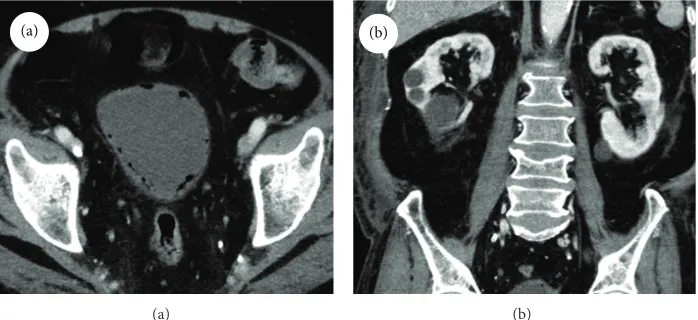
Figure 1: (a) Computed tomography of the abdomen showed multiple diffuse gas collection in the bladder wall suggestive of emphysematous cystitis. (b) Both kidneys and psoas muscle were unremarkable except multiple cortical cysts.
the following: white blood cell (WBC) count, 10,880/mm3 with 85.7% neutrophils; hematocrit, 48.2%; hemoglobin, 17.7 g/dL; platelet count, 42,000/mm3; blood urea nitro-gen, 39.1 mg/dL; serum creatinine, 1.05 mg/dL; sodium, 146 mmol/L; potassium, 3.3 mmol/L; chloride, 116 mmol/L; total protein, 4.5 g/dL; albumin, 2.0 g/dL; aspartate transam-inase, 134 IU/L; alanine transamtransam-inase, 52 IU/L; erythrocyte sedimentation rate, 11 mm/hr; C-reactive protein (CRP), 20.0 mg/dL; prothrombin time, 12.3 seconds (INR 1.19); acti-vated partial thromboplastin time, 33.2 seconds; fibrinogen, 533 mg/dL; antithrombin III 58%; FDP,>20 ug/mL; and lactic acid 3.15 mmol/L. The urinalysis revealed protein 2+, glucose 1+, nitrite positive, red blood cells 3+/HPF, and WBC 3+/HPF. The arterial blood gas analysis showed pH 7.54, PaCO2 24 mmHg, PaO2 77.5 mmHg, and oxygen saturation 96.6%. Plain chest and abdominal radiography was unremarkable. Computed tomography (CT) scan of the abdomen and pelvis revealed multiple diffuse gas collection along the bladder wall and multiple cortical cysts in both kidneys (Figure 1). Other organs were unremarkable. These results were suggestive of severe sepsis caused by emphysematous cystitis.
Vancomycin (1.0 g q12 h) and meropenem (1.0 g q8 h) were administered for empirical antibiotic therapy. Bladder catheterization was delayed due to urethral stricture. It could be done only after incision of urethral opening by the urolo-gist. Intravenous immunoglobulin was administered concur-rently as an adjuvant therapy for severe sepsis. Urine culture was negative but blood culture grew extended-spectrum𝛽 -lactamase nonproducingK. pneumoniaewhich was identified by automated Vitek2 system. This organism was sensitive to amikacin, aztreonam, cefotaxime, ceftazidime, ciprofloxacin, piperacillin/sulbactam, and imipnemem but resistant to ampicillin. Meropenem was changed to levofloxacin (500 mg q24 h). The patient’s condition deteriorated with high fever, tachypnea, hypoxemia, and shock. Endotracheal intubation was done on hospital day (HD) 10. Follow-up CT of the abdomen on HD 12 showed bilateral multiple abscess for-mation in the psoas muscle (Figure 2) which did not exist on the initial imaging study. Vancomycin and levofloxacin
were changed to teicoplanin (400 mg q12 h three times and then 400 mg q24 h) and meropenem; however, fever was sustained and CRP was kept high. We stopped meropenem and teicoplanin and started tigecycline (100 mg one time and then 50 mg q12 h) on HD 23. The patient’s condition was improved and endotracheal tube was removed on HD 26.
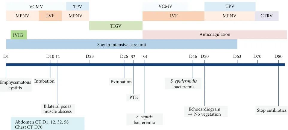
Afterwards pulmonary thromboembolism and bactere-mia caused by methicillin resistant cogaulase negative staphy-lococci developed during hospitalization which were treated successfully with anticoagulation and glycopeptides. Follow-up CT scan on HD 58 showed resolution of bilateral psoas muscle abscess and emphysematous cystitis. The patient was moved to general ward from medical intensive care unit (ICU) on HD 63. Antibiotics were stopped on HD 80 and he was discharged on HD 99. The clinical course and events were described inFigure 3.
3. Discussion
Complicated UTIs such as EC are predisposed to patients with chronic UTIs, indwelling urethral catheters, neurogenic bladders, or urinary tract outlet obstructions [1]. The diagno-sis can be made radiologically by simple plain film or CT scan, through direct visualization on cystoscopy, or pathologically on tissue from bladder biopsy or autopsy. Etiologic organisms are usually identified by urine or blood cultures. The present case was diagnosed with CT scan.K. pneumoniaegrew from blood but not from urine. Delayed bladder catheterization due to urethral stricture made it impossible to conduct urine culture before antibiotic use. This is why the urine culture was negative. Meropenem had been injected 2 times before successful urethral catheterization.
(a)
(a)
(b)
(b)
Figure 2: Follow-up abdominal computed tomography showed bilateral multiple abscess formation in the psoas muscle. (a) Frontal plane, (b) Transverse plane.
VCMV TPV
MPNV LVF MPNV
TIGV
IVIG
VCMV TPV
MPNV
LVF CTRV
Male/81, emphysematous cystitis and bilateral psoas muscle abscess
D1 D10 12 D23 D26 32 34 D46 D50 D63 D70 D80
Stay in intensive care unit
Anticoagulation
Stop antibiotics Echocardiogram
→No vegetation
S. epidermidis bacteremia
S. capitis bacteremia Extubation
PTE Intubation
Emphysematous cystitis
Bilateral psoas muscle abscess
Abdomen CT D1, 12, 32,58
Chest CT D70
Figure 3: Timeline for clinical events, radiology, and antibiotic treatment. VCMV, vancomycin; TPV, teicoplanin; MPNV, meropenem; LVF, levofloxacin; CTRV, ceftriaxone; TIGV, tigecycline; IVGV, intravenous immunoglobulin; PTE, pulmonary thromboembolism; CT, computed tomography.
prevailing causative organism followed by K. pneumoniae
(21%),Enterobacter aerogenes(7%), andClostridium perfrin-gens (6%). Most patients (90%) were treated with medical treatment, including a combination of antibiotics, bladder drainage, and glycemic control. The overall mortality rate in this review was 7%. Although 14 cases (10%) were reported to have another gas-forming urinary tract infection, none of them had PsA. Our patient was a nondiabetic man of old age but had urinary stasis secondary to bladder outlet obstruction which might be predisposed to EC. Bilateral PsA was developed as a complication.
4 Case Reports in Medicine
tumors [5, 10]. Renal abscess, perinephric abscess, and emphysematous pyelonephritis are reported predisposing UTIs associated with PsA [3,6–8]. Also, bilateral PsA was reported to be less than 3% of all PsA [11, 12]. This is the first case of PsA associated with EC. We suppose that this PsA resulted from hematogenous spread of K. pneumonia
because of intact perivesicular lining, normal ureter, and preserved kidney capsules as well as bilaterality of PsA. The abundant blood supply of the psoas muscle is likely to permit hematogenous spread [10]. The absence of no psoas pathology at admission and presence of bilateral PsA on HD 11 further support our assumption.
The clinical presentation of PsA is often variable and nonspecific. The classical clinical triad of fever, back pain, and limp is present in only 30% [4]. As the psoas muscle is innervated by L2–4, pain can radiate to hip and thigh. Other symptoms are vague abdominal pain, malaise, nausea, and weight loss. Our patient had fever and mild lumbar tenderness. Despite broad spectrum antibiotic therapy and aggressive supportive care in ICU, the patient’s condition deteriorated. We conducted abdomen-pelvis CT again to check the status of preexisting EC or other intra-abdominal comorbidities; then, we found bilateral PsA which did not exist in the initial evaluation.
The management of PsA generally involves the use of appropriate antibiotics along with percutaneous or surgical drainage [4, 10]. Some researchers emphasized PsA size in the choice of initial therapy [13]. They suggested that PsA that are smaller than 3 cm in greatest diameter may be managed successfully with antibiotics alone [13]. Eighteen primary and 23 secondary PsA cases were included in that study. Treatment was via open drainage (𝑛 = 1, 3%), CT-guided percutaneous drainage (𝑛 = 26, 63%), or antibiotics alone
(𝑛 = 14, 34%). Statistical analysis showed that the median
size of PsA in the percutaneous group was significantly larger than in the antibiotics group (6 versus 2 cm;𝑃 < 0.001). Our case had bilateral diffuse lesions and was treated successfully with medical management alone.
The reported mortality rates are 2.4% in primary PsA and 19% in secondary PsA [4]. The mortality rate in untreated patients is almost 100% [11]. Early diagnosis is the key for minimizing invasive procedure and favorable outcome.
In summary, the present case shows the importance of early diagnosis and treatment of PsA. The clinician should keep in mind that PsA can develop as a complication of bacteremia which is not uncommon in community acquired urinary tract infections.
Ethical Approval
The Institutional Review Board at National Health Insurance Corporation Ilsan Hospital approved this case report.
Consent
Written informed consent was obtained from the patient for publication of this case report and any accompanying images.
Conflict of Interests
The authors have no financial support or conflict of interests to declare.
References
[1] N. P. Patel, R. W. Lavengood, M. Fernandes, J. N. Ward, and M. P. Walzak, “Gas-forming infections in genitourinary tract,”
Urology, vol. 39, no. 4, pp. 341–345, 1992.
[2] D. Bos, P. Patal, and S. DiTullio, “Emphysematous cystitis: an atypical multi-organism presentation,”Journal of the Canadian
Urological Association, vol. 8, no. 3-4, pp. E210–E212, 2014.
[3] A. A. Thomas, B. R. Lane, A. Z. Thomas, E. M. Remer, S. C. Campbell, and D. A. Shoskes, “Emphysematous cystitis: a review of 135 cases,”BJU International, vol. 100, no. 1, pp. 17–20, 2007.
[4] I. H. Mallick, M. H. Thoufeeq, and T. P. Rajendran, “Iliopsoas abscesses,”Postgraduate Medical Journal, vol. 80, no. 946, pp. 459–462, 2004.
[5] P. Tabrizian, S. Q. Nguyen, A. Greenstein, U. Rajhbeharrysingh, and C. M. Divino, “Management and treatment of iliopsoas abscess,”Archives of Surgery, vol. 144, no. 10, pp. 946–949, 2009. [6] E. H. Chae, K. H. Kang, B. S. Lee et al., “A case of renal subcap-sular abscess complicated with psoas abscess and femoral vein thrombosis in diabetic patient,”The Journal of Korean Diabetes
Association, vol. 28, no. 3, pp. 219–224, 2004.
[7] P. H. Chuang, C. Y. Yii, K. S. Cheng, J. W. Chou, C. K. Chen, and Y. N. Lin, “Emphysematous pyelonephritis concurrent with psoas muscle abscess,”Internal Medicine, vol. 50, no. 22, pp. 2859–2860, 2011.
[8] S. Shohaib, “Nocardial psoas and perinephric abscess in a renal transplant treated by surgery and antibiotics,”Nephrology
Dialysis Transplantation, vol. 9, no. 8, pp. 1209–1210, 1994.
[9] C. S. Lim, M. Brzeski, and M. Yapanis, “A challenging case of bilateral iliopsoas abscess,”Surgical Infections, vol. 12, no. 1, pp. 69–72, 2011.
[10] B. Taiwo, “Psoas abscess: a primer for the internist,”Southern
Medical Journal, vol. 94, no. 1, pp. 2–5, 2001.
[11] M. A. Ricci, F. B. Rose, and K. K. Meyer, “Pyogenic psoas abscess: worldwide variations in etiology,” World Journal of
Surgery, vol. 10, no. 5, pp. 834–843, 1986.
[12] J. S. Bresee and M. S. Edwards, “Psoas abscess in children,”The
Pediatric Infectious Disease Journal, vol. 9, no. 3, pp. 201–206,
1990.
Submit your manuscripts at
http://www.hindawi.com
Stem Cells
International
Hindawi Publishing Corporationhttp://www.hindawi.com Volume 2014
Hindawi Publishing Corporation
http://www.hindawi.com Volume 2014 INFLAMMATION
Hindawi Publishing Corporation
http://www.hindawi.com Volume 2014
Behavioural
Neurology
Endocrinology
International Journal ofHindawi Publishing Corporation
http://www.hindawi.com Volume 2014
Hindawi Publishing Corporation
http://www.hindawi.com Volume 2014
Disease Markers
Hindawi Publishing Corporation
http://www.hindawi.com Volume 2014
BioMed
Research International
Oncology
Journal ofHindawi Publishing Corporation
http://www.hindawi.com Volume 2014
Hindawi Publishing Corporation
http://www.hindawi.com Volume 2014
Oxidative Medicine and Cellular Longevity
Hindawi Publishing Corporation
http://www.hindawi.com Volume 2014
PPAR Research
The Scientific
World Journal
Hindawi Publishing Corporation
http://www.hindawi.com Volume 2014
Immunology Research
Hindawi Publishing Corporation
http://www.hindawi.com Volume 2014
Journal of
Obesity
Journal ofHindawi Publishing Corporation
http://www.hindawi.com Volume 2014
Hindawi Publishing Corporation
http://www.hindawi.com Volume 2014 Computational and Mathematical Methods in Medicine
Ophthalmology
Journal ofHindawi Publishing Corporation
http://www.hindawi.com Volume 2014
Diabetes Research
Journal ofHindawi Publishing Corporation
http://www.hindawi.com Volume 2014
Hindawi Publishing Corporation
http://www.hindawi.com Volume 2014
Research and Treatment
AIDS
Hindawi Publishing Corporation
http://www.hindawi.com Volume 2014
Gastroenterology Research and Practice
Hindawi Publishing Corporation
http://www.hindawi.com Volume 2014
Parkinson’s
Disease
Evidence-Based Complementary and Alternative Medicine
Volume 2014 Hindawi Publishing Corporation