Proc. Natl.Acad. Sci. USA
Vol. 92, pp.8166-8170, August 1995 Immunology
Amino-terminal alteration
of the
HLA-A*0201-restricted
human
immunodeficiency
virus pol
peptide increases complex stability
and
in
vitro
immunogenicity
REBECCA R.
POGUEt, JOSEPH
ERONt,
JEFFREY A.
FRELINGERt§,
ANDMASANORI MATSUIt
Departments oftMicrobiologyandImmunology, CB# 7290, andtMedicine,University of North Carolina, Chapel Hill, NC 27599
Communicatedby RayD. Owen, California Institute of Technology, Pasadena, CA, May 10, 1995 (received forreviewJanuary23, 1995)
ABSTRACT Initial studies suggested that major histo-compatibility complex classI-restricted viral epitopes could be predicted by the presence of particular residues termed anchors. However, recent studies showed that nonanchor positions of the epitopesarealsosignificantfor class Ibinding and recognition by cytotoxic T
lymphocytes
(CTLs). We investigated if changing nonanchor amino acids couldin-crease class Iaffinity, complex stability, andT-cell
recogni-tion ofanaturalviralepitope.This concept was testedbyusing the HLA-A*0201-restricted human immunodeficiency virus type 1 epitope from reverse transcriptase (pol). Position 1
(P1) amino acid substitutionswere emphasized because P1 alterations maynotalter the T-cell receptorinteraction. The peptide with the Pl substitution of tyrosine for isoleucine
(I1Y) showed a binding
affinity
for HLA-A*0201 similar to that of thewild-typepolpeptide inacelllysateassembly
assay.Surprisingly, IlY
significantly
increased theHLA-A*0201-peptide complex stability at the cell surface. I1Y sensitized
HLA-A*0201-expressingtargetcellsforwild-typepol-specific
CTLlysisaswellaswild-type pol.Peripheralblood
lympho-cytes fromthreeHLA-A2 HIV-seropositive individualswere
stimulated in vitro with I1Y andwild-typepol. IIY stimulated
a higherwild-type pol-specific CTLresponse thanwild-type
pol in all three donors. Thus, IlY may be an "improved" epitope for use as a CTL-based human immunodeficiency
virusvaccine component. The design of improved epitopes has importantramificationsforprophylaxis and therapeutic vac-cinedevelopment.
CD8+
cytotoxic
Tlymphocytes (CTLs) play
amajor
role in the clearance of viruses(1).
The T-cellreceptor(TCR)
expressedby
theCTL
recognizesatrimolecularcomplexconsisting
ofamajorhistocompatibility complex(MHC) classIheavy chain,
032-microglobulin,
andapeptide
ofendogenous origin (2).
Thepeptideisusually8-10 aminoacidsinlengthandinteractswith theclassImolecule'sbinding pockets. Peptides restrictedto a
specific class I molecule contain predominate amino acids,
termedanchors,atparticularpositions(2). However,presence offavored residuesatthe anchorpositions doesnotnecessarily confer
high
affinity (3-5).
Peptides which bind to the same MHC molecule have a
range ofaffinities(4, 5). Among high-affinity HLA-A*0201-restricted peptides, particular amino acids were
over-representedatnonanchorpositions
(4).
Our mutationalanal-yses ofHLA-A*0201 demonstrated that mutations in any of
theMHCbinding pocketscanhaveadramaticeffectonboth
peptidebindingand
CTL
recognition (6-9).These datashowthatapeptide'snonanchorpositionscontributesignificantlyto
its free energy of binding. Improvement of a known
HLA-A*0201 epitope's affinity may be possible by changing
non-anchor amino acids, thereby enhancing the interaction
be-tweenthepeptide and the heavychain.
Theabilitytoimproveanaturalepitope throughnonanchor
substitutionswastestedby usingahumanimmunodeficiency
virus (HIV)-specific CTL response. The
HLA-A*0201-restricted HIV-1 polymerase (pol) epitope (10) was
investi-gateddue to(i) its highsequenceconservationamongHIV-1
strains(95.1%) (ref. 11 and J.A.F.,unpublished data), (ii) its relatively low affinity for HLA-A*0201 (4, 5), and (iii) the frequent observationof aCTL response tothisepitope among
HIV-1-positive individuals(12). Improvement of this epitope forimmunotherapy is beneficial, since HLA-A2 isthe most
commonhumanclass Iallele worldwide(13).
Wefocusedonposition1
(P1)
aminoacid alterationsduetoitsinteraction with both MHCand TCR. Structuralstudies of fivepeptides bound to HLA-A*0201 showed that the
orien-tation of the P1 side chainswasverysimilar(14). The majority of theP1 residuewasburied within the classIcleft.Therefore, alterations of the amino-terminal side chain may allow for
higher affinity between the peptide and the heavy chain without altering TCR interaction. Our analysis of P1-substitutedpeptides boundtoHLA-B27subtypessupportsthis
concept
(15).
Peptides with increased affinitymaybeable toenhance stimulation of natural epitope-specific CTL. Such "improved" peptidesmaybeuseful for CTL-based immuno-therapy and prophylaxis.
MATERIALS AND METHODS
Cell Lines. T2 cells are defective in antigen presentation
(16).
T2cellsweremaintained in RPMI medium 1640 (Me-diatech, Washington, DC) containing 5% calf serum(Hy-Clone),
and 2 mML-glutamine
(GIBCO).
C1R-A2 andClR-neocellsarederivativesof the HLA-A null humanB-lymphoid cell lineHmy2.ClR
(ClR),
transfected with HLA-A*0201 and pSV2-neo or pSV2-neoalone,
respectively(9).
The ClRtransfectants were maintained in the same media supple-mented with G418
(GIBCO)
at400,ug/ml.
Peptides. Peptides were synthesized by the University of
North
Carolina/Program
inMolecular Biologyand Biotech-nology Micro Protein Chemistry Facility, usingfluorenylme-thoxycarbonyl
(Fmoc) chemistry.
TheHLA-A*0201-restrictedHIV-1 pol epitope correspondsto amino acid residues
476-484 of theHIV-1reversetranscriptase
(ILKEPVHGV) (10).
The HLA-A*0201-restricted HIV-1 p17 gagepitope (amino acid residues 77-85:SLYNTVATL)
is usedas acontrol(17).
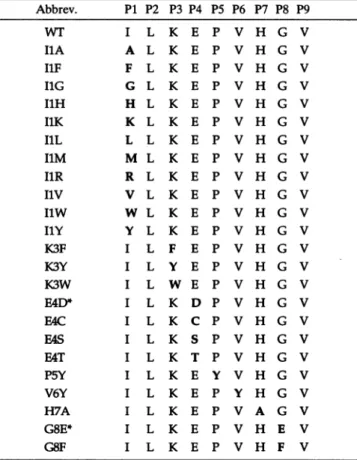
Sequences
of the substitutedpeptides are showninTable 1. Thepeptideswerepurifiedby HPLC, andpeptideconcentra-tion was determined by amino acid
analysis
at the ProteinChemistry
Laboratory
attheUniversityof NorthCarolinaat Abbreviations: MHC, major histocompatibility complex; CTL, cyto-toxic T lymphocyte; HIV, human immunodeficiency virus; TCR, T-cell receptor; P1,position 1; PBL, peripheral blood lymphocyte; WT,wild type;W, vacciniavirus; EBV, Epstein-Barrvirus.§To
whomreprintrequestsshould be addressed.Table 1. pol substitution peptide sequences
Abbrev. P1 P2 P3 P4 P5 P6 P7 P8 P9
WT I L K E P V H G V
I1A A L K E P V H G V
IlF F L K E P V H G V
I1G G L K E P V H G V
I1H H L K E P V H G V
IlK K L K E P V H G V
IlL L L K E P V H G V
i1M M L K E P V H G V
I1R R L K E P V H G V
Iiv V L K E P V H G V
I1W W L K E P V H G V
IlY Y L K E P V H G V
K3F I L F E P V H G V
K3Y I L Y E P V H G V
K3W I L W E P V H G V
E4D* I L K D P V H G V
E4C I L K C P V H G V
E4S I L K S P V H G V
E4T I L K T P V H G V
P5Y I L K E Y V H G V
V6Y I L K E P Y H G V
H7A I L K E P V A G V
G8E* I L K E P V H E V
G8F I L K E P V H F V
Abbreviations for the substitutionpeptidesusethesingle-lettercode and conformtothefollowing order: the wild-type (WT) amino acid, thepositionatwhich the substitution occurs, and finally the substituted amino acid.Inthe sequences, boldface type indicates the substitutions. Previousstudies have shownpositions2and9tobeanchor residues (1).
*A naturalvariant of thepol epitope.
Chapel
Hill/National
Institute of Environmental HealthSci-ences.
Generation ofHIV-Specific CTL Lines. HIV pol-specific CTL linesweregenerated from peripheral blood lymphocytes
(PBLs)
ofHIV-seropositive HLA-A2-positive
individuals(do-nors25and32) asdescribedpreviously(18, 19).
Cell Surface StabilizationAssay. The stabilizationassay was
performedasdescribed(20). Briefly,T2cellsweresuspended in AIMVserum-free medium
(GIBCO)
supplemented with100 nM human 82-microglobulin and peptide (Sigma). The peptide-pulsed cells were incubated overnight at 37°C. The
cells werestained for conformationallycorrectHLA-A*0201
with the monoclonal antibody BB7.2 (21), followed by fluo-rescein isothiocyanate (FITC)-labeled goat anti-mouse IgG antibody
(Southern
Biotechnology Associates). Cells wereanalyzed by
flow cytometry(FACScan,
BectonDickinson).
Themeanchannel for eachpeptide peakwasdetermined on alinear scaleby usingCYCLOPSsoftware(Cytomation).Assembly Assay.Peptide bindingwasanalyzed with theT2
assembly
assayessentially
as described(15)
except the heat denaturation of the lysate prior to peptide addition waseliminated. The HLA-A*0201 complexes were immunopre-cipitated with BB7.2(21). Immunoprecipitateswereanalyzed by electrophoresis on a reducing
SDS/12%
polyacrylamide gel. The gels were exposedovernight
to a PhosphorImagerscreen
(Molecular Dynamics).
The screen was analyzed andquantitated by
IMAGEQUANT
3.3(Molecular Dynamics).
Thevolumes of the bands were quantitated, subtracting local
background. The resulting pixel
valueis
directly proportional
todpm andwasplotted asdescribed(15).
Complex Stability Assays. T2cellswereincubated with 100
,uMpeptide and human/32-microglobulinasdescribed for the
cell surfacestabilizationassay. After theovernight incubation, the cells were incubated in RPMI medium 1640 containing 10% fetal calf serum and brefeldin A (Sigma) at 10,ug/ml to block the egress of new class I molecules. After a 1-hr incubation at 37°C in C02, theblock of Golgi to cell surface egress wasmaintained in mediumcontaining brefeldin A at 0.5
,ug/ml.At the indicated time points, an aliquot was stained
withBB7.2 (21) asdescribed above. All antibodies andwash
solutions contained brefeldinA at0.5 ,ug/ml.
51CrReleaseAssay.Recognition of the substituted peptides
by pol-specific CTLswas analyzed bya51Cr release assay as
described (6).C1R-A2 or ClR-neo cells were used as targets. The cells were incubated with peptide for 1 hr at 37°C.
pol-specific CTLswereadded to each wellat an effector-to-targetratio of 4:1 unless otherwise noted. The graphs shown for donor 25 were from asinglesetofexperiments; however, the datafordonor 32 are representative of at least two assays. For assays which measure CTL activity on vaccinia virus
(W)-infected targets, targetcells were infected at a multi-plicity of infection of 5 with W-pol, W-gag p17, or the negative control, W-NP,which expresses the nucleoprotein from influenza virus. After1.5 hrof infectionat 37°C, the cells were washed twice, resuspended in medium, and incubated overnight for expression of the VV construct genes. The
infected targets were labeled with51Cras described above. In Vitro Immunization. Frozen PBLs from HIV-seroposi-tive/HLA-A2-positive individuals (donors4, 9, and32)were thawed and stimulatedweeklywithautologous Epstein-Barr virus (EBV)-transformed B cells. These B cells were
previ-ouslyincubated with 10 ,uMpeptide and irradiated[3000rads
(30 Gy)] as described for thegeneration of CTL lines. Four
peptidesweretestedforproduction of pol-specificCTLs(WT
pol, HF,
I1Y, andp17). Starting
atday
14, the PBLs wereanalyzed for their CTL reactivityin a standard51Cr release assay. Peptide-pulsed targets were autologousBcells pulsed
withwild-type pol orp17peptides. For testing recognition of
endogenously processed antigen, cells ofanHLA-A2+ B-cell
line AR
(HLA-A1,2;
B8, 44; Cw5) were infected with VV constructs asdescribed above.SequencingofHLA-A2Subtypes.RNA wasisolatedfrom 4 x 10⁶cells, either EBV-transformed B cells or PBLs, using the
MicroFastTrack RNAisolationkit(Invitrogen). TheHLA-A2 RNA wasthenamplified by using the GeneAmpRNAPCRkit
(Perkin-Elmer).
Thefollowing antisense primer fromexon5 wasused (5'-GCTCCAAAGAGAACCAGGCCAGCAATG-3'), coupled with the sense primer from exon 1(5'-GCCGAGGATGGCCGTCATGGCGCCCCGAACC-3').
Theamplification followed thesupplier's instructions,except
thattheMgCl2 concentrationinthe PCRreaction mixturewas
increasedto5 mM. The894-bp productwaspurified byWizard
PCR Preps(Promega).DNA wassequencedattheUniversity
ofNorth CarolinaatChapelHillAutomatedDNASequencing Facilityon a model 373A DNA Sequencer (Applied
Biosys-tems).
RESULTS
Affinity
Analysis
ofpol Position 1 Substitutions.ElevenP1amino acid substitutions of WT pol were analyzed for cell surface stabilization of HLA-A*0201 (Table 1). This assay measuresthe stabilization ofemptycell surfaceheavy chains
orthe exchange of
peptide
onto heavy chains(20).
The Phe(I1F) and Tyr
(I1Y)
substitution peptidesweretheonlyonesProc. Natl.Acad. Sci. USA 92 (1995)
120
100
E
80
E
60
E40
20o
0.1 1 10 100
Peptide,
l&M
B
0.01 0.1 1
Peptide, ,uM
C
80-E~~~~
E 60 /
40
, 20- /
I~~~~~~~~~~~~~~~~~~~~~~~~~~~~~~~~~~~~0
10 0.01 0.1 1 10 100
Peptide,
ALM
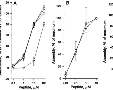
FIG. 1. Binding of P1 substitu-tion pol peptides. Allbinding
ex-periments used T2 cells (HLA-A2, -B51) (16). (A) Cell surface stabilization assay. Peptides are
representedasfollows: 0,WT; El,
HF;A,I1Y; andv,11W. Errorbars indicate SEM for three
experi-ments. (B and C)T2assembly as-says. WTpolis indicated as0 in
both graphs. o denotes IlF in B and IlY in C. Errorbars indicate SEM for three (B) or five (C)
assays.
and I1Y substitutionswere analyzed further. The kinetics of
peptide bindingtonewlysynthesized HLA-A*0201was
inves-tigated inaninvitroassemblyassay(15). In the assemblyassay,
thepeptideconcentrationofhalf-maximal assemblyrepresents
therelativepeptide affinity (15, 23). Both I1Y andIlF showed
anaffinity similar tothatofWTpol (Fig. 1 B and C). The half-lives of thepeptide/classIcomplexesat37°Cwere
determined bycomplex stability assays. The stability of cell
surface HLA-A2wassignificantly increased by both the I1Y
and I1F substitutions (Fig. 2). The half-life of the WT
pol/
heavy chaincomplex isapproximately10hr, whereas both the
I1Fand I1Y substitutions increase the complex's half-life to
approximately 30 hr. Treating the cells with brefeldin A showedthat the number ofconformationallycorrectHLA-A2
cannotbe duetoreloading of thepeptideontonewlyegressing molecules. Since theantibody isconformationally sensitiveto
thebinding cleft(21), only class I molecules binding peptide
weredetected.
100
.)a
cn
C)
.E
o
0.
E
0
0
10
Effect of P1SubstitutionsonWTpol-Specific CTL
Recog-nition. Ourmutational analysis of HLA-A*0201 showed dif-ferencesbetween binding affinity and functional recognition
0.
Cu (a
Cu
0.
Cu CO
4
co 're
Q-R
Peptide,nM
C)
&P
oo
° oi - ° Peptide,nM
3
2
0
. Ii
Peptide, nM
.r1Cu) UL)
C)Q1
Ien
- 10 0
E/T
C
WT10
ol N
0
0
-Peptide, nM ,45
F
40
35
30
25
20
15
10
0
X0 N U) I)
O Cq _
5 10 15 20
Timeat370C,hr
25 30
FIG. 2. Complex stability is increased with IlY and I1F.Acell
surfacecomplex stabilityassaywasconducted.Afterthepeptide (0,
WTpol;El,I1Y;v,I1F;a,none)had been loadedovernight,thecells
weretreated with brefeldinAtoblock theexitofnewclassImolecules. After 1hr,thecellsweremaintained in mediumcontainingalower, lesstoxic, concentration of brefeldin A which maintained the egress
block.Attheindicated timepoints, cellswerestainedfor conforma-tionally correct HLA-A2 by indirect immunofluorescence using BB7.2. Themeanchannelfluorescencewasdeterminedforeachpeak. Theerrorbars indicated SEM for threeexperiments.Ifonlyasymbol
isapparent, theerrorissmaller than thesymbol.
FIG. 3. CTLrecognitionofIlFandI1Y.Recognitionof WTpol, I1F, and I1Yby pol-specific CTLwas analyzed byastandard 51Cr releaseassay.Eachpeptideconcentrationwasanalyzedinduplicate. (AandB) Recognition bydonor 25CTLline.(CandD)Recognition by donor 32 CTL line. The negative control, C1R-neo (Neo) was
incubated with WTpol. Percentspecific lysisof C1R-neoincubated withsubstitutedpeptideswassimilartothat of Neo(datanotshown). In all panels WT pol recognition is denoted by *, and Neo is
represented as *. o indicates IlF (A andC) and I1Y (B andD). Effector-to-targetratios(E/T)were asfollows:5(A-C)and 3(D).The
CTL lines from donor 25(E)and32(F)wereassayedonVV-infected C1R-A2orClR-neotargets.Alltargetswereinfectedatamultiplicity ofinfectionof 5.For E and F thenegativecontrol ofC1R-neotargets infected withVV-polisnotedas 0.C1R-A2 cells infected withno
virus(0),W-pol (*),andVV-gagp17 (El)arenoted. 120
cn 0
0) 'a 100
E
0 0
80
E
E3
60E
40
0
CF 20
0
N 0 O
C2
-20
(6-9). CTL recognition of thesubstituted peptides was there-fore analyzed with two independent WT pol-specific CTL lines. I1F was recognized like WT pol by both CTL lines (Fig. 3 A andC). Donor 32 CTLs recognized I1Y (Fig. 3D), while the CTL line from donor 25showed reducedI1Y recognition (Fig. 3B). These CTL lines are capable of recognizing endog-enous antigen (Fig. 3 E and F). The difference in I1Y recognition was not due to HLA-A2 subtypedifferences, since both donors have theHLA-A*0201 subtypeasdeterminedby DNAsequencing. Thedistinction inI1Y recognition suggests adifference in T-cell repertoires.
In Vitro Immunogenicity ofI1Y and I1F. The crucial
ques-tion for generating "improved" epitopes iswhetherthe sub-stitutionpeptide can stimulateTcells which recognize the WT epitope. To address this question, PBLs were stimulated in
vitro todetermine if the substitutedpeptide could elicit a WT pol-specific CTL response. Since CTLgeneration fromanaive
individual'sPBLsis difficult, HLA-A2+ PBLs were used from three HIV-1-seropositive donors: 4, 9, and 32. HIV-specific CTLscanoften be detected directlyfrom seropositive PBLs
(12, 24);however, these donors exhibited minimal pol-specific
CTLreactivity aftera singlein vitro stimulation. pol-specific
CTLreactivity of WT pol peptide-pulsed, autologous EBV-transformed B cells was less than 9% for all three donors.
Significant pol-specific CTLactivity from these donorswas not
detectable after 2 weeks of in vitro stimulation with WTpol, suggesting low initial numbers of CTL precursors. Other donors, like donor 25, exhibited high CTL activity (25%
specificlysis)after the initialweek in culture. Thishighinitial CTLactivitytotheWTepitope providesahigh backgroundfor in vitro stimulation; thus, a believable increase over this high responsewould be difficulttodetect. The low responders were
specifically chosen to provide a large dynamic range for
detecting improved peptide stimulation.PBLs werestimulated
witheach of thefollowing peptidesonautologousBcells:WT pol, I1F, I1Y, and p17.
Starting atday 14,the stimulated PBLs from donor 32were
analyzed for reactivity againstautologous Bcells pulsed with either WTpolorp17(Fig. 4A).Thesubstituted peptideswere
therefore tested for theirabilitytostimulateaWTpol-specific
CTL response. Whilesignificantpol-specific reactivitywas not seen in the WT pol-stimulated PBLs, IlY-stimulated PBLs showedsignificantly higherWTpol-specific CTLreactivityat
day14(Fig. 4A). Significant pol-specificCTLreactivitywas not seen in IF-stimulated PBLs (Fig. 4A). As a control,
p17-50T A
U)
.Fa
0
.6
(0)
O
60T B
U)
0
0.
U)
U)
.)
U)
Q
stimulated PBLs from donor 32 showed p17-specific CTL reactivity and nopol-specific reactivity(Fig. 4A). At 4 weeks, asimilar trend was apparent (datanotshown).
Stimulation of PBLs from donors 4 and 9 with the I1Y peptide also resulted inhigher pol-specific CTLreactivity at
day 16 compared with PBLs stimulated with WT pol. For donor4, thepol-specific CTL reactivity of WT pol-stimulated PBLs and IlY-stimulated PBLs was 35% and 47%, respec-tively, at an effector-to-target ratio of 30 (Fig. 4B). The stimulated PBLs werealso able to recognize the pol peptide when it was produced endogenously by a VV construct ex-pressing pol (Fig. 4C). In donor 9, WT pol-stimulatedPBLs
and IlY-stimulated PBLs showed pol-specific reactivity of 12% and 23%,respectively, ataneffector-to-target ratio of 30 (data notshown).Unlike donor 32, donor 4 was stimulated for aWTpol-specific responseby I1F.The difference in immu-nogenicity of I1F between the two donors may be due to
HLA-A2 subtype or TCR repertoire differences. Thus, I1Y stimulated a WT pol-specific response better than WT pol from three donors' PBLs. These data suggest that I1Y is more immunogenic than the WTpol peptide in vitro. Donor
differ-encesintheamountofCTLstimulationbyI1F and I1Yclearly demonstratethe role of T-cellrepertoireontheavailabilityof
reactive Tcells.
Changes at Other Nonanchor Positions Lower Binding and/orCTLRecognition. We tested 10moresubstitutions at
positions 3 through 8 which were based on amino acids frequently found at these positions in high-affinity HLA-A*0201peptides(Table 1)(4). The two natural variants of the pol epitope, E4D and G8E,were also analyzed (11). All the
position 3 substitutions resulted in higher affinity peptides
which were not recognized by pol-specific CTLs (data not
shown). Almostallof theposition 4-8substitutionsdisplayed
asimilarphenotype:decreasedbindinginboth assays and little
to norecognitionby pol-specific CTLs
(data
notshown).
P5Y bound HLA-A*0201 like WT pol, andE4Swasrecognized byoneCTLlinedespitealowerbinding affinity.Interestingly,the natural variants bound HLA-A*0201 withanaffinitysimilarto
that of WT pol; however, the recognition was reduced or
abolished (data notshown).
DISCUSSION
Anepitope's affinityforanMHC molecule is believedtobe
animportant determinant foranimmuneresponse. Inaddition
C
pol 11Y 11F p17
FIG.4. Recognition ofWTpol and p17 by in vitro stimulated PBLs. PBLfrom donor32(A) or
donor4(B) werestimulatedwith autologous EBV-transformed B cells which were previously incu-bated with WT pol, I1F,
IlY,
or p17 (only A). Atday 14, the cellsweretested for theirrecognitionof autologousBcells incubated with WT pol (filled bars), p17 (open
bars),
or nopeptide
(hatched bars),
inastandard
51Cr
release assay(6).
Stimulated PBLs from donor 4
were also assayedfor recognition of VV-infected targets (C). AR B-celltargetswereinfected as
fol-lows:novirus
(hatched bars),
VV expressing p17 gag (open bars), andVVexpressing
pol(filled
bars).
Thex-axis showsthe
peptide
used in stimulation. The effector-to-- q targetratiowas50 (A),30 (B), orpol llY 12(C).
Proc.Natl. Acad. Sci. USA 92 (1995)
to peptide binding, CTL recognition of foreign antigen is dependent on aproductive interaction between the TCR and
MHC/peptidecomplexes. Given a productive(nonantagonist)
T-cellinteraction, twomodels mightdescribe the relationship between peptide affinity and T-cell response. One model suggeststhatapeptide'simmunogenicity is proportional to its affinity for MHC.Alternatively, immune responses occur once athresholdpeptide affinity or complex avidity is reached. In
this secondmodel,increasing thepeptide affinitybeyond this threshold would not enhance the specific immune response. Understanding which model is more accurate will lead to
better developments in peptide therapeutics. The WT pol
epitope must possess a binding affinity above the T-cell activation threshold, since many HLA-A2 HIV-positive indi-viduals mountaresponse tothis epitope(12).Byimproving the pol epitope's affinity, the relationship betweencomplex
sta-bility andimmunogenicity can be examined.
Given theimportanceofnonanchor amino acids inpeptide affinitytoMHC(3-5),improving the binding and recognition of the HIV-1 pol peptide was investigated by altering the nonanchor side chains. Since themajority of the P1 side chain isburied within theclass Icleft(14),P1is an excellentposition
to investigate increasing affinity without altering TCR inter-action.Of the 11 P1 substitutions investigated, only 2 aromatic substitutions, I1Y and I1F,bound cell surface HLA-A*0201 as wellas did WTpol (Fig. 1A).Inthe assembly assay, I1F and I1Y showed similar affinities to WT pol(Fig. 1 Band C).
Both I1Y and I1F displayed an approximately 3-fold in-crease inpeptide-MHC complex half-livesat 37°C at the cell surface. Thisprolongation of thehalf-life shouldincreasethe peptide's cell surface concentration, thereby increasing the
avidityof thecell-cellinteraction. Anepitope presentathigh
cell surface concentration may be able to better stimulate
peptide-specificTcells. Recentstudies inMHCclassImodels
have shownacorrelationbetween
peptide/MHC
affinityandthe abilityto detectaresponse (22).
Since theP1sidechain's orientation issimilaramongseveral HLA-A*0201-restricted peptides (14), substitutions at P1 shouldnotaffect the overallpeptide orientation. This idea is supportedbythe CTLrecognition of IlF andIlY(Fig.3).CTL lines from twodifferent donors were able torecognize these peptides. For the altered peptidestobe effectiveas immuno-gens,theymustberecognized bythe hostasthe WTepitope.
PBLs from HLA-A2, HIV-positive individuals were
stimu-latedin vitrowithWTpol, I1Y, and I1F. TheresultingPBLs wereanalyzed forrecognition of the WT pol peptide.I1Ywas
able to stimulate WT pol-specific CTLs better than WTpol
fromthe PBLs of three seropositive donors(Fig. 4).These data suggestthatI1Y ismoreimmunogenicthan WTpol,
presum-ablyduetothe dramatic increaseinthe cell surfacecomplex stability.However, differencesbetweendonors in theamount
of CTL stimulation by I1F and I1Y clearly demonstrate the important role of T-cell repertoire on the availability of reactive T cells. Inaddition,theinvitrostimulatedlymphocytes
wereable torecognizethepol peptidewhen itwasproduced endogenouslybyVVconstructs (Fig.4C). Thus, our stimula-tion condistimula-tions did not preferentially stimulate T cells with
low-affinityreceptorswhichrecognize peptide-pulse,butnot
endogenouslyprocessed, antigen.
Thein vitroimmunization analysiswasconductedonPBLs from seropositive donors. The question of previous T-cell priming is evident. Unlike many donors (12, 24), the three donorsinvestigated allexhibited very lowpol-specific reactiv-itieseven after 1 week ofstimulation with WT pol peptide.
Thissuggested low initial numbers of CTL precursors in these donors. It is unlikely that three independent donors were
previously primedin vivowithI1ForI1Y,since the
HLA-A2-restricted polepitope isextremelyconserved and neither theI1Y
northe I1F substitution has been reported in HIV-1 strains(11). Peptide immunizationusing I1Ymaybe abletoboostormaintain
pol-specific CTL activity in HIV-1-infected individuals. From theseexperiments, it is unclear if IlY would stimulate CrLs ina
naive individual that could cross-react with WT pol. In the HLA-A*0201/H-2 Kb transgenic mouse model, most high-affmity peptides tested were also highly immunogenic (22).
We have identified apeptide, I1Y, which has (i) increased complex stability for HLA-A*0201, (ii) maintains recognition by WTpol-specific CTL lines,and (iii) is more stimulatory for WT pol-specific CTLs in vitro. Thus, I1Y shows potential as a vaccine component. Optimizing peptidesthrough amino acid substitution andimmunogenicityanalysis hassignificant ther-apeutic ramifications for infectious disease, tumor immunol-ogy, and autoimmunity.
We thank Dr. P. Cresswell fortheClR and T2cell lines; Dr. A. McMichael forthenatural variantpeptides andVVconstructs; Drs. G.Matsushima, R.Johnston, and A. Reed for criticalreading of the manuscript; and Dr. C. van der Horst for blood samples. We give special thanks toAmy Peace-Brewerfor help with the cell surface complex stability assay and the University of North CarolinaatChapel HillAutomated DNASequencingFacility. This workwassupported by grants from the National Institutes of Health (AI-20288 and AI-29324), theAIDS Clinical Trails Unit (AI-25868), and the General Clinic Research Center(RR-00046). R.R.P. is supportedby National InstitutesofHealthTrainingGrant AI-07273.
1. Doherty, P. C., Allan, W.,Eichelberger,M.&Carding, S.R.(1992)
Annu. Rev.Immunol. 10, 123-151.
2. Rammensee, H.-G., Falk, K. & Rotzschke, 0. (1993)Annu. Rev.
Immunol. 11,213-244.
3. Parker, K. C.,Bednarek,M. A.,Hull,L.K.,Utz, U.,Cunningham,B., Zweerink, H. J.,Biddison,W. E. &Coligan,J. E.(1992) J. Immunol.
149, 3580-3587.
4. Ruppert, J.,Sidney,J.,Celis,E.,Kubo,R.T.,Grey,H. M. &Sette,A.
(1993) Cell 74,929-937.
5. Parker, K. C.,Bednarek, M. A.&Coligan, J.E.(1994)J.Immunol.
152, 163-175.
6. Matsui,M.,Hioe, C. E. &Frelinger, J. A. (1993) Proc. Natl. Acad. Sci. USA90,674-678.
7. Matsui, M.,Moots, R.J.,McMichael,A. J.&Frelinger,J. A.(1994)
Hum.Immunol. 41,160-166.
8. Tussey,L.G.,Matsui, M.,Rowland-Jones, S.,Warburton, R., Frelinger,
J. A. &McMichael, A. J.(1994)J. Immunol. 152, 1213-1221.
9. Moots,R.J.,Matsui,M.,Pazmany, L.,McMichael,A. J.&Frelinger,
J. A.(1991)Immunogenetics 34, 141-148.
10. Tsomides,T.J., Walker, B. D. &Eisen,H. N.(1991)Proc.Natl. Acad.
Sci. USA88,11276-11280.
11. Myers, G., Korber, B., Wain-Jobson, S., Smith, R. F. & Pavlakis, G. N.,eds.(1993)Human RetrovirusesandAIDS1993I-IV(Theor. Biol. Biophys., LosAlamos Natl. Lab., Los Alamos,NM).
12. Lamhamedi-Cherradi,S.,Culmann-Penciolelli,B.,Buy, B.,Kieny,M.,
Dreyfus, F.,Saimot, A., Sereni, D.,Sicard, D., Levy,J.-P.&Gomard,
E.(1992)AIDS 6, 1249-1258.
13. Ferandez-Vina,M.A., Falco, M.,Sun,Y.&Stastny,P.(1992)Hum.
Immunol. 33,163-173.
14. Madden, D.R., Garboczi, D. N. & Wiley, D.C. (1993) Cell 75,
693-708.
15. Colbert,R. A.,Rowland-Jones, S. L.,McMichael,A. J.&Frelinger,
J. A.(1994)Immunity 1, 121-130.
16. Salter, R. D.,Howell,D. N.&Cresswell,P.(1985) Immunogenetics21, 235-246.
17. Nixon,D.F. &McMichael,A. J.(1991) AIDS5,1049-1059. 18. Nixon, D. F., Townsend, A.R.M.,Elvin, J. G., Rizza, C. R.,Gallwey,
J.&McMichael,A.J.(1988)Nature (London) 336, 484-487.
19. Warburton,R.J.,Matsui, M.,Rowland-Jones, S. L.,Gammon,M.C., Katzenstein, G. E., Wei, T., Edidin, M., Zweerink,H.J.,McMichael, A. J. &Frelinger, J. A.(1994)Hum.Immunol. 39, 261-271. 20. Stuber, G., Leder, G. H., Storkus,W.J., Lotze,M.T., Modrow, S.,
Szekely, L.,Wolf, H., Klein, E.,Karre,K.&Klein, G.(1994)Eur.J. Immunol. 24, 765-768.
21. Parham,P.&Brodsky,F. M.(1981)Hum. Immunol. 3, 277-299.
22. Sette, A.,Vitiello, A., Reherman, B.,Fowler, P.,Nayersina, R., Kast,
W.M.,Melief, C.J.M.,Oseroff,C., Yuan, L.,Ruppert, J.,Sidney,J.,
delGuercio, M.-F.,Southwood, S., Kubo,R.T.,Chesnut,R.W.,Grey,
H. M. &Chisari,F. V.(1994)J.Immunol. 153, 5586-5592. 23. Cerundolo,V.,Elliot, T., Elivin, J.,Bastin, J.,Rammensee,H.-G. &
Townsend,A.(1991)Eur.J. Immunol. 21, 2069-2075.
24. Cease, K. B. & Berzofsky, J.A. (1994) Annu. Rev. Immunol. 12, 923-989.