SELECTIVE RECOGNITION IN POTENTIOMETRIC SENSING
BASED ON TWO COMPETITIVE RECOGNITION SITES FOR
STATIC AND HYDRODYNAMIC DETERMINATION OF
CAMYLOFIN AS A SMOOTH MUSCLE RELAXANT
Ayman H. Kamel
*, Wagiha H. Mahmoud, Marwa S. Mostafa
Keywords: camylofin; smooth muscle relaxant; potentiometric sensor; flow injection analysis.
A poly (vinyl chloride) matrix membrane sensors for selective determination of camylofin (CY) in pharmaceutical formulations were developed based on the use of dibenzo-18crown-6 (DB18C6) as a neutral carrier and ion-association complex ([CY]2[PM])of (CY) cation with phosphomolybdate anion (PM). Subsequently, these electroactive materials were dispersed in dioctyl sebacate (DOS) as solvent mediator designed and can be easily used in flow injection system. Under static mode of operation, the sensors revealed a near Nernstain response over a wide CY+concentration range 8.5x 10⁻⁶and 5 x 10⁻⁶to 1.0 x 10⁻²mol L-1with a detection limit of 6.0x10⁻⁶and 2.5x10⁻⁶mol
L-1, respectively. In flow injection potentiometry, excellent reproducibility (RSD %±0.7%), fast response, high sensitivity with a
near-Nernstian 53.9±1.1 and 40.2±0.8 mV decade-1, linear range 1.0x10⁻⁴-1.0x10⁻²mol L-1, detection limit 16.4±0.3 and 5.9±0.3 μg mL-1, high
sampling rate (20-22 and 40-45 sample h-1) and stable baseline was observed in the presence of 0.05 mol L-1citrate buffer, pH 4.5 as a
carrier for ([CY]2[PM] and (DB18C6) membrane based sensors, respectively. The utility of the sensors was tested for field monitoring of CY+ in different pharmaceutical formulations collected from the local market.
*Corresponding author. Tel.: +201000743328 E mail address: [email protected]
[a] Chemistry Department, Faculty of Science, Ain Shams University, Abbasia, Cairo, Egypt
Introduction
Camylofin (CY); N-[2-(diethylamino)ethyl]-2-phenyl-glycine isopentyl ester is a smooth muscle relaxant with both anticholinergic action as well as direct smooth muscle action. Anticholinergic action is produced by inhibiting the binding of acetylcholine to muscarinic receptors, but the action is less pronounced. Direct smooth muscle relaxation is achieved by inhibiting phosphodiesterase type IV, which leads to increased cyclic AMP and eventually reduced cytosolic calcium. Thus camylofin has a comprehensive action to relieve smooth muscle spasm.
Only a few methods for the determination of camylofin (CY) in pharmaceutical analysis were reported, including high performance liquid chromatography (HPLC),1-3 gas chromatography (GC)4 and atomic-absorption spectrometry (AAS).5 Most of these methods, however, require expensive instrumentation, suffer from lack of selectivity, involve careful control of the reaction conditions or derivatization reactions, and require time-consuming pretreatment steps which affect their usefulness for routine analysis. On the other hand, application of potentiometric sensors in the field of pharmaceutical and biomedical analysis has been advocated.6,7 The approach provides simple, fast, and selective technique for determination of various drugs.8-13 However, as far as the available literature is concerned, no potentiometric methods have yet been reported for the determination of camylofin.
The present work describes preparation, characterization and application of two potentiometric tubular membrane sensors for continuous determination of camylofin in
pharmaceutical preparations. These sensors based on the incorporation of the camylofin phosphomolybdate (CY-PMA) ion pair complex and dibenzo-18-crown-6 in plasticized PVC matrix membranes (Fig.1). Performance characteristics of both sensors reveal low detection limit, high sensitivity, fast response, long life span and application for accurate determination of camylofin in pharmaceutical preparations under static and hydrodynamic (FIA) modes of operation.
Figure 1. Structure of the CY-PMA ion association and DB18C6 ionophore.
Experimental
Equipments
All potentiometric measurements were carried out at 25±1 ºC using an EDT instruments DR 359 TX ion concentration pH meter. The sensors were used in conjunction with a double-junction Ag/AgCl reference electrode (Model 90-02) containing potassium nitrate (10% w/w) in the outer
H2N
NH
O O
2
HPMo12O40
Camylofin-phosphomolybdate ion association
O O
O
O O
O
compartment. A combination Ross pH electrode (Orion 81-02) was used for all pH measurements. Flow injection analysis (FIA) manifold consisted of a two-channel Ismatech Ms-REGLO model peristaltic pump, polyethylene tubing (0.71 mmi.d.) and an Omnifit injection valve (Omnifit, Cambridge, UK) with a sample loop of 100 μL volume. The potential signals were recorded using an Orion Model 720 SA (Cambridge, MA, USA) pH/mV meter connected to a PC through the interface ADC 16 (Pico Tech, UK) and Pico Log for windows (version 5.07) software.
Materials
All chemicals used were of analytical reagent (AR) grade and were used without further purification unless otherwise stated. Twice distilled water was used throughout all experiments. Pure grade camylofin was supplied by Drug Control Authority, Al-Haram, Giza, Egypt. Dioctyl phthalate (DOP), dioctyl sebacate (DOS), potassium p-chloro tetraphenyl borate (pCl-TPB) and poly (vinyl chloride) (PVC) were obtained from Sigma (St. Louis, MO). Dibenzo-18-crown-6 was purchased from Aldrich Chemical Campany Inc. A 0.01 mol L-1stock camylofin solution was prepared by dissolving 0.393g in 100 mL citrate buffer (0.05 mol L-1) of pH 4.5.
Potentiometeric determination of camylofin
Camylofin-phosphomolybdate ion associate complex
Camylofin phosphomolybdate ion associate complex (CY-PMA) was prepared by mixing 50 mL of 10-2 mol L-1 aqueous camylofin solution with 25 mL of 10-2 mol L-1 phosphomolybdic acid (PMA). The mixture was shaken well; the precipitate was filtered off through G4 sintered glass crucible, washed with de-ionized bi-distilled water, dried at room temperature and grounded to a fine powder. Elemental analysis of the complex gave C 38.4, H 5.4, N 4.7% for the camylofin- PMA ion-pair complex [(C19H32N2O2]2[HPMo12O40], C 38.65, H 5.51, N 4.75%).
Camylofin-PVC membrane sensors
The plastic membranes were prepared by mixing 10 mg portion of ion pair (CY-PMA) with 190 mg of PVC, 350 mg of DOP and 7 mL THF in a glass Petri dish (5 cm diameter) covered with a filter paper and left to stand overnight to allow evaporation of the solvent at room temperature. Semi-transparent master PVC membranes of approximately 0.1 mm thick were obtained. Similarly, an another membrane sensor is made by mixing 10 mg dibenzo-18-crown-6 ionophore, 190 mg PVC, 5 mg p,Cl-TPB as anionic additive, 350 mg of DOP plasticizer and 7 mL THF. A disc (8 mm diameter) was cut using a cork borer and glued to polyethylene tube (3 cm x 8 mm i.d.) which was clipped onto the end of the electrode glass body. The electrode was filled with the internal reference solution consisted of an equal volume of 1×10⁻²mol L-1of camylofin and potassium chloride solutions. An Ag/AgCl internal reference wire electrode (1 mm diameter) was immersed in the internal solutions. Camylofin sensor was finally preconditioned by soaking in 1.0×10⁻²mol L-1 camylofin for one day before use and was stored in the same solution when not in use.
The emf of the sensors were measured in buffer solutions obtained by transferring 0.5 mL aliquots of 1.0×10⁻² – 1.0x10⁻⁶ mol L-1CY+ aqueous solutions to 50 mL beaker containing 10.0 mL of 0.05 mol L-1citrate buffer of pH 4.5. Potential readings were recorded after stabilization to ± 0.2 mV and emf was plotted as a function of logarithm CY+ concentration. Calibration graphs were used for subsequent determination of unknown CY+ concentrations. General working characteristics of the CY selective electrodes were evaluated after calibration procedures carried out.
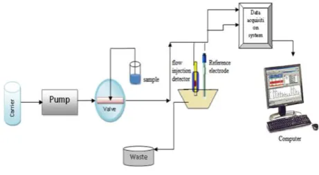
For continuous measurements (FIA), the flow cell used for detection of camylofin was designed with a constant geometry and minimum ‘dead’ space to accommodate small sensor size to avoid large dispersion of the sample in the cell and to give high response with short recovery time. With short tubing (10 cm) between the injector and detector, and using an injection volume of 100 μL, the dispersion in the system was kept to minimum (Fig. 2). A flow stream of 0.05 mol L-1citrate buffer of pH 4.5 carrier solution were allowed to pass through the flow-cell at a flow rate 3.0 mL min-1. Successive 100 μL aliquots of the standard camylofin and unknown test sample solutions were injected into the flowing stream. The corresponding potential change was measured and recorded versus time. A typical calibration plot was made and used to determine the concentration of camylofin in the unknown samples.
Figure 2. FIA manifold for the evaluation of camylofin. A 0.05 mol L-1 carrier citrate buffer solution pH 4.5; loop sample 100 μL;
and flow rate 3 mL min-1
Camylofin assessment
Potentiometric analysis was conducted on oral dosage forms of pharmaceutical preparations, commercially designated as Spasmopyralgin-M tablets (Kahira Pharm., Cairo, Egypt). Five tablets were reduced to a homogeneous fine powder in an agate mortar, accurately weighed, transferred to a 100 mL calibrated flask and completed to the mark with water. The contents of the flask were sonicated for 10 min to ensure complete dissolution. A 10.0 mL aliquot of the clear supernatant was diluted with 0.05 mol L-1citrate solution of pH 4.5 in 50 mL measuring flask. For drug measurements under static mode of operation, a 10 mL aliquot of the drug solution was potentiometrically measured.
Potentiometric assessment of CY in urine samples
Table 1. Response characteristics of camylofin membrane sensors in 0.05 mol L-1 citrate buffer of pH 4.5.
Parameter CY-PMA plasticized with
DOP (sensor I) DOS (sensor II) DOP (without additive) (sensor III)
DOP (with additive) (sensor IV)
DOS (without additive) (sensor V)
Slope, mV decade-1 69.2 66.6 53.2 51.4±0.7 57.5±0.8
Coefficient, r (n=3) 0.9998 0.9996 0.9992 0.998 0.998
Detection limit, mol L-1 2.5 × 10⁻⁶ 2.0 × 10⁻⁶ 2.5 × 10⁻⁵ 1.6 × 10⁻⁵ 6.0 × 10⁻⁶
Linear range, mol L-1 5.0×10⁻⁶- 1.0 × 10⁻² 5.0×10⁻⁶-1.0 ×10⁻² 4.5×10⁻⁵-1.0 ×10⁻² 4.5×10⁻⁵-1.0 ×10⁻² 8.5×10⁻⁶-1.0 ×10⁻²
Response time, s 10 - 20 10 - 20 10 - 20 10 - 20 10 - 20
Working range, pH 3.0 – 6.5 3.0 – 6.5 3.6 – 6.4 3.6 – 6.4 3.6 – 6.4
Standard deviation (%) 1.2 1.8 1.5 1.3 1.1
Accuracy (%) 99.3 99.1 98.7 98.8 99.1
Precision (%), Cvw(%) 0.9 0.8 1.1 1.2 1.1
Between-day variability, Cvb (%)
1.1 1.2 0.9 0.8 0.9
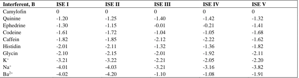
Table 2. Selectivity coeffients (Kpot
CY, J) of camylofine PVC membrane based sensors
and shaken well. A 10.0 mL portion of the diluted urine solution was transferred into a 25 mL beaker and then 0.5 mL of 10-2, 10-3and 10-4mol L-1standard drug solution were added, respectively. The working and reference electrode were immersed, and the potential readings were recorded after reaching the equilibrium response (10-20 s) and compared with the calibration plot.
Results and Discussion
Performance characteristics of the sensors
Camylofin-phosphomolybdate (CY-PMA) and Dibenzo-18-crown-6 (DB18CC6) membrane based sensors were prepared and examined for potentiometric camylofin determination with the composition 2:34:64 wt% of the ion pair complex (or ionophore), PVC and plasticizer, respectively 14. The sensors based on (CY-PMA) exhibit a potentiometric response towards camylofin ions with near Nernstian slope of 69.2± 0.9 mV (r2=0.999) and 66.6± 0.8 mV decade-1with a detection limit 2.5x10⁻⁶and 2.0x10⁻⁶mol L-1for the membrane plasticized with DOP [sensor I] and DOS [sensor II], respectively. For the sensors based on (DB18C6), they exhibit a near Nernstian response towards CY+ with slope of 53.2±1.1 mV (r2=0.999) and 51.4±0.7
mV (r2=0.998) decade-1 with a detection limits 2.5x10⁻⁵and 1.6x10⁻⁵ mol L-1 for the membrane plasticized with DOP [sensor III] and DOS [sensor IV], respectively (Fig. 3). From the previous results, it is obvious that the dielectric
constant of plasticized PVC is important when considering the material for use in ion selective electrode membrane. As shown in Fig. 3, it was found that low dielectric constant plasticizer DOS (ε=4.8) is desirable in the detection limit when measuring CY ions than DOP (ε=8). These results confirm what was reported in literature, that low dielectric constant plasticizer is desirable when monovalent ions are to be selected against divalent ions 15, and the nature of the plasticizer can influence the dielectric constant of the membrane phase, the mobility of the ionophore molecules and the state of ligands 16-18. Addition of TPB- (30 mmol% relative to the ionophore) to membrane plasticized with DOS and incorporating (DB18C6) [sensor V] improve the response towards camylofin ions with a slope 57.5±0.8 mV (r2=0.998) decade-1and detection limit 6.0x10⁻⁶mol L-1. All potentiometric response characteristics of the sensors are shown in Table 1.
The validity of the proposed potentiometric method for determining camylofin was assessed by measuring the range, lower limit of detection (LOD), accuracy (recovery), precision or repeatability (CVw), between-day variability (CVb), linearity (correlation coefficient) and sensitivity (slope) 19. Data obtained with six batches (six determinations each) of CY+ solutions are shown in Table 1. From the results, it can be concluded that, the response of sensors based on (DB18C6) is based on a neutral carrier mechanism, in which the ionophore exhibits strong affinity towards CY+to create positively charged complexes in the membrane phase.
Interferent, B ISE I ISE II ISE III ISE IV ISE V
Camylofin 0 0 0 0 0
Quinine -1.20 -1.25 -1.40 -1.42 -1.32
Ephedrine -1.30 -1.15 -0.01 -0.21 -1.41
Codeine -1.61 -1.72 -1.04 -1.05 -1.68
Caffein -1.82 -1.85 -2.12 -2.22 -1.62
Histidin -2.01 -2.11 -1.32 -1.36 -1.82
Glycin -2.10 -2.15 -2.01 -1.92 -2.11
K+ -3.21 -3.22 -2.21 -2.05 -2.20
Na+ -4.01 -4.03 -3.21 -3.16 -3.82
Figure 3. Potentiometric response of Camylofin membrane sensors I, II, III, IV and V using 0.05mol L-1 citrate buffer of pH 4.5.
To stabilize the presence of such complexes in the membrane, lipophilic anionic sites must be present [sensor V]. For sensors based on (CY-PMA), their response is mainly based on an ion exchanger mechanism, in which addition of anionic sites has no significant improvement in detection limit of the sensor.
Effect of pH and response time
The influence of pH on the potentiometric response of the proposed sensors were examined with standard 10-4and 10-3 mol L-1camylofin solutions over a pH range of 2–9. The pH of the solution was adjusted with either hydrochloric acid and/or sodium hydroxide solutions. From the pH/mV profile (Fig. 4), it is apparent that camylofin membrane based sensors reveal good stability with pH range 3.0 - 6.5 and 3.6-6.4 for sensor I and III, respectively. The potentials of both sensors considerably declined with negative drift at higher pH values due to progressive precipitation of the free camylofin base. The time required to achieve a steady potential response (±3mV) using the proposed sensors in 8.5x10⁻⁶mol L-1 camylofin solutions with a rapid 10-fold increase in concentration were < 15 s for sensors based on [CY]2[PM] and < 30 s for sensors based on DB18C6, respectively. After several calibrations for each sensor, low potential drift, long-term stability and negligible change in sensors response were observed. When not in use, the sensors were stored and conditioned in 10-3mol L-1. For all sensors examined, the detection limits, response times, linear range and calibration slopes were reproducible within ±3% of their original values over a period of at least 8 weeks.
Sensor selectivity
The effect of interferents on the potentiometric determination of camylofin was investigated by the fixed interference ion method (FIM) 20. In this method, the
selectivity coefficients of CY sensors were evaluated with a fixed concentration of interferent (10-3 mol L-1) adjusted to pH 4.5 with 0.05 mol L-1 citrate buffer solution. The selectivity coefficients were calculated using the following equation:
where aA is the varying activity of the primary ion (camylofin) and aBis the constant activity of the interfering ion.
For electrodes with membranes based on a less polar plasticizer (DOS), better selectivity toward CY+ ions over interfering cations, is obtained compared to membranes with higher polar plasticizers (DOP). The selectivity order for these plasticizers were CY+ > Quinine > Ephedrine > Codeine > Caffeine > Histidine > Glycine > K+ > Na+ > Ba2+ (sensor I), CY+ > Ephedrine > Quinine > Codeine > Caffeine > Histidine > Glycine > K+ > Na+ > Ba2+(sensor II), CY+= Ephedrine > codeine> Ba2+ > Histidine > Quinine > Glycine > Caffeine > K+> Na+(sensor III), and CY+~ Ephedrine > codeine > Ba2+ > Histidine > Quinine > Glycine > K+> Caffeine > Na+(sensor IV).
8 7 6 5 4 3
-20 0 20 40 60 80 100 120 140 160 180
mV
-log[CY], mol L-1
sensor
sensor
sensor
sensor V sensor V
(1) ,
a
pot A
KA B
zA zB aB
2 3 4 5 6 7 8 9 10
-20 0 20 40 60 80 100 120 140
sensor
10-4
mol L-1
10-3 mol L-1
EMF
,mV
pH
2 3 4 5 6 7 8 9 10
0 20 40 60 80 100 120 140
sensor
10-4
mol L-1
10-3
mol L-1
EMF
, mV
Figure 4. Effect of pH for Camylofin membrane sensor using sensors I and V.
Table 3. Response characteristics of camylofin sensors using FIA operation.
Parameter Sensor I Sensor IV
Slope (mV decade -1) 53.9±1.1 40±0.8
Correlation coefficient, r 0.9997 0.9991 Lower limit of
detection , µg mL-1
16.4±0.3 5.9±0.1
Linear range, mol L-1 1×10⁻⁴ -1.0×10⁻² 1×10⁻⁴ -1.0×10⁻²
Flow rate (mL min-1) 3.0 3.0
Sample loop volume , µL 100 100 Carrier solution 0.05 mol L-1
citrate buffer
0.05 mol L-1
citrate buffer pH of the carrier solution 4.5 4.5
Sample output, h-1 40–45 20–22
Table 4. Potentiomeric determination of camylofin in pharmaceutical preparation (Spasmograprin-M, Kahira Pharm., Egypt.) using membrane sensors I and V.
*Average of 6 measurements
The influence of the lipophilic anionic sites on the selectivity of the membrane (sensor IV) was shown in Table 2. The presence of lipophilic anionic sites helps reduce membrane resistance and limit the interference from anions at high sample activities. In addition to influencing the concentration of free carrier available for the complexing cations, they can also improve the selectivity of the ISEs 21. The optimum concentration of such lipophilic additives in the membrane phase is dependent in part on the charge of the primary ion and its complexing stoichiometry with the carrier as compared to that of the interfering ion 22. The selectivity order for sensor IV was CY+> Quinine> Ephedrine> Caffeine> Codeine> Histidine> Ba2+> Glycine > K+> Na+. This reflects that the presence of anionic sites in addition to the neutral carrier can affect on the mechanism by which the membrane responds. It will responds by the so called “Mixed mode mechanism”, i.e. ion exchanger response mechanism and neutral carrier mechanism.
Flow injection potentiometry
For the routine control of an analyte, FIA setup is of regular selection, in view of their versatility, simplicity and suitability for large-scale analyses. The flow assembly was double-channel, and the potentiometric sensor was accommodated in a flow cell of tubular configuration, allowing full membrane/sample contact. Sensors I and V were used in this study for showing the best analytical features for the simplest membrane composition.
A sample loop (100 μL) for camylofin solution ranging from 1.0 × 10⁻⁴to 1.0 × 10⁻²mol L-1at pH 4.5 with a 0.05 mol L-1 citrate carrier buffer, flow rate 3.0 mL min-1 was
chosen to study the potentiometric response (slope in mV decade-1) of the proposed sensors. Main analytical features recorded under optimum flow conditions are presented in Table 3. Sensors I and V gave slopes of 53.9±1.1 and 40.0±0.8 mV decade-1with detection limits of and 16.4±0.3 and 5.9±0.1 μg L-1and lower limits of linear range of 1.0 × 10⁻⁴mol L-1for both, as shown in Fig.5.
Analytical applications
Determination of CY in pharmaceutical preparations (Spasmopralgin-M tablets [Kahira Pharm, Egypt]) collected from local market and labeled with amount of 25 mg CY tablet-1, and analyzed by direct potentiometric analysis using sensors I and V. The average concentration was 25.3 and 22.3 mg per tablet, corresponding to recoveries of 101.1 and 89.2, respectively. Under hydrodynamic mode of operation, the same samples were also analyzed. The mean recovery values obtained were 99.4±1.1 and 94.9±0.8 for sensors I and V, respectively. All results for static and hydrodynamic potentiometric analysis for CY were shown in Table 4.
Figure 5. Typical FIA signals obtained by injecting pure camylofin standard solutions; 10-5, 10-4, 10-3 and 10-2 mol L-1 using sensors I
and V.
Application of the method for determining camylofin in biological fluids was tested by spiking aliquots of human urine samples with a known concentration of standard CY+ in 1.0x10⁻² mol L-1Citrate buffer of pH 4.5. Internal QC sample from certified reference material (10-2mol L-1) were spiked into 10 mL of urine test solutions to evaluate the method procedure and recovery (, in %) using equation (2):
where xs, x and xadd are the results of spiked sample, mean results of un-spiked sample and of added (spiked) reference,
Found (mg tablet -1)*
Labeled,
mg/tablet Batch FIA
Sensor V Sensor I
Sensor V Sensor I
23.4±1.2 24.7±0.9
22.3±1.1 25.3±0.6
25
100xs x (2)
xadd
-5 -4 -3 -2
-140 -120 -100 -80 -60 -40 -20
0 500 1000 1500 2000 2500 -160
-140 -120 -100 -80 -60 -40 -20 0
E
M
F
,
m
V
log[CY+
], mol L-1
Time,s
E
M
F
,m
V
10-5
mol L-1
10-4
mol L-1
10-3
mol L-1
10-2
mol L-1
respectively. The results reveal average recoveries of 97.5±0.3 and 99.1± 0.7 % and mean precision of ±0.05 and (n = 10) for batch and FIA mode of operations, respectively.
With the standard addition method applied to urine samples, the average recoveries for sensor I were 95.5± 0.3 % and 93.0± 0.5 % for batch and FIA mode of operations, respectively. For sensor V the average recoveries were 95.8± 0.5 % and 95.7± 0.4 % for batch and FIA mode of operations, respectively. The mean recovery obtained by spiking of 20.0 μg mL-1 internal quality control sample to 100.0 μg mL-1is 97.8±0.8 % .This confirms the applicability of the method for accurate routine analysis of camylofin in biological fluids.
Conclusions
A CY potentiometric sensors based on the use of dibenzo-18 crown-6 (DBdibenzo-18C6) as a neutral carrier, and ion-association complex of (CY) cation with phosphomolybdate
(PMA) anion, exhibited excellent potentiometric
performances such as quick response, a wide range of working pH, high sensitivity, long-term stability, good selectivity and self feasibility. The use of these sensors as detectors for the continuous monitoring of CY offered an advantage of simple design, ease of construction and possible application in the routine control of pharmaceutical drug solutions. The detectors displayed a wide range of dynamic measurement for the drug 1.0 × 10⁻⁴–1.0 × 10⁻²mol L-1with detection limits 5.9±0.1 and 16.4±0.3 μg mL-1under a continuous mode of operation at a flow rate of 3.0 mL min-1 and a sample outputs of 20–22 and 40-45 samples h-1 for sensors I and V, respectively.
References
1El-barbry, F. A. , Mabrouk, M. M., El-Dawy, M. A., J.AOAC. Int.,
2007, 90, 94-101.
2Kadam,N. N., Patil, P. C. , Singh, R. R. , Int. J. Pharm. & Pharm.
Sci., 2011, 3, 153-158.
3Rathnam, M. V., Singh, R. R., Pharm. Anal. Acta, 2010, 1, 1-4.
4Crombez, E., Van den Bossche, W., De Moerloose, P., J.
Chromatogr. A, 1976, 117, 161-166.
5C. Nerin, A. Garnica, J. Cacho, Anal. Chem., 1985, 57, 34-38.
6Kamel, A. H., J. Pharm. & Biomed. Anal., 2007, 45, 341-348.
7Hassan, S. S. M., Sayour, H. E. M., Kamel, A. H., Anal. Chim.
Acta 640 (2009) 75-81.
8Kamel, A. H., Mahmoud, W. H., Mostafa, M. S., Anal. Meth.,
2011, 3, 957-964.
9Kamel, A. H., Moreira, F. T. C., Sales, M. G. F., Anal. Lett., 2011,
44, 2107-2123.
10Moreira, F. T. C., Guerreiro, J. R. L., Azevedo, V. L. O., Kamel,
A. H., Sales, M. G. F., Anal. Meth., 2010, 2, 2039-2045.
11Kamel, A. H., Sayour, H. E. M., Electroanalysis, 2009, 21,
2701-2708.
12Kamel, A. H., Moreira, F. T. C., Sales, M. G. F., Sen. lett., 2011,
9, 1654-1660.
13Kamel, A. H., Sales, M. G. F., Almeida, S. A. A., Moreira, F. T.
C., Anal. Sci., 2009, 25, 365-371.
14Bakker, E., Buhlmann, P., Pretsch, E., Chem. Rev., 1997, 97,
3083-3123.
15Armstrong, R. D., Electrochim. Acta, 1990, 35, 1-7.
16Peper, S., Gonczy, C., Int. J. Electrochem., 2011, 2011, 1-8.
17Mousavi, M. F., Sahari, S., Alizadeh, N., Shamsipur, M., Anal.
Chim. Act, 2002, 414, 189-194.
18Fakhari, A. R., Ganjali, M. R., Shamsipur, M., Anal. Chem., 1997,
69, 3693-3696.
19Taylor, J. K., Quality Assurance of Chemical Measurements,
CRC Press, Florida, 1987.
20Umezawa, Y., Buhlmann, P., Umezawa, K., Tohoda, K.,
Amemiya, S., Pure Appl. Chem., 2000, 72, 1851-2082.
21Eugster, R., Gehrig, P. M., Morf, W. F., Spichiger, U. F., Simon,
W., Anal. Chem., 1991, 63, 2285-2289.
22Oggenfuss, P., Morf, W. F., Oesch, U., Ammann, D., Pretsch, E.,
Simon, W., Anal. Chim. Acta., 1998, 180, 299-311.