659
© 2017 by the Serbian Biological Society How to cite this article: Vujičić M, Saksida T, Stojanović I. Salvianolic acid B:
In vitro and in vivo effects on the immune system. Arch Biol Sci. 2017;69(4):659-63.
Salvianolic acid B:
in vitro
and
in vivo
effects on the immune system
Milica Vujičić, Tamara Saksida and Ivana Stojanović*
Institute for Biological Research “Siniša Stanković”, University of Belgrade, Belgrade, Serbia
*Corresponding author: [email protected]
Received: February 16, 2017; Revised: March 14, 2017; Accepted: March 24, 2017; Published online: April 4, 2017
Abstract: Type 1 diabetes (T1D) is an autoimmune disorder with a strong inflammatory component. Autoreactive cells specifically target insulin-producing β-cells, which leads to loss of glucose homeostasis. T1D remains incurable and versatile; potentially beneficial therapeutics are being tested worldwide. Possible candidates for the treatment of autoimmune diabetes are plants and their extracts since they are rich in biophenols, substances that act as secondary metabolites, and have veri-fied antioxidant and antiinflammatory effects. Salvianolic acid B (SalB) is a biophenol and one of the major constituents of Origanum vulgare ssp. hirtum (Greek oregano) extracts which in our previous studies was shown to exhibit an antidiabetic effect in mice. The aim of the present study was to determine whether SalB is responsible for the observed effects of Greek oregano extracts. SalB was applied in vitro to macrophages and lymphocytes isolated from C57BL/6 mice, as well as in vivo in the model of T1D induced by multiple low doses (MLD) of streptozotocin (STZ). SalB did not affect the viability of cells, but it significantly decreased secretion of nitric oxide (NO) and TNF in lipopolysaccharide (LPS)-stimulated macrophages, as well as the secretion of IFN-γ in concanavalin A (ConA)-stimulated lymphocytes. However, when applied in vivo, SalB at a dose of 2.5 mg/kg b.w., applied for 10 consecutive days, failed to protect mice from diabetes development. In conclu-sion, SalB exerts immunomodulatory effects in vitro, but is not effective in prevention of T1D in vivo. It probably requires cooperation with some other substances for the maximum efficacy exhibited by oregano extracts.
Key words: salvianolic acid B; type 1 diabetes; macrophage; lymphocyte; immune modulation
INTRODUCTION
Type 1 diabetes (T1D) is an autoimmune disorder where autoreactive immune cells specifically target insulin-pro-ducing pancreatic β-cells [1]. Destruction of β-cells leads to impaired insulin secretion and ultimately to a loss of glucose homeostasis [2]. Combined genetic and envi-ronmental factors lead to the immune response against β-cells [3]. The autoimmune response directs autoreac-tive immune cells towards pancreatic islets and insulitis occurs [4]. Macrophages and autoreactive T lymphocytes make up most of the immune infiltrate [5], and their effector molecules, such as pro-inflammatory cytokines and nitric oxide, lead to the creation of a pro-inflam-matory milieu, which is detrimental for β-beta cells [1].
While specific subsets of T helper (CD4+) cells, such as
IFN-γ-producing Th1 cells and IL-17-producing Th17 subsets, lead to T1D progression, IL-4-producing Th2 cells and IL-2-producing Treg cells have an antiinflam-matory role and can prevent T1D development [5-8].
T1D incidence is about 3% and is rapidly rising [9]. Currently, T1D is treated with daily insulin ad-ministration [1] and constant efforts are being made in the search for novel therapeutic strategies to treat T1D. Plants and their extracts are becoming increas-ingly interesting candidates for novel treatments of various ailments, including autoimmune diseases. This is mainly because plants are rich in biophenols and are considered to be less toxic and have fewer side effects than synthetic drugs. Biophenols are sub-stances abundantly found in plants where they act as secondary metabolites [10]. A number of studies has linked intake of biophenols through diet with benefi-cial effects in chronic diseases [11]. In our previous studies, we showed the antidiabetic effects of metha-nolic and ethyl-acetate oregano extracts (Origanum
vulgare ssp. hirtum) on the multiple low doses of
possesses anti-oxidant and anti-inflammatory effects [14]. The aim of this study was to investigate whether SalB is the substance responsible for the antidiabetic effects of methanolic and ethyl-acetate extracts of Origanum vulgare ssp. hirtum.
MATERIALS AND METHODS
Salvianolic acid B
Salvianolic acid B was a commercial sample obtained from Fluka (Buchs, Germany).
Animals
Male C57BL/6 mice, 8-12-weeks old were housed in the animal facility of the Institute for Biological Research. Their use and all experimental procedures were approved by the Ethics Committee of the in-stitute (App. No. 01-07/15), which is in accordance with Directive 2010/63/EU. The mice were kept under standard conditions at a 12 h light/dark cycle with free access to a standard pelleted diet and tap water.
Cell preparation and culture
In vitro analysis was performed on male C57BL/6 mice. To obtain cervical lymph node cells (CLNC), organs were mechanically disrupted by gentle teas-ing through a cell strainer (BD Bioscience, Bedford, USA), and the cells were collected by centrifugation. Peritoneal cells (PC) were collected by washing the peritoneal cavity of mice with cold PBS. Samples of conditioned medium, used for in vitro detection of cytokines and NO, were obtained by seeding the cells
(5×10⁶ CLNC mL-1 well-1, or 1×10⁶ PC mL-1 well-1)
for 24 h (PC) and 48 h (CLNC) in 24-well culture plates (Sarstedt, Nümbrecht, Germany) in RPMI-1640 medium (with 25 mmol/L HEPES, 2 mmol/L L-glu-tamine) supplemented with 10% FCS (PAA Chemi-cals, Pasching, Austria), penicillin/streptomycin (Sigma Aldrich, St. Louis, MO, USA), and 5 µmol/L
β-mercaptoethanol at 37°C in a 5% CO2 incubator.
Cells were cultured in the presence of mitogen activa-tors ConA (1 µg/mL, Sigma Aldrich; CLNC) and LPS (5 ng/mL, Sigma Aldrich; PC).
Multiple low-dose streptozotocin-induced diabetes
Immunoinflammatory T1D was induced in male 8-12-week-old C57BL/6 mice with multiple low
dos-es of STZ (MLDS, 40 mg kg-1 day-1, intraperitoneally
(i.p.) for five consecutive days, Sigma Aldrich). Each experimental group comprised 7 animals. Salvianolic acid B was administered i.p. for 10 consecutive days
(2.5 mg kg-1 day-1), starting from the day of diabetes
induction (prophylactic regimen). Mice were moni-tored for diabetes by weekly measurement of blood glucose levels using a glucometer (Sensimac, IMA-CO GmbH, Lüdersdorf, Germany). Clinical diabetes was defined by hyperglycemia in non-fasted animals (blood glucose>10 mmol/l).
Measurement of cytokine levels
Concentrations of cytokines in cell culture super-natants were determined by a sandwich ELISA us-ing MaxiSorp plates (Nunck, Rochild, Denmark) and anti-mouse-paired antibodies according to the manufacturer’s instructions. Samples were analyzed in duplicate for murine IL-17, IL-1β, TNF (BD Biosci-ences), IFN-γ (eBioscience) and IL-4 (R&D Systems). Absorbance was measured by a LKB 5060-006 micro-plate reader (LKB Instruments, Vienna, Austria) at 450 and 570 nm.
Determination of nitrite levels
Nitrite accumulation, an indicator of NO production, was measured in cell culture supernatants using the Griess reagent [15]. Absorbance was measured by a mi-croplate reader (LKB Instruments) at 540 and 650 nm.
Cell viability assay
Statistical analysis
Data are presented as means±SD. Statistical analy-sis was performed using Statistica 6.0 (StatSoft Inc., Tulsa, Okla., U.S.A.) software. Comparisons between the groups were performed by one-way ANOVA, fol-lowed by Student-Newman-Keuls post hoc test and finally Student’s t-test or Mann-Whitney U-test, where appropriate. Analysis of diabetes incidence was
per-formed by the χ2 test. A p-value less than 0.05 was
considered to be statistically significant.
RESULTS
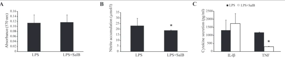
The effect of SalB on peritoneal cells in vitro To evaluate the potential influence of SalB on immune cells in vitro, PC rich in macrophages isolated from C57BL/6 mice and stimulated with LPS were treated with SalB at a concentration of 50 µg/mL. While SalB had no effect on the viability of peritoneal cells (Fig. 1A), it significantly reduced their pro-inflammatory potential, judging by the reduced levels of NO (Fig. 1B) and reduced secretion of the pro-inflammatory cyto-kine TNF (Fig. 1C). The concentration of SalB for in vitro experiments was determined on the basis of our previous work with Greek oregano extracts [12,13].
The effect of SalB on lymphocytes in vitro
While macrophages are the first cells to enter pan-creatic islets, lymphocytes are the key players in T1D pathogenesis [5]. We therefore assessed the effect of SalB on lymphocytes. SalB (50 µg/mL) showed no cy-totoxic effect on lymphocytes stimulated with ConA, judging by the viability assay (Fig. 2A). The effector
function of lymphocytes was determined by measur-ing the secretion of pro- and anti-inflammatory cyto-kines (Fig. 2B). While SalB had no effect on the secre-tion of anti-inflammatory IL-4 and pro-inflammatory IL-17, it significantly reduced the secretion of the pro-inflammatory cytokine IFN-γ (Fig. 2B) and thereby lowered their total pro-inflammatory potential.
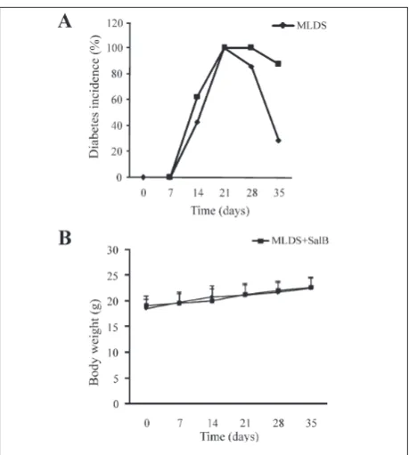
SalB does not prevent T1D development in vivo
To investigate the ability of SalB to prevent T1D de-velopment in vivo, MLDS-challenged C57BL/6 mice were treated with SalB (i.p.) at a dose of 2.5 mg/kg b.w. in a prophylactic regime, every day for ten days. The dose of SalB for in vivo experiments was deter-mined on the basis of our previous work with Greek
Fig. 1. In vitro effect of SalB on macrophage viability and function. PC isolated from C57BL/6 mice were stimulated in vitro by LPS (5
ng/mL) in the presence or absence of SalB (50 µg/mL). PC viability (A), secretion of NO (B) and cytokines (C) were measured after 24
h of incubation. Results are presented as the means±SD. *P<0.05 refers to LPS+SalB-treated vs. LPS-treated PC.
Fig. 2.In vitro effect of SalB on lymphocyte function and viabil-ity. Lymphocytes isolated from cervical lymph nodes of C57BL/6
mice were stimulated in vitro with ConA (1 μg/mL) in the
pres-ence or abspres-ence of SalB (50 µg/mL). Lymphocyte viability (A) and
secretion of cytokines (B) were measured after 48 h of
oregano extracts [12,13]. Our results showed that SalB did not prevent T1D development (Fig. 3A), in fact the incidence of T1D was higher throughout the en-tire experiment, although not statistically significant, than in the MLDS group (Fig. 3A). Additionally, the treatment with SalB showed no visible side effects on mice in terms of body weight gain (Fig 3B), behavior and general appearance (data not shown).
DISCUSSION
In this study, we present evidence that polyphenol SalB exerts an anti-inflammatory effect on both mac-rophages and lymphocytes in vitro, but that it was un-able to alter the autoimmune response in vivo and thus prevent T1D pathogenesis in C57BL/6 mice.
In our previous studies, we showed the antidiabetic effects of the methanolic and ethyl-acetate extracts of
Origanum vulgare ssp. hirtum in an MLDS model of T1D [12,13]. However, search for the active component of these extracts was less successful, since neither rosma-rinic acid nor carvacrol (constituents of these extracts) prevented the development of T1D [12,13, respectively]. SalB is another compound present in oregano extracts at a high concentration. It is frequently found in potentially immunomodulatory substances that are active in vitro, however, its effects do not appear to be translatable to an in vivo system [17]. In the present research, the in vitro anti-inflammatory potential of SalB was mediated by a reduction of TNF from macrophages, which is in accordance with the study of Sun et al. (2016), who also showed that SalB downregulated TNF secretion [18]. Macrophages also downregulated their NO produc-tion after SalB treatment. Evidence from the literature describes the inhibitory effect of a similar compound, salvianolic acid A, on macrophage-derived NO [19]. However, SalB displayed an opposite effect in vivo in the model of rat hepatitis, where macrophages expressed higher levels of inducible NO synthase, the enzyme cru-cial for induced NO synthesis [20].
SalB also exerted its in vitro immunomodulatory effect through the inhibition of lymphocyte-derived
IFN-γ that is produced by Th1 and cytotoxic CD8+
(and to a smaller extent by NK cells). The effect of SalB on lymphocytes has been poorly studied and there are only data from one in vivo study where SalB inhibited Th1 activity during experimental autoim-mune encephalomyelitis (EAE), a model of multiple sclerosis in rats [21].
Despite the obvious anti-inflammatory effect of SalB in vitro, a prophylactic treatment of MLDS-chal-lenged mice with SalB failed to protect the mice from T1D development. In contrast, one study claims that SalB is antidiabetic in vivo, since it prevented the devel-opment of hyperglycemia and subsequently increased insulin levels in Wistar rats [22]. However, there are several major differences that could be responsible for the different outcomes: the authors used outbred rats in the animal model, the doses of SalB were much higher (20 and 40 mg/kg b.w.), and were applied con-tinuously for 3 weeks. Cells that contribute the most in the creation of an inflammatory milieu in T1D are
T helper cells (Th1 and Th17), cytotoxic CD8+
lym-phocytes [6,23] and inflammatory macrophages [24]. Since SalB suppressed the IFN-γ-producing
popula-Fig. 3.In vivo effect on SalB on T1D. T1D was induced in male C57BL/6 mice by the administration of STZ (40 mg/kg b.w., i.p.) for five consecutive days. One group of animals was simultaneous-ly treated with SalB (2.5 mg/kg b.w., i.p) for 10 consecutive days (prophylactic treatment). The animals were monitored weekly
for changes in blood glucose levels (A) and body weight (B). The
tion and not the IL-17-producing Th17 cells, it can be speculated that the failure of SalB to prevent T1D development in vivo is due to the fact that SalB showed no effect on the pathogenic Th17 population. There-fore, in order to provide the most effective treatment of autoimmune/inflammatory disorders it might be more effective to use SalB in combination with some other substances that can downregulate the Th17 immune response. Since our previous research showed the an-tidiabetic effect of the oregano extracts that contain SalB among other substances, possible candidates for synergistic studies could be found in the substances present in the oregano extracts.
Acknowledgments: This work was supported by the Ministry of Education, Science and Technological Development, Republic of Serbia, Project No. OI 173013.
Authors’ contribution: MV, TS and IS performed the experiments and analyzed the data. IS and MV wrote and edited the text. IS conceived and designed the study.
Conflict of interest disclosure: We declare no conflict of interest.
REFERENCES
1. Anderson RP, van Heel DA, Tye-Din JA, Barnardo M, Salio M, Jewell DP, Hill AVS. T cells in peripheral blood after glu-ten challenge in coeliac disease. Gut 2005;54(9):1217-23. 2. Maahs DM, Rewers M. Editorial: mortality and renal
dis-ease in type 1 diabetes mellitus − progress made, more to be done. J Clin Endocrinol Metab. 2006;91:3757-9.
3. Hakonarson H, Grant S. Genome-wide association studies (GWAS): impact on elucidating the aetiology of diabetes. Diabetes Metab Res Rev. 2011;27(7):685-96.
4. Makino S, Kunimoto K, Muraoka Y, Mizushima Y, Katagiri K, Tochino Y. Breeding of a non-obese, diabetic strain of mice. Jikken Dobutsu. 1980;29:1-13.
5. Bluestone J, Herold K, Eisenbarth G. Genetics, pathogen-esis and clinical interventions in type 1 diabetes. Nature. 2010;464:1293-300.
6. Emamaullee JA, Davis J, Merani S, Toso C, Elliott JF, Thiesen A, Shapiro AM. Inhibition of Th17 cells regulates autoim-mune diabetes in NOD mice. Diabetes. 2009;58(6):1302-11. 7. Hill NJ, Van GK, Sarvetnick N. Th1 and Th2 pancre-atic inflammation differentially affects homing of islet-reactive CD4 cells in nonobese diabetic mice. J Immunol. 2003;170:1649-58.
8. Tarbell KV, Yamazaki S, Olson K, Toy P, Steinman RM.
CD25+ CD4+ T cells, expanded with dendritic cells
present-ing a spresent-ingle autoantigenic peptide, suppress autoimmune diabetes. J Exp Med. 2004;199:1467-77.
9. Cavan D, Fernandes JR, Makaroff L, Ogurtsova K, Webber
S, editors. IDF DIABETES ATLAS. 7th ed. Brussels:
Interna-tional Diabetes Federation; 2015. 144p.
10. Jaganath IB, Crozier A. Overview of health promoting com-pounds in fruits and vegetables. In: Chichester FC, editor. Phenolic Compounds of Plant Origin and Health: The Bio-chemistry Behind Their Nutritional and Pharmacological Value. United Kingdom: Wiley; 2009. p. 1-48.
11. Del Rio D, Rodriguez-Mateos A, Spencer JPE, Tognolini M, Borges G, Crozier A. Dietary (poly)phenolics in human health: structures, bioavailability, and evidence of protec-tive effects against chronic diseases. Antioxid Redox Signal. 2013;18;(14):1818-92.
12. Vujicic M, Nikolic I, Kontogianni VG, Saksida T, Charisia-dis P, Orescanin-Dusic Z, Blagojevic D, Stosic-Grujicic S,
Tzakos AG, Stojanovic I. Methanolic extract of Origanum
vulgare ameliorates type 1 diabetes through antioxidant, anti-inflammatory and anti-apoptotic activity. Br J Nutr. 2015;113(5):770-82.
13. Vujicic M, Nikolic I, Kontogianni VG, Saksida T, Charisiadis P, Vasic B, Stosic-Grujicic S, Gerothanassis I P, Tzakos AG, Stojanovic I. Ethyl Acetate Extract of Origanum vulgare L. ssp. hirtum Prevents Streptozotocin-Induced Diabetes in C57BL/6 Mice. J Food Sci. 2016;81:H1846-H1853. 14. Wang J, Xiong X, Feng B. Cardiovascular Effects of
Sal-vianolic Acid B. Evid Based Complement Alternat Med. 2013;2013:247948.
15. Stojanovic I, Saksida T, Nikolic I, Nicoletti F, Stosic-Grujicic S. Macrophage migration inhibitory factor deficiency pro-tects pancreatic islets from cytokine-induced apoptosis in vitro. Clin Exp Immunol. 2012;169:156-63.
16. Mosmann T. Rapid colorimetric assay for cellular growth and survival: application to proliferation and cytotoxicity assays. J Immunol Methods. 1983;65:55-63.
17. Jimal R, Shimogawara R, Yamamoto K, Ohta N. Anti-try-panosome effects of nutritional supplements and vitamin D3: in vitro and in vivo efficacy against Trypanosoma brucei brucei. Trop Med Health. 2016;44:26.
18. Sun B, Li C, Zuo L, Liu P. Protection of SAL B with H9C2 cells. Pharm Biol. 2016;54(5):889-95.
19. Oh KS, Oh BK, Mun J, Seo HW, Lee BH. Salvianolic acid A suppress lipopolysaccharide-induced NF-κB signal-ing pathway by targetsignal-ing IKKβ. Int Immunopharmacol. 2011;11(11):1901-6.
20. Zhao X, Jia H, Yang S, Liu Y, Deng B, Xu X, Zhang T, Zhou H, Zu C, Yin H, Li T, Song Y, Wang Y, Li P, Zou Z, Cai D. Salvianolic Acid B reducing portal hypertension depends on macrophages in isolated portal perfused rat livers with chronic hepatitis. Evid Based Complement Alternat Med. 2012;2012:786365.
21. Donga Z, Maa D, Gong Y, Yu T, Yao G. Salvianolic acid B ameliorates CNS autoimmunity by suppressing Th1 responses. Neurosci Lett. 2016;619(21):92-9.
22. Raoufi S, Baluchnejadmojarad T, Roghani M, Ghazanfari T, Khojasteh F, Mansouri M. Antidiabetic potential of salvi-anolic acid B in multiple low-dose streptozotocin-induced diabetes. Pharm Biol. 2015;53(12):1803-9.
23. Shao S, He F, Yang Y, Yuan G, Zhang M, Yu X. Th17 cells in type 1 diabetes. Cell Immunol. 2012;280(1):16-21.