© 2020 by the Serbian Biological Society How to cite this article: Pandrc MS, Ristić A, Kostovski V, Milin-Lazović J, Milić 137 N, Ćirić J. The role of echocardiography in monitoring the therapeutic effect of
levothyroxine replacement therapy in subclinical hypothyroidism. Arch Biol Sci. 2020;72(1):137-46.
The role of echocardiography in monitoring the therapeutic effect of levothyroxine
replacement therapy in subclinical hypothyroidism
Milena S. Pandrc1,*, Andjelka Ristić2, Vanja Kostovski3, Jelena Milin-Lazović4, Nataša Milić4,5 and Jasmina Ćirić6
1Department of Cardiology, Military Medical Academy, Belgrade, Serbia
2Department of Urgent Internal Medicine, Military Medical Academy, Belgrade, Serbia
3Clinic for Thoracic Surgery, Military Medical Academy, Belgrade, Serbia
4Institute for Medical Statistics and Informatics, Clinical Center of Serbia, Belgrade, Serbia
5Division of Nephrology and Hypertension, Mayo Clinic, Rochester, Minnesota, United States of America
6Clinic for Endocrinology, Diabetes and Metabolic Diseases, Clinical Center of Serbia, School of Medicine University of Belgrade,
Belgrade, Serbia
*Corresponding author: [email protected]
Received: October 29, 2019; Revised: January 5, 2020; Accepted: February 11, 2020; Published online: February 19, 2020
Abstract: Current controversies related to the treatment of subclinical hypothyroidism (SCH) with thyrotropin (TSH) < than 10 mU/L are based on the lack of evidence that levothyroxine therapy has beneficial effects. The aim of our study is to estimate the effect of levothyroxine treatment on cardiac morphology and function in subclinical hypothyroidism. Body mass index, waist circumference, blood pressure, electrocardiographic and standard echocardiographic parameters were measured before levothyroxine therapy and 3 months after TSH normalization. Significant reduction in systolic and diastolic blood pressure, PR, QT and QT corrected intervals, as well as increase in heart rate were recorded in the group on levothyroxine therapy. The following parameters of the left and right ventricle were significantly decreased in the treat-ment group: left ventricular mass index and volume, systolic and diastolic time intervals, and mitral annular plane systolic excursion (MAPSE). The increase was recorded as fractional shortening and pressure rise in early systole (dP/dt), right atrial wall thickness and diameters. Our study did not confirm differences in basic echocardiographic parameters between the treated and control groups, apart from an echocardiographic improvement of cardiac structure and function in treated individuals. The findings suggest electrocardiographic and echocardiographic screening in monitoring the therapeutic effect.
Keywords: subclinical hypothyroidism; electrocardiography; echocardiography
Abbreviations: subclinical hypothyroidism (SCH); thyroid-stimulating hormone (TSH); levothyroxine (LT4); free thyroxine (FT4); free triiodothyronine (FT3); thyroid antibodies (tAbs); ejection fraction (EF); fractional shortening (FS); early/late transmittal peak velocity ratio (E/A ratio); myocardial performance index (TEI index); mitral annular plane systolic excur-sion (MAPSE); pressure rise in early systole (dP/dt); amplitude of systolic motion of the lateral tricuspid annulus segment (TAPSE); systolic pressure in the right ventricle (SPRV)
INTRODUCTION
Subclinical hypothyroidism (SCH) is defined biochemi-cally as a persistent increase in serum thyroid-stimulat-ing hormone (TSH) and normal serum thyroxine (FT4) and triiodothyronine (FT3) levels [1]. A clinical picture of SCH is generally absent or could be mild. Whickam survey data registered SCH in 7.5% of women and 2.8% of men, with the highest incidence in women aged over 60 years [2-3]. Although there are controversies about
the treatment of SCH, current guidelines clearly sug-gest a thyroxine replacement trial in individuals aged less than 70 if mild symptoms of hypothyroidism are present, when TSH is between 4.12 and 10 mIU/L [4-6].
wall thickening and stiffening, endothelial dysfunc-tion and heart failure are clearly linked with SCH, but the beneficial effects of LT4 treatment have not been confirmed [3-12]. Clarification of the influence of LT4 treatment on cardiovascular risk in SCH would contribute to planning the strategy in cardiovascular prevention for these patients.
Some studies showed that morphological altera-tions of the heart, global, as well as systolic and diastol-ic cardiac dysfunctions and pulmonary hypertension found in SCH were improved after substitutive thy-roxine therapy [13-16]. More recent data underlined measurable differences in some echo parameters when the SCH group was compared to the control group at baseline, and in the SCH group tested before and after thyroxine treatment [6,17]. These findings point to the need for monitoring cardiac morphology and function in individuals with SCH on substitutive therapy in or-der to provide better objectivity in the assessment of LT4 effects and the rationale for its early introduction.
The aim of our study was to specify the type and the extent of changes in cardiac morphology and func-tion and the effect of LT4 on its reversion in individu-als with SCH who had persistently increased serum TSH levels between 4.5-10 mIU/L, positive thyroid antibodies and symptoms of mild hypothyroidism as reason for treatment. The finding could be of interest for supporting early introduction of an LT4 trial treat-ment and better validation of its effects, not only on symptoms but also on cardiac structure and function.
MATERIALS AND METHODS
Ethics statement, patient samples and clinical information
This is a pilot study within a prospective open-label study [5-6]. Informed consent was obtained from all participants included in the study. All procedures per-formed in studies involving human participants were in accordance with the ethical standards of the Ethical Commission of Belgrade University of Medical Sci-ences and with the 1964 Helsinki declaration and its later amendments or comparable ethical standards.
The criteria for inclusion were the presence of un-treated SCH defined as TSH levels between an upper
normal level and 10 mIU/L for more than 3 months, normal FT4, positive thyroid antibodies (tAbs) and/or an ultrasound scan characteristic for chronic autoim-mune thyroiditis. The exclusion criteria were missing vital data (TSH), previous history of thyroid disease and treatment, conditions that affect thyroid status and lipid metabolism [18], the taking of any medicine that affects the thyroid or lipid metabolism in the past 6 months [10], past or current serious medical diseases including diabetes mellitus and coronary heart disease, use of any medication, including aspirin or diuretics, that might affect the study parameters, presence of symptoms and signs of clinical bleeding, smoking. The appropriate control group had 40 healthy respondents.
Study protocol and measurements
Weight, height, body mass index (BMI), waist cir-cumference (WC) and blood pressure as well as elec-trocardiographic (ECG) variables (heart rate, PR in-terval, QT inin-terval, and corrected QT interval) were measured before intervention with thyroxine and 3 months after the euthyroid state was achieved.
The protocol included the analysis of morphology and function of the left and right heart. An IE33xMA-TRIX ultrasound machine (Philips Healthcare) with a X5-1 transducer was used for measurement cardiac morphology and function parameters.
The following cardiac morphology parameters of left ventricle were evaluated: the anteroposterior dimension of the left atrium (LA), the thickness of inter-ventricular septum (IVS), left ventricular pos-terior wall thickness, relative thickness of the poste-rior wall (calculated as: 2x left ventricular posteposte-rior wall thickness/end-diastolic diameter (EDD)), in-dex of LV hypertrophy. LV mass was calculated by using cube- function formula (LV mass = 0.8 (1.04 (EDD+posterior wall thickness) 3 -EDD3) + 0.6g). LV mass index was calculated as LV mass/height.
Beside morphology, the cardiac function param-eters were also checked. The study protocol included left ventricle global, systolic, and diastolic function parameters as well as right ventricle systolic and dia-stolic function parameters.
LV global function was expressed through the index of myocardial performance (Tei index). Tei in-dex was calculated as isovolumetric contraction time (IVCT)+isovolumetric relaxation time (IVRT))/ejec-tion time (ET)). The IVCT, IVCT/ET, pre-ejec(IVRT))/ejec-tion pe-riod (PEP) were also estimated.
Left ventricle systolic function was assessed by ejection fraction (EF) calculated by Simpson method. Ejection fraction was classified as normal, borderline, or abnormal (55%, 55-45%, and less than 45% respec-tively). Except EF, for left ventricle systolic function assessment mitral annular plane systolic excursion (MAPSE), fractional shortening (FS) (calculated by Teicholz’s formula: FS=(EDD-ESD)/EDD) in %), systolic mitral annular velocity (s), and dP/dt were measured. Doppler-derived dP/dt determined from the continuous-wave Doppler spectrum of the mi-tral regurgitation jet [19-20]. Cardiac output was also calculated as heart rate x stroke volume (EDV-ESV). LV diastolic function was expressed by the follow-ing parameters: early diastolic peak fillfollow-ing velocity (E), late diastolic peak filling velocity (A), E/A, early dia-stolic mitral annular velocity (e), late diadia-stolic mitral annular velocity(a), E/e, and some diastolic intervals (deceleration time of E velocity (DT), IVRT, PEP /ET).
RV global function was assessed by Tei index and pre-ejection period (PEP). RV systolic function was measured by FS, the amplitude of systolic motion of the lateral tricuspid annulus segment (TAPSE), and systolic tricuspid annular velocity (s). S is the aver-age value, calculated by measuring s to the septal and lateral tricuspid ring segment.
Right ventricular diastolic function was assessed by the the following measurements, done on the tri-cuspid orifice: E, A, E/A (all measured in the lateral tricuspid ring segment at the end of expirium), e, a, (obtained by measuring e and a to the septal and lat-eral tricuspid ring segment and calculating averages values), e/a, E/e, and PEP/ET.
Recommended variables for identifying diastolic dysfunction and their abnormal cutoff values are an-nular e velocity: septal e<7 cm/sec, lateral e<10 cm/ sec, average E/e ratio>14, LA volume index>34 mL/ m2, and peak TR velocity>2.8 m/s (LV diastolic dys-function is present if more than half of the available parameters meet these cutoff values) [21].
Two-dimensional echocardiography, pulsed and tissue Doppler echocardiography were used. The ventricular dimensions and systolic function were as-sessed with the help of M-mode. MAPSE is defined as the mitral annular plane systolic excursion (normal value from 0.8 to 1.4 cm). TAPSE (normal value from 2.1 to 2.9 cm) is defined as the amplitude of systolic motion of the lateral tricuspid annulus segment. The pulsed Doppler measured transvalvular velocities (E, A), and deceleration time of E velocity (DT) in the apical four-chamber view. Doppler filling velocities were measured and their ratios, which were consistent with impaired relaxation (E/A<0.7) and restrictive fill-ing (E/A>1.5). The tissue Doppler echocardiography analyzed systolic intervals (IVCT, IVRT and ET) in the apical four-chamber views. It was also used for measuring e, a, s of the mitral and tricuspid orifice.
After the initial investigation, LT4 treatment was initiated in all patients. Doses sufficient to normal-ize TSH ranged from 25 mcg to 75 mcg daily with a mean dose of 50 mcg. Three months after TSH nor-malization all tests were repeated. The control group consisted of healthy subjects with normal serum TSH levels, matched by age, sex, weight and height, inves-tigated at baseline.
Statistical analysis
or Wilcoxon’s signed rank tests were used (according to data distribution) to test the change before and after LT4 substitution for numerical variables. The Pear-son or Spearman correlation coefficient (according to data distribution) was used to analyze the relationship between study variables. The Bonferroni correction was applied according to 0.05/131, and p<0.0004 was considered statistically significant. All statistical analy-ses were performed in SPSS 20.0 (SPSS Inc., Chicago, Illinois, USA).
RESULTS
Baseline findings did not differ between the treated and control groups
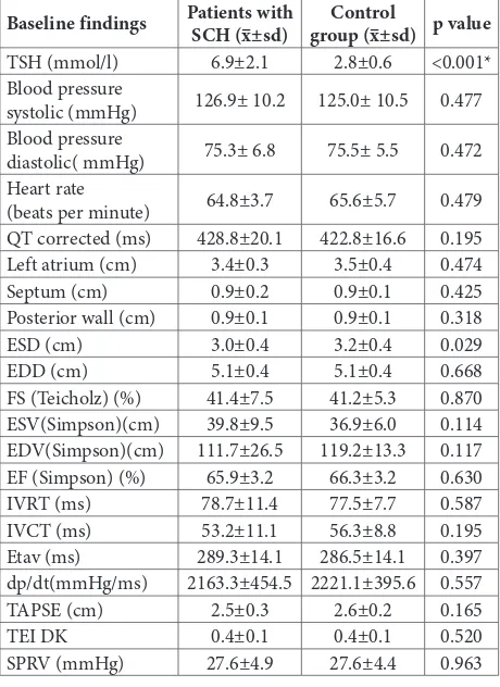
Our study included 35 patients with SCH (mean age 51.6±15.4 years, 29 females – 82.9%, 6 males – 17.1%) and 40 healthy controls matched for age (47.3±13.1 years) and sex (32 females – 80% females, 8 males – 20%). Mean WC and BMI in SCH group were 87.1±16.0 cm and 25.5± 4.0 kg/m2 respectively, and in the control group they were 86.2±15.0 cm and 24.5±3.5 kg/m2, respectively. The baseline findings did not dif-fer statistically and significantly between the SCH and control groups (Table 1). A significant reduction in systolic and diastolic blood pressure were recorded (p=0.024, p=0.019) in the SCH group on LT4 therapy.
Electrocardiographic (ECG) parameters in treated patients improved
The ECG parameters in patients with subclinical hy-pothyroidism before and after therapy are shown in Table 2. There was a statistically significant increase in heart rate (p=0.001) and a significant decrease in PR (before: 0.16±0.02, after: 0.15±0.02; p<0.001), QT (before: 389.58±10.12, after: 383.54±8.62; p<0.001) and QT corrected (before: 428.77 ±20.11, after: 411.77±14.73; p<0.001) intervals.
Echocardiographic indices exhibited a favorable trend in individuals on LT4
The following parameters of the left ventricle were decreased after the treatment in the SCH group: LV mass index (before: 76.81±13.52, after: 70.62±16.45;
Table 1. Comparison of baseline data between individuals with subclinical hypothyroidism (SCH) and control group (MV±SD) Baseline findings Patients with SCH (x–±sd) group (x–±sd) p valueControl
TSH (mmol/l) 6.9±2.1 2.8±0.6 <0.001*
Blood pressure
systolic (mmHg) 126.9± 10.2 125.0± 10.5 0.477
Blood pressure
diastolic( mmHg) 75.3± 6.8 75.5± 5.5 0.472
Heart rate
(beats per minute) 64.8±3.7 65.6±5.7 0.479
QT corrected (ms) 428.8±20.1 422.8±16.6 0.195
Left atrium (cm) 3.4±0.3 3.5±0.4 0.474
Septum (cm) 0.9±0.2 0.9±0.1 0.425
Posterior wall (cm) 0.9±0.1 0.9±0.1 0.318
ESD (cm) 3.0±0.4 3.2±0.4 0.029
EDD (cm) 5.1±0.4 5.1±0.4 0.668
FS (Teicholz) (%) 41.4±7.5 41.2±5.3 0.870
ESV(Simpson)(cm) 39.8±9.5 36.9±6.0 0.114
EDV(Simpson)(cm) 111.7±26.5 119.2±13.3 0.117
EF (Simpson) (%) 65.9±3.2 66.3±3.2 0.630
IVRT (ms) 78.7±11.4 77.5±7.7 0.587
IVCT (ms) 53.2±11.1 56.3±8.8 0.195
Etav (ms) 289.3±14.1 286.5±14.1 0.397
dp/dt(mmHg/ms) 2163.3±454.5 2221.1±395.6 0.557
TAPSE (cm) 2.5±0.3 2.6±0.2 0.165
TEI DK 0.4±0.1 0.4±0.1 0.520
SPRV (mmHg) 27.6±4.9 27.6±4.4 0.963
Thyroid-stimulating hormone (TSH), end-systolic diameter (ESD), end-diastolic diameter (EDD), fractional shortening (FS), end-systolic volume (ESV), end-diastolic volume (EDV), ejection fraction (EF), isovolumetric relaxation time (IVRT), isovolumetric contraction time (IVCT), ejection time (ET), pressure rise in early systole (dP/dt), amplitude of systolic motion of the lateral tricuspid annulus segment (TAPSE), myocardial performance index (TEI index), systolic pressure in right ventricle (SPRV); p-value from t-test for independent samples; x– arithmetic mean; SD – standard deviation; * – statistically significant difference (Bonferroni correction was applied alpha=0.0004)
Table 2. ECG parameters of patients with subclinical hypothyroid-ism before therapy and after (MV±SD).
ECG parameters before x–±sd after x–±sd p
Heart rate
(beats per minute) 64.77±3.69 67.23±4.09 0.001
PR interval ( ms ) 0.16±0.02 0.15±0.02 <0.001*
QT interval (ms) 389.58±10.12 383.54±8.62 <0.001* QT corrected
interval (ms ) 428.77±20.11 411.77±14.73 <0.001*
p<0.001 respectively), ESV (before: 38.86±9.6, after: 36.95±9.25; p<0.001), EDV (before: 114.83±20.6, af-ter: 108.43±18; p<0.001), MAPSE 2D ((before: 1.67 (1.55-1.89), after: 1.53 (1.34-1.65); p<0.001)). Frac-tional shortening increased significantly after LT4 treatment ((before: 39.40 (35.40-46.80), after: 42.40 (38.80-47.20), p<0.001)), (Table 3).
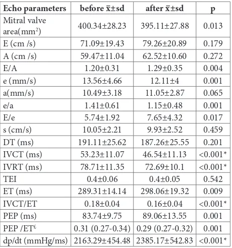
After therapy, the following parameters of mi-tral orifice decreased as follows: IVCT (before: 53.23±11.07, after: 46.54±11.13; p<0.001), IVRT (before: 78.71±11.35, after: 72.69±10.1; p<0.001), IVCT/ET (before: 0.18±0.04, after: 0.16±0.04; p<0.001). Dp/dt increased after LT4 therapy (before: 2163.29±454.48, after: 2385.17±542.83; p<0.001) (Table 4).
There was no significant change in the frequency of diastolic dysfunction after therapy compared to the initial finding (Table 5).
Table 3. Echocardiographic (ECHO) parameters of left atrium and ventricle morphology and function (MV±SD).
Echo parameters before x–±sd after x–±sd p
Left atrium (cm) 3.41±0.32 3.38±0.33 0.365
Septum in diastole
(cm) 0.89±0.16 0.85±0.16 0.012
Posterior wall in
diastole (cm) 0.89±0.14 0.9±0.16 0.586
RWT (cm) 0.4±0.08 0.4±0.08 0.624
Index of LV
hypertrophy 0.4±0.08 0.4±0.08 1.000
LV mass (Cube)(g ) 138.97±28.56 133.43±29.4 0.003
LV mass index
(g/m) 76.81±13.52 70.62±16.45 <0.001*
ESD (cm) 2.96±0.38 2.91±0.47 0.412
EDD (cm) 5.06±0.42 4.98±0.46 0.197
EF (Simpson) (%) 65.91±3.21 67.29±3.58 0.001
FS (Teicholz) (%) £ 39.40
(35.40-46.80) (38.80- 47.20) <0.001*42.40 ESV(M-mode)
(mL) 38.86±9.6 36.95±9.25 <0.001*
EDV (M-mode)
(mL) 114.83±20.6 108.43±18 <0.001*
EDV (M-mode)
(mL/m2) 62.6±11.03 60.72±10.42 0.028
ESV (Simpson)
(mL) 39.75±9.49 39.13±12 0.630
EDV (Simpson)
(mL) 111.69±26.47 108.71±24.84 0.001
EDV(Simpson)
(mL/m2) 57.61±16.72 55.48±15.92 0.001
MAPSE 2D (mm)£ 1.67(1.55-1.89) 1.53
(1.34-1.65) <0.001* Stroke volume
(mL) 75.97±11.0 71.05±8.75 <0.001*
Cardiac
output(mL/min) 2527.3±673.3 2486.8±641.4 0.244
Relative thickness of the posterior wall in diastole (RWT), left ventricle (LV), end-systolic diameter (ESD), end-diastolic diameter (EDD), ejection fraction (EF), fractional shortening (FS), end-systolic volume ( ESV), end-diastolic volume (EDV), mitral annular plane systolic excursion (MAPSE); p-value from paired t-test; £ data are presented as median (25th-75th percentile)
Wilcoxon test was performed, x– arithmetic mean; SD – standard deviation; * statistically significant difference (Bonferroni correction was applied alpha=0.0004)
Table 4. ECHO parameters of the mitral orifice (MV±SD).
Echo parameters before x–±sd after x–±sd p
Mitral valve
area(mm2) 400.34±28.23 395.11±27.88 0.013
E (cm /s) 71.09±19.43 79.26±20.89 0.179
A (cm /s) 59.47±11.04 62.52±10.60 0.272
E/A 1.20±0.31 1.29±0.35 0.004
e (mm/s) 13.56±4.66 12.11±4 0.001
a(mm/s) 10.49±3.18 11.05±2.87 0.065
e/a 1.41±0.61 1.15±0.48 0.001
E/e 5.74±1.92 7.65±4.32 0.017
s (cm/s) 10.05±2.21 9.93±2.52 0.459
DT (ms) 191.11±25.62 187.26±25.55 0.201
IVCT (ms) 53.23±11.07 46.54±11.13 <0.001*
IVRT (ms) 78.71±11.35 72.69±10.1 <0.001*
TEI 0.4±0.06 0.4±0.05 0.542
ET (ms) 289.31±14.14 298.06±19.32 0.009
IVCT/ET 0.18±0.04 0.16±0.04 <0.001*
PEP (ms) 83.74±9.75 89.06±13.55 0.001
PEP /ET£ 0.31 (0.27-0.34) 0.29 (0.27-0.32) 0.001
dp/dt (mmHg/ms) 2163.29±454.48 2385.17±542.83 <0.001*
E (early diastolic peak filling velocity), A (late diastolic peak filling velocity), e (early diastolic mitral annular velocity), a (late diastolic mitral annular velocity), s (systolic mitral annular velocity), DT (deceleration time of E velocity), isovolumetric contraction time (IVCT), isovolumetric relaxation time (IVRT), myocardial performance index (TEI index), ejection time (ET), pre-ejection period (PEP), pressure rise in early systole (dP/dt); p-value from paired t-test, £ data
are presented as median (25 th-75th percentile) Wilcoxon test was
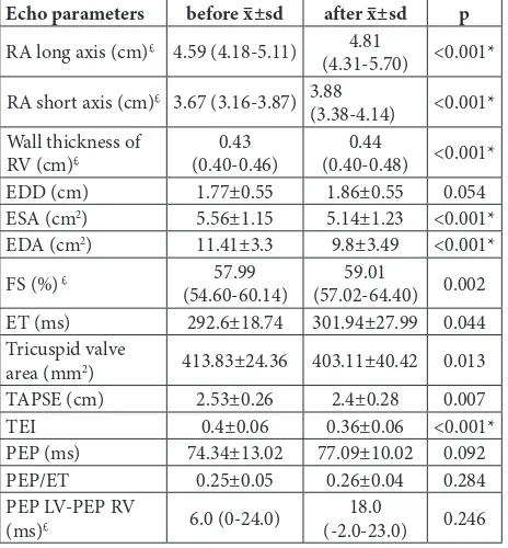
After LT4 substitution therapy, there was a re-duction of the following parameters: ESA (before: 5.56±1.15, after: 5.14±1.23; p<0.001), EDA (before: 11.41±3.3, after: 9.8±3.49; p<0.001), TEI (before: 0.4±0.06, after: 0.36±0.06; p<0.001). A statistically sig-nificant increase was recorded in the RA long axis ((be-fore: 4.59 (4.18-5.11), after: 4.81 (4.31-5.70); p<0.001)) and short axis ((before: 3.67 (3.16-3.87), after: 3.88 (3.38-4.14); p<0.001)), wall thickness ((before: 0.43 (0.40-0.46), after: 0.44 (0.40-0.48); p<0.001)) (Table 6).
After LT4 therapy there was an increment in the following parameters of the tricuspid and pulmonary orifice: E (before: 48.82 ± 9.66, after: 51.75 ± 9.82; p<0.001), e (before: 8.54 ± 2.44, after: 10.36 ± 2.66; p<0.001), Ep (before: 10.65 ± 3.35, after: 11.83 ± 3.79; p<0.001) and decrease of Ap (before: 27.6 ± 4.88, after: 25.49 ± 4.84; p<0.001) (Table 7).
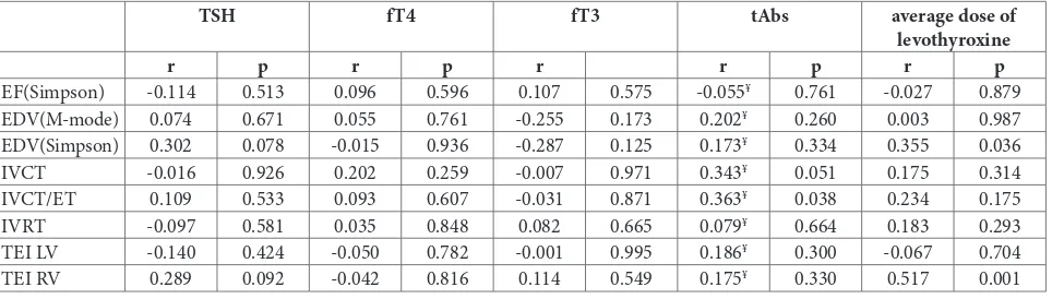
There was no correlation between changes in thy-roid and echocardiographic parameters (Table 8).
DISCUSSION
Some studies have shown that subclinical thyroid dysfunction results in changes in heart rate, blood pressure, cardiac output and contractility, as well as vascular resistance [18,22]. These alterations in car-diac morphology and function may be objectified by
Table 7. ECHO parameters of the tricuspid et pulmonary orifice (MV±SD).
Echo parameters before x–±sd after x–±sd p
Et (cm/s) 48.82±9.66 51.75±9.82 <0.001*
A (cm/s) 36.5±10.35 38.18±9.84 0.096
E/A 1.4±0.31 1.4±0.23 0.967
e (mm/s) 8.54±2.44 10.36±2.66 <0.001*
a (mm/s) 10.12±5.45 9.48±4.75 0.035
e/a£ 1.0 (0.9-1.4) 1.(0.7-2.0) 0.108
E/e 6.04±1.6 5.3±1.47 0.001
E pulmonary (cm/s) 10.65±3.35 11.83±3.79 <0.001*
A pulmonary (cm/s) 27.6±4.88 25.49±4.84 <0.001*
E (early diastolic peak filling velocity ), A (late diastolic peak filling veloc-ity), e (early diastolic tricuspid annular velocveloc-ity), a (late diastolic tricuspid annular velocity); p-value from paired t-test, £ data are presented as median
(25 th-75th percentile) Wilcoxon test was performed; x– arithmetic mean;
SD – standard deviation; * – statistically significant difference (Bonferroni correction was applied alpha=0.0004)
Table 5. The frequency of diastolic dysfunction baseline and after the therapy.
Criteria for diastolic
dysfunction before n (%) after n (%) p-value
e septal<7 cm/s 10(28.6) 8(22.9) 0.625
e lateral<10 cm/s 5 (14.3) 3(8.58) 0.615
average E/e>14 0(0) (0) NA
Left atrium volume
index>34ml/m2 2(5.7) 0(0) 0.500
Peak TR velocity>2.8 m/s 5(14.3) 4(11.4) 1.000
Diastolic dysfunction 13(37.1) 11(31.4) 0.625
e (early diastolic mitral annular velocity), E (early diastolic peak filling velocity), tricuspid regurgitation (TR); p-value from McNemar test; NA – not applicable
Table 6. Echocardiographic (ECHO) parameters of right atrium and ventricle morphology and function (MV±SD).
Echo parameters before x–±sd after x–±sd p
RA long axis (cm)£ 4.59 (4.18-5.11) 4.81
(4.31-5.70) <0.001* RA short axis (cm)£ 3.67 (3.16-3.87) 3.88
(3.38-4.14) <0.001* Wall thickness of
RV (cm)£ (0.40-0.46)0.43 (0.40-0.48) <0.001*0.44
EDD (cm) 1.77±0.55 1.86±0.55 0.054
ESA (cm2) 5.56±1.15 5.14±1.23 <0.001*
EDA (cm2) 11.41±3.3 9.8±3.49 <0.001*
FS (%) £ 57.99
(54.60-60.14) (57.02-64.40) 0.00259.01
ET (ms) 292.6±18.74 301.94±27.99 0.044
Tricuspid valve
area (mm2) 413.83±24.36 403.11±40.42 0.013
TAPSE (cm) 2.53±0.26 2.4±0.28 0.007
TEI 0.4±0.06 0.36±0.06 <0.001*
PEP (ms) 74.34±13.02 77.09±10.02 0.092
PEP/ET 0.25±0.05 0.26±0.04 0.284
PEP LV-PEP RV
(ms)£ 6.0 (0-24.0) (-2.0-23.0)18.0 0.246
Right atrium (RA), right ventricle ( RV), left ventricle (LV), end-diastolic diameter (EDD), end-systolic area (ESA), end-end-diastolic area (EDA), fractional shortening (FS), ejection time (ET), amplitude of systolic motion of the lateral tricuspid annulus segment (TAPSE), myocardial performance index (TEI index), pre-ejection period (PEP); p-value from paired t-test, £ data are presented as median (25th-75th
observing changes in several echocardiographic pa-rameters: increased LV mass index, increments of the index of myocardial performance (IVCT, IVCT/ET), mild systolic dysfunction, impaired diastolic func-tion (prolonged IVRT, higher PEP/ET, reduced E/A) [13-15,23-29]. Most of these shifts are responsive to LT4 treatment, so the restoration of normal thyroid function principally leads to the normalization of car-diovascular hemodynamics [13,28].
Hypothyroidism is accompanied by a rise in dia-stolic blood pressure and consequently low cardiac output and narrowed pulse pressure [30]. Normal-ization of thyroid hormone levels in our study group was associated with blood pressure reduction, heart rate increment and stroke volume extenuation, all resulting in unchanged cardiac output. Bradycardia is also a common feature of SCH, and stimulation of β-adrenergic receptors by T3 was found to improve it [22]. Prolongation of QT interval, which is a risk fac-tor for malignant ventricular irritability and sudden cardiac death, is also found to be linked with a rise in TSH and is shown to be completely reversible with LT4 treatment [31-33]. We have also demonstrated the potential of thyroxine therapy to significantly reduce PR and QT intervals.
Beside ventricular irritability, it was suggested that substitutive therapy improves decreased LV global contractility evaluated by dP/dt ratio [34-35]. Our results have confirmed previously listed study data. Isovolumic phase measurement was previously used as an index of LV function estimated by noninvasive methods; recently dP/dt measurement has been
pro-posed as being less load-dependent and more accu-rate. Some studies evaluated LV global contractility by the dP/dt ratio as we did, and also demonstrated that decreased LV function in SCH was improved by LT4 treatment [34-35].
Recent findings have reported higher values of mean left ventricular wall thickness (IVS and left ven-tricular posterior wall (LVPW), and a greater increase of LV mass and mass index in SCH individuals in comparison to controls [15,36-37]. It was also high-lighted that LT4 therapy can reduce LV wall thickness and LV mass [15,36]. We also demonstrated a favor-able reduction in left ventricular mass index after LT4 substitutive therapy.
Myocardial performance index (Tei index) re-duction was also noted after LT4 replacement in ac-cordance with previous data [27-28]. The Tei index combines both systolic (IVCT and ET) and diastolic (IVRT) time intervals as indices of the peripheral hy-pothyroidism and echocardiographic markers of SCH [27-28]. Impaired development of myocardial force was the most likely explanation for the increases in IVCT and the IVCT/ET ratio, which is also observed in our patients with SCH and which was improved after thyroxine replacement. This points to the pos-sible role of tissue hypothyroidism or the autoimmune process per se in events leading to the upgrading of systolic time intervals. It also appears to be important that improvements of both RV and LV Tei indices did not correlate with the dose of LT4, and higher initial doses may not lead to faster improvement of RV and LV pump functioning.
Table 8. The correlation of the changes in parameters of cardiac function with changes in TSH, FT4, FT3, tAbs and the average dose of LT4.
TSH fT4 fT3 tAbs average dose of
levothyroxine
r p r p r r p r p
EF(Simpson) -0.114 0.513 0.096 0.596 0.107 0.575 -0.055¥ 0.761 -0.027 0.879
EDV(M-mode) 0.074 0.671 0.055 0.761 -0.255 0.173 0.202¥ 0.260 0.003 0.987
EDV(Simpson) 0.302 0.078 -0.015 0.936 -0.287 0.125 0.173¥ 0.334 0.355 0.036
IVCT -0.016 0.926 0.202 0.259 -0.007 0.971 0.343¥ 0.051 0.175 0.314
IVCT/ET 0.109 0.533 0.093 0.607 -0.031 0.871 0.363¥ 0.038 0.234 0.175
IVRT -0.097 0.581 0.035 0.848 0.082 0.665 0.079¥ 0.664 0.183 0.293
TEI LV -0.140 0.424 -0.050 0.782 -0.001 0.995 0.186¥ 0.300 -0.067 0.704
TEI RV 0.289 0.092 -0.042 0.816 0.114 0.549 0.175¥ 0.330 0.517 0.001
Ejection fraction (EF), end-diastolic volume (EDV), isovolumetric contraction time (IVCT), ejection time (ET), isovolumetric relaxation time (IVRT), myocardial performance index (TEI index), left ventricle (LV), right ventricle (RV); Pearson’s rank correlation coefficient; ¥Spearman’s rank correlation
We also separately estimated the systolic and dia-stolic left ventricular functions that are often impaired in SCH. The left ventricular systolic function is influ-enced by changes in preload, afterload and myocardial inotropic function. Without invoking simultaneous impairment in myocardial inotropic function, the changes in preload and afterload were not sufficient to explain the decrease in systolic pump performance observed in our SCH patients [32,34]. Substitutive therapy in patients with SCH contributed to higher EF, but within a referent range. After comparison of base-line findings of the SCH group with the control group, we could not demonstrate any significant difference. The more precise findings of magnetic resonance im-aging study are in accordance with our results [38].
In order to provide objective interpretation of the data obtained before and after treatment and to make the relation of the findings to subclinical hypothyroid-ism more certain, we used the Bonferroni correction for confirmation of statistical significance. In addition, all measured ultrasound parameters were in the nor-mal range. This is why we believe that the significance of the measured improvements in our study is actually compatible with the known effects of serum and tissue LT4 on cardiac performance.
After the normalization of TSH by LT4 treatment, participants in our study showed a reduction in EDV (decreased preload as a result of LT4-induced systemic vasodilatation), higher heart rate and higher FS. De-spite EDV reduction, central and peripheral thyroid hormone effects synergistically improved systolic LV performance measured by the FS of individuals in the follow-up group.
The slowing of LV relaxation is associated with aging [39]. The greatest changes are observed in se-niors, overlapping with the hallmarks seen in mild diastolic dysfunction in younger patients (40-60 years of age). Our study was designed to minimize age as a confounding factor by choosing a population of early middle age.
The alterations of left ventricular diastolic func-tion in SCH were represented by the prolongafunc-tion of the isovolumetric relaxation time and deceleration time, an increased pre-ejection/ejection time ratio and reduced early diastolic mitral flow velocity/late diastolic mitral flow velocity ratio [14-15,17,28,36,40].
Although the diastolic time intervals and the mitral flow velocity ratio have been proven to be significantly impaired only in overt primary hypothyroidism, the improvement of these parameters by thyroxine was found in both overt and subclinical primary hypo-thyroidism [41].
Despite the positive trend that has been observed in other trials, in our study the treatment with LT4 did not influence the frequency of diastolic dysfunction, although IVRT was improved [14-15,22,28-29,35-36].
CONCLUSIONS
Current controversies about the treatment of sub-clinical hypothyroidism are related to insufficient evidence that therapy with LT4 has beneficial effects. According to the guidelines, a thyroxine replacement trial is clearly suggested only in symptomatic SCH patients, aged less than 70 years. Comparing the find-ings obtained by basic heart ultrasound examination, our study did not confirm significant difference be-tween SCH patients and the control group. Our study demonstrated improvements in cardiac output, heart rate, rhythm, contractility, blood volume and systemic vascularresistance, as well as in cardiac morphology, global, systolic, diastolic and pulmonary function in patients with SCH three months after normaliza-tion of TSH by thyroxine replacement therapy. These changes did not correlate with the dose of thyroxine used for TSH correction. The improvement of cardiac structure and function, measured by electrocardio-graphic and echocardioelectrocardio-graphic indicators, should be a useful factor in monitoring the therapeutic effect. This may also be a better approach when deciding whether a patient should be permanently treated with LT4, and more objective than loss or reduction of symptoms.
be interpreted as a tendency to become risk factors. Future studies with larger numbers of participants are needed to ascertain the clinical and hemodynamic significance of these findings.
Acknowledgments: This work received no funding.
Author contributions: MSP, AR, VK, JML and NM contributed to data acquisition, data analysis and data interpretation. MP and JĆ designed the study and wrote the paper. All authors approved the final version of the manuscript.
Conflict of interest disclosure: The authors declare no conflict of interest.
REFERENCES
1. Van Vliet G, Deladoëy J. Interpreting minor variations in thyroid function or Echostructure: Treating Patients, Not Numbers or Images. Pediatr Clin North Am. 2015;62(4):929-42.
2. Vanderpump MP, Tunbridge WM, French JM, Appleton D, Bates D, Clark F, Evans JG, Hasan DM, Rodgers H, Tun-bridge FK. The incidence of thyroid disorders in the com-munity: a twenty-year follow-up of the Whickham survey. Clin Endocrinol (Oxf) 1991;43:55-69.
3. Razvi S, Weaver JU, Vanderpump MP, Pearce SHS. The inci-dence of ischemic heart disease and mortality in people with subclinical hypothyroidism: reanalysis of the Whickham survey cohort. J Clin Endocrinol Metab, 2010;95:1734-40. 4. Pearce SHS, Brabant G, Duntas LH, Monzani F, Peeters RP,
Razvi S, Wemeau JL. 2013 ETA Guideline: Management of Subclinical Hypothyroidism. Eur Thyroid J. 2013;2:215-28. 5. Pandrc M, Ristić A, Kostovski V, Stanković M, Antić V, Milin-Lazović J, Ćirić J. The effect of the early substitution of subclinical hypothyroidism on biochemical blood param-eters and the quality of life. J Med Biochem. 2017;36:127-36. 6. Pandrc M S, Ristić A, Kostovski V, Randjelović-Krstić V,
Milin-Lazović J, Nedeljković-Beleslin B, Ćirić J. Evaluation of a three-month trial of thyroxine replacement in symp-tomatic subclinical hypothyroidism: the impact on clini-cal presentation, quality of life and adoption of long-term therapy. Vojnosanit Pregl. 2020;https://doi.org/10.2298/ VSP180708157P
7. Pyati A, Dhuttargi S, Das D. Assessment of the Cardiovas-cular Risk in Subclinical Hypothyroidism. Int J Pharm Biol Sci. 2012;2(2):128-34.
8. Kottagi SS, Rathi DB, Dongre NN. Evaluation of LDLCho-lesterol / HDL-ChoLDLCho-lesterol Ratio as Predictor of Dyslipid-emia in Subclinical Hypothyroidism. J Krishna Inst Medical Sci Univ. 2014;3(1):34-40.
9. Hak AE, Pols HA, Visser TJ, Drexhage HA, Hofman A, Witteman JC. Subclinical hypothyroidism is an indepen-dent risk factor for atherosclerosis and myocardial infarc-tion in elderly women: the Rotterdam study. Ann Int Med. 2000;132:270-8.
10. Walsh JP, Bremner AP, Bulsara MK, O’Leary P, Leedman PJ, Feddema P, Michelangeli V. Subclinical thyroid dysfunction as a risk factor for cardiovascular disease. Arch Intern Med. 2005;165:2467-72.
11. Feldt-Rasmussen U. Subclinical Hypothyroidism and Car-diovascular Risk - An Overview of Current Understanding. European Endocrinol. 2011;7(1):53-7.
12. Yao K, Zhao T, Zeng L, Yang J, Liu Y, He Q, Zou X. Non-invasive markers of cardiovascular risk in patients with subclinical hypothyroidism: A systematic review and meta-analysis of 27 case control studies. Sci Rep. 2018;8:4579. 13. Biondi B, Palmieri EA, Lombardi G, Fazio S. Effects of
sub-clinical thyroid dysfunction on the heart. Ann Intern Med. 2002;137:904-14.
14. Arem R, Rokey R, Kiefe C, Escalante DA, Rodriguez A. Cardiac systolic and diastolic function at rest and exercise in subclinical hypothyroidism: effect of thyroid hormone therapy. Thyroid 1996;6:397-402.
15. Monzani F, Di Bello V, Caraccio N, Bertini A, Giorgi D, Giusti C, Ferrannini E. Effect of levothyroxine on cardiac function and structure in subclinical hypothyroidism: a double blind, placebo-controlled study. J Clin Endocrinol Metab. 2001;86:1110-5.
16. Curnock AL, Dweik RA, Higgins BH, Saadi HF, Arroliga AC. High prevalence of hypothyroidism in patients with pri-mary pulmonary hypertension. Am J Med Sci. 1999;318:289-92.
17. Nakova VV, Krstevska B, Kostovska ES, Vaskova O, Ismail LG. The effect of levothyroxine treatment on left ventricular function in subclinical hypothyroidism. Arch Endocrinol Metab. 2018;62(4):392-8.
18. Sun Z, Ojamaa K, Coetzee WA, Artman M, Klein I. Effects of thyroid hormone on action potential and repolarization currents in rat ventricular myocytes. Am J Physiol Endocri-nol Metab. 2000;278:E302-7.
19. Bargiggia GS, Bertucci C, Recusani F, Raisaro A, de Servi S, Valdes-Cruz LM, Sahn DJ, Tronconi L. A new method for estimating left ventricular dP/dt by continuous wave Doppler-echocardiography. Validation studies at cardiac catheterization. Circulation. 1989;80:1287-92.
20. Kolias TJ, Aaronson K D, Armstrong FW. Doppler-derived dP/dt and −dP/dt predict survival in congestive heart failure. J Am Coll Cardiol. 2000;36(5):1594-9.
21. Nagueh SF, Smiseth OA, Appleton CP, Byrd BF, Dokainish H, Edvardsen T. Recommendations for the Evaluation of Left Ventricular Diastolic Function by Echocardiography: An Update from the American Society of Echocardiography and the European Association of Cardiovascular Imaging. J Am Soc Echocardiogr. 2016;29:277-314.
22. Klein I, Danzi S. Thyroid Disease and the Heart. Circulation. 2007;116:1725-35.
23. Papi G, Degli Uberti E, Betterle C, Carani C, Pearce EN, Braverman LE, Roti E. Subclinical hypothyroidism. Curr Opin Endocrinol Diabetes Obes. 2007;14:197-208. 24. Biondi B, Palmieri EA, Lombardi G, Fazio S.
Sub-clinical hypothyroidism and cardiac function. Thy-roid.2002;12:505-10.
Increased left ventricular mass is a risk factor for the devel-opment of a depressed left ventricular ejection fraction within five years: the Cardiovascular Health Study. J Am Coll Cardiol. 2004;43:2207-15.
26. Gardin JM, Siscovick D, Anton-Culver H, Lynch JC, Smith VE, Klopfenstein HS, Bommer WJ, Fried L, O’Leary D, Manolio TA. Sex, age, and disease affect echocardiographic left ventricular mass and systolic function in the free-liv-ing elderly. The Cardiovascular Health Study. Circulation. 1995;91:1739-48.
27. Yazici M, Gorgulu S, Sertbas Y, Yazici M, Gorgulu S, Sertbas Y, Uyan C. Effects of thyroxin therapy on cardiac function in patients with subclinical hypothyroidism: index of myocar-dial performance in the evaluation of left ventricular func-tion. Int J Cardiol. 2004;95:135-43.
28. Brenta G, Mutti LA, Schnitman M, Fretes O, Perrone A, Matute ML. Assessment of left ventricular diastolic func-tion by radionuclide ventriculography at rest and exercise in subclinical hypothyroidism and its response to l-throxine therapy. Am J Cardiol. 2003;91:1327-30.
29. Biondi B, Fazio S, Palmieri EA, Carella C, Panza N, Cittadini A, Bonè F, Lombardi G, Saccà L. Left ventricular diastolic dysfunction in patients with subclinical hypothyroidism. J Clin Endocrinol Metab. 1999;84:2064 -7.
30. Danzi S, Klein I. Thyroid hormone and the cardiovascular system. Minerva Endocrinologica. 2004;29:139-50. 31. Unal O, Erturk E, Ozkan H, Kiyici S, Guclu M, Ersoy C,
Yener F, Imamoglu S. Effect of Levothyroxine Treatment on QT Dispersion in Patients with Subclinical Hypothyroidism. Endocrine Practice. 2007;13(7):711-5.
32. Klein I. Endocrine disorders and cardiovascular disease. In: Zipes DP, Braunwald E, editors. Heart Disease: a textbook of cardiovascular. 7th ed. Philadelphia: Saunders; 2005. p. 2051-64.
33. Fredlund BO, Olsson SB. Long QT interval and ventricular tachycardia of “torsade de pointe” type in hypothyroidism. Acta Med Scand. 1983;213(3):231-5.
34. Klein I, Ojamaa K. Thyroid hormone and the cardiovascular system. Engl J Med. 2001;344:501-9.
35. Gao C, Li T, Liu J, Guo Q, Tian L. Endothelial Function-ing and Hemodynamic Parameters in Rats with Subclini-cal Hypothyroid and the Effects of Thyroxine Replacement. PLoS ONE. 2015;10(7):e0131776.
36. Ilić S, Tadić M, Ivanović B, Čaparević Z, Trbojević B, Čelić V. Left and right ventricular structure and function in subclini-cal hypothyroidism: The effects of one-year levothyroxine treatment. Med Sci Monit. 2013;19:960-8.
37. Dorr M, Wolff B, Robinson DM, John U, Ludemann J, Meng W, Felix SB, Völzke H. The association of thyroid function with cardiac mass and left ventricular hypertrophy. J Clin Endocrinol Metab. 2005;90:673-7.
38. Ripoli A, Pingitore A, Favilli B, Bottoni A, Turchi S, Osman NF, De Marchi D, Lombardi M, L’Abbate A, Iervasi G. Does subclinical hypothyroidism affect cardiac pump perfor-mance? Evidence from a magnetic resonance imaging study. J Am Coll Cardiol. 2005;45(3):439-45.
39. Carrick-Ranson G, Hastings JL, Bhella PS, Shibata S, Fuji-moto N, Palmer MD, Boyd K, Levine BD. Effect of healthy aging on left ventricular relaxation and diastolic suction. Am J Physiol Heart Circ Physiol. 2012;303(3):H315-22. 40. Chen X, Zhang N, Zhang WL, Shi JP. Meta-analysis on the