http://www.cmbl.org.pl
DOI: 10.2478/s11658-006-0046-y Received: 07 June 2006
Revised form accepted: 19 July 2006
© 2006 by the University of Wrocław, Poland
* Author for correspondence: e-mail: [email protected]
Abbreviations used: dcEF – direct current electric field; MW – molecular weight; PBS – phosphate buffered saline; PEG – polyethylene glycol; RBC – red blood cells
A NEW METHOD FOR THE PREPERATIVE AND ANALYTICAL ELECTROPHORESIS OF CELLS
ANNA WILK, KINGA ROŚKOWICZ and WŁODZIMIERZ KOROHODA
Department of Cell Biology, Faculty of Biotechnology, Jagiellonian University ul. Gronostajowa 7, 30-378 Kraków, Poland
Abstract: In this paper, a new method is described for the horizontal electrophoresis of cells on a density cushion under near-isopycnic conditions. When cell sedimentation is minimized, the electrophoresis of red blood cells (RBC) used as model cells within an anti-convective porous matrix (with pores over 300 μm in diameter) was capable of separating a mixture of human and chicken RBC according to their electrophoretic mobilities. Samples taken from the separated RBC bands show over 90% purity for each species. The simultaneous electrophoresis of several RBC samples carried out under identical conditions permitted the use of comparative data based on the electrophoretic mobility of cells which differ in their surface properties. We believe that this relatively simple system, in which cell sedimentation and convection are minimized, has the potential to be modified and adapted for the separation of other cell types/organelles.
Key words: Cell electrophoresis, Cell separation, Cell surface, Red blood cells
INTRODUCTION
pathological conditions, changes in electrophoretic mobility are often associated with cell surface changes [14-19]. The heterogeneity of cell populations isolated from animal tissues and in cell lines expanded in vitro has been shown via microelectrophoretic methods [18, 20-22]. Cell surface properties can also be experimentally modified in a variety of ways, and this can be traced by measurements of cell electrophoretic mobility [11, 16, 17, 23-25].
The application of microscopic methods to monitor cell electrophoresis revealed the potential of electrophoresis in the study of cell surface properties. The differences in electrophoretic mobility between different cells have been postulated to be useful for the separation of cell subpopulations differing in their surface properties. However, electrophoretic methods suitable for preparative cell separations, although studied for many years, are not as yet in common use in cell biology laboratories. The most advanced studies using preparative and analytical electrophoresis of cells and cellular organelles have so far been carried out with vertical density gradient electrophoresis [18, 26-28], and in particular with the free-flow curtain electrophoresis apparatus designed by Heidrich and Hannig [5, 20, 29, 30], subsequently modified for commercial markets [4, 31-33]. Those methods showed that electrophoresis could effectively subfractionate cells and/or cellular organelles, and enabled the study of biological particle surfaces [22, 34, 35]. However, the high cost and complex equipment required for density gradient electrophoresis and the difficulties and laboriousness associated with free-flow electrophoresis instruments have limited more common usage of these methods [31]. In recent years, more attention has been paid to developing micro-methods suitable for the rapid separation and identification of small samples of cell suspensions [36].
The goal of our experiments was to develop a simple eletrophoretic method for the separation of cell subpopulations and for studies of cell surface properties. Our primary experiments, presented here, were carried out with RBC, which are commonly used for the standardization of cell electrophoresis methods [37-43] and in research on blood cells with diagnostic and therapeutic purposes [15, 43-45].
MATERIAL AND METHODS
Cell preparation
or sedimentation solution, and the number of cells was determined using a Bürker haemacytometer (Superior, Marienfeld, Germany).
Solutions
The sedimentation and electrophoresis solutions contained: dextrans, MW 15000-20000 Da (MP Biomedicals, LLC, Eschwege, Germany); sucrose (PoCh, Gliwice, Poland); and PBS with Ca, Mg (Biomed, Lublin, Poland).
Each solution was characterized for: osmolarity determined with a freezing-point osmometer (Marcel os 3000, Marcel, Warszawa, Poland); density with a glass pycnometer, viscosity with an Ostwald’s type viscometer or Ubbelohole viscometer (Schott, Mainz, Germany); and conductivity with an electric conductivity meter (Radeliks, Hungary). The reagents used to prepare the electrode solutions and agar bridges were: PBS with Ca, Mg (Biomed, Lublin, Poland); agar (Sigma-Aldrich Chemie, Steinheim, Germany); and NaCl, NaHCO3 and phenol red (POCh, Gliwice, Poland).
The reagents used to prepare the various dyes and fluorescent mixtures were: Trypan blue (Lachema, Praga, Czech Republic), Lissamine green (BDH chemicals LTD, Poole, England), Calcein (Sigma-Aldrich Chemie GmbH, Steinheim, Germany), Azur II (IE, Berkshire, England), Neutral red (Sigma-Aldrich Chemie GmbH, Steinheim, Germany), and Rhodamine S (Serva, Heidelberg, Germany). To visualize the fluorescent dyes, the electrophoresis chamber was illuminated with UV light (EMITA VP-60, Łódź, Poland).
Human RBC sedimentation
RBC sedimentation was observed in calibrated 15 ml test tubes. One ml of RBC suspension, mixed 1:1 with the sedimentation solutions described in Tab. 1, was gently layered on the top of 9 ml of the sedimentation solution. The cell density was 3.5 x 10⁵ RBC per ml, and sedimentation was observed and photographed
with a digital camera (Camedia C-3040 ZOOM, Olympus Optical co., LTD., Tokyo, Japan) at 30 min intervals for 3 h at room temperature.
Electrophoresis equipment and RBC electrophoresis conditions
solutions described in the Results section below. Voltage was applied (GibcoBRL type Electrophoresis Power Supply LTI PS304, France) at 5-7 V/cm and 21 mA both before and after the addition of RBC; the temperature of the electrophoresis medium was measured with a digital thermoelectric couple (Amaren Electronic, Kreuzwertheim, Denmark).
Fig. 1. Diagram of the electrophoresis chamber and the Ag/AgCl electrodes.
The PU sponge shown in Fig. 1 was first covered and saturated with 13 ml of high density dextran solution (12 g dextran and 6 g sucrose in 100 ml of 10 times diluted PBS in deionized H2O), taking care to eliminate all the air bubbles.
This “density cushion” approximately half-filled the electrophoresis chamber, and was carefully overlaid with an upper sucrose solution (7.6 g sucrose in 100 ml of 10 times diluted PBS in deionized H2O), and adjusted for osmolarity to
preserve cell viability. 3 ml of the high density solution was then aspirated to lower the level of the “density cushion” and allow penetration of the sucrose solution into the PU sponge. A voltage gradient of 5-7 V/cm was applied to equilibrate the system for 20 min. prior to the addition of RBC.
An aliquot of the RBC suspension (approx. 25 μl and 2x10⁵ RBC) was carefully
layered with a finely drawn Pasteur pipette onto the density cushion of the PU sponge. Generally, 3 to 5 samples were applied and electrophoresed at 5-7 V/cm and 21 mA for 2-3 h, for which the temperature of the electrophoresis solution and the polyurethane sponge were 25ºC ± 1ºC. The position of the RBC bands was monitored using a digital camera.
RBC analysis
Each RBC species, before and after electrophoresis, was assessed using a Bürker haemacytometer and Coulter counter (Beckman Coulter 2TM, 2 Particle
RESULTS
To determine the optimal electrophoretic conditions for cell separations, it was necessary to overcome the problems of cell sedimentation, loss of viability and heat generation during the electrophoresis.
Sedimentation
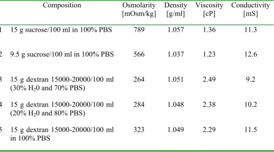
Sedimentation studies of human RBC cells were done in five solutions representing different physical properties, as described in Tab. 1. RBC sedimentation is shown over a 3 h period (Fig. 2). While hypertonic sucrose-containing solutions did not prevent sedimentation, it was minimized in dextran-containing solutions, especially slightly hypotonic ones. Moreover, under such conditions, the RBC did not aggregate, and under isopycnic conditions (where the densities of the solution and RBC are similar), they remained suspended as a relatively thin band (Fig. 2, lane 3; solution 3 from Tab. 1).
By contrast, other solutions containing polymers such as ficoll, starch, agarose and polyethylene glycol of different molecular weights (data not shown) were all inferior to the slightly hypotonic dextran solution shown to be non-toxic for RBC over 3 h. The physical properties of solution 3 (Tab. 1) were shown to be suitable for supporting RBC, and these were retained by the addition of sucrose (for viability) and reduced PBS (for conductivity) to provide a “density cushion” (shown as electrophoresis medium I in Fig.1).
Tab. 1. The physical properties of the solutions used in the cell sedimentation experiment. (cf. Fig. 2).
Composition Osmolarity
[mOsm/kg] Density [g/ml] Viscosity [cP] Conductivity [mS]
1 15 g sucrose/100 ml in 100% PBS 789 1.057 1.36 11.3
2 9.5 g sucrose/100 ml in 100% PBS 566 1.037 1.23 12.6
3 15 g dextran 15000-20000/100 ml (30% H20 and 70% PBS)
264 1.051 2.49 9.2
4 15 g dextran 15000-20000/100 ml (20% H20 and 80% PBS)
284 1.048 2.38 10.2
5 15 g dextran 15000-20000/100 ml in 100% PBS
15 min 60 min 120 min 180 min
Fig. 2. Sedimentation of RBC for indicated period. The test tubes contained solutions 1-5, of the composition given in Tab. 1.
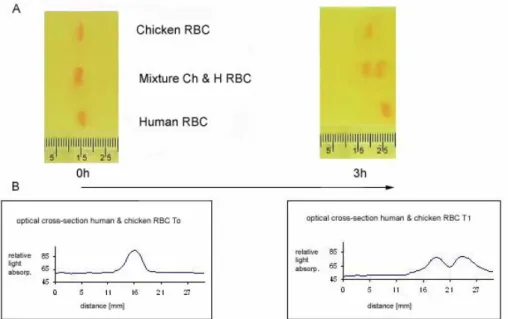
The separation of human and chicken RBC via horizontal, nearly isopycnic electrophoresis on a PU sponge
Fig. 3A shows the distribution of human and chicken RBC, and an equal mixture of both before and after electrophoresis. After 3 h, the mixed sample showed two distinct bands. The one with greater mobility corresponded to that for the human RBC, while the slower one was relative to the chicken RBC band. Fig. 3B shows the absorbance profiles of the mixed RBC preparation before and after 3 h of electrophoresis, confirming the resolution of the mixed sample into two distinct bands. Complete separation of the two bands could be achieved by extending the time for electrophoresis.
RBC analysis
Each RBC band was examined for purity using Coulter counter analysis and microscopy. The distribution of cell size is shown in Fig. 4, where the average diameter for the human RBC is shown as 5.2 µm, compared to approximately 5.5 µm for the chicken RBC. The profile shown in Fig. 4A for the mixed RBC sample before electrophoresis demonstrates two peaks of cell size which correspond to the two RBC species. Moreover, the difference in nucleation was evident in the microscopic haemacytometer analysis of the different RBC fractions (Fig. 5). The purity of the separated RBC bands required the sampling of 5-10 μl directly from the middle part of each band with a finely drawn Pasteur pipette (Ø approx. 0.5 mm). Tab. 2 shows the haemacytometric/microscopic analysis of the two RBC peaks, separated as shown in Fig. 3. The faster-moving band contained 97.6% human RBC, while the slower-moving band was 90% nucleated chicken RBC, as determined via microscopic haemacytometer analysis. The slower-moving band also contained a small proportion of larger contaminating cells such as leukocytes.
Fig. 4. Cell size distribution in the sample taken before RBC electrophoresis and in samples taken from the fast and slowly moving bands as determined using a Coulter counter.
Tab. 2. Purity of the erythrocyte fractions.
Results from Bürker haematocytometer Human RBC number (%) Chicken RBC number (%)
Faster band 244 (97.6) 6 (2.4)
Slower band 25 (10) 225 (90)
Dye electrophoresis
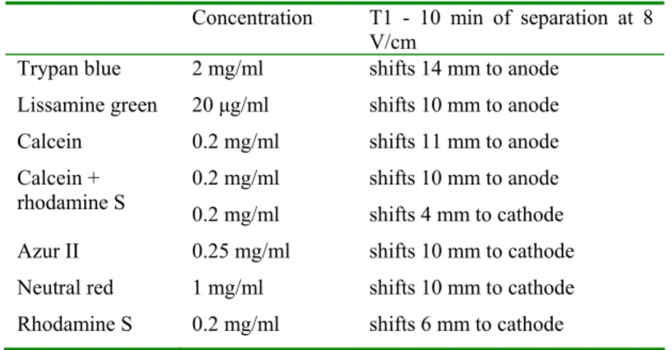
To demonstrate the electrophoresis system stability for the RBC separation described here, similar experiments were performed with the same system and solutions using dyes with different charges. The negatively charged dyes trypan blue, lissamine green and calcein all moved towards the anode (+ve), while the positively charged dyes azur II, neutral red and rhodamine S moved towards the cathode (-ve) (Fig. 6, Tab. 3). The dyes (all less than 1 kDa) became separated in less than 30 min under conditions of 8 V/cm. The negatively charged dyes showed different electrophoretic mobilities (TB > calcein > LG) as determined by their different charges. Some of the positively charged dyes had an affinity with, and stained, the PU matrice, and at least part of those dyes remained stationary. However, this did not prevent the separation of a mixed solution of calcein (-ve) and rhodamine S (+ve) as shown in Fig. 6 and Tab. 3. The results obtained show that the described horizontal electrophoresis system designed for the separation of cells is also suitable for the separation of much smaller molecules, especially those with a net negative charge.
Tab. 3. The concentration of the fluorescent dye solutions and band positions after 10 min. electrophoresis at 8 V/cm.
Concentration T1 - 10 min of separation at 8 V/cm
Trypan blue 2 mg/ml shifts 14 mm to anode Lissamine green 20 μg/ml shifts 10 mm to anode Calcein 0.2 mg/ml shifts 11 mm to anode 0.2 mg/ml shifts 10 mm to anode Calcein +
rhodamine S 0.2 mg/ml shifts 4 mm to cathode
Azur II 0.25 mg/ml shifts 10 mm to cathode Neutral red 1 mg/ml shifts 10 mm to cathode Rhodamine S 0.2 mg/ml shifts 6 mm to cathode
DISCUSSION
RBC isolated from various species have often been used as standards to examine cell electrophoretic separations [11, 24, 40-42, 46, 47], since their preparation does not require enzyme treatment and their cell surface properties are well characterized with the microscopic methods of cell electrophoresis. Human RBC have a two-fold higher electrophoretic mobility than rabbit RBC [26, 46, 48, 49]. As they are easily visually separable (chicken RBC are nucleated and thus easily identified by microscopy), this study used human and chicken RBC, which have approximately a 30% difference in their electrophoretic mobility.
The main obstacles to the development of a simple electrophoretic system for cell separations include: cell sedimentation/gravity; heat production and thermal convection; and factors which contribute to droplet sedimentation, pH instability and changes in electric conductivity. Such factors are associated with the good resolution of electrophoretically separated cells in free-flow apparatus under microgravity conditions [4, 29, 50, 51].
where: S – sedimentation constant, r – cell radius, ρp – cell (particle) density, ρm
– solution density, η – solution viscosity [52].
We chose RBC to examine our horizontal electrophoretic system for cell separation as RBC are cells which show relatively fast sedimentation in various methods based on cell separation in density gradients [52].
This simplified equation does not take into account the interaction of the cell surface with the molecules used to modify the solution density. Such events are very complex and cell surface specific [43, 49, 53], but our RBC sedimentation studies identified dextran 15000-20000 MW as the most suitable to form a “density cushion” for the RBC. These lower MW dextrans are osmotically active, whereas higher MW dextrans cause cell aggregation and RBC rouleaux formation, and coat the cell surfaces [37, 39, 44]. Other macromolecular solutions tested included ficolls or PEGs of MW ranging from 1000 to 400000, but these also coated the cell surface and modified cell permeability and electrophoretic mobility, as judged by haemoglobin leakage which compromised and even reversed the direction of cell electrophoresis [23, 43, 54-56].
Separation matrices for cell electrophoresis have to be optically transparent, hydrophilic, highly porous to allow free cell movement, and preferably electrically neutral to avoid or at least reduce electroosmosis. In addition, they should be non-adhesive for cells. Conventional matrices used for the electrophoretic separation of macromolecules were reported to have pore size values ranging from 2-3 nm to 200 nm [3]. After checking many spongy and/or random network-forming materials, we found that polyurethane synthetic sponges with pore sizes ranging between 200-5000 μm met these requirements (i.e. with pores 1000 to 2000 times greater than the porous materials used for the separation of proteins or nucleic acids). Such spongy hydrophilic matrices were found suitable as anti-convective supports suitable for the separation of cells with a diameter 5 to 20 μm. Polyurethane is reported to be non-adhesive for RBC and lymphocytes, but adhesive for macrophages [57], an observation confirmed in our experiments.
Ag/AgCl electrodes were used in preference to platinum since the latter caused changes in pH when DC current was applied. The addition of phenol red to the electrode solutions not only confirmed pH stability during electrophoresis but also provided assurance that the diffusion of the electrode solutions did not transgress the agar bridges. Moreover, although glucose or glycine were used for previous cell electrophoresis separation [20, 32], they were found to be inferior to sucrose for the best adjustment of media osmolarity and retention of RBC viability.
9
η
)
ρ
(
ρ
2r
S
p m2
−
To avoid the rise of temperature during electrophoresis, we reduced the conductivity of the separation solutions and the voltage/current employed to effectively separate viable RBC, as judged by microscopy, lack of haemoglobin release and trypan blue exclusion. Future studies using cooling blocks (water-perfused) or even cold-room operations should facilitate the separation of cells at a higher ionic strength than used in the described experiments. One of the advantages in using RBC in our horizontal electrophoretic system was the knowledge that the passive electrical parameters of the cell surface show little change despite any changes in cell shape, a conclusion based on radiowave dielectric spectroscopy [58].
In conclusion, we report here on a simple electrophoretic system which permits the separation of cells which differ in their electrophoretic mobilities and surface charge. Horizontal separations in near-isopycnic conditions appear to be more important than other considerations such as thermal convection and system instability. The main advantage of the described method is the facility to perform simultaneous electrophoresis of a few samples in parallel. This makes it easy to compare electrophoretic mobilities and/or separate experimental and control samples in one experiment. It appears that this horizontal electrophoretic separation of cells has many advantages over established vertical systems and may provide new opportunities for the electrophoresis of various cell types, not least by applying modifications already developed for the separation and study of smaller particles and macromolecules.
Acknowledgements. This study was partly supported by a grant from the Polish Committee for Scientific Research (3P040-02923). The authors cordially thank Prof. D. Woolley, University of Manchester, UK for discussions and language corrections.
REFERENCES
1. Abramson, H.A., Moyer, L.S. and Goris, M.H. Electrophoresis of proteins and the chemistry of cell surfaces, Reinhold NY 1942, 1-307.
2. Blanco, S., Clifton, M.J., Joly, J.L. and Peltre, G. Protein separation by electrophoresis in nonsieving amphoteric medium. Electrophoresis 17
(1996) 1126-1133.
3. Chiari, M. and Righetti, P.G. New types of separation matrices for electrophoresis. Electrophoresis 16 (1995) 1815-1829.
4. Roman, M.C. and Brown, P.R. Free-flow electrophoresis as a preparative separation technique. Anal. Chem. 66 (1994) 86-94.
5. Wang, Y., Hancock, W.S., Weber, G., Eckerskorn, C. and Palmer-Toy, D. Free-flow electrophoresis coupled with liquid chromatography/mass spectrometry for a proteomic study of the human cell line (K562/CR3).
6. Zakharov, S.F., Chang, H.T. and Chrambach, A. Reproducibility of mobility in gel electrophoresis. Electrophoresis 17 (1996) 84-90.
7. Ambrose, E.J. Cell Electrophoresis. J&A Churchill Ltd., London 1965. 8. Chaubal, K.A. Cell electrophoretic mobility as an aid to study biological
systems, in: Cell Electrophoresis (Schütt, W., Klinkmann, H., Ed.), Walter de Gruyter, Berlin (NY) 1985, 515-526.
9. Fürész, J., Pál, K., Budavári, I. and Lapis, K. The physico-chemical properties of tumor cells with different metastatic potential. Neoplasma 32
(1985) 689-693.
10. Korohoda, W. Electrophoretic studies on plant cells III. Electrophoretic mobilities of cell-forms of Myxomycetae Physarum nudum Macbride. Folia Biologica 11 (1963) 465-472.
11. Mehrishi, J.N. and Bauer, J. Electrophoresis of cells and the biological relevance of surface charge. Electrophoresis 23 (2002) 1984-1994.
12. Mori, T. and Shimizu, M. The changes of lymphocyte electrophoretic mobility in cancer patient. in: Cell Electrophoresis (Schütt, W., Klinkmann, H., Ed.),Walter de Gruyter, Berlin ( NY), 1985, 355-366.
13. Preece, A.W. and Sablović, D. in: Cell electrophoresis: clinical application and methodology. North-Holland Publishing company, Amsterdam 1979.
14. Abercrombie, M. and Ambrose, E.J. The surface properties of cancer cells: a review. Cancer Res. 22 (1962) 332-245.
15. Jovtchev, S., Djenev, I., Stoeff, S. and Stoylov, S. Role of electrical and mechanical properties of red blood cells for their aggregation. Colloids and Surfaces A:Physicochem. Engineer. Asp. 164 (2000) 95-104.
16. Vransky, V.K. Die zellelektrophorese. in: Fortschritte der experimentellen und theoretischen Biophysik Band 18. (Beier, W., Ed.) Leipzig 1974. 17. Masui, M., Takata, H. and Kominami, T. Cell adhesion and negative cell
surface charges in embryonic cells of the starfish Asterina pectinifera.
Electrophoresis 23 (2002) 2087-2095.
18. Platsoucas, C.D., Good, R.A. and Gupta, S. Separation of human T lymphocyte subpopulations (Tμ, Tγ) by density gradient electrophoresis.
Proc. Natl. Acad. Sci. USA 76 (1979) 1972-1976.
19. Rychly, J., Anders, O., Eggers, G. and Schulz, M. Electrophoretic mobility distribution of cells in leukaemia. in: Cell Electrophoresis (Schütt, W., Klinkmann, H., Ed.),Walter de Gruyter, Berlin (NY) 1985, 477-483.
20. Heidrich, H.G. and Hannig, K. Separation of cell population by free-flow electrophoresis. Methods Enzymol. 171 (1989) 513-531.
22. Sengeløv, H. and Borregaard, N. Free-flow electrophoresis in subcellular fractionation of human neutrophils. J. Immunol. Methods 232 (1999) 145-152.
23. Neu, B., Armstrong, J.K., Fisher ,T.C. and Meiselman, H.J. Surface characterization of poly(ethylene glycol) coated human red blood cells by particle electrophoresis. Biorheology 40 (2003) 477-487.
24. Seaman, G.V.F. and Cook, G.M.W. Modification of the electrophoretic behavior of the erythrocyte by chemical and enzymatic methods. in: Cell Electrophoresis (Ambrose, E.J., Ed.). J&A Churchill Ltd., London 1965, 48-65.
25. Wilson, W.W., Wade, M.M., Holman, S.C. and Champlin, F.R. Status of methods for assessing bacterial cell surface charge properties based on zeta potential measurements. J. Microbiol. Methods 43 (2001) 153-164.
26. Catsimpoolas, N. and Griffith, A.L. Transient electrophoresis and sedimentation analyses of cells in density gradients. In: Methods of Cell Separation, vol.2. (Catsimpoolas, N., Ed.) Plenum Press NY 1979, 1-63. 27. Pertoft, H. and Lauren, T.C. Isopycinc separation of cells and cell organelles
by centrifugation in modified colloidal silica gradients. in: Methods of Cell Separation, vol.1. (Catsimpoolas, N., Ed.) Plenum Press NY 1977, 25-65. 28. Pretlow II, T.G. and Pretlow, T.P. Separation of viable cells by velocity
sedimentation in an isokinetic gradient of ficoll in tissue culture medium. in:
Methods of Cell Separation, vol. 1. (Catsimpoolas, N., Ed.) Plenum Press NY 1977, 171-191.
29. Akiba, T., Nishi, A., Takaoki, M., Matsumiya, H., Tomita, F., Usami, R. and Nagaoka, S. Separation of bacterial cells by free flow electrophoresis under microgravity: a result of the spacelab – Japan project on space shuttle flight sts – 47. Acta Astron. 36 (1995) 177-181.
30. Zeiller, K., Löser, R., Pascher, G. and Hannig, K. Free-flow electrophoresis II: Analysis of the method with respect to preparative cell separation.
Hoppe-Seyler’s Z Physiol. Chem. 356 (1975) 1225-1244.
31. Eggleton, P. Separation of cells using free flow electrophoresis. in: Cell Separation. A Practical Approach. (Fisher, D., Francis, G.E. and Rickwood, D., Ed.) Oxford University Press, Oxford, New York, Tokyo 1998, 213-252.
32. Hansen, E. Preparative free flow electrophoresis of lymphoid cells: A review. in: Cell Electrophoresis (Schütt, W., Klinkmann, H., Ed.) Walter de Gruyter, Berlin (NY) 1985, 287-304.
33. Kuhn, R., Wagner, H., Mosher, R.A. and Thormann, W. Experimental and theoretical investigation of the stability of stepwise pH gradients in continuous flow electrophoresis. Electrophoresis 8 (1987) 503-508.
35. Morré, D.J., Morré, D.M. and van Alstine, J.M. Separation of endosomes by aqueous two-phase partition and free-flow electrophoresis. J. Chromatogr. B 711 (1998) 203-215.
36. Toner, M. and Irimia, D. Blood-on-a-chip. Annu. Rev. Biomed. Eng. 7 (2005) 77-103.
37. Barshtein, G., Tamir, I. and Yedgar, S. Red blood cell rouleaux formation in dextran solution: dependence on polymer conformation. Eur. Biophys. J. 27
(1998) 177-181.
38. Bäumler, H., Donath, E., Krabi, A., Knippel, W., Budde, A. and Kiesewetter, H. Electrophoresis of human red blood cells and platelets. Evidence for depletion of dextran. Biorheology 33 (1996) 333-351.
39. Gardner, B. The effect of dextrans on zeta potential. Proc. Soc. Exp. Biol. Med. 131 (1969)1115- 1118.
40. Ichiki, T., Ujiie, T., Shinbashi, S., Okuda, T. and Horiike, Y. Immunoelectrophoresis of red blood cells performed on microcapillary chips. Electrophoresis 23 (2002) 2029-2034.
41. Lu, W.H., Deng, W.H., Liu, S.T., Chen, T.B. and Ra, P.F. Capillary electrophoresis of erythrocytes. Anal. Biochem. 314 (2003) 194-198.
42. Omasu, F., Nakano,Y. and Ichik, T. Measurement of the electrophoretic mobility of sheep erythrocytes using microcapillary chips. Electrophoresis
26 (2005) 1163-1167.
43. Walter, H. and Widen, K.E. Differential electrophoretic behavior in aqueous polymer solutions of red blood cells from Alzheimer patients and from normal individuals. Biochim. Biophys. Acta 1234 (1995) 184-190.
44. Bäumler, H., Neu, B., Donath, E. and Kiesewetter, H. Basic phenomena of red blood cell rouleaux formation. Biorheology 36 (1999) 439-442.
45. Schüt, W., Thomaneck, U., Knippel, E., Rychly, J. and Klinkmann, H. Suitability of automated single cell electrophoresis (ASCE) for biomedical and clinical applications: General remarks. in: Cell Electrophoresis (Schütt, W., Klinkmann, H., Ed.) Walter de Gruyter, Berlin ( NY) 1985, 313-332. 46. Seaman, G.V.F. Electrokinetic behavior of red cells. in: The Red Blood
Cellsvol. 2. (Mac, D., Surgenor, N., Ed.), Academic Press, New York 1975, 1-135.
47. Slivinsky, G.G., Hymer, W.C., Bauer, J. and Morrison, D.R. Cellular electrophoretic mobility data: A first approach to a database.
Electrophoresis 18 (1997) 1109-1119.
48. Josefowicz, J.Y. Electrophoretic light scattering and its application to the study of cells. in: Methods of Cell Separation, vol. 2. (Catsimpoolas, N., Ed.) Plenum Press NY 1979, 67-91.
49. Walter, H. Cell partitioning in two-polymer aqueous phase systems. TIBS (1978) 97-100.
51. Todd, P. Microgravity cell electrophoresis experiments on the space shuttle: a 1984 overview. in: Cell Electrophoresis. (Schütt, W., Klinkmann H., Ed.), Walter de Gruyter, Berlin ( NY) 1985, 3-19.
52. Patel, D., Ford, T.C. and Rickwood, D. Fractionation of cells by sedimentation methods. in: Cell Separation. A Practical Approach.
(Fisher, D., Francis, G.E. and Rickwood, D., Ed.) Oxford University Press, Oxford, New York, Tokyo 1998, 43-89.
53. Malström, P., Nelson, K., Jönsson, A., Sjögren, H.O., Walter, H. and Albertsson, P.A. Separation of rat leukocytes by countercurrent distribution in aqueous two-phase systems. Cell Immunol. 37 (1978) 409-421.
54. Arnold, K., Herrmann, A., Pratsch, L. and Gawrisch, K. The dielectric properties of aqueous solutions of poly(ethylene glycol) and their influence on membrane structure. Biochim. Biophys. Acta 815 (1985) 515-518. 55. Hansen, P.L., Cohen, J.A., Podgornik, R. and Parsegian, V.A. Osmotic
properties of poly(ethylene glycols): quantitative features of brush and bulk scaling lows. Biophys. J. 84 (2003) 350-355.
56. Sabolovic, D., Sestier, C., Perrotin, P., Guillet, R., Tefi, M. and Boynard, M. Covalent binding of polyethylene glycol to the surface of red blood cells as detected and followed up by cell electrophoresis and rheological methods.
Electrophoresis 21 (2000) 301-306.
57. Higuchi, A., Yamamiya, S., Yoon, B.O., Sakurai, M. and Hara, M. Peripheral blood cell separation through surface modified polyurethane membranes. J. Biomed. Materials Res.A 68A (2004) 34-42.
58. Di Basio, A. and Cametti, C. Effect of the shape of human erythrocytes on the evaluation of the passive electrical properties of the cell membrane.