Address for correspondence
Dr. Lubna Khondker, Assistant Professor, Department of Dermatology and Venereology Bangabandhu Sheikh Mujib Medical University (BSMMU)
Dhaka, Bangladesh
Email: [email protected]
Original Article
Comparative efficacy of topical mometasone
furoate 0.1% cream vs topical tacrolimus 0.03%
ointment in the treatment of atopic dermatitis
Md Alauddin Khan*, Lubna Khondker**, Dilshad Afroze*
*Department of Medicine, Bangabandhu Sheikh Mujib Medical University (BSMMU), Dhaka, Bangladesh
**Department of Dermatology and Venereology, Bangabandhu Sheikh Mujib Medical University (BSMMU), Dhaka, Bangladesh
Abstract
Objective To compare the efficacy of mometasone furoate and tacrolimus in the treatment of atopic dermatitis.Methods Sixty patients of atopic dermatitis were treated with mometasone furoate 0.1% (n=30) and tacrolimus 0.03% (n=30). Both treatments were applied twice daily for 12 weeks. Patients were followed up monthly. The disease severity assessed by SCORAD index. A 4-point scale was used to measure the level of response to treatment.
Results Before treatment the respective mean SCORAD was 30.57±13.62 and 30.90±17.17 in group A and B and at the end of treatment decreased to 11.87±12.04 and 11.20±13.85, respectively (p>0.05). Percent reduction of severity from baseline to final follow-up was 69.20±23.41 in group A and 74.77±23.30 in group B (p=0.360). At final follow-up 56.7% of group A and 63.3% of group B achieved excellent response, 13.3% of group A and 16.7% of group B achieved good response.
Conclusion We conclude that both treatments, mometasone furoate and tacrolimus, are effective in the treatment of atopic dermatitis.
Key words
Efficacy, mometasone furoate, tacrolimus, atopic dermatitis.
Introduction
Atopic dermatitis (AD) is an itchy, chronic or chronically relapsing, inflammatory skin condition. The lesion is characterized by itchy papules, occasionally vesicles which become
excoriated and lichenified.1,2 Atopy is a
syndrome which may be defined as a
genetically determined immune system
maturation disorder of unknown origin, in which there is increased liability to form IgE antibodies and is frequently associated with
personal or family history of atopic dermatitis, allergic rhinitis or asthma.3,4 In AD there is
activation of the T helper 2 (Th2) immune response, with synthesis of cytokines 4, IL-5, IL-10 and IL-13 and inhibition of T helper 1 (Th1) response. IL-4 and IL-5 produces elevated IgE level and eosinophilia in tissue and peripheral blood.5,6 Pruritus is the hallmark
of atopic dermatitis in all stages. 60% infantile AD patients present in first year of life.7,8
Pruritus is paroxysmal and a constant feature.
Skin biopsy for histopathology show
spongiosis and edema of the epidermis, hyperkeratosis and acanthosis in chronic stage along with perivascular infiltrate of in upper
dermis.9,10 Topical corticosteroids are very
have many side effects.11 Mometasone furoate
is a medium potency corticosteroid, indicated for the relief of the inflammatory and pruritic manifestations of atopic dermatitis.12 Topical
calcineurin inhibitors like tacrolimus may be used as alternate to steroid. Topical tacrolimus suppresses inflammation in a similar way to steroids and is equally as effective as a medium potency steroid.10 It does not cause
skin thinning or other steroid related side-effects.13 To the best of my knowledge, no
study exploring the efficacy and safety of topical mometasone furoate comparing with topical tacrolimus in the treatment of atopic dermatitis has yet been conducted in Bangladesh. So, to know and to treat the patient with atopic dermatitis in an effective way, such kind of study was conducted in Bangladesh.
Methods
A clinical trial was conducted in department of Dermatology and Venereology, Bangabandhu Sheikh Mujib Medical University, Dhaka. The duration of the study was from September 2011 to February 2012. Patients of atopic dermatitis attending outpatient department of Dermatology and Venereology, Bangabandhu Sheikh Mujib Medical University, Dhaka were the study population. Purposive type of non-probability sampling method was followed in this study. Inclusion criteria were patients of atopic dermatitis diagnosed clinically, patient who gave informed written consent, age more than 2 years and patients of either gender.
Exclusion criteria were: known
hypersensitivity to mometasone or tacrolimus,
patients suffering from hepatic, renal,
cardiovascular and hematological diseases, patients with co-existing acute infections, neoplasia, uncontrolled hypertension and diabetes mellitus, patients suffering from any food allergy and other skin morbidity causing acute onset of skin rash, skin disorders likely to affect drug absorption or disorders requiring
medical treatment within 5 days before the start of the study, pregnancy and lactation.
Procedure of data collection
A total number of 60 patients were primarily selected and they were randomized using computer-generated codes into two groups (group A and group B), each of which included 30 patients. Complete history,
general physical and dermatological
examinations were done for all enrolled patients. History and physical findings were recorded in a structured proforma. Patients, who matched the inclusion and exclusion criteria and freely gave their informed consent, were selected for the study.
Intervention
Group A was treated with mometasone furoate 0.1% ointment and group B with tacrolimus 0.03% ointment. Both preparations were applied twice daily for a period of 12 weeks. Patients were clinically assessed monthly for three months. Each time the severity of the disease was recorded and clinical photographs were taken. The severity of disease was measured by Scoring of Atopic Dermatitis (SCORAD) index. For calculating SCORAD index, six clinical signs were recorded for each
case: erythema, edema/papulation,
is defined as the subjective symptoms (0-20). The maximum SCORAD score is 103 (i.e. patients with high score are rated “worse").
A 4-point scale was used to measure the level of response to treatment, if >75% clear - excellent response; if 50-75% clear - good response; if 25-50% clear fair response; if <25% clear - poor response.
Data analysis
Data analysis was performed by Statistical Package for Social Science (SPSS), version-12. Level of significance (p value) was set at 0.05 and confidence interval at 95%.
Results
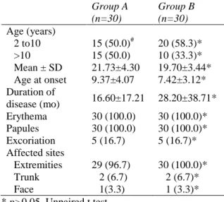
Table 1 shows the clinical characteristics in the two groups. Both groups were comparable in all parameters (p<0.05). Mean age of group A patients was 21.73±4.30 and group B was 19.70±3.44 years. 50% of group A and 58.3% of group B were from the 2 to10 year age group and 50.0% of group A and 33.3% of group B wee older than 10 years. Mean age of onset of disease was 9.37±4.07 years and 7.42±3.12 years in group A and group B,
respectively (p=0.420). Mean duration of
disease was 16.60±17.21 months and
28.20±38.71 months in group A and group B, respectively (p=0.139).
All patients of both groups presented with erythema, papules and 16.7% presented with excoriation (p=0.999). Lesions on extremities were present in 96.7% of group A and 100.0% of group B, lesions on trunk were present in 6.7% and lesions on face in 3.3% (p>0.05).
In Table 2 effect of both treatments on three cardinal signs of diseases is compared. Both groups showed a comparable improvement
(p>0.05). At baseline, mean number of
erythematous lesions in group A and group B
Table 1 Demographic and clinical data of patients. Group A
(n=30)
Group B (n=30) Age (years)
2 to10 15 (50.0)# 20 (58.3)* >10 15 (50.0) 10 (33.3)* Mean ± SD 21.73±4.30 19.70±3.44* Age at onset 9.37±4.07 7.42±3.12* Duration of
disease (mo) 16.60±17.21 28.20±38.71* Erythema 30 (100.0) 30 (100.0)* Papules 30 (100.0) 30 (100.0)* Excoriation 5 (16.7) 5 (16.7)* Affected sites
Extremities 29 (96.7) 30 (100.0)*
Trunk 2 (6.7) 2 (6.7)*
Face 1(3.3) 1 (3.3)*
* p>0.05, Unpaired t test.
Table 2 Extent of score of erythematous lesion, papules and excoriation in different follow-up.
Group A Group B Erythematous lesion
Baseline 12.77±4.01 11.80 ±3.93 1st follow-up 7.80±4.11 7.77 ±4.08 2nd follow-up 6.10±4.03 5.63 ±4.16 Final follow-up 4.17±4.02 3.47 ±4.00 Papules
Baseline 17.30±10.29 18.57±13.88 1st follow-up 12.40±9.46 13.10± 12.67 2nd follow-up 9.97±8.73 10.10± 11.17 Final follow-up 7.63±8.08 7.73±9.98 Excoriation
Baseline 0.50±1.33 0.53±1.28 1st follow-up 0.30±0.88 0.30±0.75 2nd follow-up 0.17±0.59 0.10±0.31 Final follow-up 0.07±0.37 0.00 *p>0.05, Unpaired t test.
Table 3 SCORAD (Mean of total scoring of Atopic dermatitis) in different follow up.
Group A Group B Baseline 30.57±13.62 30.90±17.17* 1st follow-up 20.50±13.64 21.17±16.94* 2nd follow-up 16.23±12.74 15.83±15.29* Final follow-up 11.87±12.04 11.20±13.85* % reduction from
baseline to final follow-up
69.20±23.41 74.77±23.30*
*p>0.05, Unpaired t test.
was 12.77±4.01 and 11.80±3.93, respectively (p=0.350). At 1st follow-up mean number of
erythematous lesions in group A and group B
Table 4 Clearance level of disease at different follow-ups.
Group A Group B 1st follow up
Excellent 1 (3.3)# 1 (3.3)
Good 3 (10.0) 8 (26.7)
Fair 18 (60.0) 12 (40.0)
Poor 8 (26.7) 9 (30.0)
2nd follow up
Excellent 4 (13.3) 9 (30.0) Good 14 (46.7) 12 (40.0)
Fair 9 (30.0) 7 (13.3)
Poor 3 (10.0) 2 (6.7)
3rd follow up
Excellent 17 (56.7) 19 (63.3)
Good 4 (13.3) 5 (16.7)
Fair 7 (23.3) 5 (16.7)
Poor 2 (6.7) 1 (3.3)
*p>0.05, Chi square test.
3.47±4.00 (p>0.05). At baseline mean number
of papules in group A and group B was 17.30±10.29 and 18.57±13.88, respectively (p=0. 690). At 1st follow-up, mean number of papules in group A and group B was 12.40±9.46 and 13.10±12.67, respectively. At
2nd follow up, it was 9.97±8.73and
10.10±11.17 and at final follow-up 7.63±8.08 and 7.73±9.98 (p>0.05). At baseline, mean number of excoriation in group A and group B was 0.50±1.33 and 0.53±1.28, respectively (p=0. 922). At 1st follow up mean number of excoriation in group A and group B was
0.30±0.88 and 0.30 ± 0.75 respectively, at 2nd
follow up it was 0.17±0.59 and 0.10±0.31 and at final follow up 0.07±0.37 and 0.00 (p>0.05).
At baseline mean of total score of atopic dermatitis was 30.57±13.62 and 30.90±17.17 in group A and B, at 1st follow up it was 20.50±13.64 and 21.17±16.94, respectively in
group A and B, at 2nd follow up it was
16.23±12.74 and 15.83±15.29 and at final follow up it was 11.87±12.04 and 11.20±13.85
respectively in group A and B (p>0.05).
Percent reduction of severity from base line to final follow up was 69.20±23.41 in group A
and 74.77±23.30 in group B (p=0.360).
At 1st follow up 3.3% of both group got
excellent response, 10.0% of group A and 26.7% group B got good response, 60.0% of group A and 40.0% of group B got fair response and 26.7% of group A and 30.0% of
group B got poor response (p=0.317).
At 2nd follow up 13.3% of group A and 30.0%
of group B got excellent response, 46.7% of group A and 40.0% of group B got good response, 30.0% of group A and 13.3% of group B got fair response and 10.0% of group A and 6.7% of group B got poor response (p=0.470). At final follow up 56.7% of group A and 63.3% of group B achieved excellent response, 13.3% of group A and 16.7% of group B achieved good response, 23.3% of group A and 16.7% of group B achieved fair response and 6.7% of group A and 3.3% of
group B achieved poor response (p=0.828).
Discussion
In our study, both treatments showed a significant improvement in mean scores of three important clinical signs of AD, mean SCORAD index and level of clearance in their respective groups (p<0.05). The improvement in all these parameters was similar in two groups (p>0.05), showing that both drugs are equally effective in children as well adults suffering from AD. The efficacy of both drugs is proven in children9,10 but few studies14
directly compared these two drugs in AD.
Gradman et al.14 compared the suppressive
effects of topical mometasone furoate and tacrolimus on skin prick testing in 12 children with atopic eczema before and after 2 weeks of treatment with topical mometasone furoate and tacrolimus. Both treatments significantly suppressed the allergen wheal size.
Pei et al.15 conducted a study to observe the
fluticasone proprionate ointments in the treatment of moderate to severe atopic dermatitis in children. There was significant improvement in the disease severity from baseline during the first 2 weeks of the open
application arm (p=0.043).15 In another
vehicle-controlled trial in AD by Schnopp et
al.16 mometasone furoate 0.1% ointment was
significantly better than vehicle (p<0.01).
Hoeger et al.17 determined the efficacy of
tacrolimus vs. vehicle in children with mild-moderate facial AD dependent on/intolerant of topical corticosteroids. Investigators' global
assessment (p=0.004) and median time to
clearance was significantly better in favour of tacrolimus treated group.17
Zuberbier et al.18 studied whether treatment of
patients with AD with tacrolimus can decrease the development of flares necessitating the use of a topical corticosteroid on the face and thus
reduce the need for use of topical
corticosteroids in this sensitive skin area. Patients in the vehicle group needed prednicarbate treatment on the face on 20.7% of the days vs. 11.7% of the study days in the
tacrolimus group (p=0.0024). Fifty per cent of
patients in the tacrolimus group had no flare on the face during the treatment period compared with 37.5% of patients in the
vehicle group (p=0.012). Long-term
intermittent treatment of facial AD in children and adolescents with tacrolimus cream 1% does significantly reduce the need for topical corticosteroids.18
Conclusion
In the light of the findings of the study we conclude that each of the treatment of
mometasone furoate and tacrolimus is
individually effective in the treatment of atopic dermatitis. The efficacy of mometasone furoate 0.1% is almost same as that of
tacrolimus in the treatment of atopic dermatitis.
References
1. Atherton DJ. Topical corticosteroids in atopic dermatitis. Recent research reassures that they are safe and effective in the medium term. BMJ. 2003;327: 942- 3.
2. Green C, Colquitt JL, Kirby J et al. Clinical and cost-effectiveness of once daily versus more frequent use of same potency topical corticosteroids for atopic eczema: a systematic review and economic evaluation. Health Technol Assess. 2004;8:1-120.
3. Hauser C, Wuetrich B, Matter L et al. Staphylococcus aureus skin colonization in atopic dermatitis. Dermatologica. 1985;170:35-9.
4. McFadden JP, Noble WC, Camp RD. Superantigenic exotoxin secreting potential of staphylococci isolated from eczematous skin. Br J Dermatol. 1993;128:631-2.
5. Dhar S. Should topical antibacterials be routinely combined with topical steroids in the treatment of atopic dermatitis? Indian J Dermatol Venereol Leprol. 2005;71:71-2.
6. Proksch E. The role of emollients in the management of diseases with chronic dry skin. Skin Pharmacol Physiol. 2008;21:75-80.
7. Eichenfield LF, Hanifin JM, Luger TA et al. Consensus conference on pediatric atopic dermatitis. J Am Acad Dermatol. 2003;49:1088-95.
8. Gambichler T. Narrowband UVB phototherapy in skin conditions beyond psoriasis. J Am Acad Dermatol. 2005;52:660-70.
9. Rajka G, Avrach W, Gartner L, Overgaard-Petersen H. Mometasone furoate 0.1% fatty cream once daily versus betamethasone valerate 0.1% cream twice daily in the treatment of patients with atopic and allergic contact dermatitis. Cur Ther Res Clin Exp. 2005;54:23-9.
10. Hoybye S, Balk MS, De Cunha BF et al. Continuous and intermittent treatment of atopic dermatitis in adults with mometasone furoate versus hydrocortisone 17-butyrate. Cur Ther Res Clin Exp. 2004;50:67-72.
12. Williams H. New treatments for atopic dermatitis. BMJ. 2002;324:1533-4. 13. Lagos BR, Maibach HI. Frequency of
application of topical corticosteroids: an overview. Br J Dermatol. 2003;139:763-6. 14. Gradman J, Wolthers OD. Suppressive effects of topical mometasone furoate and tacrolimus on skin prick testing in children. Pediatr Dermatol. 2008;25:269-70.
15. Pei AY, Chan HH, Ho KM. The effectiveness of wet wrap dressings using 0.1% mometasone furoate and 0.005% fluticasone proprionate ointments in the treatment of moderate to severe atopic dermatitis in children. Pediatr Dermatol. 2001;18:343-8.
16. Schnopp C, Holtmann C, Stock S et al. Topical steroids under wet-wrap dressings in atopic dermatitis—a vehicle-controlled trial. Dermatology. 2002;204:56-9. 17. Hoeger PH, Lee KH, Jautova J et al. The
treatment of facial atopic dermatitis in children who are intolerant of, or dependent on, topical corticosteroids: a randomized, controlled clinical trial. Br J Dermatol. 2009;160:415-22. Epub 2008 Nov 25.