Original Article
Impacted Mandibular Third Molar, Associated Pathoses, and Their
Relation to Angulation and Impaction Depth: A Cone Beam CT
Study
Movahhedian Na; Shahidi Shb*; Jozari Sc; Mosharaf Ad; Naderi Ae
aAssistant Professor, Department of Oral and Maxillofacial Radiology, Faculty of Dentistry, Shiraz University of
Medial Science, Shiraz, Iran
bProfessor, Department of Oral and Maxillofacial Radiology, Biomaterials Research Center, School of Dentistry,
Shiraz University of Medical Sciences, Shiraz, Iran
cUndergraduate Student, Student Research Committee, School of Dentistry, Shiraz University of Medical Sciences,
Shiraz, Iran
dpostgraduate student of oral and maxillofacial Surgery, Department of Oral and Maxillofacial Surgery, school of
dentistry, Shiraz University of Medical Sciences, Shiraz, Iran
epostgraduate student of oral and maxillofacial radiology, Department of Oral and Maxillofacial Radiology, school
of dentistry, Shiraz University of Medical Sciences, Shiraz, Iran
ARTICLE INFO Abstract
Article History:
Received: 16 September 2017 Accepted: 12 November 2017
Statement of problem: prophylactic removal of the impacted lower third molar (ILTM) is controversial and accompanying pathologic conditions play an important role.
Objectives: The aim of the present study is to evaluate the prevalence of commonly found pathoses associated with ILTM in relation to angulation and impaction depth in cone beam computed tomography (CBCT).
Materials and Methods: We evaluated CBCT of 500 ILTMs from 235 females (57%) and 177 males (43%) for the presence of caries on the second and third molars, external root resorption (ERR) of the second molar, and follicular spaces (FS) >5 mm in diameter in relation to angulation and impaction depth according to Pell and Gregory and Winter’s classifications, respectively. Results: We observed that 55.6% of ILTM had at least one detectible lesion. ERR was the most frequent pathologic condition (31.2%), followed by caries on the second (26%) and third (13.4%) molars, and FS >5 mm (2.4%). ERR was the only pathology influenced by angulation. There was significantly more ERR in mesioangular ILTMs (40.5%, P<0.001). Most ERR occurred in direct contact with the third molar. Class C showed a lower risk for second and third molar caries (P<0.001), but higher risk for ERR (P=0.008) and FS >5 mm (P=0.035). There were more caries on the second molar (P=0.013) and FS >5 mm (P<0.001) in class III.
Conclusions: Prophylactic removal of ILTMs (especially in mesioangular or horizontal impactions) could be suggested considering the potential for pathologic changes in ILTMs and the propensity for these teeth to cause ERR in second molars.
Key words:
Impacted mandibular third molar
Cone beam computed tomography (CBCT) External root resorption Caries
Follicular space Corresponding Author: Shoaleh Shahidi
Professor, Department of Oral and Maxillofacial Radiology, Biomaterials Research Center, School of Dentistry, Shiraz University of Medical Sciences, Shiraz, Iran
Email: shoalehshahidi@yahoo. com
Tel: +98-9173119066 Fax: +98-7136270325
Cite this article as: Movahhedian N, Shahidi Sh, Jozari S, Mosharaf A, Naderi A. Impacted Mandibular Third Molar, Associated Pathoses, and Their Relation to Angulation and Impaction Depth: A Cone Beam CT Study.J Dent Biomater, 2018;5(1):520-527.
Introduction
Tooth impaction is defined as the condition in which a tooth fails to erupt into its normal position after root completion due to pathological or developmental factors such as jaw size, eruption pattern, and any barrier to the eruption path [1]. In a survey, researchers have reported that 28.3% of the population had at least one impacted tooth, 82.5% of which was the mandibular third molar [2].The decision regarding prophylactic removal or retention of the impacted third molar has been a challenging and controversial subject for dental professionals [3-6] and accompanying pathologic conditions play an important role in this decision. While some of these impacted lower third molars (ILTMs) may remain with no pathologic or clinical complications, others may develop pathologic conditions that include infection, external root resorption (ERR) or caries of the adjacent teeth, and cystic/tumoral transformation of the follicle [3,7,8]. Radiographs are necessary to evaluate impacted teeth. Compared to conventional techniques, three-dimensional imaging modalities such as computed tomography (CT) and cone beam CT (CBCT) provide more information to analyze impacted teeth, which would not be available otherwise. Additionally, because of less effective radiation dosage, the relatively lower cost, and fewer artifacts, CBCT has proven to be the technique of choice for evaluation of impacted teeth and related pathologic conditions [9-11]. Most data that pertain to the pathos of ILTM is available through panoramic studies [3,7,12-14]. This modality has a number of shortcomings that include lack of detail, distortion, superimposition, and projection errors, which in turn reduces its validity [12].
The objective of this study is to assess the prevalence of the most common pathoses associated with ILTMs that include caries on the second and third molars, ERR of the second molar, and follicular spaces (FS) >5 mm in diameter in relation to their impaction depth and angulation by means of CBCT images. The results of this study may help dental professionals with the risk assessment of removal or retention of an ILTM. Materials and Methods
In this retrospective cross-sectional study, we analyzed 500 ILTMs from 412 patients. These patients had preoperative radiologic examinations of their third molars over a two-year time frame (February 2014 to February 2016) in a private oral and maxillofacial radiology clinic in Shiraz, Iran. The Ethics and Research Committee of Shiraz University of Medical Sciences approved the study protocol (# 03-9504). All CBCT images were obtained with a NewTom VGi (Quantitative Radiology, Verona, Italy) CBCT unit that operated at 90 kVp and 6 mA with an exposure time of 10 seconds, and voxel size set for 0.3 mm in a 15×15 field of view. The images were evaluated with NNT viewer software (version 4.6, NewTom, Verona, Italy) in all three orthogonal planes (axial, coronal, and sagittal) by two oral and maxillofacial radiologists with consensus. In instances where it was difficult for the two radiologists to reach an agreement, a third expert reviewed the images. The parameters evaluated in the present study were caries on the distal surface of the mandibular second molar, caries of the mandibular third molar, FS of more than 5 mm in diameter around the third molar, and ERR of the second molar (Figure 1). Different degrees of resorption from a mild blunting of the root curvature to resorption that invaded the pulpal structure were considered as the stages of root resorption.The lower third molar impaction depths in relation to the ramus and occlusal plane have been determined according to the Pell and Gregory classification [15], as follows. Class I: the third molar is completely anterior to the anterior border of the ramus; class II: up to half of the third molar crown is covered by the ramus; class III: more than half of the third molar crown is covered by the ramus. Moreover, Class A: the third molar occlusal plane is at the same level or above the second molar occlusal plane; class B: the third molar occlusal plane is between the cervical line and occlusal plane of the second molar; and class C: the third molar is below the cervical line of the second molar.We used the Winter classification [16] to determine the ILTM inclination according to which the angle between the longitudinal axis of the ILTM to the occlusal plane defined the inclination, as follows: 0-30: horizontal; 31-60: mesioangular; 61-90: vertical; and >90: distoangular.This study excluded subjects
Figure 1: (A, B) External root resorption (ERR) in the sagittal and axial planes. (C, D) Enlarged follicular space (FS) in the sagittal and axial planes. (E) Caries on the second molar. (F) Caries on the third molar
with gross carious lesions, extensive restoration in the second molar, third molars simultaneously in classes I and A, or root development of less than two thirds.
Statistical analysis
We used the chi-square test to assess the relationship between variables. SPSS version 18.0 (SPSS Inc., Chicago, IL, USA) was applied for statistical analysis. P<0.05 was considered statistically significant.
Results
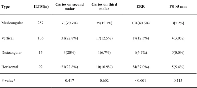
The sample for this study consisted of 500 ILTMs from 412 CBCT images of which there were 235 (57%) females and 177 (43%) males. Most cases (51.4%, 257 teeth) were in a mesioangular position, followed by vertical (27.2%, 136 teeth), horizontal (18.4%, 92 teeth), and distoangular (3%, 15 teeth)
positions. Class A comprised 6.4% of the ILTMs, followed by 56.0% for class B, and 37.6% for class C. The impaction depth in the images of the samples were 23.6% (class I), 54.4% (class II), and 22% (class III). Among the evaluated pathologic conditions, ERR of the second molar was the most frequent with a prevalence of 31.2%, followed by caries on the second (26%) and third (13.4%) molars, and FS >5 mm in diameter (2.4%). By taking into consideration the results, we concluded that 222 (44.4%) cases did not have any of the evaluated pathologies and 278 (55.6%) had at least one detectible lesion. Table 1 shows the distribution of evaluated pathologies according to angulation. The only pathologic factor influenced by the angulation of the teeth was ERR (P<0.001), which was significantly more prevalent in mesioangular ILTMs (40.5%). The occurrence of caries and FS were not significantly related to ILTM angulation (Table 1). Only one out of 12 enlarged FS was
Table 1: Frequency of pathologic findings in terms of the angulation of the third molar
Type ILTM(n) Caries on second molar Caries on third molar ERR FS >5 mm
Mesioangular 257 75(29.2%) 39(15.2%) 104(40.5%) 3(1.2%)
Vertical 136 31(22.8%) 17(12.5%) 17(12.5%) 4(3.0%)
Distoangular 15 3(20%) 1(6.7%) 1(6.7%) 0(0.0%)
Horizontal 92 21(22.8%) 10(10.9%) 34(37.0%) 5(5.4%)
P-value* 0.417 0.602 <0.001 0.115
*Chi-square test (base on Monte-Carlo method)
ILTM = Impacted mandibular third molar, ERR = external root resorption, FS= Follicular space associated with a horizontally impacted third molar
and caused ERR on the second molar. Analysis of the relationship between pathologic conditions and impaction status showed that class C had a lower risk for caries on the second and third molars (both
Table 2: Frequencies of pathologic findings in terms of impaction status
Type ILTM(n) second molarCaries on Caries on third molar ERR FS >5 mm
Class A 32(6.4%) 11(34.4%) 7(21.9%) 3(9.3%) 0(0.0%)
Class B 280(56.0%) 105(37.5%) 55(19.6%) 84(30.0%) 2(0.7%)
Class C 188(37.6%) 14(7.4%) 3(1.6%) 68(36.2%) 8(4.2%)
P-value* <0.001 <0.001 0.008 0.035
Class I 118(23.6%) 38(32.2%) 18(15.2%) 32(27.1%) 1(0.8%)
Class II 272(54.4%) 74(27.2%) 41(15.1%) 86(31.6%) 1(0.3%)
Class III 110(22.0%) 17(15.4%) 7(6.3%) 38(34.5%) 8(7.2%)
P-value* 0.013 0.054 0.474 <0.001
*Chi-square test (base on Monte-Carlo method)
ILTM= Impacted mandibular third molar, ERR = external root resorption, FS=Follicular space
P<0.001). However, as Table 2 shows, we observed an increasing risk for ERR (P=0.008) and FS >5 mm (P=0.035) gradually from class A to class C. Table 2 also shows that class III had significantly more caries on the second molar (P=0.013) and FS
>5 mm (P<0.001). Discussion
The decision to prophylactically remove or retain an ILTM is a matter of great debate with divergent opinions among professionals [3-6]. The complications following extraction of the impacted third molar have tremendous influence on this debate [3]. Oral surgeons should weight the benefits of removing a third molar against the risks. While there is no documented evidence for prophylactic surgery of an asymptomatic third molar, some researches have rejected this idea. However, there is strong indication for removal of the third molar in cases with accompanied pathologic changes [12,17]. In support, the American Association of Oral and Maxillofacial Surgeons (AAOMS) have provided a guideline for the extraction of third molars according to associated lesions [18]. AAOMS has also advocated new studies that concern the evaluation of pathologic conditions accompanied with third molars [12]. Two-dimensional characteristics, as well as the lack of sufficient details, distortion, and potential errors make it difficult for panoramic radiography to detect lesions as precisely as CBCT [8,12,17,19,20]. CBCT allows for the examination of a subject free from overlapping structures in all three orthogonal planes with higher spatial resolution [21]. Studies that examined the ability of panoramic and CBCT radiography to diagnose ERR [22,23] and proximal and occlusal caries [24,25] have confirmed the favorable position of CBCT over panoramic radiography. Oenning et al. [8] reported an agreement of only 4.3% between CBCT and panoramic radiography in ERR detection. They recommended CBCT for its evaluation. Another study by Alqerban et al. [23] had similar results in the ERR of incisor teeth. The higher prevalence of ERR in the present study (31.2%) compared to panoramic studies supported this fact. Similar studies reported incidents for ERR that ranged from 0.3% [7] to 1.4% [13]. Although the overall prevalence of ERR in this study was 31.2%, there were significantly higher incidents of third molars that had mesioangular (40.5%) and horizontal (37%) inclinations. The results of the present study were closer a study by Oenning
et al. that reported 49.43% ERR associated with mesioangular ILTM on CBCT [12]. Other studies supported the results of this study regarding the higher frequency of ERR in mesially inclined third molars [8,12]. These findings could be related to the eruptive forces of mesially inclined impacted third molars and probability of more contact area with second molars. However, the relationship between impaction depth and ERR has been inconsistently reported in the literature. Our findings revealed a significant tendency of class C (36.2%) and B (30%) impactions for ERR, while Wang et al. [17] reported more ERR in class A and C, and Oenning et al. [12] showed a higher frequency of ERR in class A and B impactions. This discrepancy, as well as lower frequencies of ERR reported in other CBCT studies [8,17], could be due to differences in study inclusion criteria, sample size, definition of ERR and its complicating resemblances to proximal caries, particularly in partially erupted third molars. According to the current study data, 26% of second molars and 13.4% of third molars showed carious lesions; distal caries of the second molars were the most common pathos evaluated in the present study after ERR. Al-Khateeb et al. [7] reported similar findings regarding the prevalence of caries on the third molar (13.6%). Our findings of distal caries on the second molar were higher than some studies that reported frequencies of 7.9% [7], 12.6% [3], and 17.2% [26]. Our findings were similar to those reported by Ozec et al. (20%) [27]. However, several studies reported higher frequencies of 42% [28] and 52% [24]. The results of this study revealed higher frequencies of impaction depth in class A and B. This finding supported previous studies [3, 24,26], which found classes A and B more afflicted with distal caries on the second molar. This variation in findings must be due to a number of factors. First, the aforementioned studies, except for one [24], used less precise two-dimensional modalities compared to CBCT [19,20]. This frequency could be affected by oral hygiene as well as the eruption status of the third molar. We excluded cases with multiple restorations or carious lesions to minimize the effect of oral hygiene. Hence, the distal caries of the second molar could be related to the third molar position more so than the potentially higher risk for caries. However, it is not accurate to place the
blame completely on the status of the third molars. In terms of the enlarged FS, the results of this study showed a relatively low frequency of 2.4%. Chu et al. [2] Guven et al. [29] and Patil et al. [30] reported the incidence of cystic/tumoral transformation of the FS to be as low as 1%, 3.1%, and 3.4%, respectively. These findings supported reports by other studies on cyst formation around an impacted third molar [29,31]. However, these studies were not exactly comparable to this study, due to the use of panoramic radiography and the different criteria used to define the enlargement. In the present study, we have defined the pathologic FS, according to White et al. [32] as an area radiographically larger than 5 mm in diameter. Others defined it as 2, 2.5, or 4 mm in diameter or utilized pathological evaluation as the criteria. Dentigerous or follicular cysts, compared to other cystic lesions, have more potential for ERR on adjacent teeth. Therefore, it is expected that FS size may be a defining factor and possibly have a direct effect on the extent of ERR of the adjacent teeth. However, the present study did not support this expectation. The results of the current study revealed that almost none of the enlarged FS, except for one, had an association with ERR on the adjacent second molar. This finding suggested that ERR mostly occurred by direct contact of the second and third molars rather than through the enlargement of dental follicle. This result reinforced the findings of Ericson et al. [33] which concluded that the ERR of maxillary incisors was most probably due to direct contact with canine in its eruption course rather than the dental follicle. To the extent of our knowledge, the present study used the largest sample size and variety of conditions evaluated among CBCT studies of the pathoses associated with the third molar. However, since the cases were selected from the CBCT images taken for pre-surgical evaluation, it might not be an ideal representation of the entire population. In addition, we did not assume the length of impaction period as a factor, which might cause some bias in the results of pathoses associated with impaction. We suggest that additional studies should be conducted to consider these factors.
Conclusions
Due to the prevalence of pathoses related to ILTMs and high potential for inducing ERR in the second molars; we encourage dental professionals to consider prophylactic removal of these teeth, especially those with mesioangular or horizontal impactions.
Acknowledgments
The authors thank the Vice-Chancellery of Research, Shiraz University of Medical Sciences, for supporting this research (Grant# 9504). This manuscript has been extracted from the DDS Thesis of Dr. Sadegh Jozari, which was conducted under the supervision of Dr. Najmeh Movahhedian and the advisory of Dr. Shoaleh Shahidi. The authors would like to express their gratitude to Dr. Mehrdad Vossoughi of the Center for Research Improvement of the School of Dentistry for the statistical analysis and Ms. Marziyeh Setayesh for improving the use of English in this paper.
Conflict of Interest: None declared
Refrences
1. Tamimi D, ElSaid K. Cone beam computed tomography in the assessment of dental impactions. Seminars in Orthodontics 2009; 15:57-62.
2. Chu FC, Li TK, Lui VK, et al. Prevalence of impacted teeth and associated pathologies-a radiographic study of the Hong Kong Chinese population. Hong Kong Med J. 2003; 9:158-163.
3. Polat HB, Özan F, Kara Is, et al. Prevalence of commonly found pathoses associated with mandibular impacted third molars based on panoramic radiographs in Turkish population. Oral Surg Oral Med Oral Pathol Oral Radiol Endod. 2008; 105:41-47.
4. Richards D. Management of unerupted and impacted third molar teeth. A National Clinical Guideline. Evidence-Based Dentistry. 2000; 2:44-46.
5. Dodson TB. Surveillance as a management strategy for retained third molars: Is it desirable? J Oral Maxillofac Surg. 2012;70:20-24.
6. Mettes TG, Nienhuijs ME, van der Sanden WJ, et al. Interventions for treating asymptomatic impacted wisdom teeth in adolescents and adults. Cochrane Database Syst Rev. 2005; 18:CD003879
7. Al-Khateeb TH, Bataineh AB. Pathology associated with impacted mandibular third molars in a group of Jordanians. J Oral Maxillofac Surg. 2006; 64:1598-1602.
8. Oenning AC, Neves FS, Alencar PN, et al. External root resorption of the second molar associated with third molar impaction: comparison of panoramic radiography and cone beam computed tomography. J Oral Maxillofac Surg. 2014; 72:1444-1455.
9. Haney E, Gansky SA, Lee JS, et al. Comparative analysis of traditional radiographs and cone-beam computed tomography volumetric images in the diagnosis and treatment planning of maxillary impacted canines. Am J Orthod Dentofacial Orthop. 2010;137:590-597. 10. Pauwels R, Beinsberger J, Collaert B, et al.
Effective dose range for dental cone beam computed tomography scanners. Eur J Radiol. 2012; 81:267-271.
11. Miracle AC, Mukherji SK. Conebeam CT of the head and neck, part 1: physical principles. AJNR Am J Neuroradiol. 2009; 30:1088-1095. 12. Oenning AC, Melo SL, Groppo FC, et al.
Mesial Inclination of Impacted Third Molars and Its Propensity to Stimulate External Root Resorption in Second Molars—A Cone-Beam Computed Tomographic Evaluation. J Oral Maxillofac Surg. 2015;73:379-386.
13. Akarslan ZZ, Kocabay C. Assessment of the associated symptoms, pathologies, positions and angulations of bilateral occurring mandibular third molars: Is there any similarity? Oral Surg Oral Med Oral Pathol Oral Radiol Endod. 2009;108:26-32.
14. Zamiri B, Shahidi SH, Shoeleh S. Assessment of Anatomical Relationship between Impacted Lower Third Molar Tooth and Mandibular Canal in Panoramic View of Men and Women between Ages 20-70 Years Old. J Dent (Shiraz). 2003;4:29-38.
15. Pell GJ, Gregory B. Impacted mandibular third molars: classification and modified techniques for removal. Dent Digest 1933; 39:330-338.
16. Winter GB. Principles of exodontia as applied to the impacted mandibular third molar: a complete treatise on the operative technic with clinical diagnoses and radiographic interpretations. American medical book company; 1926.
17. Wang D, He X, Wang Y, et al. External root resorption of the second molar associated with mesially and horizontally impacted mandibular third molar: evidence from cone beam computed tomography. Clin Oral Investig. 2017;21:1335-1342.
18. The Management of Impacted Third Molar Teeth [Internet]. Illinois: American Association of Oral and Maxillofacial Surgeons. c 2017. Available from: https://www.aaoms.org/ docs/practice_resources/clinical_resources/ impacted_third_molars.pdf
19. Charuakkra A, Prapayasatok S, Janhom A, et al. Diagnostic performance of cone-beam computed tomography on detection of mechanically-created artificial secondary caries. Imaging Sci Dent. 2011;41 :143-150. 20. Kayipmaz S, Sezgin ÖS, Saricaoğlu ST, et al.
An in vitro comparison of diagnostic abilities of conventional radiography, storage phosphor, and cone beam computed tomography to determine occlusal and approximal caries. Eur J Radiol. 2011; 80:478-482.
21. Shahidi S, Zamiri B, Bronoosh P. Comparison of panoramic radiography with cone beam CT in predicting the relationship of the mandibular third molar roots to the alveolar canal. Imaging Sci Dent. 2013; 43:105-109.
22. Alqerban A, Jacobs R, Souza PC, et al. In-vitro comparison of 2 cone-beam computed tomography systems and panoramic imaging for detecting simulated canine impaction-induced external root resorption in maxillary lateral incisors. Am J Orthod Dentofacial Orthop. 2009;136:764. e1-11.
23. Alqerban A, Jacobs R, Fieuws S, et al. Comparison of two cone beam computed tomographic systems versus panoramic imaging for localization of impacted maxillary canines and detection of root resorption. Eur J Orthod. 2011; 33:93-102.
24. Kang F, Huang C, Sah MK, et al. Effect of Eruption Status of the Mandibular Third Molar
on Distal Caries in the Adjacent Second Molar. J Oral Maxillofac Surg. 2016; 74:684-692. 25. Tyndall DA, Rathore S. Cone-beam CT
diagnostic applications: caries, periodontal bone assessment, and endodontic applications. Dent Clin North Am. 2008; 52 :825-841. 26. Chang SW, Shin SY, Kum KY, et al. Correlation
study between distal caries in the mandibular second molar and the eruption status of the mandibular third molar in the Korean population. Oral Surg Oral Med Oral Pathol Oral Radiol Endod. 2009; 108:838-843. 27. Ozeç I, Hergüner Siso S, Taşdemir U, et al.
Prevalence and factors affecting the formation of second molar distal caries in a Turkish population. Int J Oral Maxillofac Surg. 2009; 38:1279-1282.
28. Allen RT, Witherow H, Collyer J, et al. The mesioangular third molar–to extract or not to extract? Analysis of 776 consecutive third
molars. Br Dent J . 2009; 206:E23.
29. Güven O, KeskIn A, Akal ÜK. The incidence of cysts and tumors around impacted third molars. Int J Oral Maxillofac Surg. 2000; 29:131-135.
30. Patil S, Halgatti V, Khandelwal S, et al. Prevalence of cysts and tumors around the retained and unerupted third molars in the Indian population. J Oral Biol Craniofac Res. 2014; 4:82-87.
31. Lysell L, Rohlin M. A study of indications used for removal of the mandibular third molar. Int J Oral Maxillofac Surg. 1988; 17:161-164. 32. White SC, Pharoah MJ. Oral radiology:
principles and interpretation(7nd edn): Elsevier Health Sciences 2014:696.
33. Ericson S, Bjerklin K, Falahat B. Does the canine dental follicle cause resorption of permanent incisor roots? A computed tomographic study of erupting maxillary canines. Angle orthod. 2002; 72:95-104.