A s s o c i a t i o n o f C a n i n e O s t e o s a r c o m a a n d M o n o c y t e P h e n o t y p e a n d
C h e m o t a c t i c Fu n c t i o n
J.L. Tuohy, B.D.X. Lascelles, E.H. Griffith, and J.E. Fogle
Background: Monocytes/macrophages are likely key cells in immune modulation in dogs with osteosarcoma (OSA). Increased peripheral monocyte counts are negatively correlated with shorter disease-free intervals in dogs with OSA. Under-standing the monocyte/macrophage’s modulatory role in dogs with OSA can direct further studies in immunotherapy devel-opment for OSA.
Hypothesis/Objectives: That OSA evades the immune response by down-regulating monocyte chemokine receptor expres-sion and migratory function, and suppresses host immune responses.
Animals: Eighteen dogs with OSA that have not received definitive treatment and 14 healthy age-matched controls
Methods: Clinical study—expression of peripheral blood monocyte cell surface receptors, monocyte mRNA expression and cytokine secretion, monocyte chemotaxis, and survival were compared between clinical dogs with OSA and healthy con-trol dogs.
Results: Cell surface expression of multiple chemokine receptors is significantly down-regulated in peripheral blood mono-cytes of dogs with OSA. The percentage expression of CCR2 (median 58%, range 2–94%) and CXCR2 expression (median 54%, range 2–92%) was higher in control dogs compared to dogs with OSA (CCR2 median 29%, range 3–45%,P=0.0006; CXCR2 median 23%, range 0.2–52%, P =0.0007). Prostaglandin E2 (PGE2) (OSA, median 347.36 pg/mL, range 103.4– 1268.5; control, 136.23 pg/mL, range 69.93–542.6, P=.04) and tumor necrosis factor-alpha (TNF-a) (P=.02) levels are increased in OSA monocyte culture supernatants compared to controls. Peripheral blood monocytes of dogs with OSA exhi-bit decreased chemotactic function when compared to control dogs (OSA, median 1.2 directed to random migration, range 0.8–1.25; control, 1.6, range of 0.9–1.8,P=.018).
Conclusions and Clinical Importance:Dogs with OSA have decreased monocyte chemokine receptor expression and mono-cyte chemotaxis, potential mechanisms by which OSA might evade the immune response. Reversal of monomono-cyte dysfunction using immunotherapy could improve survival in dogs with OSA.
Key words: Bone tumor; Chemoreceptors; Chemotaxis; Immunoregulation.
A
ppendicular osteosarcoma (OSA), the most
com-mon primary bone malignancy in dogs, has a high
rate of metastasis and a poor prognosis.
1,2The overall
median survival time from time of diagnosis for dogs
that receive standard-of-care therapy (primary tumor
removal and adjuvant chemotherapy) is approximately
10
–
12 months, with a 20% 2-year survival rate.
1Metas-tasis is the primary cause of death in dogs with OSA,
with approximately 90% of dogs already harboring
occult metastatic disease at diagnosis.
1Controlling
meta-static disease is crucial to improving outcomes in OSA.
Tumor-induced immune suppression is one of the
mechanisms by which malignancies ensure survival. For
example, tumors recruit monocytes and macrophages
and induce pro-tumorigenic changes in their phenotypes
and function leading to a microenvironment that favors
tumor growth.
3OSA cells from dogs secrete
Transform-ing
Growth
Factor-beta
(TGF-
b
),
which
likely
From the Department of Population Health and Pathobiology, College of Veterinary Medicine, North Carolina State University, Raleigh, NC (Tuohy, Fogle); the Comparative Pain Research Laboratory, Department of Clinical Sciences, College of Veterinary Medicine, North Carolina State University, Raleigh, NC (Lascelles); the Comparative Medicine Institute, College of Veterinary Medicine, North Carolina State University, Raleigh, NC (Lascelles, Fogle); the Center for Pain Research and Innovation, University of North Carolina School of Dentistry, Chapel Hill, NC (Lascelles); and the Department of Statistics, College of Agriculture and Life Sciences, North Carolina State University, Raleigh, NC (Griffith).
Corresponding author: J.E. Fogle, NCSU CVM, 1060 William Moore Drive, Raleigh, NC 27607; e-mail: [email protected].
Submitted July 27, 2015; Revised February 26, 2016; Accepted May 4, 2016.
Copyright © 2016 The Authors. Journal of Veterinary Internal Medicine published by Wiley Periodicals, Inc. on behalf of the Ameri-can College of Veterinary Internal Medicine.
This is an open access article under the terms of the Creative Commons Attribution-NonCommercial License, which permits use, distribution and reproduction in any medium, provided the original work is properly cited and is not used for commercial purposes.
DOI: 10.1111/jvim.13983
Abbreviations:
APC allophycocyanin COX-2 cyclooxygenase-2
DMEM Dulbecco’s Modified Eagle Medium FACS flow cytometry
FBS fetal bovine serum FITC fluorescein isothiocyanate
fMLP N-Formyl-L-methionyl-L-leucyl-L-phenylalanine FSC forward scatter
GAPDH glyceraldehyde-3-phosphate dehydrogenase IACUC Institutional Animal Care and Use Committee IL-10 interleukin-10
IL-12 interleukin-12 LPS lipopolysaccharide
MCP-1 monocyte chemoattractant protein-1 NCSU North Carolina State University OSA osteosarcoma
PBMCs peripheral blood mononuclear cells PBS phosphate-buffered saline PE phycoerythrin
PGE2 prostaglandin E2 qPCR quantitative RT-PCR SSC side scatter
contributes to immune suppression as TGF-
b
is
consid-ered an inhibitory immunoregulatory cytokine.
4–6Inter-estingly, dogs with OSA who develop infections after
limb-spare surgery have been observed to almost double
their median survival time, potentially via up-regulation
of antitumor immunity.
7Dogs with OSA and infections
were half as likely to die, half as likely to develop
metastasis, and these survival effects were because of a
delay in metastasis rather than control of local tumor
recurrence.
7Studies in mice have confirmed these
observations and suggest that the antitumor effects of
infection could be modulated via monocytes and
macro-phages. Mice with chronic osteomyelitis that developed
OSA experienced improved survival, had increased
numbers of inflammatory monocytes, and depletion of
monocytes/macrophages in these mice reversed the
sur-vival benefit afforded by osteomyelitis.
8Understanding
the role of immunomodulatory cells such as monocytes
in creating a protumorigenic environment in dogs with
OSA will inform future investigations to identify
thera-peutic targets to reverse tumor-induced immune
dys-function and inhibit metastatic disease. To this end, we
sought to identify phenotypic and functional differences
in monocytes between dogs with OSA and healthy dogs.
The objectives of our study were (1) to compare cell
surface receptor expression between monocytes from
OSA and healthy dogs, (2) to compare mRNA
expres-sion and cytokine secretion between monocytes from
OSA and healthy dogs, (3) to compare chemotactic
dif-ferences between monocytes from OSA and healthy
dogs, and (4) to analyze associations between OSA
sur-vival and peripheral blood monocyte counts/receptor
expression. Our hypothesis was that OSA evades the
immune response by down-regulating monocyte
chemo-kine receptor expression and migratory function, and
suppresses host immune responses.
Materials and Methods
Patient Enrollment and Sample Collection
Client-owned dogs diagnosed with OSA that had not received any tumor-directed therapy or immunomodulatory drugs were enrolled into the study. Because of the enrollment of dogs at a ter-tiary referral center, some dogs with OSA had been prescribed anti-inflammatory medications by their primary referring veteri-narian. A similar population of age-matched medium and large-breed control dogs without known disease that were also not receiving any anti-inflammatory or immunomodulatory medica-tions were recruited for the study by the North Carolina State University (NCSU) Clinical Studies Core. Verbal and written owner consent were obtained for all dogs participating in the study, and peripheral blood samples were collected under an approved Institutional Animal Care and Use Committee (IACUC) protocol.
Isolation of Peripheral Blood Mononuclear Cells
(PBMCs) and of a Purified Monocyte Population
Approximately 25 mL of peripheral blood was collected into EDTA blood collection tubes from 14 control dogs and 18 dogs with OSA via venipuncture. Five mL blood was submitted for complete blood counts (CBC) in 10 healthy controls and 18 dogs
with OSA. Peripheral blood mononuclear cells (PBMCs) were isolated by density gradient centrifugation using HistopaqueÒ -1077.a A portion of the PBMCs from 13 healthy controls and 18 dogs with OSA was preserved for cell surface staining, and a por-tion underwent cell sorting to isolate a purified monocyte popula-tion using a MoFloÒ high-speed cell sorter.b Monocyte cell isolation was performed based on forward (FSC) versus side scat-ter (SSC), and CD14+expression, and the phenotype of the sorted monocytes was confirmed via light microscopy.9 Approximately 19106sorted monocytes from 7 healthy controls and 7 dogs with OSA were centrifuged and resuspended in RNAprotectc cell reagent for PCR analysis. Sorted monocytes from 8 healthy con-trols and 7 dogs with OSA were used for stimulation cultures, and sorted monocytes from 14 healthy controls and 5 dogs with OSA were used for chemotaxis assays, as outlined below.
Flow Cytometry (FACS) Analysis
A portion of the PBMCs from 13 healthy controls and 18 dogs with OSA were centrifuged, resuspended in 2 mL Phosphate-Buffered Saline (PBS), then stained with the following antibodies against cell surface receptors: anti-CD14,d anti-CD16,e anti-CD62L,f anti-CD32e, anti-CD11c,g anti-CCR2,h anti-CCR7h, anti-CD43h, anti-CX3CR1h, and anti-CXCR2h (Table 1). A tube containing unstained cells served as a negative control, and the appropriate isotype controls (IgG2a PEf, IgG1 FITCf, rabbit poly-clonaleconjugated antibodies) were applied. Single color compen-sation controls were established at the start of these experiments, and the data shown here represent multicolor analysis. All antibodies were directly conjugated to fluorescein isothiocyanate (FITC), phycoerythrin (PE), or allophycocyanin (APC). The stained cells were incubated for 20 minutes on a shaker at low-speed, in the dark, at room temperature, after which the cells were washed twice with PBS. A FacsCaliberi flow cytometer was used to perform FACS analysis, and the data were analyzed using the accompanying CellQuestTM
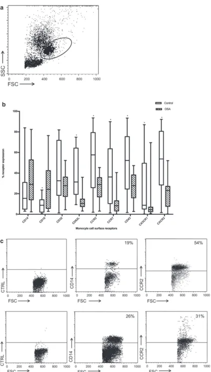
software. Monocytes were gated based on FSC and SSC (Fig 1a), and gates for determining the percent-age of monocytes that stained positive for antibody were set using isotype controls as guidelines.
Total RNA Extraction, Reverse Transcription, and
Quantitative RT-PCR
Sorted monocytes from 7 healthy controls and 7 dogs with OSA were used for PCR analysis. The RNeasy minikitcwas used to extract total RNA for quantitative RT-PCR (qPCR) from the sorted monocytes according to the manufacturer’s protocol. We verified the quality and quantity of the purified RNA using a
Table 1.
List of antibodies used for flow cytometry.
Antibodies Clone Isotype Fluorescence labeling
CD14 TUK4€ mIgG2a APC, FITC
CD16 LNK16 mIgG1 PE
CD62L SK11 mIgG2a PE
CD32 AT10 mIgG1 FITC
CD11c BU15 mIgG1 FITC
CCR2 Polyclonal Rabbit FITC CCR7 Polyclonal Rabbit FITC CD43 Polyclonal Rabbit FITC CX3CR1 Polyclonal Rabbit FITC CXCR2 Polyclonal Rabbit FITC
a
b
c
NanoDrop 2000c spectrophotometerg, and also used gel elec-trophoresis to confirm RNA quality (Figure S1a). cDNA was sub-sequently synthesized in a Techne TC4000j thermal cycler using the Promega©RT kitkaccording to manufacturer’s directions. Pri-mers for qPCR to detect mRNA expression levels of Interleukin-10 (IL-Interleukin-10), Interleukin-12 (IL-12), Tumor Necrosis Factor-alpha (TNF-a), and Cyclooxygenase-2 (COX-2) were selected based on previous reports (Table 2),10,11 and purchased from Integrated DNA Technologies.lThe PerfeCTaÒSYBRÒGreen FastMixÒfor iQTM
PCR kitmwas used for mRNA expression detection, and the reactions performed with the iCycler iQTM
real-time PCR detection system.n All samples were run in triplicate. The thermal cycling conditions were as follows: cycle 1 (95.0°C for 8 minutes) 1X, cycle 2 (95.0°C for 30 seconds, 56.0°C for 20 seconds, 72.0°C for 40 seconds) 40X, cycle 3 (melting curve starting at 60.0°C for 10 seconds, with stepwise increases of 0.5°C for 70X to achieve 95°C), and cycle 4 (hold at 4.0°C). The housekeeping gene, Glycer-aldehyde-3-Phosphate Dehydrogenase (GAPDH), was used for normalization of results. The CT value of GAPDH was consistent across experiments and between animals for all PCR results reported here (CT= ~21). The PCR product was separated using agarose gel electrophoresis, the DNA was sequenced by GENE-WIZÒDNA sequencing services, and the sequences were subjected to alignment searches in the NCBI database to confirm matches with the gene of interest (Figure S1b). The 2DDCT method was used to derive the normalized relative mRNA expression level of the target genes.
Prostaglandin ELISA
Sorted monocytes from 8 healthy controls and 7 dogs with OSA were used for stimulation experiments—half of these mono-cytes served as unstimulated controls and incubated for 6 hours at 37°C and 5% CO2. The other half were stimulated with 100 ng/ 106 monocytes of lipopolysaccharide (LPSa) and incubated simi-larly for 6 hours at 37°C and 5% CO2. The resultant supernatants were collected, stored at20°C until they were assessed for PGE2 levels using a commercially available ELISA kito according to manufacturer’s directions.
IL-10, IFN-
c
, TNF-
a
Magnetic Bead Panel Assay
Sorted monocytes from 5 healthy controls and 5 dogs with OSA were used for stimulation experiments as described for the prostaglandin ELISA. The resultant supernatants were collected,
stored at 20°C until they were assessed for IL-10, IFN-c, and TNF-alevels using a commercially available canine cytokine mag-netic bead panel kit (MILLIPLEXÒMAP)paccording to manufac-turer’s directions. IL-12 was not available as an analyte in the kit, thus IFN-clevels were analyzed as a surrogate, as IL-12 is known to induce IFN-csecretion.
Chemotaxis Assay
Sorted monocytes from 14 healthy controls and 5 dogs with OSA were used for migration assays. Monocyte migration was assessed using a NeuroProbe ChemoTxq 96-well disposable cell migration system with 8lm polycarbonate framed filters. Sorted monocytes were resuspended in 106/mL of cell culture media, labeled with 1lg calcein-AMr/106monocytes, and the cells incu-bated for 30 minutes in the dark at room temperature. Following incubation, the cells were resuspended at 106/mL in Dulbecco’s Modified Eagle Medium (DMEMs ). Twenty-five lL of the cell suspension were deposited on each filter. The lower chamber, ie, the 96-well plate, contained the following: 100% heat-inactivated fetal bovine serum (FBSg), 100 ng/mL monocyte chemoattractant protein-1 (MCP-1t ), 100 nM N-Formyl-L-Methionyl-L-Leucyl-L-Phenylalanine (fMLPa), 100 ng/mL CCL19t as positive chemoattractants, and PBS as a negative control. The positive chemoattractants were either placed individually in separate wells, or combined together in a single well. For total cell counts, 25lL of cells was placed directly into the lower well to quantify maximal fluorescence. The plate was incubated for 4 hours at 37°C, after which the nonmigrated cells on top of the filters were gently wiped off using a cell scraper. Cellular fluorescence in the bottom wells was quantified using a fMax fluorescence microplate readeru at 485 and 538 nm wavelength, and the corresponding SoftMax Pro software. Filter fluorescence was also quantified to confirm that cells were not adherent to the underside of the filter. Degree of migration was calculated as a percentage of migrated cells out of the known cell numbers placed in bottom wells. Cell migration was expressed as a migration index, calculated by dividing directed cell chemotaxis (cells migrating in response to a chemokine, quan-tified by degree of fluorescence) by random cell chemotaxis (cells migrating in response to media alone, quantified by degree of fluo-rescence). Each sample was assayed in triplicate.
Statistical Analyses
Statistical analyses were performed to compare monocyte cell surface receptor expression levels between groups using the non-parametric Mann-WhitneyU-test because of the non-normal dis-tribution of the data. Comparison of relative mRNA expression of each gene and of PGE2levels between groups were analyzed with the Mann-WhitneyU-test. All statistical tests were carried out as 2-sided tests, and a P-value of <.05 was considered significant. Comparison of peripheral blood monocyte counts between dogs with OSA and healthy controls was performed using a 2-tailed T-Test. Pearson’s correlation coefficient was used to determine the strength and direction of a possible linear relationship between monocyte surface receptors and peripheral blood monocyte counts. Two-way ANOVAs and least-squares means were used to test for differences in monocyte surface receptors because of status and monocyte counts (high versus low). To analyze the chemotaxis data, one-way ANOVAs were run and means were compared using Welch’s adjustment for heterogeneous variation. Kaplan-Meier survival analysis was used to compare survival between groups, with proportional hazards regression analysis to test the effect of chemokine receptor expression on survival. Survival was calculated as the number of days from diagnosis of disease to death attributed to disease, or last follow-up if the patient was still
Table 2.
Quantitative
RT-PCR
primers
to
detect
mRNA expression levels of proinflammatory and
anti-inflammatory cytokines, and COX-2.
Target (50–30)
Position (bp)
IL-10 (F) GCGACGCTGTCACCGATT 379–396 IL-10 (R) CTGGAGCTTACTAAATGCGCTCTT 460–437 IL-12 (F) TGGCTGCTATTCACAAGCTCAAGT 638–661 IL-12(R) TGGTTTGATGATGTCTCTGATGAAG 708–684 TNF-a
(F)
AGCCAGTAGCTCATGTTGTAGCAA 759–781
TNF-a (R)
GGCACTATCAGCTGGTTGTCTGT 358–380
COX-2 (F)
CTGTTCCCACCCATGTCAA –
COX-2 (R)
GCAGTTTTCGCCGTAGAATC –
alive. A dog was determined to have died of disease if metastasis or local tumor recurrence was the cause, and dogs with an unknown cause of death were presumed to be dead because of dis-ease. Dogs were censored from the analysis if they were still alive at last follow-up. All analyses were performed using SAS software (Version 9.4, Cary, NC). All statistical analyses were performed in consultation with the NCSU Biostatistics consulting group (Dr. E. Griffith).
Results
Study Population
The mean (
SD) age at time of OSA diagnosis was
8.7 (2.1) years. All dogs with OSA were purebred,
with the most common pure breeds being Labrador
Retriever (5 [28%]), Rottweiler (3 [17%]), Great Dane
(3 [17%]), and Golden Retriever (2 [11%]). There
were 8 (44%) spayed females, and 10 (56%) castrated
males.
Monocyte Surface Receptor Expression
The primary flow cytometry canine monocyte gates
were based on typical forward and side scatter
charac-teristics. Once that forward versus side light scatter gate
was established, we ensured that greater than 98%
of the CD14
+
cells were found within this gate. As
CD14 expression on peripheral blood monocytes varies
greatly, this ensured that we included the CD14
l,omonocytes for surface receptor analysis (Fig 1a).
Mono-cytes exhibited positive surface expression of CD14,
CD16, CD32, CD62L, CCR2, CCR7, CD43, CX3CR1,
CXCR2, with low levels of CD11c. Irrelevant isotype
controls were used for each experiment
—
the isotype
control binding of monocytes was
<
5%. The median
percentages and ranges of positively staining cells for
each antibody are listed in the legend accompanying
Fig 1b. Comparison of monocyte surface receptors
between untreated dogs with OSA and healthy controls
revealed a significant decrease in surface receptor
expression of CD62L (
P
<
.00001), CCR2 (
P
=
.0006),
CCR7
(
P
=
.0004),
CD43
(
P
=
.008),
CX
3CR1
(
P
=
.002), CXCR2 (
P
=
.0007) in dogs with OSA, and
a significant increase in CD16 expression in dogs with
OSA (
P
=
.005) (Fig 1b). Fig 1c depicts representative
dot plots with the percentage of cell surface receptor
expression in monocytes of a healthy control versus an
OSA dog.
A peripheral blood monocyte count of
>
400 cells/
l
L
has been significantly associated with decreased
disease-free interval in dogs with OSA.
12Others have reported
associations between peripheral blood monocyte counts
and outcomes in humans with OSA.
13Hence, we
evalu-ated the flow cytometry data in conjunction with
peripheral blood monocyte counts, using 400 cells/
l
L
as a differentiation point between high and low
mono-cyte counts according to previously reported findings.
12There was no significant difference in the peripheral
blood
monocyte
counts
of
dogs
with
OSA
(mean
=
514.7 cells/
l
L, SD
=
377.9 cells/
l
L) compared
to
healthy
controls
(mean
=
331.9 cells/
l
L,
SD
=
192.6 cells/
l
L;
P
=
.16). Comparison of receptor
expres-sion between dogs with high (
>
400 cells/
l
L) versus low
(
<
400 cells/
l
L) monocyte counts revealed a dichotomy
of CD14, CD16 expression in dogs with OSA
—
CD14
expression is high (
P
=
.01) and CD16 expression is low
(
P
=
.02) when monocyte counts are high (Table 3).
Least-squares means from the 2-way ANOVA
compar-ing relationships between monocyte counts and receptor
expression are shown in Table S1.
In healthy controls, there were strong significant
posi-tive correlations between chemokine receptors, whereas
in dogs with OSA, there were fewer and less robust
sig-nificant positive correlations between chemokine
recep-tors (Fig 2). The Pearson’s correlation values with
corresponding
P
-values are represented in Table S2.
Relative mRNA Expression of Monocyte 10,
IL-12, TNF-
a
, and COX-2
TNF
a
, IL-10, and IL-12 are inflammatory mediators
secreted by monocytes and macrophages, and
differen-tial levels of TNF
a
, IL-10, and IL-12 secretion have
been described for the various human monocyte
sub-sets,
14,15reflecting subset-specific functional differences.
COX-2 expression in monocytes reflects a
proinflamma-tory state.
16–18We compared relative mRNA expression
of IL-10, IL-12, TNF
a
, and COX-2 in dogs with OSA
versus healthy controls to discern potential variation in
cytokine secretion that might reflect functional
differ-ences. mRNA expression analysis of monocytes from
untreated dogs with OSA and healthy controls revealed
no significant differences in relative mRNA expression
levels of IL-10 (
P
=
.44), IL-12 (
P
=
.41), and TNF-
a
(
P
=
.14) between the 2 groups. The relative mRNA
expression of COX-2 was decreased in dogs with OSA
compared to healthy controls, but the difference was
not significant (
P
=
.08).
Evaluation of IL-10, IFN-
c
, TNF-
a
, and prostaglandin
E
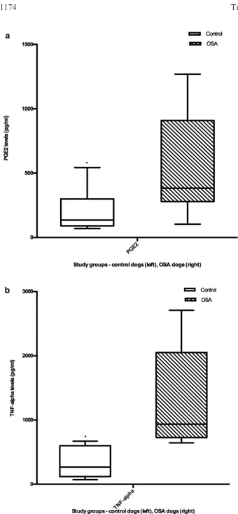
2secretion from monocytes
As mRNA levels might not consistently correspond
with protein secretion, we analyzed monocyte secretion
of the cytokines IL-10, IFN-
c
, TNF-
a
, and PGE
2.
Simi-lar to our mRNA evaluation, we compared cytokine
secretion from monocytes of dogs with OSA versus
healthy controls to assess for functional differences.
Secretion of cytokines such as TNF-
a
, IL-10, IFN-
c
, and
PGE
2has been noted to differ between human monocyte
subsets, and can be affected by disease.
15,17PGE
controls. Levels of IL-10 and IFN-
c
were low to
unde-tectable in both groups.
Monocyte Chemotaxis
The results of our phenotypic analysis of monocytes
between dogs with OSA and healthy controls revealed
marked decreases in chemokine receptor expression in
dogs with OSA (Fig 1b,c). Therefore, we asked whether
monocyte chemotaxis might be altered by OSA. As
described in our methods, monocytes from OSA and
healthy dogs were assessed by the NeuroProbe
Che-moTx
Òcell migration system. Comparison of monocyte
chemotaxis between dogs with OSA and healthy
con-trols revealed significantly decreased chemotaxis in dogs
with OSA when CCL19 (
P
=
.018), and when a mixture
of all 4 chemoattractants (
P
=
.018) was used (Fig 4).
With CCL2 or fMLP as chemoattractants, there was
also decreased monocyte chemotaxis in dogs with OSA
compared to healthy controls, but the difference did not
reach significance (CCL2
P
=
.067; fMLP
P
=
.067).
Survival Analysis
As a peripheral monocyte count of
>400 cells/
l
L has
been significantly associated with decreased disease-free
interval in dogs with OSA,
12we performed a
Kaplan-Meier survival analysis comparing dogs with OSA with
high or low monocyte counts, using 400 cells/
l
L as a
cut-off point. One dog died of unknown cause, and was
assumed to have died from OSA. We did not find a
sta-tistically
significant
difference
in
median
survival
between the 2 groups (
P
=
.15, Fig 5). Our regression
analysis revealed that increasing percentages of CCR2
expression on peripheral monocytes was significantly
associated with increasing survival in dogs with OSA
(
P
<
.0001).
Discussion
The goal of this study was to determine phenotypic
and functional differences between peripheral blood
monocytes from untreated dogs with OSA versus
healthy age-matched controls. Our hypothesis was that
evasion of the immune response by OSA is due in part
to down-regulation of monocyte chemokine receptor
expression and migratory function, and suppression of
host immune responses. We demonstrated a significant
decrease in monocyte chemokine receptors, increased
PGE
2and
TNF-
a
secretion
by
monocytes,
and
decreased monocyte chemotaxis in dogs with OSA.
Monocytes display a variety of cell surface receptors,
including leukocyte chemokine receptors. CD62L, also
known as L-selectin, is a cell adhesion molecule present
on immune cells such as monocytes, lymphocytes, and
NK cells. CD62L mediates the trafficking of these cells
to sites of inflammation and peripheral lymphoid tissue
by binding to glycoproteins.
19,20CCR2, a chemokine
receptor for MCP-1, is especially prominent in
mediat-ing monocyte migration.
21Similarly, CCR7 regulates
migration of leukocytes to secondary lymphoid organs
via binding of its ligands CCL19 and CCL21.
20CD43
can be found on a variety of immune cells including
lymphocytes, granulocytes, monocytes, macrophages,
and NK cells, and it plays a role in cell adhesion and
migration.
22,23CX
3CR1, and CXCR2, both G
protein-coupled
chemokine
receptors,
mediate
lymphocyte,
monocyte and granulocyte migration.
20There are few
reports describing the effect of OSA on monocyte
surface receptor expression in dogs. For example,
down-regulation of MHC class II and CD80 in canine
myeloid cells when exposed to tumor cell lines including
OSA in vitro has been observed.
24In humans, the
prevalence of an immunosuppressive phenotype in
peripheral blood monocytes of OSA patients has been
observed.
13However, to the authors’ knowledge, there
are no reports evaluating phenotypic changes in
mono-cytes of clinical dogs with OSA.
We assessed the expression of monocyte surface
receptors using flow cytometry, and demonstrated that
peripheral blood monocytes from dogs with OSA
exhib-ited significantly decreased chemokine receptor
expres-sion,
namely
of
CD62L,
CCR2,
CCR7,
CD43,
CX
3CR1, and CXCR2. This pronounced
down-regula-tion of monocyte chemokine receptors of dogs with
OSA suggests a peripheral sequestration of monocytes,
inhibited from migrating to sites of need such as the
primary tumor or metastatic lesions. Two of the
chemo-kine receptors, CCR2 and CXCR2, had significantly
higher expression on monocytes of healthy controls
with low monocyte counts compared to monocytes of
dogs with OSA that had high or low monocyte counts.
This supports our theory that the decreased expression
of chemokine receptors in dogs with OSA reflects a
peripheral sequestration. There was no difference in the
mean peripheral monocyte counts between dogs with
Table 3.
Comparison of receptor expression between dogs with high (>400 cells/
l
L, n
=
11) versus low (<400 cells/
l
L,
n
=
17) monocyte counts. In dogs with OSA, when monocyte counts are high, there is a dichotomy of CD14, CD16
expression
—
CD14 is high (
P
=
.01) and CD16 is low (
P
=
.02).
Chemokine receptor Monocyte counts
Control Dogs Dogs with OSA
Median (% expression) P-value Median (% expression) P-value
CD14 High 11.9 .90 37.5 .01
Low 7.7 13.9
CD16 High 2.8 .15 5.7 .02
OSA and healthy controls, which could indicate that
this parameter is not as sensitive for detecting a
periph-eral sequestration compared to analyzing the
relation-ship between chemokine receptors and monocyte counts
in both groups of dogs. Alternatively, the lack of
differ-ence in mean monocyte counts could also refute our
theory of peripheral monocyte sequestration in OSA.
We found a strong positive correlation between
expres-sion
of
chemokine
receptors
in
healthy
controls,
whereas the correlation was less robust in dogs with
OSA, suggesting a disturbance in the regulation of these
chemokine receptors.
A retrospective study of dogs with OSA found that
dogs with peripheral monocyte counts above 400 cells/
l
L
had a significantly shorter disease-free interval.
12Simi-larly, in human OSA patients, a low peripheral
lympho-cyte-to-monocyte ratio (
≤
3.43) was associated with
poorer prognosis for survival and disease-free interval.
25One possible explanation is that higher monocyte
counts reflect a peripheral sequestration of monocytes
in dogs that had more rapid tumor progression, and
our findings of suppressed expression of multiple
mono-cyte chemokine receptors in dogs with OSA suggest
such an association. We did not detect a significant
overall difference in peripheral monocyte counts of dogs
with OSA versus healthy controls in our study
popula-tion. Our Kaplan-Meier survival analysis comparing
dogs with OSA with high (
>
400 cells/
l
L) versus low
(
<
400 cells/
l
L) monocyte counts did not reveal
signifi-cant differences in survival between groups. However,
we did find that increasing percentages of CCR2
expres-sion on monocytes of dogs with OSA was significantly
associated with increasing survival, which, in
conjunc-tion with our chemotaxis data, suggests that
suppres-sion of chemokine receptors and chemotaxis is a form
of tumor-mediated monocyte dysfunction.
Additionally, we found that dogs with OSA with a
peripheral monocyte count of
>
400 cells/
l
L exhibited a
dichotomy of CD14, CD16 expression on monocytes
—
CD14 expression was high and CD16 expression was
low, when compared to dogs with monocyte counts
<
400 cells/
l
L. Human monocytes have been classified
into subsets, with different phenotypes and functions.
Earlier classification schemes split human monocytes
into
CD14
highCD16-
“inflammatory,
classical”
and
CD14
lowCD16
+
“resident, nonclassical” monocytes.
26,27Recent studies have shown that the CD16
+
monocytes
can be further characterized into intermediate and
non-classical subsets, with the intermediate subset expressing
higher CD14 and lower CD16 levels, and the
nonclassi-cal subset conversely expressing lower CD14 and higher
CD16 levels.
15The function of these monocyte subsets
has not been fully defined, but the intermediate subset
appears to produce proinflammatory cytokines such as
TNF-
a
, IL-1
b
, IL-6, express high levels of MHC II
antigen processing and presentation genes, and are
potent stimulators of T cells.
15There are no
corre-sponding studies differentiating canine monocytes into
subsets. Based upon human classification, our data
sug-gest that there is an increase in intermediate monocytes
in dogs with OSA with monocyte counts
>
400 cells/
l
L.
Potentially, these intermediate monocytes, with their
proinflammatory and immunostimulatory capacity, are
effective antitumor monocytes that are sequestered in
the periphery in OSA.
Monocyte migration can reportedly be affected by
neoplasia. Peripheral monocyte migration is inhibited
by lactate concentrations common in tumor
microenvi-ronments, and this loss of migratory ability is not
because of a reduction in cell viability.
28Chemokine
Fig 3. Monocyte PGE2 and TNF-a secretion are increased inreceptors such as CCR5 are down-regulated in
mono-cytes and macrophages in human patients with head
and neck squamous cell carcinoma, leading to
dysregu-lation of chemotactic receptor-ligand signaling and
decreased monocyte/macrophage migratory ability.
29Based on findings by other investigators and our
demonstration
of
profound
down-regulation
of
monocyte chemokine receptors in dogs with OSA, we
hypothesized that OSA inhibits monocyte chemotaxis as
an immunosuppressive strategy. Using an in vitro
migration assay, we demonstrated decreased monocyte
chemotaxis in dogs with OSA compared to healthy
con-trols. This difference was significant when CCL19 was
used as a chemoattractant, and when supraphysiologic
combinations of chemoattractants were used
—
a
combi-nation of FBS, CCL2, CCL19, and fMLP. CCL19 is a
ligand for the chemokine receptor CCR7, and is a
known monocyte chemoattractant.
30There was also
decreased monocyte migration in dogs with OSA when
other known chemokines such as CCL2 or fMLP were
used, and even though the differences were not
signifi-cant, the proximity of the
P
-value (.067) to significance
(.05) suggests that use of these chemokines deserves
fur-ther evaluation. CCL2 is also known as monocyte
chemotactic protein, binds CCR2, and together with
fMLP, which binds to G protein-coupled receptors, is
used as chemoattractants for monocytes. The significant
down-regulation of CCR2 and CCR7 on monocytes of
dogs with OSA demonstrated by our flow cytometry
data supports the observation that monocyte migration
is decreased in dogs with OSA. The primary
disadvan-tage of our method for assessing migration was the high
level of random migration observed, potentially because
of excessive stimulation of monocytes by the cell sorting
process. As a result, we included supraphysiologic
com-binations of chemoattractants to counter this potential
issue.
Fig 4. Monocyte chemotaxis is decreased in OSA: Box-and-whisker plot comparing the mean levels of random: directed monocyte migra-tion for the various chemokines used, either alone or in combinamigra-tion (CCL2, CCL19, FBS, fMLP) between healthy controls (n=14) and dogs with OSA (n=5). Monocyte migration is significantly reduced in dogs with OSA when CCL19 (P=.018) or a combination of chemokines (P=.018) was used as chemoattractants. Decreased migration in dogs with OSA was also observed with CCL2 or fMLP as chemoattractants, but the difference did not reach significance (CCL2P=.067; fMLPP=.067).
There could be a multi-faceted explanation for the
down-regulation of monocyte chemokine receptors in
dogs with OSA. Similar to our findings, the loss of
CX
3CR1 expression in human monocytes increased
glioma growth.
31However, in contrast to our
observa-tions, this tumor-promoting effect is not because of loss
of migratory capability. In fact, the absence of CX3CR1
was associated with an increase in monocyte infiltration
into the tumor microenvironment, where the monocytes
were theorized to differentiate into tumor-associated
macrophages for promotion of tumor growth. Evaluation
of the relationship between monocyte chemokine receptor
expression and the infiltration of
monocytes/macro-phages in the tumor microenvironment in OSA is needed
to explore whether this phenomenon occurs in dogs.
The higher PGE2
levels that we observed in monocyte
culture supernatants of dogs with OSA are consistent
with our hypothesis that OSA suppresses monocyte
chemotaxis and the host immune response. PGE
2is a
main metabolite resulting from the conversion of
arachidonic acid by cyclooxygenases (1 and
COX-2), and increased levels of COX-2 and PGE
2have been
observed in various tumors.
32–35PGE2
secretion from
human carcinoma cell lines inhibits monocyte
chemo-taxis by down-regulating cell surface expression of
che-mokine and adhesion receptors CCR5 and Mac-1,
respectively.
36Our finding of increased PGE
2secretion
from monocytes of dogs with OSA correlates with our
observation that monocyte chemotaxis is inhibited
in OSA. Increased PGE2
levels have been shown to
effect
global
immunosuppression,
contributing
to
immunopathology in many cancers.
37For example,
PGE
2disables the innate immune response by inhibiting
neutrophils, monocytes and macrophages, and disrupts
the cross-talk between dendritic cells and T cells that is
essential
for
activation
of
the
adaptive
immune
response. PGE
2also skews T helper cells toward a type
2 pro-tumorigenic instead of a type 1 antitumor
response, and promotes the accumulation of T
regula-tory and myeloid-derived suppressor cells, which can be
utilized by the tumor to suppress an effective antitumor
response.
37A recent study in human OSA showed
inhi-bition of human OSA cells by decreasing PGE
2levels
via microRNA modulation, demonstrating the role of
PGE
2in tumor promotion.
38Specifically in OSA of
dogs, PGE2
is increased in both canine OSA cell lines
and in naturally occurring tumors,
39COX-2,
microso-mal PGE2
synthase-1, and PGE2
receptor are
demon-strated
to
be
increased
in
OSA
lesions
using
immunohistochemistry staining,
40and OSA cells from
dogs secrete PGE
2.
41Our findings of increased PGE
2in
dogs with OSA are in accordance with these previous
reports, and emphasize the ability of PGE
2to induce
immunopathology in OSA.
We did not find significant differences in relative
mRNA expression of IL-10, IL-12, TNF-
a
, and COX-2
in monocytes from OSA compared to healthy dogs.
There was a decreasing trend in COX-2 expression in
dogs with OSA, but the difference from healthy controls
was not significant. This is contrary to what we
expected,
given
the
higher
PGE
2secretion
from
monocytes of dogs with OSA, and the overexpression
of COX-2 that has been reported in OSA of dogs.
40,42Potentially, there might be other enzymes such as
microsomal PGE
2synthase-1 that could be concurrently
influencing the level of PGE
2secretion. However, as the
difference in COX-2 expression between OSA and
healthy dogs was not statistically significant, whether
COX-2 expression in monocytes of dogs with OSA is
truly decreased needs to be further evaluated, as other
influences such as miRNA activity can affect mRNA
transcription. Because of potential disparities between
mRNA and protein expression, we evaluated monocyte
secretion of TNF-
a
, IFN-
c
, and IL-10, using IFN-
c
as
representative of IL-12 activity. There were low to
undetectable levels of IFN-
c
and IL-10 from both
groups, and higher levels of TNF-
a
secreted from
monocytes of dogs with OSA. This was an unexpected
finding, as TNF-
a
is a proinflammatory cytokine.
How-ever, TNF-
a
induces a number of other changes,
partic-ularly in the context of the tumor microenvironment.
TNF-
a
induces the up-regulation of IL-34 expression in
OSA cell lines, and IL-34 has been associated with
pro-gression of disease and promotion of angiogenesis in
OSA.
43TNF-
a
has also been shown to increase the
pro-duction of PGE2
as a mechanism for effecting bone
resorption.
44Potentially, the increase in TNF-
a
in
monocytes of dogs with OSA reflects tumor-driven
mechanisms for OSA promotion rather than induction
of
a
proinflammatory
monocyte
response.
Other
immunomodulatory cytokines such as TGF-
b
are
uti-lized by OSA for tumor promotion. TGF-
b
, generally
known to dampen immune responses and promote
tumor growth, is secreted by OSA cells from dogs,
6and
has also been implicated in the pathogenesis of human
OSA.
45Collectively, these findings indicate a complex
microenvironment that promotes immune dysfunction
and tumor growth.
One potential limitation of this study is the use of
clinical dogs and random source healthy controls,
instead of purpose-bred dogs which provide a
geneti-cally uniform study population. However, one strong
advantage of using nonpurpose-bred dogs is the genetic
diversity that accurately reflects that of the general pet
population. This diversity, together with the exposure
to shared environmental variables with humans,
con-tributes to the unique canine model of spontaneously
occurring OSA that bears striking similarities to the
human disease. Such a model is extremely valuable for
studying the disease to improve outcomes for both
humans and dogs.
exert a multitude of immunopathologic effects such as
interfering with lymphocyte proliferation and the
antitu-mor cytotoxicity of T cells, inhibit NK cell, dendritic
cell, neutrophil, monocyte and macrophage effector
functions. Increased TNF-
a
secretion could be a
tumor-promoting mechanism through manipulation of the
tumor microenvironment and elevating PGE2
produc-tion. Understanding the mechanisms by which OSA
causes dysregulation of the immune response will
pro-vide insight into developing novel immunotherapeutics
to reverse tumor-induced immunopathology and reduce
metastatic disease in OSA.
Acknowledgments
This work was done at North Carolina State
Univer-sity, Raleigh, NC.
This work was funded by an American Kennel Club
Canine Health Foundation Acorn grant (grant
#01903-A), an American Kennel Club Canine Health
Founda-tion Clinician-Scientist Fellowship, and by the Ruth L.
Kirschstein T32 training grant.
Part of this study was presented as an oral abstract
at the 2014 ACVIM Forum and the 2014 Veterinary
Society of Surgical Oncology conference, and as a
pos-ter at the 2014 North Carolina State University College
of Veterinary Medicine Research Forum.
The authors thank Ms. Janet Dow and Ms. Sarah
Bianco for their assistance with cell sorting.
Conflict of Interest Declaration:
Authors declare no
conflict of interest.
Off-label Antimicrobial Declaration:
Authors declare
no off-label use of antimicrobials.
Footnotes
aSigma-Aldrich, St. Louis, MO (Histopaque, fMLP, LPS) bBeckman-Coulter, Atlanta, GA (Cell sorter)
cQiagen, Germantown, MD (RNAProtect, RNEasy minikit) dAbDSerotec, Raleigh, NC (CD14)
eAbCam, Cambridge, MA (CD16, CD32, rabbit polyclonal) fBD BioSciences Pharmigen, San Diego, CA (CD62L, IgG2a PE,
IgG1 FITC) g
Thermo Scientific,Waltham, MA (CD11c, FBS) h
Bioss Inc, Woburn, MA i
BD Biosciences, San Jose, CA (FACS Calibur) j
Bibby Scientific, Burlington, NJ (TC4000 thermal cycler) k
Promega, Madison, WI (Promega RT kit) l
Integrated DNA Technologies, IO (Primers for PCR) m
Quanta Biosciences, Gaithersburg, MD (PCR SYBR Green kit) n
Bio-Rad, Hercules, CA (iCycler)
oCayman Chemical, Ann Arbor, MI (PGE2 ELISA kit#514010) pEMD Millipore, Billerica, MA (CCYTOMAG-90K
MILLI-PLEX MAP kit)
qNeuroProbe, Gaithersburg, MD (Chemotaxis assay) rAnaSpec Inc., Fremont, CA (Calcein)
sCorning CellGroÒ(Mediatech Inc), Manassas, VA (DMEM) tR&D Systems, Minneapolis, MN (MCP-1, CCL19)
uMolecular Devices, Sunnyvale, CA (Plate-reader)
References
1. Mueller F, Fuchs B, Kaser-Hotz B. Comparative biology of human and canine osteosarcoma. Anticancer Res 2007;27:155–164. 2. Withrow SJ, Powers BE, Straw RC, et al. Comparative aspects of osteosarcoma. Dog versus man. Clin Orthop Relat Res 1991;270:159–168.
3. Chanmee T, Ontong P, Konno K, et al. Tumor-associated macrophages as major players in the tumor microenvironment. Cancers 2014;6:1670–1690.
4. Miller MM, Petty CS, Tompkins MB, et al. CD4+CD25+T regulatory cells activated during feline immunodeficiency virus infection convert T helper cells into functional suppressors through a membrane-bound TGFbeta/GARP-mediated mechanism. Virol J 2014;11:7.
5. Petty CS, Tompkins MB, Tompkins WA. Transforming growth factor-beta/transforming growth factor-betaRII signaling may regulate CD4+CD25+T-regulatory cell homeostasis and sup-pressor function in feline AIDS lentivirus infection. J Acquir Immune Defic Syndr 2008;47:148–160.
6. Portela RF, Fadl-Alla BA, Pondenis HC, et al. Pro-tumori-genic effects of transforming growth factor beta 1 in canine osteosarcoma. J Vet Intern Med 2014;28:894–904.
7. Lascelles BD, Dernell WS, Correa MT, et al. Improved sur-vival associated with postoperative wound infection in dogs trea-ted with limb-salvage surgery for osteosarcoma. Ann Surg Oncol 2005;12:1073–1083.
8. Sottnik JL, U’Ren LW, Thamm DH, et al. Chronic bacterial osteomyelitis suppression of tumor growth requires innate immune responses. Cancer Immunol Immunother 2010;59:367–378.
9. Hood SFT, Thompson EM, Akaronu NO, et al. Monocyte-derived dendritic cells from feline immunodeficiency virus positive cats are productively infected and maintain CD8+T cell stimula-tory capacity. Int J Virol AIDS 2015;2:007.
10. Sanchez-Robert E, Altet L, Alberola J, et al. Longitudinal analysis of cytokine gene expression and parasite load in PBMC in Leishmania infantum experimentally infected dogs. Vet Immunol Immunopathol 2008;125:168–175.
11. Clemente M, Sanchez-Archidona AR, Sardon D, et al. Dif-ferent role of COX-2 and angiogenesis in canine inflammatory and non-inflammatory mammary cancer. Vet J 2013;197:427–432.
12. Sottnik JL, Rao S, Lafferty MH, et al. Association of blood monocyte and lymphocyte count and disease-free interval in dogs with osteosarcoma. J Vet Intern Med 2010;24:1439–1444.
13. Hingorani P, Maas ML, Gustafson MP, et al. Increased CTLA-4(+) T cells and an increased ratio of monocytes with loss of class II (CD14(+) HLA-DR(lo-neg)) found in aggressive pedi-atric sarcoma patients. J Immunother Cancer 2015;3:35.
14. Smedman C, Ernemar T, Gudmundsdotter L, et al. Flu-oroSpot analysis of TLR-activated monocytes reveals several dis-tinct cytokine secreting subpopulations. Scand J Immunol 2011;75:249–258.
15. Wong KL, Yeap WH, Tai JJ, et al. The three human monocyte subsets: implications for health and disease. Immunol Res 2012;53:41–57.
16. Ma X, You X, Zeng Y, et al. Mycoplasma fermentans MALP-2 induces heme oxygenase-1 expression via mitogen-acti-vated protein kinases and Nrf2 pathways to modulate cyclooxyge-nase 2 expression in human monocytes. Clin Vaccine Immunol 2013;20:827–834.
17. Beyan H, Goodier MR, Nawroly NS, et al. Altered mono-cyte cyclooxygenase response to lipopolysaccharide in type 1 dia-betes. Diabetes 2006;55:3439–3445.
19. Ivetic A. Signals regulating L-selectin-dependent leucocyte adhesion and transmigration. Int J Biochem Cell Biol 2013;45:550–555.
20. Thomas S, Baumgart DC. Targeting leukocyte migration and adhesion in Crohn’s disease and ulcerative colitis. Inflam-mopharmacology 2012;20:1–18.
21. Yadav A, Saini V, Arora S. MCP-1: chemoattractant with a role beyond immunity: a review. Clin Chim Acta 2010;411:1570– 1579.
22. Steiniger B, Stehling O, Scriba A, et al. Monocytes in the rat: phenotype and function during acute allograft rejection. Immunol Rev 2001;184:38–44.
23. Axelsson B, Perlmann P. Persistent superphosphorylation of leukosialin (CD43) in activated T cells and in tumour cell lines. Scand J Immunol 1989;30:539–547.
24. Wasserman J, Diese L, VanGundy Z, et al. Suppression of canine myeloid cells by soluble factors from cultured canine tumor cells. Vet Immunol Immunopathol 2012;145:420–430.
25. Liu T, Fang XC, Ding Z, et al. Pre-operative lymphocyte-to-monocyte ratio as a predictor of overall survival in patients suf-fering from osteosarcoma. FEBS Open Bio 2015;5:682–687.
26. Gordon S, Taylor PR. Monocyte and macrophage hetero-geneity. Nat Rev Immunol 2005;5:953–964.
27. Varol C, Yona S, Jung S. Origins and tissue-context-depen-dent fates of blood monocytes. Immunol Cell Biol 2009;87:30–38.
28. Goetze K, Walenta S, Ksiazkiewicz M, et al. Lactate enhances motility of tumor cells and inhibits monocyte migration and cytokine release. Int J Oncol 2011;39:453–463.
29. Chakraborty K, Bose A, Chakraborty T, et al. Restoration of dysregulated CC chemokine signaling for monocyte/macro-phage chemotaxis in head and neck squamous cell carcinoma patients by neem leaf glycoprotein maximizes tumor cell cytotoxic-ity. Cell Mol Immunol 2010;7:396–408.
30. Frascaroli G, Varani S, Moepps B, et al. Human cytomega-lovirus subverts the functions of monocytes, impairing chemokine-mediated migration and leukocyte recruitment. J Virol 2006;80:7578–7589.
31. Feng X, Szulzewsky F, Yerevanian A, et al. Loss of CX3CR1 increases accumulation of inflammatory monocytes and promotes gliomagenesis. Oncotarget 2015;6:15077–94.
32. Eberhart CE, Coffey RJ, Radhika A, et al. Up-regulation of cyclooxygenase 2 gene expression in human colorectal adeno-mas and adenocarcinoadeno-mas. Gastroenterology 1994;107:1183–1188.
33. Wolff H, Saukkonen K, Anttila S, et al. Expression of cyclooxygenase-2 in human lung carcinoma. Cancer Res 1998;58:4997–5001.
34. Pugh S, Thomas GA. Patients with adenomatous polyps and carcinomas have increased colonic mucosal prostaglandin E2. Gut 1994;35:675–678.
35. Rigas B, Goldman IS, Levine L. Altered eicosanoid levels in human colon cancer. J Lab Clin Med 1993;122:518–523.
36. Zeidler R, Csanady M, Gires O, et al. Tumor cell-derived prostaglandin E2 inhibits monocyte function by interfering with CCR5 and Mac-1. FASEB J 2000;14:661–668.
37. Kalinski P. Regulation of immune responses by prostaglan-din E2. J Immunol 2012;188:21–28.
38. Xu H, Mei Q, Shi L, et al. Tumor-suppressing effects of miR451 in human osteosarcoma. Cell Biochem Biophys 2014;69:163–168.
39. Mohammed SI, Coffman K, Glickman NW, et al. Prosta-glandin E2 concentrations in naturally occurring canine cancer. Prostaglandins Leukot Essent Fatty Acids 2001;64:1–4.
40. Millanta F, Asproni P, Cancedda S, et al. Immunohisto-chemical expression of COX-2, mPGES and EP2 receptor in nor-mal and reactive canine bone and in canine osteosarcoma. J Comp Pathol 2012;147:153–160.
41. Shor S, Fadl-Alla BA, Pondenis HC, et al. Expression of nociceptive ligands in canine osteosarcoma. J Vet Intern Med 2015;29:268–275.
42. Mullins MN, Lana SE, Dernell WS, et al. Cyclooxygenase-2 expression in canine appendicular osteosarcomas. J Vet Intern Med 2004;18:859–865.
43. Segaliny AI, Mohamadi A, Dizier B, et al. Interleukin-34 promotes tumor progression and metastatic process in osteosar-coma through induction of angiogenesis and macrophage recruit-ment. Int J Cancer 2015;137:73–85.
44. Zhang W, Dziak R. Tumor necrosis factor alpha stimualtes arachidonic acid metabolism in human osteoblastic osteosarcomal cells. Prostaglandins Leukot Essent Fatty Acids 1996;54:427–431.
45. Xu S, Yang S, Sun G, et al. Transforming growth factor-beta polymorphisms and serum level in the development of osteosarcoma. DNA Cell Biol 2014;33:802–806.