Abstract:
The Wnt signaling pathway is necessary for development, cell fate regulation,
homeostasis, and proliferation. Mis-regulation of Wnt signaling is associated with many cancer types, particularly colorectal carcinomas. catenin is the main effector of Wnt signaling. β-catenin levels are regulated by the multi-protein β-β-catenin destruction complex, which consists of the scaffolding protein Axin, the tumor suppressor Adenomatous polyposis coli (APC), glycogen synthase kinase 3 (GSK-3), and casein kinase 1 (CK1). After the destruction complex
phosphorylates β-catenin, an E3 ubiquitin ligase recognizes phosphorylated β-catenin and targets it for destruction by the proteasome. Despite over 30 years of research, the mechanism by which the destruction complex transfers β-catenin to the E3 ligase remains largely unknown. We hypothesize that interaction between Axin and Slimb (a member of the SCFF-Box E3 ligase in Drosophila) aids in the transfer of β-catenin from the destruction complex to the E3 ligase. To
investigate the relationship between Axin and Slimb we co-expressed fluorescently tagged wild-type and mutant forms of Drosophila Axin and Slimb in a human colon cancer cell line. We examined co-localization and used co-immunoprecipitations (co-IPs) to investigate a close association between Axin and Slimb. Our results suggest that the E3 ligase and the destruction complex co-localize in cells, and that Axin recruits Slimb to the destruction
Introduction:
The highly conserved Wnt signaling pathway regulates important developmental events and is mutated in many human diseases (Gordon and Nusse 2006). Wnt signaling mutations are found in more than 80% of colon and rectal carcinomas and nearly 30% of uterine corpus endometrial carcinomas (Peifer and Polakis 2000; Kandoth et. al 2013).
Figure 1. Simplified diagram of OFF (A) and ON (B) states of the Wnt signaling pathway. A) When Wnt is OFF β-catenin is phosphorylated by the
destruction complex composed of Axin, GSK3, APC, and CK1. β-catenin is targeted for degradation by the proteasome after ubiquitination by the E3 ligase. B) When Wnt is ON the destruction complex is
inhibited; leading to
accumulation of β-catenin in the cytoplasm. β-catenin then translocates to the nucleus and activates gene transcription.
When Wnt signaling is off, the destruction complex maintains low levels of cytoplasmic β-catenin. The core β-catenin destruction complex is comprised of the scaffolding protein Axin, the tumor suppressor adenomatous polyposis coli (APC), glycogen synthase kinase 3β (GSK-3), and casein kinase 1 (CK1) (Gordon and Nusse 2006; Roberts et al. 2012) (Fig. 1A). In normal cells, this multi-protein complex phosphorylates β-catenin via CK1 and GSK-3 (Gordon and Nusse 2006). Phosphorylation of β-catenin allows it to be recognized by an SCF (Skp
1-Cullin-Destruction complex: OFF APC CK1 TCF Nucleus APC CK1 Destruction complex: ON Frizzled Wnt Receptor Wnt OFF
Frizzled Wnt Receptor
Wnt Wnt ON
F-box protein) E3 ligase where it is ubiquitylated and subsequently passed to the proteasome for destruction (Fig. 1A) (Gordon and Nusse 2006).
When Wnt ligand is present, it binds to a multi-protein receptor on the cell surface. This leads to the inhibition of the β-catenin destruction complex; thus, preventing β-catenin
phosphorylation and subsequent degradation (Gordon and Nusse 2006) (Fig. 1B). In these conditions cytoplasmic β-catenin accumulates and enters the nucleus (Gordon and Nusse 2006). Once in the nucleus, β-catenin binds to TCF/LEF family DNA binding proteins and is able to act as a transcriptional coactivator of genes involved in cell proliferation and fate (Peifer and Polakis 2000) (Fig. 1B).
In human colon cancer, the destruction complex function is severely reduced due to a truncation of APC. This truncation reduces APC’s ability to bind Axin, preventing proper destruction complex function and thus inducing cell proliferation due to high levels of
cytoplasmic β-catenin (Peifer and Polakis 2000). Yet in colon cancers some β-catenin must be destroyed since too much β-catenin will induce cell apoptosis (Kim et al. 2000). This suggests that some β-catenin is still being transferred to the E3 ligase. However, the mechanism of how phosphorylated β-catenin is transferred from the destruction complex to the E3 ligase in normal or APC mutant cells remains largely unknown.
suggested that APC protects β-catenin from dephosphorylation by PP2A to ensure that
phosphorylated β-catenin is recognized by the SCF E3 ligase (Su et al. 2008). Previous results in the Peifer Lab have suggested that the E3 ligase and the destruction complex co-localize in cells. We hypothesize that the SCFSlimb E3 ligase may be recruited to the destruction complex,
regardless of the presence of phosphorylated β-catenin, by one of the core components of the destruction complex. An E3 ligase closer to the destruction complex would allow for faster ubiquitination of β-catenin and prevent dephosphorylation of β-catenin by PP2A leading to its ultimate destruction. In this study, we found that Axin recruits Slimb to the destruction complex. Our results also suggest that the RGS domain of Axin may interact with Slimb.
Methods:
Cell Culture:
SW480 cells, a human colon cancer cell line, were used to express Drosophila Axin and Slimb constructs. Cells were grown on glass cover slips in a 6 well plate. Cells were incubated in L15 medium at 37°C.
Preparation of Constructs:
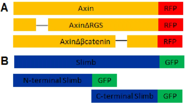
Constructs (Fig. 2) were prepared by first isolating genomic DNA from Drosophila. Axin and Slimb were then PCR amplified from the genomic DNA.
Figure 2. Axin (A) and Slimb (B) constructs. A) Full length Axin with C-terminal RFP tag. The RGS domain was removed (AxinΔRGS) and the β-catenin binding site was removed (AxinΔβcatenin). Each
Mutagenesis was performed using the QuikChange XL kit (Agilent Technologies). Protocol provided with kit was followed. Mutant constructs were verified by DNA sequencing.
For Transfection:
4 μl of Lipofectamine was added to 250 μl of Opti-MEM and allowed to sit for five minutes. 2 μl of DNA construct was added to a separate aliquot of 250 μl Opti-MEM. The Lipofectamine and Opti-MEM solution was then added to the DNA and Opti-MEM solution for a total of 500 μl. The solution was allowed to sit for twenty minutes before adding 500 μl to each well. After addition of the 500 μl Lipofectamine solution, cells were incubated overnight at 37°C.
Fluorescent Staining:
L15 media was removed from coverslips and cells were washed three times with Phosphate-buffered saline (PBS). At room temperature, cells were fixed in 3.7% formaldehyde and placed on a nutator for five minutes. Formaldehyde was removed and cells were washed two times with PBS. Cells were placed on a nutator for five minutes in 1% Triton-X in PBS. Cells were blocked for 30 minutes in NGS:PBS (1:1000) and incubated in primary antibody for one hour on a nutator at room temperature. After three more washes with PBS, secondary antibody (1:1000) was added for 30 minutes. Cells were washed three more times with PBS and then mounted on slides. Images were taken using a PASCAL confocal microscope and analyzed using Image J.
Immunoprecipitation:
SW480 cells were transfected with Axin and/or Slimb constructs and incubated
Cells were scraped, re-suspended in lysis buffer, transferred to a cold Eppendorf tube, and centrifuged at 13,200 rpm for 30 minutes at 4°C. Sepharose beads were prepared by washing three times with lysis buffer, 100 μl of Sepharose beads were then added to each Eppendorf tube, and incubated on nutator at 4°C for one hour. Sepharose beads were removed and a small
amount of supernatant (50μl) was saved as whole cell lysate. Antibody (1μl) was added to each Eppendorf tube and incubated overnight at 4°C on nutator. Sepharose beads were washed three times with PBS and incubated overnight at 4°C in 1 mL of PBS and 100μl of 50% BSA. The next day, beads were washed two times with PBS, one time with lysis buffer, re-suspended in 1 mL of lysis buffer and added (100μl) to each IP tube to incubate for two hours. Samples were centrifuged (30 seconds at 800 rpm) and washed five times with lysis buffer (300μl) at 4°C. After the final wash, the supernatant was removed, leaving only the Sepharose beads. SDS buffer was added to the whole cell lysate and IP tubes and both were boiled for 10 minutes.
Classification of cells based on percentage of Axin puncta that recruited Slimb
Results:
Earlier work provided a foundation for our studies. In SW480 cells, a human colon cancer cell line carrying a truncated APC, increasing protein expression of Axin and FWD1 (F-box/WD40-repeat protein 1), the mouse Slimb homolog, increased β-catenin destruction
(Kitagawa et al. 1999). Using human embryonic kidney (HEK) 293T cells, FWD1 was shown to co-IP with both Axin and β-catenin (Kitagawa et al. 1999). Furthermore, Kitagawa et al. (1999) suggested that Axin is required for FWD1 to interact with β-catenin. To gain insight into the mechanism of how β-catenin is transferred to the E3 ligase we first wanted to visualize where the
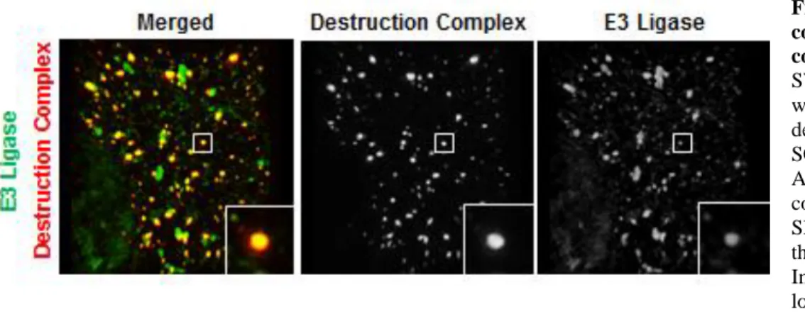
SCF complex localizes in cells in relation to the destruction complex. To do so, we co-expressed parts of the destruction complex (Axin and APC) along with different components of the
SCFSlimb E3 ligase (SkpA, Lin19, the Drosophila homolog of Cullin, and Slimb) in SW480 cells. We observed co-localization of the E3 and Axin or APC (Fig. 2). These data suggest that the SCFSlimb E3 ligase and the destruction complex come into close proximity with one another. Since Axin, the scaffolding protein of the destruction complex, has the ability to recruit the other core protein components of the destruction complex, we thus hypothesized that Axin directly interacts with Slimb in the E3 ligase.
Figure 2. The destruction complex and the E3 ligase co-localize. SIM image of a SW480 cell co-transfected with proteins from the destruction complex and the SCFSlimb E3 ligase. APC and Axin of the destruction complex are both RFP tagged. Slimb, SkpA, and Lin19 of the E3 ligase are GFP tagged. Insets highlight
To investigate a possible interaction between Axin and Slimb we co-expressed
fluorescently tagged Drosophila Axin and Slimb in SW480 cells. When transfected alone, Axin self-polymerizes and forms cytoplasmic puncta (Fig. 3A), as previously shown
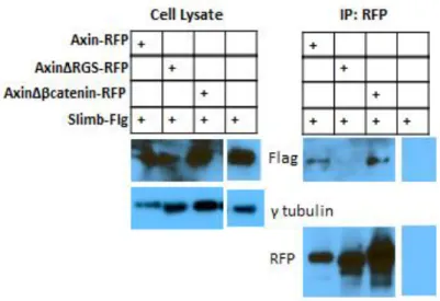
(Schwarz-Romond et al. 2007). When Slimb was transfected alone, it was diffuse throughout the cytoplasm (Fig. 3B). When Axin and Slimb were co-transfected, Axin recruited Slimb into Axin puncta (Fig. 3C), suggesting the two proteins may interact in some manner. To further investigate a possible Axin-Slimb interaction, we performed immunoprecipitations (IPs). An IP for Axin pulled down Slimb, suggesting a stable Slimb-Axin interaction (Fig. 4).
Crystallography studies have shown both Axin and the E3 ligase bind directly to β -catenin, although Slimb only binds when β-catenin is phosphorylated (Xing et al. 2003; Liu et al. 1999). Since both Axin and Slimb directly interact with β-catenin, it is possible that
phosphorylated β-catenin acts as a bridge between Axin and Slimb. To test this hypothesis, we deleted Axin’s β-catenin binding domain (AxinΔβ-cat) and performed an IP for AxinΔβ-cat. Loss of Axin’s β-catenin binding domain did not eliminate Axin’s ability to pull down Slimb (Fig. 4) suggesting that β-catenin is not facilitating this association.
Figure 4: Slimb
immunoprecipitates with Axin. Cells were
Figure 5. Both the N-terminal and the C-terminal halves of Slimb are recruited to Axin puncta. SW480 cells transfected with A) N-terminal Slimb-GFP, B-F) N-terminal Slimb-GFP and Axin-RFP, G) C-terminal Slimb-GFP, and H-L) C-terminal Slimb-GFP and Axin-RFP. Insets show magnified view of highlighted puncta. B-F) shows decreasing recruitment of N-terminal Slimb GFP to Axin-RFP puncta. Cells were classified into three groups based on the percentage of Axin puncta that recruited Slimb. G-L) shows decreasing recruitment of C-terminal Slimb-GFP to Axin-RFP puncta. Cells were also classified into three groups based on the percentage of Axin puncta that recruited Slimb. M) Percentage of cells classified into each category based on the percentage of Axin puncta that recruited either N-terminal Slimb or C-terminal Slimb
To further investigate the interaction between Axin and Slimb two different pieces of Slimb were constructed; one composed of the N-terminal half, containing the F-box domain, and another composed of the C-terminal half, containing the WD40 repeats. Previous experiments in HEK cells have suggested that the N-Terminal half of FWD1, the mouse Slimb homolog, does not coIP with β-catenin or Axin and neither does the C-Terminal half of FWD1 (Kitagawa et al. 1999). However, when only the F-box domain of FWD1 was deleted FWD1 coIPs with β-catenin and Axin (Kitagawa 1999). Based on these data we hypothesized that both the N-terminal and C-N-terminal half of Slimb would remain diffuse in the cytoplasm when co-transfected with Axin-RFP.
Fluorescently tagged N-terminal and C-terminal Slimb were transfected into cells alone and with full length Axin. When transfected alone, both N-terminal and C-terminal Slimb were diffuse (Fig. 5A and G), similar to full length Slimb (Fig. 3B). Both N-terminal and C-terminal Slimb were recruited to Axin puncta after co-transfection (Fig. 5B and H). This suggests that Slimb may have multiple binding sites on Axin or within the destruction complex. Interestingly, there was variation in the percentage of Axin puncta that recruited Slimb (Fig. 5 B-F and H-L). Some cells displayed a phenotype in which greater than 80% of Axin puncta recruited either N-terminal Slimb or C-N-terminal Slimb (Fig. 5B and H), other cells displayed a phenotype where approximately 40-80% of Axin puncta recruited Slimb (Fig. 5C-E and I-K), and the third phenotype observed was when less than 40% of Axin puncta recruited Slimb (Fig. 5F and L). Cells were classified into these three groups based on the percentage of Axin puncta that recruited Slimb as described in the methods section.
π-helix in α-π-helix 9 of Axin-RGS (Spink et. al 2000). Helicies are a common in protein secondary structure. A helix is defined by the number of residues and atoms per single turn of the helix. An
α-helix is defined by 3.6 residues and 13 atoms and a π-helix is defined by 4.4 residues and 16 atoms (Weaver 2001). π-helices are not observed often in protein structures due to their unstable conformation; however, previous studies have suggested a correlation exists between the
conservation of a π-helix and a unique function (Weaver 2000). Spink et al. (2000) mutated conserved residues of the Axin RGS domain to observe their effect on SAMP binding. The mutation Tyr247Ala, within the π-loop in human Axin, had no effect on Axin and APC binding (Spink et al. 2000). Since this residue did not contribute to the RGS-SAMP interaction, and Slimb may interact with Axin-RGS we hypothesized that Axin and Slimb may interact through the π-loop of Axin.
To test this hypothesis, we mutated Drosophila Axin residue 171, the equivalent of human Axin Tyr 247. We created two mutations to residue 171, a deletion and a substitution. The deletion of Tyr171 (AxinΔ171) was made in order to see if the structure of the π-helix was
necessary for interaction between Axin with Slimb. The substitution of Tyr171Ala (Axin Y171A) was made to test if residue 171 is necessary for the Axin-Slimb interaction.
To verify first that our mutations in Axin-RGS did not interfere with the Axin-APC interaction, we co-transfected AxinY171A or AxinΔ171 with APC and observed co-localization. Both Axin constructs recruited APC into Axin puncta (Fig. 6B; Fig. 7B) suggesting that a
mutation or deletion at residue 171 does not affect Axin-APC interaction.
Next, we co-transfected either AxinY171A or AxinΔ171 with Slimb to test if Slimb was still recruited into Axin puncta. Preliminary data suggests that AxinY171A-RFP is able to co-localize with Slimb-GFP (Fig. 6C). However, when AxinΔ171-RFP was co-transfected with Slimb-GFP, Slimb-GFP remained diffuse (Fig. 7C). This observation suggests that the structure of the π-helix is necessary for the Axin Slimb interaction.
Figure 7. Axin mutant Δ171 co-localizes with APC but not with Slimb.
Discussion:
Although Wnt signaling has been studied for many years the mechanism by which phosphorylated β-catenin is transferred to the SCFSlimb
E3 ligase remains unknown. Determining how the destruction complex and E3 ligase interact is a key component in determining the mechanism by which β-catenin is transferred to the SCF E3 ligase complex and subsequently destroyed by the proteasome. Previous results in the Peifer Lab suggested that the E3 ligase and destruction complex co-localize in cells (Fig 2). This led us to investigate how the E3 ligase and the destruction complex interact. We found that Axin is able to recruit Slimb into Axin puncta. This suggested that Axin may recruit Slimb to aid in the transfer of β-catenin to the SCFSlimb complex and prevent dephosphorylation of β-catenin by PP2A.
Our results suggest that the Axin and Slimb interaction is not bridged by β-catenin, but instead occurs through the RGS domain of Axin (Fig. 4). We found that both the N-terminal and C-terminal halves of Slimb are recruited to Axin puncta (Fig. 5) suggesting that Slimb may have multiple binding sites on Axin or with other components of the destruction complex. Another possibility is that the N-terminal half of Slimb has an Axin recognition site and the C-terminal half is recruited to the destruction complex because of the WD40 domain’s affinity for
phosphorylated β-catenin which is bound to Axin. To distinguish between these two models we will perform a co-IP to see if the N-terminal half of Slimb, the C-terminal half of Slimb, or both associate with Axin. If the N-terminal half of Slimb co-IPs with Axin and the C-terminal half of Slimb does not, this would suggest that the N-terminal half of Slimb associates with Axin.
to recruit Slimb into the Axin puncta (Fig. 6C), suggesting that the π-helix structure is necessary for Slimb-Axin interaction. To verify these observations we will perform a co-IP to determine if our Axin mutants associate with Slimb.
These data allow us to propose a model of Axin-Slimb interaction. I hypothesize that Slimb binds to Axin to be in close proximity to phosphorylated β-catenin. This allows for faster transfer of phosphorylated β-catenin to the E3 ligase for ubiquitination and its subsequent degradation (Fig. 8) and it prevents dephosphorylation of β-catenin by PP2A. To further test this model, we can use FRAP (fluorescence recovery after photobleaching) to determine if Slimb is turning over quickly. A quick Slimb turnover would suggest that the E3 dissociates from the destruction complex to travel to the proteasome with phosphorylated β-catenin. Conversely, if Slimb turnover is slow this could suggest that the proteasome is being recruited to the E3 ligase. Defining how the destruction complex and the E3 ligase interact is important to fully understand the mechanism by which β-catenin is destroyed. Knowing this information will provide new
insight into the key regulated step of β-catenin transfer from the destruction complex to the E3 ligase.
Acknowledgements:
References:
Fuchs, S. Y.; Spiegelman, V. S. and Kumar, KG S. "The Many Faces of β-TrCP E3 Ubiquitin Ligases: Reflections in the Magic Mirror of Cancer." Oncogene 23.11 (2004): 2028-036.
Gordon, M. D., and Nusse, R. "Wnt Signaling: Multiple Pathways, Multiple Receptors, and Multiple Transcription Factors." Journal of Biological Chemistry 281 (2006): 22429-2433.
Kandoth, C; McLellan, M. D.; Vandin, F; Kai, Y; Beifang, N; Lu, C; Qunyuan Z; Mingchao, X; McMichael, J. F.; Wyczalkowski, M. A.; Leiserson, M. D.; Miller, C. A.; Welch, J. S.; Walter, M. J.; Wendl, M. C.; Ley, T. J.; Wilson, R. K.; Raphael, B. J.; and Ding, L. "Mutational Landscape and Significance across 12 Major Cancer Types." Nature 502.7471 (2013): 333-39.
Kitagawa, M; Hatakeyama, S; Shirane, M; Matsumoto, M; Ishida, N; Hattori, K; Nakamichi, I; Kikuchi, A; Nakayama, K; and Nakayama, K. "An F-box Protein, FWD1, Mediates Ubiquitin-dependent Proteolysis of β-catenin." The EMBO Journal 18.9 (1999): 2401-410.
Kim K; Pang KM; Evans M; and Hay ED. “Overexpression of β-Catenin Induces Apoptosis Independent of Its Transactivation Function with LEF-1 or the Involvement of Major G1 Cell Cycle Regulators.” Molecular Biology of the Cell. (2000);11(10):3509-3523. Liu, C; Kato, Y; Zhang, Z; Do, V; Yanker, B and He, X. “β-Trcp couples β-catenin
Peifer, M and Polakis, P. "Wnt Signaling in Oncogenesis and Embryogenesis--a Look Outside the Nucleus." Science 287.5458 (2000): 1606-609.
Schwarz-Romond, T.; Metcalfe, C. and Bienz, M. "Dynamic Recruitment of Axin by Dishevelled Protein Assemblies." Journal of Cell Science 120.14 (2007): 2402-412. Spink, K; Polakis, P and Weis, W. “Structural basis of the Axin-adenomatous polyposis coli
interaction.” The EMBO Journal. 19.10 (2000): 2270-2279.
Su, Y; Fu, C; Ishikawa, S; Stella, A; Kojima, M; Shitoh, K; Schreiber, E; Day, B and Liu, B. “APC Is Essential for Targeting Phosphorylated β-Catenin to the SCFβ-TrCP Ubiquitin Ligase.” Molecular Cell. 32.5 (2008): 652-661
Roberts, DM.; Pronobis, MI.; Alexandre, KM.; Rogers, GC.; Poulton, JS.; Schneider, DE.; Jung, KC; Mckay, DJ; and Peifer, M. "Defining Components of the ßcatenin Destruction Complex and Exploring Its Regulation and Mechanisms of Action during Development." PLoS ONE 7.2 (2012): E31284.
Weaver, T. “The π-helix translates structure into function.” Protein Science (2001): 201-206 Wu, G; Xu, G; Schulman, BA; Jeffrey, PD; Harper, JW; Pavletich, NP. Structure of a
β-TrCP1-Skp1-β-Catenin Complex: Destruction Motif Binding and Lysine Specificity of the SCFβ-TrCP1 Ubiquitin Ligase, Molecular Cell, 11. 6 (2003): 1445-1456.