Charles G Durbin Jr MD FAARC
Introduction
Why Perform a Tracheostomy? Benefits of Tracheostomy
Improved Comfort?
Faster Weaning and Shorter Stay? Increased Patient Safety?
Fewer Lung Infections With Tracheostomy? When to Perform a Tracheostomy?
Do Head-Injured or Other Trauma Patients Benefit from Early Tra-cheostomy?
Do Medical Patients Benefit Form Early Tracheostomy? How Should a Tracheostomy Be Performed?
Surgical Technique for Placing a Tracheostomy Tube Techniques for Percutaneous Dilational Tracheostomy Modifications of PDT Technique
Bedside or Operating Room Tracheostomy Placement? Summary
Tracheostomy is one of the most frequent procedures performed in intensive care unit (ICU) patients. Of the many purported advantages of tracheostomy, only patient comfort, early movement from the ICU, and shorter ICU and hospital stay have significant supporting data. Even the belief of increased safety with tracheostomy may not be correct. Various techniques for tracheostomy have been developed; however, use of percutaneous dilation techniques with bronchoscopic control continue to expand in popularity throughout the world. Tracheostomy should occur as soon as the need for prolonged intubation (longer than 14 d) is identified. Accurate prediction of this duration by day 3 remains elusive. Mortality is not worse with tracheotomy and may be improved with earlier provision, especially in head-injured and critically ill medical patients. The timing of when to perform a tracheostomy continues to be individualized, should include daily weaning assessment, and can generally be made within 7 days of intubation. Bedside techniques are safe and efficient, allowing timely tracheostomy with low morbidity. Key words: tracheostomy; intubation; intensive care. [Respir Care 2010;55(8):1056 –1068. © 2010 Daedalus Enterprises]
Introduction
Tracheostomy is one of the most frequently performed surgical procedures in intensive care unit (ICU) patients. As many as 10% of patients requiring at least 3 days of mechanical ventilation will eventually receive a tracheos-tomy for prolonged mechanical ventilation or airway sup-port. While prolonged respiratory failure is probably the most common reason for performing tracheostomy, other
Charles G Durbin Jr MD FAARC is affiliated with the Department of Anesthesiology, University of Virginia, Charlottesville, Virginia. Dr Durbin presented a version of this paper at the 25th New Horizons Symposium, “Airway Management: Current Practice and Future Direc-tions,” at the 55th International Respiratory Congress of the American Association for Respiratory Care, held December 5–8, 2009, in San Antonio, Texas.
indications such as decreased level of consciousness, poor airway protective reflexes, and severe alterations in phys-iology associated with trauma and medical illness are also indications for tracheostomy. With development of less invasive tracheostomy techniques that can be performed safely at the patient’s bedside, the frequency of performing tracheostomy appears to be increasing. This paper will review the information regarding tracheostomy indications, timing, and techniques for performing the procedure. The primary focus of this review will be recently published trials and meta-analyses of studies that include appropriate comparison groups. However, where such quality data are lacking, the best available information will be used to answer the questions:, In whom, when, and how to per-form a tracheostomy?
Why Perform a Tracheostomy?
Placement of a tracheostomy is usually an elective pro-cedure and there are generally 4 reasons for performing it: • To relieve upper-airway obstruction due to tumor,
sur-gery, trauma, foreign body, or infection
• To prevent laryngeal and upper airway damage due to prolonged translaryngeal intubation
• To allow easy or frequent access to the lower airway for suctioning and secretion removal
• To provide a stable airway in a patient who requires pro-longed mechanical ventilation or oxygenation support
Two important patient benefits may result from replac-ing a conventional translaryngeal endotracheal tube (ETT) with a tracheostomy tube: improved comfort and increased patient safety. Although there are no comparative studies of tracheostomy (or other invasive surgical approach) for emergency relief of airway obstruction, direct entry into the trachea remains the recommended approach when man-ual ventilation and intubation attempts have failed and complete cessation of gas exchange occurs.1,2The perfor-mance, safety, complications, and effective use of surgical emergency airways are not the subjects of this review. However, familiarity with these methods is important for all who manage airway intubations.
Benefits of Tracheostomy
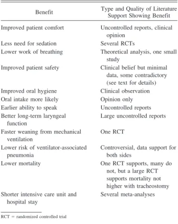
Table 1 lists potential reasons that a tracheostomy would be considered the preferred prolonged artificial airway
when compared to continued translaryngeal intubation. While some of these benefits have considerable support in the published literature, there are very few data to support most of them.
Protection of the larynx and the upper airway from pro-longed intubation is an important reason to perform a tra-cheostomy, and to consider early provision of this surgical airway. Many anatomical structures are at risk from trans-laryngeal intubation. Vocal cord edema and damage, la-ryngeal mucosal erosions, lala-ryngeal scarring and stenosis, and recurrent laryngeal nerve damage can lead to per-manent disability. The potential for recovery or surgical repair of many of these injuries is less with continued prolonged intubation. Direct laryngeal examination dem-onstrates marked airway changes within several days of translaryngeal intubation. Usually these early changes are reversible and there is gradual improvement in airway ex-amination after the tube is removed from the larynx.3An example of a damaged larynx with fused vocal cords, which occurred after only 3 days of translaryngeal intu-bation, is seen in Figure 1. This patient recovered com-pletely after a tracheostomy was placed and the oral ETT was removed.
The author has disclosed no conflicts of interest.
Charles G Durbin Jr MD FAARC, Department of Anesthesiology, Uni-versity of Virginia, Box 800710, Charlottesville VA 22908. E-mail: [email protected].
Table 1. Reputed Benefits of Changing From a Translaryngeal Endotracheal Tube to a Tracheostomy Tube in a Patient Who Requires Prolonged Intubation
Benefit Type and Quality of Literature
Support Showing Benefit Improved patient comfort Uncontrolled reports, clinical
opinion
Less need for sedation Several RCTs
Lower work of breathing Theoretical analysis, one small study
Improved patient safety Clinical belief but minimal data, some contradictory (see text for details)
Improved oral hygiene Clinical observation
Oral intake more likely Opinion only
Earlier ability to speak Uncontrolled reports Better long-term laryngeal
function
Large uncontrolled reports Faster weaning from mechanical
ventilation
One RCT Lower risk of ventilator-associated
pneumonia
Controversial, data support for both sides
Lower mortality One RCT supports, many do
not, but a large RCT supports mortality not higher with tracheostomy Shorter intensive care unit and
hospital stay
Several meta-analyses
Improved Comfort?
Patients experience discomfort with persistent transla-ryngeal intubation and are more comfortable following tracheostomy.4Improved patient comfort and less require-ment for sedation have been reported in several studies following placement of a tracheostomy. In a follow-up study of patients who were randomized to remain intu-bated translaryngeally for a prolonged period or to receive an early tracheostomy, Blot and colleagues reported that oral comfort scores, mouth uncleanliness, perception of change in body image, feelings of safety, and overall com-fort were lower in the prolonged translaryngeal intubation group, compared to those who were randomized to early tracheostomy.5 Thirteen patients in that study who sur-vived to hospital discharge and had undergone both trans-laryngeal intubation and tracheostomy reported tracheos-tomy as the more comfortable airway of the two. Patient comfort alone may be enough to justify tracheostomy rather than continuing with prolonged translaryngeal intubation if the risks of the 2 approaches are comparable.
Faster Weaning and Shorter Stay?
Besides protecting the larynx, tracheostomy may shorten the duration of mechanical ventilation because of reduced work of breathing,6the need for less sedation and analge-sia, or because once a secure airway is in place clinician weaning behavior changes.7In a prospective trial that in-cluded 74 surgical/trauma patients who were unable to complete a spontaneous breathing trial after 72 hours of mechanical ventilation, Boynton and colleagues randomly divided these patients into 2 groups: those in whom it was decided to continue translaryngeal intubation, and those in whom it was decided to proceed with immediate trache-ostomy prior to any attempt at weaning.8 Weaning from mechanical ventilation was carried out in both groups
us-ing the same protocol of gradual reduction in pressure-support ventilation. There were 21 patients in the imme-diate-tracheostomy group and 54 in the continued-translaryngeal-intubation group, 25 of whom eventually underwent tracheostomy. Only immediate tracheostomy before weaning and having a lower rapid shallow breath-ing index predicted more rapid weanbreath-ing, with the odds ratio for immediate tracheostomy being 2.1 for being weaned in 3 versus 6 days. A meta-analysis of randomized trials comparing earlier versus later tracheostomy done by Griffiths et al9 confirmed that weaning was more rapid with early tracheostomy.
Faster weaning may reduce ICU and hospital stay, but so may other tracheostomy attributes. Patients requiring only extended mechanical ventilation or airway support are often moved from an ICU to a less intensive care environment once a tracheostomy is in place. This appears on the surface to be reasonable, because if the tube be-comes dislodged, no special equipment or skills are needed to replace it. After the stomal tract has matured, usually within 3–7 days, the tube can usually be easily reinserted without difficulty. In some institutions it is routine to elec-tively change a tracheostomy tube after a week or two following initial insertion. This is done to confirm the stability of the tract and reduce the likelihood of difficulty at reinsertion should the tube inadvertently become dis-lodged or for scheduled tube changes at a later time. Ear-lier transition of a patient from the ICU remains a major demonstrable effect of tracheostomy. This transition to a lower level care area may not be without risk.
Increased Patient Safety?
The presumed increased safety of having a tracheos-tomy in a patient managed outside of an ICU or special care unit has come under scrutiny. In a prospective obser-vational cohort study, Martinez and colleagues recently reported that patients discharged to the ward with a tra-cheostomy in place had almost 3 times the mortality of those who had received a tracheostomy but who were decannulated prior to ward placement.10In that study, how-ever, the difference was only shown as a trend withP⫽.10. Multivariate analysis identified 3 highly significant factors associated with increased ward mortality: lack of decan-nulation at ICU discharge (odds ratio 6.76, 95% CI 1.21– 38.46,P⫽.03), body mass index⬎30 kg/m2(odds ratio 5.81, 95% CI 1.24 –27.24,P⫽.03), and tenacious sputum at ICU discharge (odds ratio 7.27, 95% CI 1.00 –55.46, P ⫽ .05). Most of the deaths on the ward were due to unexpected cardiorespiratory arrests, usually in the early morning hours. Despite the fact that only one episode of tube-associated problem was reported in the study group, inadequate monitoring of patients and failure of early treat-ment of tube problems may have played a part in the Fig. 1. Fused vocal cords were seen after only 3 days of intubation
in this patient. This lesion resolved several days after the trache-ostomy was placed and the endotracheal tube removed.
increased mortality in that group. Other groups have re-ported similar findings in less well designed trials,11,12 whereas some have not identified having a tracheostomy at ward discharge a risk for increased mortality when cor-rected for other risk factors.13
Other authors have noticed the additional risks of hav-ing a tracheostomy tube, as compared to a standard trans-laryngeal tube in place, even when the patient resides in an ICU environment.14,15 In 2000, Kapadia and colleagues reported airway accidents occurring in all intubated pa-tients in a 16-bed multidisciplinary ICU. The study pop-ulation included 5,046 patients intubated for 9,289 days during a 4-year period. They prospectively collected data, including the number and timing of airway accidents, the type of tracheal tube used, the duration of intubation, de-scription of the type of accident, the severity of the acci-dent, impact on the course of the patient’s illness, and whether the accident was preventable. The total number of accidents for the entire study period was only 36, and 26 occurred in the 5,043 endotracheally intubated patients during their 8,446 days of intubation. None of those acci-dents were severe, and no deaths occurred. There were 10 tracheostomy-related accidents in the 79 patients with tra-cheostomies during their 843 days of intubation. Six of those events had severe consequences, and one resulted in death. Thus, even when monitored in an ICU, airway ac-cidents associated with tracheostomy tubes occurred more frequently and resulted in higher mortality (10%) than in patients with conventional ETTs. The clinician’s reassur-ance of a secure airway by having a tracheostomy may not translate into actual greater safety.
Fewer Lung Infections With Tracheostomy?
Microaspiration of oral secretions past the tube cuff is thought to contribute to development of pneumonia. It was believed that incidence of ventilator-associated pneumonia (VAP) would be decreased by placing a tracheostomy early in respiratory failure. This would allow laryngeal compe-tence to recover and lessen the quantity of aspirated se-cretions. Reported data are mixed in this regard: some studies have suggested a reduced VAP rate, whereas oth-ers, mostly older studies, indicate an increase in lower-respiratory-tract infections following tracheostomy. Over-all, if there is an influence on the incidence or course of VAP from tracheostomy, it is small. The meta-analysis by Griffiths et al mentioned previously demonstrated only an insignificant trend to lower incidence of VAP when tra-cheostomy was performed earlier rather than later.9A re-cent review and meta-analysis of early versus later trache-ostomy, by Durbin and colleagues, confirmed the observation of minimal effect on the incidence of VAP.16
When to Perform a Tracheostomy?
Tracheostomy is indicated when the need for endotra-cheal intubation is or is projected to be prolonged. How long is “prolonged” continues to evolve. In older guide-lines, tracheostomy was recommended for consideration only if extubation did not occur by 21 days; more recently it has been suggested that tracheostomy be considered within 2–10 days of intubation, and that a projected need for 14 days of intubation be used as the criterion for the procedure. In selected patients with severe multi-trauma and/or head injury with low Glasgow coma score, trache-ostomy at the earliest convenient time, often within 3– 4 days of intubation, appears to afford some benefits. This early placement of a tracheostomy corresponds to that proposed by the otorhinolaryngologists who suggested that tracheostomy be performed within several days of intubation, to prevent laryngeal injury from even very short periods of intubation.17 Because having a tracheostomy tube in place may add additional risks to patient safety, qualified caregivers proximate to the patient are needed to prevent injury or death when tube accidents occur.
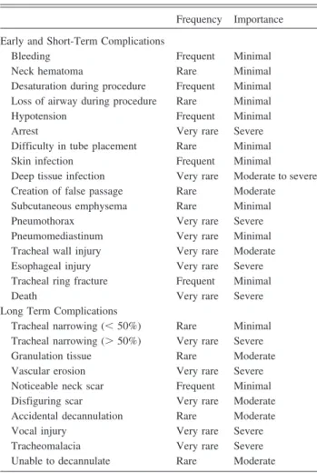
A continuing controversy surrounding tracheostomy is when in the course of critical illness is it appropriate to perform it. In attempting to balance the risks and benefits of the procedure, an individualized patient approach has been recommended. A balance of the risks and benefits of the procedure in a particular patient should be used to determine if and when a tracheotomy should be placed. Although this approach would appear to have merit, the risks of tracheotomy in an individual patient are difficult to predict and often delayed, and are dependent on the technique selected, individual patient anatomy, comorbidi-ties, and the experience of the operator. As discussed above, the meaningful benefits of tracheostomy are few, so the balance is greatly affected by estimation of risk. A com-pilation of observed problems and acute and longer-term complications of tracheostomy are listed in Table 2. Some of these are very rare but very important (eg, death), and others are frequent but carry no long-term impact on the patient (eg, transient oxygen desaturation). The long-term laryngeal and tracheal outcomes following decannulation have not been well studied, as large registries with detailed outcome data have not been established. A recently com-pleted study performed in an attempt to provide some information on early versus late tracheostomy has pro-vided some interesting and useful material informing the decision of when to perform a tracheostomy. The data collected during this study remains under peer review and (hopefully) will soon be published. Early release of infor-mation presented at international meetings forms the basis for the material reported here.
The Tracheostomy Management in Critical Care (Trac-Man) study (http://www.tracman.org.uk) was designed to
determine if there are any important differences in patients receiving a tracheostomy within 4 days or following 10 or more days of translaryngeal intubation. Patients were screened at 72 hours after being intubated, and if their clinicians believed that they would require at least 14 days of mechanical ventilation, consent was obtained and they were entered into the study. Patients were randomized to an “early” or “late” tracheostomy group. The early group received a tracheostomy immediately, whereas the proce-dure was delayed for at least 10 days in the late group. This multi-institution study, carried out within the United Kingdom, involved 72 ICUs and lasted over 4 years, eventually enrolling over 900 patients. The technique of tracheostomy was not controlled by protocol, but the great majority were preformed percutaneously at the patient’s bedside. Initial data analysis suggests that randomization was successful; the groups had an equal average age (⫾ 63 y), severity of illness (via Acute Physiology and Chronic Health Evaluation II [APACHE] scores), and type of ICU (approximately 80% medical units and 20% surgical units). Important observations include an iden-tical mortality between the groups (approximately 30%),
very few minor procedural complications, and no trache-ostomy-related deaths. A positive outcome in the early-tracheotomy group was fewer days of patient sedation. However, there were no differences in ICU or hospital stay. An important, highly significant difference was seen in the number of tracheostomies actually performed in the 2 groups: over half of the patients assigned to receive tracheostomy later than 10 days never received one. Most of these patients randomized to the late-tracheostomy group were weaned and extubated prior to the 14-day projection, and a few others had died. A major implication of this study is that clinicians are poor at predicting who will need prolonged intubation and ventilation. Another important conclusion is that performing a tracheostomy appears not to increase mortality or long-term complications (patients were followed for at least 12 months to identify compli-cations and late outcomes); however, the complete story from this study is not yet published and available for scrutiny.
A weakness of the TracMan trial protocol is that the process for identifying who would need prolonged intu-bation was not objectively specified. Also, the distribution of participating units largely excluded surgical and neuro-logically injured patients. Are there methods or markers that can accurately predict which patients will require pro-longed intubation and therefore could benefit from earlier tracheostomy? An early report by Qureshi and colleagues suggested that patients with poor Glasgow Coma Score, brainstem dysfunction, or supratentorial lesions 3 days fol-lowing intubation would either die or require tracheotomy for prolonged recovery because of poor airway protective reflexes.18This has been confirmed by others, including Bouderka and colleagues, who compared prolonged intu-bation with early tracheostomy in patients with isolated head injury with Glasgow coma scores ⬍ 8, brainstem deficits, and cerebral contusion on computed tomogram.19 They randomized 62 patients to tracheostomy at day 4 or 5 or to continue with translaryngeal intubation. The 2 groups were comparable in terms of age, sex, and severity of illness via the Simplified Acute Physiologic Score. The average duration of mechanical ventilatory support was shorter in the tracheostomy group (14.5⫾ 7.3 d), com-pared to the translaryngeal intubation group (17.5⫾10.6 d) (P ⫽ .02). In those patients who developed pneumonia, mechanical ventilation time was shorter in the tracheos-tomy group (6⫾4.7 d) than in the translaryngeal-intuba-tion group (11.7⫾ 6.7 d) (P⫽.01). However, there was no difference in frequency of pneumonia or mortality be-tween the 2 groups.
Do Head-Injured or Other Trauma Patients Benefit from Early Tracheostomy?
Recognizing the contribution severe head injury makes to outcome from traumatic injury, the Eastern Association
Table 2. Short-Term and Long-Term Complications of Tracheostomy
Frequency Importance Early and Short-Term Complications
Bleeding Frequent Minimal
Neck hematoma Rare Minimal
Desaturation during procedure Frequent Minimal Loss of airway during procedure Rare Minimal
Hypotension Frequent Minimal
Arrest Very rare Severe
Difficulty in tube placement Rare Minimal
Skin infection Frequent Minimal
Deep tissue infection Very rare Moderate to severe
Creation of false passage Rare Moderate
Subcutaneous emphysema Rare Minimal
Pneumothorax Very rare Severe
Pneumomediastinum Very rare Minimal
Tracheal wall injury Very rare Moderate
Esophageal injury Very rare Severe
Tracheal ring fracture Frequent Minimal
Death Very rare Severe
Long Term Complications
Tracheal narrowing (⬍50%) Rare Minimal
Tracheal narrowing (⬎50%) Very rare Severe
Granulation tissue Rare Moderate
Vascular erosion Very rare Severe
Noticeable neck scar Frequent Minimal
Disfiguring scar Very rare Moderate
Accidental decannulation Rare Moderate
Vocal injury Very rare Severe
Tracheomalacia Very rare Severe
for the Surgery of Trauma (EAST) has authored recent guidelines recommending that early tracheostomy be per-formed in these patients.20This Level II recommendation states that fewer days of mechanical ventilation will be needed and ICU stay will be shorter with early (within 5 days of injury) tracheostomy. The recommendation is supported by class II data, which includes clinical studies in which the data were collected prospectively, and retro-spective analyses that were based on clearly reliable data. Types of studies classified as such included observational studies, cohort studies, prevalence studies, and case-con-trol studies. Level II recommendations are defined as rea-sonably justifiable by available scientific evidence, strongly supported by expert opinion, and supported by class I or class II data.
The only Level I recommendation from that guideline is that there is no mortality difference between early or late tracheostomy, which is consistent with the TracMan trial described above. The final Eastern Association for the Surgery of Trauma recommendation from this literature evaluation process is that since early tracheostomy may decrease the total days of mechanical ventilation and ICU stay in trauma patients without head injuries, and early tracheostomy may decrease the rate of pneumonia in trauma patients, it is recommended that early tracheostomy should be considered in all trauma patients who are anticipated to require mechanical ventilation for at least 7 days. That is a Level III recommendation, suggesting only that the ma-jority of practitioners consider it reasonable. This would apply to those patients with respiratory failure or neuro-logic impairment without primary head injuries.
A counterpoint argument to early tracheostomy in pa-tients with neurologic abnormalities has been elegantly advanced by King and colleagues.21They have reviewed reports and examined the impact of extubation in those patients who remained intubated purely for airway protec-tion concerns due to a poor level of consciousness. In their review it appears that poor mental status alone is insuffi-cient to require prolonged intubation and thus tracheos-tomy. Cough effectiveness, secretion quantity, and secre-tion viscosity all impact on the success of extubasecre-tion in this group and other types of patients as well. Those au-thors correctly point out that many studies demonstrate that failed extubation carries increased morbidity as well as mortality, although the magnitude of the increase and reasons for it are not clear. While it is clear that delaying extubation is associated with increased morbidity and mor-tality, the medical literature also demonstrates harm from extubation failure. Over 55 studies, involving more than 30,000 patients, suggest that the overall rate of extubation failure is approximately 12% (range 2–25%).22One study from a medical ICU found that re-intubation resulted in an average 12 additional days of mechanical ventilation, 21 ICU days, 30 hospital days, and an increased need for
tracheostomy and post-acute-care hospitalization.23 Inter-estingly, while patients requiring re-intubation tend to be sicker, multivariate analyses have shown that premorbid health status, severity of illness, and complications di-rectly associated with re-intubation do not explain the in-creased mortality associated with extubation failure.24A direct correlation between increasing time to re-intubation and mortality has led some authors to suggest that clinical deterioration prior to re-institution of mechanical ventila-tory support is responsible for the increased mortality as-sociated with extubation failure.25It has been suggested that careful monitoring and rapid intervention for respira-tory failure developing following extubation may prevent this excessive mortality. However, this hypothesis has not yet been tested.
A report by Salam and colleagues, who assessed the ability of 88 patients who had successfully completed a spontaneous breathing trial to perform 4 simple tasks (open eyes, follow with eyes, grasp hand, and stick out tongue) prior to extubation.26 Patients unable to complete the 4 commands were 4 times more likely to require re-intuba-tion (risk ratio 4.3, 95% CI 1.8⫺10.4). Depressed cough peak flow (risk ratio 4.8, 95% CI 1.4 –16.2) and secretions greater than 2.5 mL/h (risk ratio 3.0, 95% CI 1.0 – 8.8) were also independent predictors of extubation failure in this study. In a prospective cohort study specifically as-sessing brain-injured patients, Coplin and colleagues fol-lowed the course of mechanical ventilation, weaning, and extubation in 136 patients.27 Ninety-nine patients (73%) were extubated on meeting readiness criteria, and the re-maining 37 patients (27%) remained intubated for an ad-ditional average of 3 days (range 2–19 d) following meet-ing weanmeet-ing criteria. Patients with prolonged intubation had a lower median Glasgow coma score (7 vs 10,P⬍.001). Glasgow coma score was not found to be predictive of the need for re-intubation. That study is often cited as sup-porting the safety of extubating patients with acute brain injury, but caution should be used in interpreting those data. Given that the prolonged intubation group was de-layed in their time to extubation, the natural history of that group was altered and may not reflect the outcome that would have been obtained if they had been extubated at the time they first passed a weaning trial.
Do Medical Patients Benefit Form Early Tracheostomy?
Medical patients requiring prolonged intubation may have lower mortality with early tracheostomy. A study by Rumbak and colleagues performed in 3 institutions ran-domized severely ill patients (APACHE score⬎25) to a percutaneous tracheostomy at 48 hours or to a tracheos-tomy at 14 days or longer. Low-tidal-volume ventilation, a ventilator weaning protocol, and daily spontaneous
breath-ing trials were specified in the research protocol.28 Unfor-tunately, how clinicians were able to predict the need for 14 or more days of intubation was not reported. Prediction was very good, and only 10% of the late-tracheostomy group did not receive a tracheostomy (2 died before 14 days and 8 were weaned and extubated), which is much better than the prediction in the TracMan trial, where more than 50% of the late group did not need tracheostomy at 14 days. Unlike any other published study, the mortality benefit of early tracheostomy was remarkable, with mortality only 32% in the early tracheostomy group and over 61% in the late group. Infections and other complications were more common in the late-tracheostomy group. An important question is, can the need for prolonged intubation in pa-tients with medical disorders be predicted with accuracy? In the case of respiratory failure requiring mechanical ventilation in patients with acute lung injury/acute respi-ratory distress syndrome the duration of intubation may be predicted by the initial degree of injury and by the pro-gression of disease within the first few days. As long ago as 1990, Heffner and Zamora suggested that by combining repeated evaluations of physiology and anatomy in pa-tients with acute respiratory distress syndrome on day 7, a prediction of needing prolonged intubation (and trache-ostomy) could be made. They used the ratio of arterial to alveolar PO2, presence of high PEEP (defined asⱖ10 cm
H2O), and the chest radiograph percentage of lung-field infiltrates on day 7 to predict continued need for intubation and ventilation on day 14 (Table 3).29While these mea-sures were able to identify all patients requiring prolonged intubation (sensitivity), they also detected many patients who were able to be weaned (false positives). As expected, the presence of more severe disease for a longer period of time is predictive of longer intubation. This was also noted in Rumbak’s study, as those patients with high APACHE scores required longer intubation.28Accuracy of these and other indices in predicting the need for long-term airway support has not been assessed in a systematic way. Current recommendations for tracheostomy time are: continued
need for intubation should be assessed daily, and if im-provement is not observed, a decision to perform a trache-ostomy should be made when the need for intubation is believed to extend for at least 14 days in patients with medical diseases and respiratory failure. The expected res-olution of the particular disease and the patient’s comor-bidities will influence this decision. The algorithm repro-duced in Figure 2 regarding the decision of tracheostomy timing used at the Mayo Clinic provides useful guidance in many patients.30Fear of the risk of unnecessary trache-ostomy should be less with the reassuring mortality pre-liminarily reported from the TracMan study and data from large trauma registries.
How Should a Tracheostomy Be Performed?
There are many ways to perform a tracheostomy, but these can be classified into several general approaches. One common division of techniques is “open” or surgical tracheostomy and the other is “percutaneous” or percuta-neous dilatory tracheostomy (PDT). Ever since Ciaglia first described his use of a guide wire and serial dilation technique for PDT in 1985 (which was an adaptation of the percutaneous nephrostomy tube placement technique, and is a variant of the vascular access technique described by Seldinger31in 1953), the popularity of this technique has grown dramatically.32Early comparisons between sur-gical tracheostomy and PDT suggest more (but generally
Table 3. Prediction of Need for More Than 14 Days of Mechanical Ventilation
Predictive Value Sensitivity
(%)
Specificity (%)
Positive (%)
Negative (%)
PEEP⬎10 cm H2O 71 100 100 71
PaO2/PaO2⬍0.40 57 80 80 57
No radiographic improvement 67 100 100 67
⬎50% of lung fields with radiographic alveolar infiltrates
78 100 100 75
(Data from Reference 29.)
Fig. 2. Approach to the timing of tracheostomy in patients receiv-ing mechanical ventilation. APACHE ⫽ Acute Physiology and Chronic Health Evaluation. (Adapted from Reference 30, with per-mission.)
less severe) early complications during PDT placement, but fewer late problems.33,34 A recent meta-analysis by Higgins and Punthakee, which included almost 1,000 pa-tients, identifies several outcome differences between sur-gical tracheostomy and PDT.35The findings are summa-rized in Table 4. In their report, pooled odds ratios revealed more difficulties during decannulation and from tracheos-tomy tube obstruction after PDT. These issues were minor, none were considered life-threatening, and they may have been related to differences in the type of tube used (ie, PDT rarely placed tubes with removable inner cannulas). Mortality was not clearly different between the techniques, but the trend favored PDT. There were significantly fewer stomal wound infections and smaller neck scars after PDT placement. There was no statistically significant differ-ence between the techniques in creation of a false passage, causing minor or major hemorrhage, or developing late subglottic stenosis. There was no significant difference in overall complications between the techniques; however, there was a trend in complications favoring the percuta-neous technique. PDT performance time was shorter over-all by 4.6 min (not clinicover-ally important), and costs (when reported) were less by approximately $456 per procedure; however, data were available from only 4 studies.
Initially, PDT was reserved for patients with few risk factors and favorable neck anatomy. With growing expe-rience, the indications for PDT have been expanded and the patient exceptions which mandate a surgical tracheos-tomy have decreased. In the case of morbid obesity and moderate coagulopathy, PDT may afford better survival with fewer complications.36,37Internationally, countries dif-fer in the predominant tracheostomy technique; however, PDT is increasingly the technique of choice for critically ill patients in ICUs throughout the world (Table 5).38-43
Surgical Technique for Placing a Tracheostomy Tube
To perform a surgical tracheostomy, the patient’s shoul-ders are elevated and the head is extended unless contra-indicated by cervical disease or injury. This position ele-vates the larynx and exposes more of the upper trachea. As with most surgical procedures, prophylactic antibiotics specific for skin pathogens are usually administered 30 – 60 min prior to skin incision. The skin from the chin to below the clavicles is sterilely prepared with either an iodine-based disinfectant or a solution of chlorhexidine. If excessive hair is present, it should be removed with elec-tric clippers immediately prior to skin preparation. Sterile drapes are placed, creating an opening from the top of the larynx to the patient’s suprasternal notch. Local anesthesia with a vasoconstrictor is infiltrated into the skin and deeper neck tissues to reduce the amount of bleeding and provide analgesia during the procedure.
The skin of the neck over the 2nd tracheal ring is iden-tified, and a vertical incision about 2–3 cm in length is created. Care must be taken to avoid cutting deeper than the subcutaneous tissues to prevent lacerating the thyroid isthmus or a large neck vein. Sharp dissection following the skin incision is used to cut across the platysma muscle, with bleeding controlled by hemostats and ties or electro-cautery. Blunt dissection parallel to the long axis of the trachea is then used to spread the submuscular tissues until the thyroid isthmus is identified (Fig. 3). If the thyroid gland lies superior to the 3rd tracheal ring, it can be bluntly undermined and retracted superiorly to gain access to the trachea. If the isthmus overlies the 2nd and 3rd ring of the trachea, it must be mobilized and either a small incision made to clear a space for the tracheostomy (Fig. 4) or complete transection of the isthmus must be accomplished (Fig. 5).
Blunt dissection is continued longitudinally through the pretracheal fascia, and the desired ring (usually the 2nd ring) is identified. One of 2 types of tracheal entry is
Table 4. Outcome From Meta-analysis of Open Surgical Tracheostomy Versus Percutaneous Dilational Tracheostomy
Favored Percutaneous Technique
Favored Open Surgical Tracheostomy Wound infection (P⬍.001) Decannulation/obstruction
(P⫽.009)
Unfavorable scarring (P⫽.01) False passage (P⫽.08) Cost-effectiveness (P⬍.001) Minor hemorrhage (P⫽.77) Case length (P⬍.001)
Overall complications (P⫽.05)
Major hemorrhage (P⫽.17) Subglottic stenosis (P⫽.19) Death (P⫽.50)
Data from Reference 35.
Table 5. Tracheostomy Techniques and Use of Bronchoscopy By Country
Country Routine Use of
PDT (%)
Routine Use of FOB With PDT (%)
Preferred PDT Technique
France38 21 ND Modified Ciaglia
Germany39 86 98 Modified Ciaglia
Netherlands40 62 36 Classic Ciaglia
Spain41 72 16 Griggs Forceps
Switzerland42 57 ND ND
United Kingdom43 97 83 Modified Ciaglia
PDT⫽percutaneous dilational tracheostomy FOB⫽fiberoptic bronchoscopy ND⫽no data available (Adapted from Reference 39.)
usually used for surgical tracheostomy. These are: com-plete removal of the anterior part of the tracheal ring to create the stoma, and creation of a rectangular flap with the severed but still attached part of the ring. In the ring-removal approach, the ring is lifted with a tracheal hook and 2 circumferential sutures are placed around the ring laterally. The portion of ring between the secured sutures is then cut and removed, leaving a hole in the anterior tracheal wall for the tracheostomy tube. The sutures are left in place and used to provide counter-traction on the trachea as the tube is forced into the lumen. This is
illus-trated in Figure 6. The ring sutures are cut long and left out of the wound or used to secure the tracheostomy tube. These sutures can be used to identify the trachea and re-insert an inadvertently dislodged tracheostomy tube. After placement of a surgical tracheostomy, the fistula tract is not stable for at least 4 –5 days, and a tube dislodged soon after placement often cannot be reinserted through the fis-tula into the trachea. The ring sutures may help if this situation occurs. A second method for entry into the tra-chea involves creating a tratra-cheal wall flap sutured to the skin. This is done by incising the fascia over the superior ring and entering the trachea along its inferior margin. This becomes the outer lip of the flap. Lateral cuts through the lower ring complete the sharp dissection. The flap thus created is reflected downward and attached with several sutures to the skin of the neck. This fistula is truly a “stoma,” with tracheal mucosa approximated to the skin. The stability of this tract is believed to be superior to the ring resection-removal technique; however, no study has convincingly demonstrated this.
Fig. 3. Creating a surgical tracheostomy. After incising the skin and dividing the strap muscles of the neck, the thyroid isthmus is mobilized with a hemostat. (From Reference 44.)
Fig. 4. Creating a surgical tracheostomy. If the thyroid cannot be retracted either superiorly or inferiorly to reveal the 2nd and 3rd tracheal rings, a small incision in the gland may be created to allow access to the trachea. (From Reference 44.)
Fig. 5. Creating a surgical tracheostomy. If the thyroid isthmus remains in the way of the site of the tracheostomy, it may be completely divided, carefully ensuring there is no bleeding. This is the most common approach and gives the greatest access to the trachea. (From Reference 44.)
Fig. 6. Creating the tracheal portal. There are 2 basic approaches to tracheal entry. As illustrated here, the 2nd tracheal ring is di-vided laterally and the anterior portion removed. Lateral sutures are used to provide counter-traction during tracheostomy-tube insertion. These are left uncut to provide assistance should the tube be accidentally dislodged later. (From Reference 44.)
Techniques for Percutaneous Dilational Tracheostomy
PDT, an alterative to surgical tracheostomy, can be per-formed in one of several different ways. Most of these are variants of the method described by Ciaglia,32 which is achieved by placing a guide wire between 2 tracheal rings entering the anterior tracheal wall in the midline.
Follow-ing this, the wire is used to direct the passage of one or more dilators over the wire to mechanically separate the rings and create the stoma for the tracheostomy tube. As with surgical tracheostomy, proper head position, sterile skin preparation, local anesthesia and epinephrine infiltra-tion, provision of sedation or anesthesia, and complete barrier precautions are used. The earliest techniques con-sisted of a series of stiff plastic dilators of increasing size, advancing and removing each over the wire sequentially until one large enough for the chosen tracheostomy tube was passed. A separate loading or inserting dilator with the snugly-fitting tracheostomy tube is then passed and re-moved, leaving the tracheostomy tube behind in place. The steps in the PDT technique are illustrated in Figures 7, 8, 9, and 10.
Early changes in this basic technique consisted of cre-ating a single dilator of appropriate size with a long taper to create the stoma with a single pass,45 and designing tracheostomy tubes with tapered tips, allowing them to Fig. 7. The trachea is entered between the appropriate tracheal
rings with an intravenous catheter, with aspiration of air to confirm correct location. The needle is withdrawn and the catheter left in place as a conduit for the guide wire. (From Reference 44.)
Fig. 8. The guide wire is threaded through the catheter to act as a guide for the dilators that follow. It also helps protect the tracheal wall by directing the pointed dilator tips down the trachea. (From Reference 44.)
Fig. 9. Through the fiberoptic bronchoscope the wire is seen en-tering between the 2nd and 3rd tracheal rings, directly in the cen-ter or the trachea.
Fig. 10. The tip of the dilator is seen entering the trachea over the wire. After the stoma is created, a loading dilator with the trache-ostomy tube is used to deliver the tube to its final position. (From Reference 44.)
more smoothly pass through the neck tissues over the in-sertion dilator. Soon after development of the PDT tech-nique, complications from bent or misplaced wires were seen. This led to the development of a plastic sheath in-serted over the wire to stiffen it and help prevent misplace-ment. Using a fiberoptic bronchoscope through the exist-ing ETT helped direct the tracheal entry point, confirm final placement, identify tracheal injuries, and improve success and safety.46,47
Modifications of PDT Technique
Several variants on the PDT have been developed and are advocated by various groups. In Europe, a wire-guided sharp, cutting forceps developed by Griggs and colleagues gained early popularity but is reported to have more seri-ous problems, including substantial bleeding and possibly long-term risk.48In order to improve safety by minimizing tracheal injury, performance of PDT under bronchoscopic control, using any of the many available techniques, has been advocated.46,47In order to reduce the likelihood of fracturing a tracheal ring during the forced dilation, Fan-toni developed a complex system of passing the dilator from inside the trachea to the outside using a specially designed tracheostomy tube and a rigid bronchoscope.49 Another approach to try to reduce tracheal trauma during PDT has been developed using a screw-like device (Per-cuTwist) to separate the trachea rings.50 Each of these variations came about as an attempt to improve some as-pect of the basic dilation technique or observed shortcom-ings of particular methods.
There have been several additional modifications of PDT described. One of these, creation of the tracheal stoma using an angiographic balloon, is likely to add an-other group of clinicians to the groups who are now per-forming PDT.51 With this technique, a sausage shaped high-pressure angiographic balloon (Ciaglia Blue Dolphin Balloon Percutaneous Tracheostomy Introducer, Cook Critical Care, Bloomington, Indiana) is used to create the stoma. The trachea is entered between tracheal ring 2 and 3 in the conventional way, with a needle; then a guide wire is placed through a 1–2 cm skin incision. Bronchoscopic control is maintained with the ETT withdrawn into the larynx. The dilating balloon is placed through the trache-ostomy tube and loading dilator and carefully advanced over the guide wire. When the balloon is located precisely in the center of the stoma, it is inflated with 11 atmo-spheres pressure for 15 seconds, spreading the tracheal rings and compressing blood vessels and other tissues. This radial pressure separates the rings and stretches the skin and may reduce bleeding and the incidence of tra-cheal ring fracture; however, in the first 20 cases, a 5% ring fracture rate was noted (1 out of 20). Long-term con-sequences of tracheal ring fractures produced during PDT
are minimal and occur with most percutaneous techniques. The effect of the dilating balloon inflation on the posterior and lateral tracheal wall integrity is not known and may pose an additional new risk associated with this technique. One patient in the initial evaluation group developed sub-cutaneous emphysema, suggesting the balloon inflation may have produced an airway injury.
A variant on the single-dilator technique of PDT place-ment begins by first creating a small surgical incision, using blunt dissection, to allow a gloved finger to palpate the trachea.52The finger tip is used to identify the cricoid ring and tracheal rings. Operators using this technique often dispense with the fiberoptic bronchoscope, relying on tactile identification of correct position for needle en-try. The needle and guide wire are than passed and if no unusual resistance is felt, the single dilator and tracheos-tomy tube are inserted blindly. In some ways this is similar to an open tracheostomy; however, it is less invasive, sim-pler, and requires little or no surgical dissection. Even when using this technique, most operators have a fiberop-tic scope available for confirmation of correct placement or to view the entire process if undue resistance to passage of the wire occurs.
Use of a laryngeal mask airway (LMA) as a conduit for the bronchoscope is very helpful if the patient can be safely extubated and maintained with an LMA while the tracheostomy is being performed. Unlike withdrawing an existing ETT and attempting not to accidently extubate the patient, planned extubation and LMA placement can be electively performed and carefully controlled. If difficulty occurs while maintaining the patient with an LMA, the trachea should be secured with a cuffed ETT. Patients requiring high airway pressures or those with a known difficult intubation are probably not good candidates for this approach. Once the LMA is in place and adequate gas exchange is established, entry into the larynx with the bronchoscope is easily accomplished as the LMA usually rests directly in front of the larynx. A specially designed supraglottic airway, the Air-Q (Mercury Medical, Clear-water, Florida), is a variant on the classic LMA: it has a larger diameter, short, rigid connecting tubes, and un-Fig. 11. Specially designed intubating supraglottic airway, which is useful as a conduit for fiberoptic bronchoscope insertion during percutaneous dilational tracheostomy. The patient can continue to breath (or be ventilated) through the large-diameter connecter with the bronchoscope in the larynx during the entire procedure.
blocked distal openings. The AirQ is recommended for blind intubation using a conventional ETT (Fig. 11). This special tube is also very useful for fiberoptic bronchoscope placement during PDT. The bronchoscope tip can be left protected in the larynx during the entire procedure, allow-ing precise identification of tracheal entry, the dilation process, and identification of complications if they occur. Recently some authors suggested using ultrasonography of the neck to identify underlying anatomy with more precision than palpation. Tracheal rings are usually easily appreciated and an overlying large vessel or thyroid gland can be seen and avoided during the procedure. As with all techniques, success using the ultrasound device and interpret-ing the images takes practice, and expertise requires substan-tial experience. With adequate experience it may be possible to correctly identify the entry point; however, confirmation of correct tube placement is usually best achieved with direct visualization with a fiberoptic bronchoscope.
Bedside or Operating Room Tracheostomy Placement?
Whatever technique is chosen, there are advantages to performing the procedure in the ICU. Unstable patients experience less deterioration in vital signs and avoid the risks of travel to the operating theater if the procedure is performed in the ICU. Multiple caregivers already familiar with the patient can monitor and care for the patient if the procedure is performed in the unit. Charges for the pro-cedure are usually less when performed in the ICU; how-ever, actual costs are difficult to calculate and payments are unrelated to charges or costs. Occasionally a PDT procedure fails or a problem arises that would benefit from ready availability of operating room resources such as high-intensity lights or electrocautery devices. Whether to move a patient to the operating room if a surgical tracheostomy is needed or to perform it in the ICU and bring required equipment is an institutional decision. Excellent results have been reported with all of the PDT techniques as well as surgical tracheostomy performed in either the operating room environment or at the patient’s bedside in the ICU. Local policy and resources should guide the decision of where to perform a tracheostomy.
Summary
Tracheostomy is the most commonly performed proce-dure in the critically ill, approaching 10% of intubated pa-tients. General indications for tracheostomy include the need for a prolonged artificial airway, poor airway protective re-flexes, and upper-airway obstruction for any of a number of reasons. Expected intubation for more than 14 days is con-sidered a common reason to consider a tracheostomy. Un-fortunately, a clinician’s ability to predict this duration is especially poor early in the course of illness.
Patients with severe traumatic injuries, especially those with head injury or altered mental status, are likely to benefit from earlier tracheostomy. Known benefits from tracheostomy are: less need for deeper sedation, shorter weaning time, and shorter ICU and hospital stay. With a few exceptions, there is no difference in mortality with a tracheostomy or continued prolonged translaryngeal intu-bation. One study of very ill medical patients demonstrated markedly lower mortality with earlier tracheostomy, but the reason for that improvement remains unclear. Patients usually can be transitioned out of an ICU earlier with a tracheostomy, and consideration of early tracheostomy should be entertained when this possibility exists. Airway accidents may be more frequent and more severe in pa-tients with tracheostomy tubes, and safety should modu-late the decision to move a patient from an ICU.
Techniques for tracheostomy are evolving and improv-ing. More tracheostomies are being performed using one of a variety of PDT techniques at the patient’s beside in the ICU. Some institutions and even entire countries are slow to adopt these changes, which are driven by clinical experience and local practice patterns. French physicians for instance, continue to perform the majority of trache-ostomies using an open surgical technique.
REFERENCES
1. American Society of Anesthesiologists Task Force on Difficult Air-way management. Practice guidelines for management of the diffi-cult airway. Anesthesiology 2003;98(5):1269-1277. Erratum in: An-esthesiology 2004;101(2):565.
2. Wong DT, Pradhu AJ, Coloma M, Imasogie N, Chung FF. What is the minimum training required for successful cricothyroidotomy? Anesthesiology 2003;98(2):349-353.
3. McWhorter AJ. Tracheostomy: timing and techniques. Curr Opin Otolaryngol Head Neck Surg 2003;11(6):473-479.
4. Nieszkowska A, Combes A, Luyt CE, Ksibi H, Trouillet JL, Gilbert C, Chastre J. Impact of tracheostomy on sedative administration, sedation level, and comfort of mechanically ventilated intensive care unit patients. Crit Care Med 2005;33(11):2527-2533.
5. Blot F, Similowski T, Trouillet JL, Chardon P, Korach JM, Costa MA, et al. Early tracheotomy versus prolonged endotracheal intuba-tion in unselected severely ill ICU patients. Intensive Care Med 2008;34(10):1779-1787.
6. Jaeger JM, Littlewood KA, Durbin CG Jr. The role of tracheostomy in weaning from mechanical ventilation. Respir Care 2002;47(4): 469-480.
7. Pierson DJ. Tracheostomy and weaning. Respir Care 2005;50(4): 526-533.
8. Boynton JH, Hawkins K, Eastridge BJ, O’Keefe GE. Tracheostomy timing and the duration of weaning in patients with acute respiratory failure. Crit Care 2004;8(4):R261-R267.
9. Griffiths J, Barber VS, Morgan L, Young JD. Systematic review and meta-analysis of studies of the timing of tracheostomy in adult pa-tients undergoing artificial ventilation. BMJ 2005;330(7502):1243. 10. Martinez GH, Fernandez R, Casado MS, Cuena R, Lopez-Reina P,
Zamora S, Luzon E. Tracheostomy tube in place at intensive care unit discharge is associated with increased mortality. Respir Care 2009;54(12):1644-1652.
11. Engoren M, Arslanian-Engoren C, Fenn-Buderer N. Hospital and long-term outcome after tracheostomy for respiratory failure. Chest 2004;125(1):220-227.
12. Clec’h C, Alberti C, Vincent F, Garrouste-Orgeas M, de Lassence A, Toledano D, et al. Tracheostomy does not improve the outcome of patients requiring prolonged mechanical ventilation: a propensity analysis. Crit Care Med 2007;35(1):132-138.
13. Combes A, Luyt CE, Nieszkowska A, Trouillet JL, Gibert C, Chastre J. Is tracheostomy associated with better outcomes for patients re-quiring long-term mechanical ventilation? Crit Care Med 2007;35(3): 802-807.
14. Kapadia FN, Bajan KB, Raje KV. Airway accidents in intubated intensive care unit patients: an epidemiological study. Crit Care Med 2000;28(3):659-664.
15. Kapadia FN, Bajan KB, Singh S, Mathew B, Nath A, Wadkar S. Changing patterns of airway accidents in intubated ICU patients. Intensive Care Med 2001;27(1):296-300.
16. Durbin CG Jr, Perkins MP, Moores LK. Should tracheostomy be performed as early as 72 hours in patients requiring prolonged me-chanical ventilation? Respir Care 2010;55(1):76-87.
17. McWhorter AJ. Tracheotomy: timing and techniques. Curr Opin Otolaryngol Head Neck Surg 2003;11(6):473-479.
18. Qureshi AI, Suarez JI, Parekh PD, Bhardwaj A. Prediction and tim-ing of tracheostomy in patients with infratentorial lesions requirtim-ing mechanical ventilatory support. Crit Care Med 2000;28(5):1383-1387.
19. Bouderka MA, Fakhir B, Bouaggad A, Hmamouchi B, Hamoudi D, Harti A. Early tracheostomy versus prolonged endotracheal intuba-tion in severe head injury. J Trauma 2004;57(2):251-254. 20. Michele H, Dunham M, Brautigan R, Clancy TV, Como JJ, Ebert JB,
et al. Practice management guidelines for timing of tracheostomy: The EAST Practice Management Guidelines Work Group. J Trauma 2009;67(4):870-874.
21. King CS, Moores LK, Epstein SK. Should patients be able to follow commands prior to extubation? Respir Care 2010;55(1):56-62. 22. Epstein SK. Decision to extubate. Intensive Care Med 2002;28(5):
535-546.
23. Epstein SK, Ciubotaru RL, Wong JB. Effect of failed extubation on the outcome of mechanical ventilation. Chest 1997;112(1):186-192. 24. Epstein SK. Preventing postextubation respiratory failure. Crit Care
Med 2006;34(5):1547-1548.
25. Epstein SK, Ciubotaru RL. Independent effects of etiology of failure and time to reintubation on outcome for patients failing extubation. Am J Respir Crit Care Med 1998;158(2):489-493.
26. Salam A, Tilluckdharry L, Amoateng-Adjepong Y, Manthous C. Neurologic status, cough, secretions and extubation outcomes. Int Care Med 2004;30(7):1334-1339.
27. Coplin WM, Pierson DJ, Cooley KD, Newell DW, Rubenfeld GD. Implications of extubation delay in brain-injured patients meeting standard weaning criteria. Am J Respir Crit Care Med 2000;161(5): 1530-1536.
28. Rumbak MJ, Newton M, Truncale T, Schwartz SW, Adams JW, Hazard PB. A prospective, randomized, study comparing early per-cutaneous dilational tracheotomy to prolonged translaryngeal intu-bation (delayed tracheotomy) in critically ill medical patients. Crit Care Med 2004;32(8):1689-1694.
29. Heffner JF, Zamora C. Clinical predictors of prolonged translaryn-geal intubation in patients with the adult respiratory distress syn-drome. Chest 1990;97(2):447-452.
30. Sameer R, Pendem S, Pogodzinski M, Hubmayr RD, Gajic O. Tracheostomy in critically ill patients. Mayo Clin Proc 2005;80(12): 1632-1638.
31. Seldinger SI. Catheter replacement of the needle in percutaneous arteriography: a new technique. Acta Radiol 1953;39(5):368-376.
32. Ciaglia P, Firsching R, Syniec C. Elective percutaneous dilatational tracheostomy. A new simple bedside procedure; preliminary report. Chest 1985;87(6):715-719.
33. Dulguerov P, Gysin C, Perneger TV, Chevrolet JC. Percutaneous or surgical tracheostomy: a meta-analysis. Crit Care Med 1999;27(8): 1617-1625.
34. Durbin CG Jr. Early complications of tracheostomy. Respir Care 2005;50(4):511-515.
35. Higgins KM, Punthakee X. Meta-analysis comparison of open ver-sus percutaneous tracheostomy. Laryngoscope 2007;117(3):447-454. 36. Groves DS, Durbin CG Jr. Tracheostomy in the critically ill: indi-cations, timing and techniques. Curr Opin Crit Care 2007;13(1):90-97.
37. Romero CM, Cornejo RA, Ruiz MH, Ga´lvez LR, Llanos OP, Tobar EA, et al. Fiberoptic bronchoscopy-assisted percutaneous tra-cheostomy is safe in obese critically ill patients: a prospective and comparative study. J Crit Care 2009;24(4):494-500.
38. Blot F, Melot C. Indications, timing, and techniques of tracheostomy in 152 French ICUs. Chest 2005(4);127:1347-1352.
39. Kluge S, Baumann HJ, Maier C, Klose H, Myer A, Nierhaus A, Kreymann. Tracheostomy in the intensive care unit: a national sur-vey. Anesth Analg 2008;107(5):1639-1643.
40. Fikkers BG, Fransen GA, van der Hoeven JG, Briede IS, van den Hoogen FJ. Tracheostomy for long-term ventilated patients: a postal survey of ICU practice in The Netherlands. Intensive Care Med 2003(8);29:1390-1393.
41. Anon JM, Escuela MP, Gomez V, Garcia de LA, Montejo JC, Lopez J. Use of percutaneous tracheostomy in intensive care units in Spain: results of a national survey. Intensive Care Med 2004(6);30:1212-1215.
42. Fischler L, Erhart S, Kleger GR, Frutiger A. Prevalence of trache-ostomy in ICU patients: a nationwide survey in Switzerland. Inten-sive Care Med 2000(10);26:1428-1433.
43. Krishnan K, Elliot SC, Mallick A. The current practice of tracheos-tomy in the United Kingdom: a postal survey. Anaesthesia 2005(6); 60:360-364.
44. Durbin CG Jr. Techniques for performing tracheostomy. Respir Care 2005;50(4):488-496.
45. Byhahn C, Wilke HJ, Halbig S, Lischke V, Westphal K. Percutane-ous tracheostomy: ciaglia blue rhino versus the basic ciaglia tech-nique of percutaneous dilational tracheostomy. Anesth Analg 2000; 91(4):882-886.
46. Marelli D, Paul A, Manolidis S, Walsh G, Odim JN, Burdon TA, Shennib H, et al. Endoscopic guided percutaneous tracheostomy: early results of a consecutive trial. J Trauma 1990;30(4):433-435. 47. Oberwalder M, Weis H, Nehoda H, Kafka-Ritsch R, Bonatti H,
Prommegger R, et al. Videobronchoscopic guidance makes percuta-neous dilational tracheostomy safer. Surg Endosc 2004;18(5):839-842.
48. Griggs WM, Worthley LI, Gilligan JE, Thomas PD, Myburg JA. A simple percutaneous tracheostomy technique. Surg Gynecol Obstet 1990;170(6):543-545.
49. Fantoni A, Ripamonti D. A non-derivative, non-surgical tracheos-tomy: the translaryngeal method. Intensive Care Med 1997;23(4): 386-392.
50. Westphal K, Maeser D, Scheifler G, Lischke V, Byhahn C. Per-cuTwist: a new single-dilator technique for percutaneous tracheos-tomy. Anesth Analg 2003;96(1):229-232.
51. Gromann TW, Birkelbach O, Hetzer R. Balloon dilatational trache-ostomy: initial experience with the Ciaglia Blue Dolphin method. Anesth Analg 2009;108(6):1862-1866.
52. Paran H, Butnaru G, Hass I, Afanasyv A, Gutman M. Evaluation of a modified percutaneous tracheostomy technique without broncho-scopic guidance. Chest 2004;126(3):868-871.