Metal Exposure during Pregnancy and Pre-eclampsia:
Links to Thyroid Hormone Signaling
Yvonne Nguyen
Honors Thesis ENHS
Department of Environmental Sciences and Engineering
Gillings School of Global Public Health
The University of North Carolina at Chapel Hill
May, 2014
1 | P a g e Abstract
Pre-eclampsia is the most common pregnancy complication that can be fatal for the
mother and fetus. There is no cure nor known cause of disease, but symptoms are treatable.
eclampsia causes 13 out of 100 maternal deaths due to pregnancy complications worldwide.
Pre-eclampsia is defined by high blood pressure and high protein urine content 20 weeks into
pregnancy. The objective of this study is to investigate the relationships between toxic metals
and key signaling pathways that are associated with placental health. This study utilized 40
samples provided through a collaboration with UNC hospital. The samples were tested for metals
levels and both gene and protein expression. We found that there is a positive, significant
association between cadmium and zinc in pre-eclampsia cases. Additionally, in thyroid hormone
receptor alpha (THRA) protein expression was significantly higher in cases than in controls. The
placental zinc levels were also significantly lower in pre-eclampsia cases. These results agree
with several other findings that have suggested the influence of thyroid hormone regulation on
placenta placement and pre-eclampsia development. This suggests that relationships between
metals and thyroid signaling may impact the development of pre-eclampsia. Based on these
results, further studies can be done to understand the role of toxic metals in the pathogenesis of
pre-eclampsia in terms of thyroid hormone dysfunction and placental formation errors.
Acknowledgements:
This study was supported by the UNC Department of Obstetrics and Gynecology Research Fund
2 | P a g e Table of Contents
Page
List of Tables 3
List of Figures 3
Introduction 4
Material and Methods 8
Results 11
Discussion 18
References 23
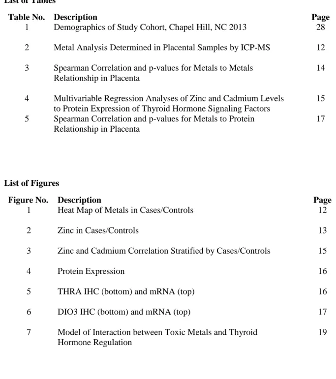
3 | P a g e List of Tables
Table No. Description Page
1 Demographics of Study Cohort, Chapel Hill, NC 2013 28
2 Metal Analysis Determined in Placental Samples by ICP-MS 12
3 Spearman Correlation and p-values for Metals to Metals Relationship in Placenta
14
4 Multivariable Regression Analyses of Zinc and Cadmium Levels to Protein Expression of Thyroid Hormone Signaling Factors
15
5 Spearman Correlation and p-values for Metals to Protein Relationship in Placenta
17
List of Figures
Figure No. Description Page
1 Heat Map of Metals in Cases/Controls 12
2 Zinc in Cases/Controls 13
3 Zinc and Cadmium Correlation Stratified by Cases/Controls 15
4 Protein Expression 16
5 THRA IHC (bottom) and mRNA (top) 16
6 DIO3 IHC (bottom) and mRNA (top) 17
7 Model of Interaction between Toxic Metals and Thyroid Hormone Regulation
4 | P a g e Introduction
We hypothesized that maternal exposure to toxic metals is associated with thyroid
hormone signaling disruption and pre-eclampsia. In order to understand this relationship, we
conducted a pilot study at UNC Hospitals to investigate the relationships and their connections to
pre-eclampsia. Pre-eclampsia is a pregnancy complication that is characterized by high blood
pressure and high urine protein content after 20 weeks of pregnancy. It is a potentially a
life-threatening condition to both the mother and the fetus if the placenta is not removed from the
body in time. Pre-eclampsia causes 13% of all maternal deaths worldwide and 18% of maternal
deaths in the United States [1]. Although pre-eclampsia occurs in part because of dysfunction of
the placenta, the biological causes are still unknown. Several risk factors have been identified
including obesity, previous history of high blood pressure, and multiple births [2]. It has been
suggested that the positioning or formation of the placenta, if incorrect, can lead to hypoxia,
which can induce the increase in protein levels and blood pressure characteristic of
pre-eclampsia [3]. Other suggestive causes include genetic links to thyroid dysfunction that affect
placental formation [4,5].
Environmental contaminants such as metals have been suggested to play potential roles in
the development of pre-eclampsia. Toxic metals such as lead and cadmium are environmental
pollutants, which pose a serious threat to human health. Humans are exposed through food,
smoking, occupational hazards, water sources, air, and soil. The average mean for lead and
cadmium blood levels in pregnant women are 19.80 µg/L and 0.53 µg/L respectively [6,7]. In
previous studies, toxic metals were associated with several pregnancy complications such as
reduced birth weight, gestational hypertension, and pre-eclampsia [6,8]. The half-lives for these
5 | P a g e able to cross the placenta, while the placenta acts as a partial barrier to cadmium so some
cadmium is prevented from reaching the fetus. Cadmium accumulates in the placenta and has
been found in umbilical cord blood [6]. Thus, the fetus is exposed to these toxic metals through
blood exchange and interaction with the placenta [6].
Studies on venous blood samples from pregnant women have shown that toxic metals
such as lead and cadmium are significantly associated with pre-eclampsia [8,10,11]. There are
various ways that metals can influence placental biology. For example, lead exposure contributes
to an oxidative stress, which leads to a decrease in placental cell proliferation and cell death [12].
This affects the development of the placenta and ultimately leads to endothelial dysfunction
characteristic in pre-eclampsia [12]. Lead acts more directly on the reproductive system and the
fetus, whereas cadmium accumulates in placental tissue. Cadmium is commonly found in
cigarette smoke, but interestingly, cadmium levels in non-smoker and smoker mothers have not
been found to be significantly different [15]. Studies have shown that cadmium can affect the
amniotic surface, which affects fetus development and mother-fetus exchange [13]. Little is
known about the relationship between cadmium, placenta formation, and pre-eclampsia. The
placental acts as a limited barrier to cadmium which makes it an ideal biomarker for cadmium
exposure during pregnancy [14].
Zinc is a key metal in placental development. Studies have found that increased intake of
zinc supplements can help prevent gestational hypertension, which is characterized by high
blood pressure when pregnancy begins [10]. However, this has not been shown to prevent
pre-eclampsia, but it does assist in alleviating one of the symptoms of the disorder. Cadmium affects
the metabolism of several metals including zinc. Cadmium interacts with zinc to result in
6 | P a g e reducing uteroplacental blood flow [15,16]. Cadmium and zinc share similar chemical properties,
so cadmium can compete for zinc binding sites in the body. There is no regulatory system to
prevent cadmium toxication, since cadmium binds to zinc binding sites such as metallothionein,
a storage protein [15]. A buildup in cadmium results in changes in the metabolism of zinc [15].
For example, cadmium-methallothionein can be a toxic factor resulting in pre-eclampsia if
cadmium levels are high [16]. Symptoms of cadmium toxicity closely resemble those of
pre-eclampsia including high protein content in urine and high blood pressure [16]
Another potential mode of action by metals that is influential in preeclampsia is thyroid
hormone regulation. Thyroid hormone regulation is important in development of the fetus.
Progesterone acts on the growth and development of uterine cells for the implantation of embryo
and formation of the placenta. It does this in part by binding to a thyroid hormone receptor.
Failure in this signaling and thus failure of the uterine tissues to develop correctly can lead to
defective placentation and disorders like preeclampsia [17]. Thyroid Hormone Receptor Alpha
(THRA) is of specific interest because it is a receptor for the thyroid hormone, thiiodothyronine
(T3), which has been found to be at low levels during embryonic development [18]. This is due
to the involvement of diodinase, iodothyronine type 3 (DIO3). This enzyme inactivates the
thyroid hormone T3. DIO3 is found at high levels in the placenta and the pregnant uterus, which
proposes the necessity of the inactivation of thyroid hormones during placental formation and
embryo development [19]. THRA regulates the expression of DIO3 by interacting with T3
[22-24]. Increased levels of thyroid hormone receptor alpha have been associated with fetal growth
restriction, an adverse outcome associated with pre-eclampsia [24]. Cadmium exposure has been
associated with disruption of a thyroid hormone signaling pathway, and cadmium has a
7 | P a g e and the potential for cadmium to take up zinc binding sites can affect THRA function in relation
to the binding of thiiodothyronine [16,21].
In human protein studies, DIO3 has been shown to deactivate the thyroid hormone,
triiodothyronine (T3). DIO3 expression is increased by the presence of T3, so they operate in a
negative feedback loop [18]. The upregulation of T3 is dependent on a binding interaction with
THRA, which is reflective in the increased expression of THRA during gestation due to the spike
in T3 [22-24]. Zinc is associated with higher levels of T3 and is needed for THRA activation
[21,26]. Zinc has similar chemical properties to cadmium, which may explain the interactions
between cadmium and thyroid hormone regulation [16]. Cadmium interrupts DIO3 function and
is associated with an increase in T3 levels [27]. Higher frequencies of low T3 levels have been
associated with pre-eclampsia [25].
Therefore, in this pilot study, we look specifically at toxic metals because they have been
linked to several other pregnancy complications that have symptoms linked to pre-eclampsia. A
focus is put on cadmium, because it has potential links to deleterious effects on thyroid function.
Zinc is necessary to facilitate the binding of THRA to DNA, and the competition between zinc
and cadmium due to chemical similarities can potentially affect THRA function in relation to
regulation of T3. These factors can combine to result in problems with placental formation and
other symptoms linked to pre-eclampsia. In order to understand these relationships, we examined
gene and protein expression in placental samples from a random, pilot cohort from UNC
Hospitals to examine the relationships between metals, thyroid hormone regulation, and
8 | P a g e Material and Methods
Study Cohort Design
The participants in the study were recruited at UNC Hospitals by Dr. Kim Boggess.
Placenta samples were collected with approval from Human Research Ethics Committees at the
University of North Carolina and Department of Obstetrics & Gynecology, UNC Hospital. Forty
samples were collected. Half were from healthy individuals to act as controls, and the other half
were from severe preeclampsia cases. The placental tissue samples were collected at delivery or
pregnancy termination and stored in 15 mL sterile conical tissue culture treated tubes.
Placental Tissue Sample Processing
The placental samples were stored in AllProtect Tissue Reagent in order to preserve the
integrity of the tissue. They were then stored at -80ºC for RNA, small RNA, DNA, and protein
extraction. Before the samples were processed, they were weighed and transferred to 7mL screw
capped polyethylene vials. The placental tissue were then digested in 300µL optima grade nitric
acid with cap loosely attached for 12 to 24 hours at room temperature. After 12-24 hours, the
samples were gradually heated to 85ºC on a heat block for 24 hours. The samples were then
cooled to room temperature, and 200µL of concentrated hydrogen peroxide (30%, w/w) was
added. High-purity deionized water was added to the samples to obtain a final volume of 5 mL.
Metal Analysis
Internal standard controls were prepared for analysis of a standard panel of metals (arsenic
(As), cadmium (Cd), chromium (Cr), lead (Pb), nickel (Ni), selenium (Se), vanadium (V), and zinc
9 | P a g e Cr, Ni, Pb, Se, V and Zn content was performed with Agilent Technologies 7500cx inductively
coupled plasma mass spectrometer (ICP-MS, Bellevue, WA, USA).
Tissue Microarray (TMA) Construction
Forty human placental specimens were fixed in 10% buffered formalin and embedded in
paraffin blocks according to a conventional tissue processing procedure. Tissue blocks were cut
into 5 thick sections and stained with hematoxylin and eosin (H&E). The H&E slides were used
to guide the TMA construction. For each specimen 6-1mm tissue cores were removed.
Protein Expression: Antibodies and Immunohistochemistry (IHC)
Rabbit polyclonal antibodies against type 3 iodothyronine deiodinase (DIO3; ab102926)
and thyroid hormone receptor alpha (THRA; ab53729) were purchased from Abcam
(Cambridge, MA). IHC was carried out in the Bond fully automated slide staining system (Leica
Microsystems Inc. Norwell, MA). Slides were de-paraffinized in Bond dewax solution
(AR9222) and hydrated in Bond wash solution (AR9590). Antigen retrieval for all antibodies
was performed for 30 min at 100ºC in Bond-epitope retrieval solution 1 (AR9961). To inhibit
non-specific staining, slides were incubated with serum-free protein block for 10 min
(DakoCytomation Inc., Carpinteria, CA) prior to the addition of the primary antibody. After
pretreatment THRA (1:500) was applied for 1h, or DIO3 (1:100) for 2h. Detection of all
antibodies was performed using the Bond Polymer Refine Detection System (DS9800). Stained
slides were dehydrated and coverslipped. Positive and negative controls (no primary antibody)
10 | P a g e Protein Expression: Imaging and Imaging Analysis
H&E and IHC stained whole tissue and TMA sections were digitally imaged (20x
objective) using the Aperio ScanScope XT (Aperio Technologies, Vista, CA). TMA slides were
de-arrayed to visualize individual cores using TMA Lab (Aperio) and the expression of all
markers was measured using Aperio image analysis algorithms tuned for each stain. DIO3 and
THRA were analyzed using Aperio cytoplasmic v2 algorithm and the color deconvolution v9
algorithm. The intensity score and the percentage of positive stained nuclei obtained with the
nuclear v9 algorithm at each intensity level were used to calculate the score using formula:
H-Score = (% at 1+) * 1 + (% at 2+) * 2 + (% at 3+) * 3.
Gene Expression
Expression of THRa and DIO3 mRNA levels were assessed by qRT-PCR using the
validated QuantiTect Primer Assay (Qiagen, Valencia, CA). Total RNA was isolated from each
of the placental samples (n=20 controls, n=20 preeclampsia) using Qiagen Allprep Mini Kits
(Qiagen, Valencia, CA, USA). The amplification conditions for THRA, DIO3, and BACTIN were
50OC for 30 min (Reverse transcription step), followed by PCR initial activation step (95OC for 15 min), 40 cycles of 94OC for 15 sec, 55OC for 30 sec, and 72OC for 30 sec on the Mx3005P Real-Time PCR system (Stratagene, La Jolla, CA). All samples (n=3) were analyzed in
duplicate. Fold changes between controls versus cases groups were calculated based on
delta-delta cycle threshold (ΔΔCt) values and normalized with BACTIN as a housekeeping gene.
Statistical significance of the transcript levels between controls versus cases were calculated
11 | P a g e Statistical Analysis
Data were analyzed using the statistical package SAS 9.3 (SAS Institute Inc., Cary, North
Carolina) and Spotfire 6.0 (TIBCO). Relationships between metals to metals and metals to
thyroid hormone regulatory proteins (THRA and DIO3) were examined using Spearman’s rank
correlation and a two-tailed t-test. Student’s t-test was used to compare the mean metal levels in
case and control placental samples. The relationships between metals (e.g. cadmium and zinc)
and thyroid hormone-related proteins (e.g. THRA and DIO3 protein expression) were analyzed
using multivariable regression models adjusting for maternal age, maternal race (non-Hispanic
black, non-Hispanic white, and other), smoking status (current smoker vs current non-smoker)
and gestational age (weeks). Statistical significance was established if P<0.05.
Results
Study Cohort
The study cohort collected by Kim Boggess consisted of 40 participants, 20 controls and 20
cases. The demographics of the participants are shown in Table 1 in the supplemental.
Metals
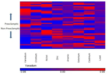
Metals were analyzed by ICP-MS with internal standard controls, and the results are displayed
on Table 2. The relationship of metals in cases and controls is also shown in the heat map in
Figure 1. There was a significant difference between zinc levels of the cases and controls. As
shown in Figure 2, cases have lower levels of zinc. The cadmium levels measured in this study is
in the lower range compared to previous studies: 4.25 ng/g versus a range of 1.2 to 53 ng/g [32].
Additionally, the average zinc levels were in the lower ranges compared to other studies:
12 | P a g e Table 2: Metal Analysis Determined in Placental Samples by ICP-MS
Metal
Mean+/std, Median, (Range)
Control (ng/g) Case (ng/g) Overall (ng/g)
Arsenic 1.77+/-1.10, 1.56, (0.94-6.20)
1.84+/-1.70, 1.40, (0.95-8.91)
1.81+/-1.41, 1.46, (0.94, 8.91)
Cadmium 4.41+/-2.74, 3.26, (1.01-10.42)
4.10+/-2.12, 4.31, (0.35-8.01)
4.26+/-2.42, 4.28, (0.35-10.42)
Chromium 32.82+/-14.31, 35.03, (11.80-59.97) 49.12+/-33.11, 36.04, (14.35-125.46) 40.97+/-26.50, 35.04, (11.80, 125.46)
Lead 5.15+/-7.47, 2.23,
(1.27-28.96)
2.77+/-0.77, 2.70, (1.12, 4.32)
3.96+/-5.37, 2.51, (1.12-28.96)
Nickel 7.86+/-4.71, 6.79, (1.66-18.58)
7.82+/-4.46, 6.39, (2.24-18.98)
7.84+/-4.53, 6.46, (1.66-18.98)
Selenium 261.33+/-40.69, 263.69, (165.26, 333.34) 267.89+/-51.51, 276.34, (159.21-351.86) 264.61+/-45.94, 269.86, (159.21-351.86)
Vanadium 0.54+/-0.16, 0.52, (0.31-0.93)
0.81+/-0.34, 0.75, (0.39-1.64)
0.67+/-0.30, 0.61, (0.31-1.64)
Zinc 8728.43+/-2543.28,
7965.58, (6431.61-17969.99) 7292.49+/-1150.34, 7372.07, (5266.23-9191.76) 8010.46+/-2079.56, 7814.76, (5266.23-17969.99)
13 | P a g e Figure 2: Zinc in Cases/Controls
Metals to Metals
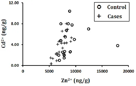
Correlation and significance of metal to metal relationships are shown in Table 3 with p-values
lower than 0.05 indicating significance and correlation (r) values higher than approximately 0.6
indicating correlation. Based on these standards, there is a significant association and a positive
correlation between zinc and cadmium. This association is shown in Figure 3, stratified by cases
14 | P a g e Table 3: Spearman Correlation and p-values for Metals to Metals Relationship in Placenta
r-value
(p-value)
Arsenic Cadmium Chromium Lead Nickel Selenium Vanadium Zinc
Arsenic 1.00000 0.29925
(0.0607) 0.23508 (0.1442) 0.23227 (0.1492) -0.1127 (0.4885) 0.07223 (0.6578) -0.06116 (0.7077) 0.22908 (0.1551)
Cadmium 0.29925
(0.0607)
1.00000 -0.22833
(0.1565) 0.14484 (0.3725) -0.0893 (0.5837) 0.42008 (0.0070) -0.00150 (0.9927) 0.59644 (<.0001)
Chromium 0.23508
(0.1442)
-0.22833
(0.1565)
1.00000 0.28462
(0.0751) 0.14221 (0.3814) -0.19212 (0.2350) 0.24090 (0.1343) -0.3084 (0.0528)
Lead 0.23227
(0.1492)
0.14484
(0.3725)
0.28462
(0.0751)
1.00000 0.33846
(0.0327) 0.00300 (0.9853) 0.21782 (0.1769) 0.08236 (0.6134)
Nickel -0.1128
(0.4885) -0.08931 (0.5837) 0.14221 (0.3814) 0.33846 (0.0327)
1.00000 0.13096
(0.4206)
0.35704
(0.0237)
0.21932
(0.1739)
Selenium 0.07223
(0.6578) 0.42008 (0.0070) -0.19212 (0.2350) 0.00300 (0.9853) 0.13096 (0.4206)
1.00000 0.01257
(0.9386)
0.49737
(0.0011)
Vanadium -0.06116
(0.7077) -0.00150 (0.9927) 0.24090 (0.1343) 0.21782 (0.1769) 0.35704 (0.0237) 0.01257 (0.9386)
1.00000 -0.0098
(0.9524)
Zinc 0.22908
15 | P a g e Figure 3: Zinc and Cadmium Correlation Stratified by Cases/Controls
Table 4: Multivariable Regression Analyses of Zinc and Cadmium Levels to Protein Expression
of Thyroid Hormone Signaling Factors
THR DIO3
Controls Cases Controls Cases
Cadmium = 0.42 (-0.99, 1.84)
= -1.18 (-4.32, 1.97)
= -1.7 (-5.3, 1.9)
= -3.5 (-8.1, 1.1)
Zinc = -0.0005
(-0.0020, 0.0010)
= -0.0021, (-0.0083, 0.0041)
= -0.0010 (-0.0050, 0.0030)
= -0.0079 (-0.017, 0.011)
Thyroid Hormone Regulation
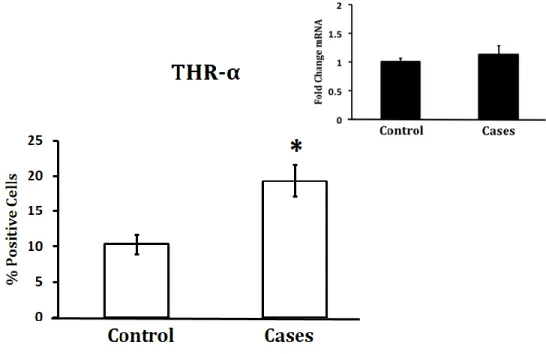
Protein expression was measured with antibodies and IHC with sample sizes of 20. Gene
expression was measured with qRT-PCR with a sample size of 3 for preliminary results. There is
an increase in THRA protein levels and a decrease in Dio3 protein levels in preeclampsia cases,
16 | P a g e THRA and DIO3 protein expression at the bottom and gene expression at the top corner for
comparison. There is a significant increase of THRA protein expression in pre-eclampsia cases,
and a significant decrease in DIO3 gene expression in pre-eclampsia cases.
Figure 4: Protein Expression
17 | P a g e Figure 6: DIO3 IHC (bottom) and mRNA (top)
Metals to Thyroid Hormone Regulatory Proteins
Correlation and significance between the metals and the thyroid hormone regulatory proteins are
shown in Table 5 with p-values lower than 0.05 indicating significance and correlation (r) values
higher than approximately 0.6 indicating correlation. Based on these standards, there are no
significant associations or correlations between metals and thyroid hormone regulatory proteins.
Table 5: Spearman Correlation and p-values for Metals to Protein Relationship in Placenta
r-value
(p-value)
Arsenic Cadmium Chromium Lead Nickel Selenium Vanadium Zinc
THRA 0.13696
(0.3994) -0.01426 (0.9304) 0.09531 (0.5585) 0.07655 (0.6387) -0.19268 (0.2336) -0.19268 (0.2336) -0.01914 (0.9067) -0.29250 (0.0670)
Dio3 -0.0777
18 | P a g e Relationship between Placental Cadmium, Zinc, THRA Protein and DIO3 Protein Levels
There was a negative trend between cadmium levels and THRA protein expression (β= -1.18,
95% CI -4.32 to 1.97) in pre-eclamptic cases, whereas in normotensive placentas the trend was
positive (β= 0.42, 95% CI 0.99 to 1.84) (Table 5). There was a negative trend between zinc
levels and THRA protein expression in preeclamptic and normotensive placentas (β= -0.0021,
95% CI -0.0083 to 0.0041; β= -0.0005, 95% CI -0.0020 to 0.0010, respectively) (Table 5). The
trend between DIO3 expression and cadmium was negative in both preeclamptic and
normotensive placentas, with preeclamptic placentas (β= -3.5, 95% CI -8.1 to 1.1) lower than
normotensive placentas (β= -1.7, 95% CI -5.3 to 1.9) (Table 5). Trends between DIO3
expression and placental zinc levels were negative in both preeclamptic and normotensive
placentas, with preeclamptic placentas (β= -0.0079, 95% CI -0.017 to 0.011) showing a greater
decrease compared to controls (β= -0.0010, 95% CI -0.0050 to 0.0030) (Table 5).
Discussion
Pre-eclampsia is a disorder that occurs during pregnancy, which results in high urine
protein content and high blood pressure as symptoms after 20 weeks of pregnancy. It can have
fatal results for both the mother and the fetus if the placenta is not removed from the body [1].
There is no current cure for pre-eclampsia, but it has been linked to problems with the placenta
[3]. To address this problem, we look at a pilot study of samples from UNC Hospitals called the
MATT cohort to examine the effects of toxic metal exposure on the thyroid hormone regulatory
system. The aim of this study was to investigate the effects of toxic metals on Thyroid Hormone
Receptor Alpha (THRA) and diodinase, iodothyronine type 3 (DIO3), which are associated with
thyroid hormone function and placenta placement. Previous studies have focused on serum
19 | P a g e of pre-eclampsia. The cohort selected provided an even distribution of case and controls to
compare exposure levels to toxic metals and the occurrence of pre-eclampsia. By investigating
this relationship, we are better able to understand the biological processes underlying the
development of pre-eclampsia and supports the theory backing the importance of placenta
placement in pre-eclampsia cases.
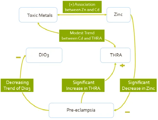
Few studies have been done to examine thyroid hormone regulation and toxic metal
interaction in blood samples. Our findings with placental tissue are summarized in Figure 7.
Figure 7: Model of Interaction between Toxic metals and Thyroid Hormone Regulation
In our trial study, we found significant decreases in zinc, increases in THRA protein levels, and
decreases in DIO3 protein and gene levels in pre-eclampsia cases. Additionally, there is a
20 | P a g e This study utilized placental tissue from healthy normotensive and pre-eclamptic women
to measure relationships between cadmium and zinc levels and thyroid hormone signaling factors
THRA and DIO3. We found a novel, positive correlation between cadmium and zinc levels in
pre-eclamptic placentas, and showed that placental zinc levels are lower in preeclampsia
compared to normotensive women. We also demonstrated that placental THRA protein
expression was elevated in pre-eclampsia and a modest positive correlation exists between
placental cadmium level and THRA protein expression in healthy placentas, suggesting an
underlying possible relationship by which cadmium is a mediator of THRA expression.
However, in pre-eclamptic cases there was an inverse trend between cadmium and THRA.
The results of the present study demonstrated a significant association and a positive
correlation between cadmium and zinc. Additionally, there was a significant decrease in zinc in
pre-eclampsia cases. This finding is supported by previous studies where zinc has been shown to
be an important metal in placental development, and higher levels of zinc are associated with
lower rates of gestational hypertension [10]. Zinc deficiency has also been associated with
pre-eclampsia in previous reports [31]. Cadmium and zinc are chemically similar, which allows
cadmium to bind to zinc binding sites in the body such as sites related to THRA binding [16, 21].
This suggests that the cadmium exposure could prevent zinc from facilitating the binding of
THRA, which is important for the inactivation of thyroid hormones to ensure correct placental
development [18, 19]. Interestingly, while we demonstrate a clear association between placental
cadmium and zinc levels in pre-eclampsia, there was not a statistically significant difference in
cadmium levels between controls and cases. We report total mean placental cadmium levels of
4.3ng/g, which is within the lower range of placental cadmium levels previously reported, which
21 | P a g e pre-eclampsia are limited, and the results varied [11], despite the body of research demonstrating
the reproductive and developmental toxicity of cadmium [33]. To our knowledge, we are the first
to present significant associations between placental cadmium and zinc levels in pre-eclampsia.
Our data shows that there is a significant THRA protein expression but a significant
decrease in DIO3 gene expression in pre-eclampsia cases. In previous studies, DIO3 has been
found at relatively high levels in the placenta during pregnancy [19]. DIO3 functions to
inactivate the thyroid hormone thiiodothyronine (T3), which functions by binding to THRA [18,
19]. The decline in DIO3 could potentially explain the increase in THRA protein expression,
since it is no longer inactivating the thyroid hormones that are essential in regulating placental
formation. The elevated placental THRA protein expression in pre-eclampsia cases could
potentially serve as a biomarker for pre-eclampsia. Interestingly, a modest positive correlation
exists between cadmium and THRA protein expression in healthy placentas, suggesting an
underlying mechanism by which cadmium is a mediator in THRA expression. However, in
pre-eclamptic cases, there was an inverse trend between cadmium and THRA. These results suggest
a dynamic relationship between toxic, environmentally ubiquitous metals and nutritionally
essential metals and expression of thyroid hormone signaling factors.
In the present study we prioritized analysis on two key players in thyroid hormone
signaling, namely THRA and DIO3. Zinc regulates thyroid target gene transcription through
THRA, and DIO3 expression is regulated by THRA, which ultimately controls T3 levels.
Moreover, cadmium affects transcription through the activation of metal responsive elements
(MRE) as well as displacing zinc and altering zinc-mediated transcription and translation, which
could impact thyroid hormone signaling [28,29]. This study highlights the differential
22 | P a g e novel study examined the relationships between thyroid hormone signaling regulation and
cadmium in the context of adverse pregnancy outcomes. The results from this study contributes
to the existing literature that is aimed at elucidating potential links between the environment and
23 | P a g e References
[1] Tsigas E. Preeclampsia Fact Sheet 2012; 2, Preeclampsia Foundation.
[2] Espinoza J, Romero R, Nien JK, Gomez R, Kusanovic JP, Goncalves LF, Medina L, Edwin
S, Hassan S, Carstens M, Gonzalez R,. Identification of patients at risk for early onset and/or
severe preeclampsia with the use of uterine artery Doppler velocimetry and placental growth
factor. Obstet.Gynecol. 2007; 196:326 e1-13.
[3] Doridot L, Houry D, Gaillard H, Chelbi ST, Barbaux S, Vaiman D,. miR-34a expression,
epigenetic regulation, and function in human placental diseases. Epigenetics; 9.
[4] Barber KJ, Franklyn JA, McCabe CJ, Khanim FL, Bulmer JN, Whitley GS, Kilby MD,. The
in vitro effects of triiodothyronine on epidermal growth factor-induced trophoblast function.
J.Clin.Endocrinol.Metab. 2005; 90:1655-1661.
[5] Kilby MD, Barber K, Hobbs E, Franklyn JA,. Thyroid hormone action in the placenta.
Placenta 2005; 26:105-113.
[6] Garcia-Esquinas E, Gomez B, Fernandez-Navarro P, Fernandez MA, de Paz C,
Perez-Meixeira AM, Gil E, Iriso A, Sanz JC, Astray J, Cisneros M, de Santos A, Asensio A,
Garcia-Sagredo JM, Garcia JF, Vioque J, Lopez-Abente G, Pollan M, Gonzalez MJ, Martinez M,
Aragones N,. Lead, mercury and cadmium in umbilical cord blood and its association with
parental epidemiological variables and birth factors. BMC Public Health; 13:841.
24 | P a g e [8] Motawei SM, Attalla SM, Gouda HE, El-Harouny MA, El-Mansoury AM,. Lead level in
pregnant women suffering from pre-eclampsia in Dakahlia, Egypt. Int J Occup Environ Med;
4:36-44.
[9] Liu KS, Hao JH, Zeng Y, Dai FC, Gu PQ,. Neurotoxicity and biomarkers of lead exposure: a
review. Chin.Med.Sci.J.; 28:178-188.
[10] Tande DL, Ralph JL, Johnson LK, Scheett AJ, Hoverson BS, Anderson CM,. First trimester
dietary intake, biochemical measures, and subsequent gestational hypertension among
nulliparous women. J.Midwifery Womens Health; 58:423-430.
[11] Kolusari A, Kurdoglu M, Yildizhan R, Adali E, Edirne T, Cebi A, Demir H, Yoruk IH,.
Catalase activity, serum trace element and heavy metal concentrations, and vitamin A, D and E
levels in pre-eclampsia. J.Int.Med.Res. 2008; 36:1335-1341.
[12] Fiore G, Capasso A,. Effects of vitamin E and C on placental oxidative stress: an in vitro
evidence for the potential therapeutic or prophylactic treatment of preeclampsia. Med Chem
2008; 4:526-530.
[13] Guiet-Bara A, Bara M, Durlach J,. Toxic metals and human amniotic ion permeability. II.
Ultrastructural study and relationship with magnesium. Magnes.Res. 1991; 4:77-81.
[14] Sakamoto M, Yasutake A, Domingo JL, Chan HM, Kubota M, Murata K,. Relationships
between trace element concentrations in chorionic tissue of placenta and umbilical cord tissue:
25 | P a g e [15] Semczuk M, Semczuk-Sikora A,. New data on toxic metal intoxication (Cd, Pb, and Hg in
particular) and Mg status during pregnancy. Med.Sci.Monit. 2001; 7:332-340.
[16] Chisolm JC, Handorf CR,. Further observations on the etiology of pre-eclampsia:
mobilization of toxic cadmium-metallothionein into the serum during pregnancy.
Med.Hypotheses 1996; 47:123-128.
[17] Gellersen B, Brosens J,. Cyclic AMP and progesterone receptor cross-talk in human
endometrium: a decidualizing affair. J.Endocrinol. 2003; 178:357-372.
[18] Baqui M, Botero D, Gereben B, Curcio C, Harney JW, Salvatore D, Sorimachi K, Larsen
PR, Bianco AC,. Human type 3 iodothyronine selenodeiodinase is located in the plasma
membrane and undergoes rapid internalization to endosomes. J.Biol.Chem. 2003;
278:1206-1211.
[19] Dentice M, Salvatore D,. Deiodinases: the balance of thyroid hormone: local impact of
thyroid hormone inactivation. J.Endocrinol.; 209:273-282.
[20] Luo J, Hendryx M,. Relationship between blood cadmium, lead, and serum thyroid
measures in US adults - the National Health and Nutrition Examination Survey (NHANES)
2007-2010. Int.J.Environ.Health Res.
[21] Maxwell C, Volpe SL,. Effect of zinc supplementation on thyroid hormone function. A case
26 | P a g e [22] Barca-Mayo O, Liao XH, Alonso M, Di Cosmo C, Hernandez A, Refetoff S, Weiss RE,.
Thyroid hormone receptor alpha and regulation of type 3 deiodinase. Mol.Endocrinol. 2011;
25:575-583.
[23] Nunes FM, Aparicio R, Santos MA, Portugal RV, Dias SM, Neves FA, Simeoni LA, Baxter
JD, Webb P, Polikarpov I,. Crystallization and preliminary X-ray diffraction studies of isoform
alpha1 of the human thyroid hormone receptor ligand-binding domain. Acta Crystallogr.D
Biol.Crystallogr. 2004; 60:1867-1870.
[24] Kilby MD, Verhaeg J, Gittoes N, Somerset DA, Clark PM, Franklyn JA,. Circulating
thyroid hormone concentrations and placental thyroid hormone receptor expression in normal
human pregnancy and pregnancy complicated by intrauterine growth restriction (IUGR).
J.Clin.Endocrinol.Metab. 1998; 83:2964-2971.
[25] Lao TT, Chin RK, Swaminathan R, Lam YM,. Maternal thyroid hormones and outcome of
pre-eclamptic pregnancies. Br.J.Obstet.Gynaecol. 1990; 97:71-74.
[26] Miyamoto T, Sakurai A, DeGroot LJ,. Effects of zinc and other divalent metals on
deoxyribonucleic acid binding and hormone-binding activity of human alpha 1 thyroid hormone
receptor expressed in Escherichia coli. Endocrinology 1991; 129:3027-3033.
[27] Chen A, Kim SS, Chung E, Dietrich KN,. Thyroid hormones in relation to lead, mercury,
and cadmium exposure in the National Health and Nutrition Examination Survey, 2007-2008.
27 | P a g e [28] Kothinti RK, Blodgett AB, Petering DH, Tabatabai NM. Cadmium down-regulation of
kidney Sp1 binding to mouse SGLT1 and SGLT2 gene promoters: possible reaction of cadmium
with the zinc finger domain of Sp1. Toxicol Appl Pharmacol 2010; 244(3):254-62.
[29] Staneviciene I, Sadauskiene I, Lesauskaite V, Ivanoviene L, Kasauskas A, Ivanov L.
Subacute effects of cadmium and zinc ions on protein synthesis and cell death in mouse liver.
Medicina (Kaunas) 2008; 44(2):131-8.
[30] de Moraes ML, de Faria Barbosa R, Santo RE, da Silva Santos F, de Almeida LB, de Jesus
EF, de Carvalho Sardinha FL, do Carmo MD. Distribution of Calcium, Iron, Copper, and Zinc in
Two Portions of Placenta of Teenager and Adult Women. Biol Trace Elem Res. 2011
[31] Brophy MH, Harris NF, Crawford IL. Elevated copper and lowered zinc in the placentae of
pre-eclamptics. Clin Chim Acta 1985;145(1):107-11.
[32] Esteban-Vasallo MD, Aragones N, Pollan M, Lopez-Abente G, Perez-Gomez B. Mercury,
cadmium, and lead levels in human placenta: a systematic review. Environ Health Perspect
2012;120(10):1369-77.
[33] Thompson J, Bannigan J. Cadmium: toxic effects on the reproductive system and the
28 | P a g e Supplemental
Table 1: Demographics of MATT Cohort, Chapel Hill, NC 2013
Characteristic Control Case Overall
N=20 N=20 N=40
Race (No., %)
Caucasian 7 (35.00%) 6 (30.00%) 13 (32.50%)
African American 6 (30.00%) 9 (45.00%) 15 (37.50%)
Asian 2 (10.00%) 0 (0.00%) 2 (5.00%)
Hispanic 5 (25.00%) 2 (10.00%) 7 (17.50%)
Other 0 (0.00%) 3 (15.00%) 3 (7.50%)
Age (Mean+/std, Median, Range)
28.35+/-5.49, 28.5,
(19-38)
28.15+/-5.6, 28.5,
(19-37)
28.25+/-5.48, 28.5,
(19-38)
Smoking Status 1 (5.0%) 2 (10.0%) 3 (7.5%)
Primipara 11 (55.0%) 13 (65.0%) 24 (60.0%)
Multipara 9 (45.0%) 7 (35.0%) 16 (40.0%)
Gestational Age 38.6 + 1.9 32.7 + 4.7 35.6 + 4.6