i
CHONDROGENIC DIFFERENTIATION OF HUMAN ADIPOSE DERIVED STEM CELLS: THE ROLES OF MECHANICAL LOADING, ELEVATED CALCIUM, AND THE EXTRACELLULAR CALCIUM SENSING
RECEPTOR.
John Michael Williams II
A thesis submitted to the faculty of the University of North Carolina at Chapel Hill in partial fulfillment of the requirements for the degree Masters of Science in the Department of
Biomedical Engineering
Chapel Hill 2012
APPROVED BY:
Elizabeth G. Loboa, Ph.D
Albert J Banes, Ph.D
ii ABSTRACT
JOHN MICHAEL WILLIAMS II: Chondrogenic Differentiation of Human Adipose Derived Stem Cells: The Roles of Mechanical Loading, Elevated Calcium, and the Calcium Sensing Receptor.
(Under the direction of Elizabeth G. Loboa).
iii
I would like to acknowledge everyone who has contributed to this research. I would like to thank my advisor, Dr Loboa, for the opportunity to work under her tutelage, and further for her patience, mentorship, and guidance, without which I could not have completed this work. I would also like to thank my committee for your time and effort in ensuring I could succeed in this
endeavor. I would like to thank Dr Susan Bernacki for her constant instruction, and encouragement, ensuring I had everything I needed to succeed. I would also like to recognize our collaborators at Duke, Dr Farshid Guilak, Dr Brad Estes, Dr Frank Moutos, and Brian Deikman, without whom, much of this research could not have been conducted.
iv
Table of Contents
List of Tables ... vii
List of Figures ... viii
List of Abbreviations and Symbols ... ix
Chapter 1: Introduction ... 1
1.1 Motivation ...1
1.2 Thesis Findings ...2
Chapter 2: Literature Review ... 3
2.1 Human Adipose Derived Stem Cells and Chondrogenesis ...3
2.2 Cartilage tissue ...4
2.3 Stem Cells ...6
2.4 Cartilage extracellular matrix. ...6
2.5 Mechanical requirements of cartilage ...8
2.6 Factors Effecting Chondrogenesis in vitro ...8
2.7 Mechanical loading ... 10
2.8 Mechanotransduction ... 18
2.9 Conclusions ... 19
Chapter 3: Elevated Extracellular Calcium Inhibits Chondrogenic Differentiation . of Human Adipose-Derived Stem Cells in Pellet Culture ... 20
3.1 Introduction ... 20
3.2 Materials and Methods ... 21
3.3 Results ... 26
3.4 Discussion ... 32
3.5 Conclusion ... 34
Chapter 4: Targeting the Calcium Sensing Receptor in Human Adipose Derived . Stem Cell Osteogenic differentiation ... 35
4.1 Introduction ... 35
4.2 Materials and Methods ... 36
4.3 Results ... 39
4.4 Discussion ... 45
4.5 Conclusion ... 48
v
5.1 Conclusions ... 49
5.2 Future Work. ... 51
References ... 52
Chapter 2 ... 52
Chapter 3 ... 69
vii List of Tables
Table
1. Studies of mechanical loading and chondrogenesis………11 2. Studies of mechanical loading and hASC………...18
viii
List of Figures
Figures
3.1 Alcian blue stain of pellets cultured in six different CDM formulas . (A-F). IHC for collagen II in pellets cultured in six different CDM . . formulas (G-L).Media formulations (M)...………23 3.2 Alcian Blue staining of 8mM Ca2+
pellet sections (A,C) and control pellet . section (B,D)……….………..28
3.3 Safranan O/FCF Fastgreen/Hematoxylin and Alizarin Red stain of 8mM . Ca2+ pellet sections (A,C) and control pellet sections (B,D)………..………….29
3.4 IHC of pellet sections for col I (A-8mM Ca2+, B-control), col II (C-8mM . Ca2+, D-control), and col X (E-8mM Ca2+, F-control)………..……….………..…30 3.5. Quantification assays: Sulfated GAG content of Pellets normalized to DNA.
Hydroxyproline contents of pellets normalized to DNA………..………….…31
4.1 Calcium to protein ratios: A) Transient knockdown of PC2 B) Allosteric . binding of CaR in elevated Ca2+………...………42
4.2 Calcium to protein ratio at Day 12………..………..43
ix
List of Abbreviations and Symbols
ALP-Alkaline phosphatase
BMP6- Bone morphogenetic protein 6 Ca2+-Calcium ions
CaR- Extracellular calcium sensing receptor CDM-Chondrogenic differentiation media CGM-Complete growth media
CHP-Cyclic hydrostatic pressure Col-Collagen
DNA-Deoxyribonucleic acid FBS-Fetal bovine serum GAG-Glycosaminoglycan
hASC- Human adipose derived stem cells
hMSC-Human bone-marrow derived mesenchymal stem cells ITS+- Insulin, transferrin, selenous acid
MMP-Matrix metalloproteinase mRNA-messenger Ribonucleic acid PC2- Polycystin 2
PCR-Polymerase chain reaction
1 Chapter 1:
Introduction
1.1 Motivation
Cartilage is an avascular, connective tissue with the critical function of supporting movement. The tissue is responsible for bearing multiples types of load repetitively, without degrading. Further, because of its avascular nature, the tissue has little regenerative capability. Cartilage defects can come about due to injury, disease, or the aging process, and can lead to reduced mobility and quality of life. While there are a number of methods of adjusting to minimize the impact of the damaged tissue, few treatments exist to effectively repair defective cartilage tissue. The Cell Mechanics Laboratory specializes in understanding the mechanical forces necessary to engineer functional musculoskeletal tissue from mesenchymal stem cells derived from both bone marrow and adipose tissue. it is our goal to develop novel techniques for cartilage tissue engineering utilizing hASC.
Some concerns with current treatments for cartilage defect repair are the integration of engineered tissue into the joint, as well as the material properties of the engineered tissue. We plan to address the issue of assimilation by the using an autologous cell source, limiting the body’s immune response. Also, mechanical loading plays a key role in native cartilage tissue metabolism and homeostasis. We plan to leverage this to condition engineered tissue with loading protocols comparable to those seen in vivo, ensuring the tissue can properly bear the expected loads.
2
1.2 Thesis Findings
The findings of this body of work have been shared in numerous professional academic conferences and are being submitted to peer-reviewed journals for publication.
Manuscripts in preparation:
The Role of Mechanical Loading in Cartilage Tissue Engineering with Human Adipose Derived Stem Cells
Williams, JM; Loboa EG
Planned journal: Tissue Engineering, Pt B
Elevated Extracellular Calcium Inhibits Chondrogenic Differentiation of hASC
Williams, JM; Kannan A; Dent MR; Estes B; Moutos F, Hluck BH, Guilack F,; Loboa EG
Planned journal: Tissue Engineering, Pt A
Targeting the Extracellular Calcium Sensing Receptor in Human Adipose Derived Stem Cell Osteogenic Differentiation
Williams JM; Bodle JC; Loboa EG
Planned journal: Journal of Bone and Mineral Research
Conference Proceedings
Elevated Extracellular Calcium Inhibits Chondrogenic Differentiation of Human Adipose Derived Stem Cells in Pellet Culture
Williams JM; Kannan A; Dent MR; Estes B; Moutos F; Hluck BH; Bernacki SH; Guilak F; Loboa EG
3 Chapter 2:
Literature Review
2.1 Human Adipose Derived Stem Cells and Chondrogenesis
Human adipose-derived stem cells (hASC) show great potential for cartilage tissue engineering applications. When exposed to appropriate chemical and mechanical stimuli, these cells provide a number of distinct advantages over previously studied cells while behaving in a comparable manner with respect to chondrogenic potential. These cells are relatively more abundant than either bone marrow derived mesenchymal stem cells, hMSC, or articular
chondrocytes, with less donor site morbidity, and greater expansion capabilities.(1-6) A number of chemical chondrogenic induction protocols previously used with other stem and progenitor cells have been adapted for use with hASC in cartilage tissue engineering, with varying levels of success (7-13). Some chondrogenic conditions, such as soluble growth factors, 3D culture, low oxygen
4 2.2 Cartilage tissue
Cartilage is one of the most critical and complex musculoskeletal materials within the body. When healthy it provides a number of key functions, varying from providing the structure for facial features as in the ears and nose, to providing critical mechanical buffering between bones at joints. The complex nature of the material provides it with the mechanical properties to perform each of these functions. Unfortunately, the same complexity and functionality that are crucial to cartilage function, has made repair and/or regeneration of defective or damaged cartilage that much more difficult. Degenerative conditions such as rheumatoid and osteoarthritis, which affect millions of people, along with common injuries, have demonstrated that damaged cartilage not only greatly diminishes mobility, but also quality of life (32-34).
One of the major issues with cartilage defects or injuries is the inability of articular cartilage to regenerate naturally. Articular cartilage tissue is avascular, leaving the material no access to many of the regenerative nutrients found within blood. When a vascular supply is present during in vivo chondrogenesis, as in wound or fracture healing, the resulting cartilage is usually a type of
loosely organized fibrocartilage associated with wound healing, which possesses significantly different mechanical properties than articular cartilage, and is less functionally capable (35-37). Additionally, the regenerative capabilities of the sparse community of living cells within the tissue are limited. The chondrocyte, which produces the extracellular matrix (ECM) of cartilage, maintains a very low level of metabolic activity, and therefore limited turnover of tissue (32,38). While
5
A variety of approaches to long term cartilage repair have been and are being explored in an effort to mimic the mechanical functions of cartilage tissue, improve mobility and quality of life. Treatments like physical therapy, bracing, and weight loss have been effective in relieving some symptoms associated with cartilage defects without surgery. While these methods are sometimes effective in providing temporary relief, they do not repair or regenerate damaged cartilage tissue(35). Microfracture, a surgical treatment involving removal of damaged cartilage around a defect and drilling holes in the subchondral bone, results in some success by reducing shear stresses in the defective tissue, reducing pain, and allowing mesenchymal stem cells to migrate into the defect site(35,36,42). This treatment must be followed up with some type of physical therapy regimen to introduce the mechanical forces needed to heal the site. Unfortunately, while this procedure can lead to chondrogenesis, it often results in fibrocartilage formation.(36)
Today, a number of approaches to cartilage repair focus on regenerating functional cartilage tissue, instead of temporarily supplementing defects. Such approaches include implantation of donated or engineered cartilage in the form of an allograft.(35,41) These allografts are generally successful in that the material will have similar mechanical properties of native cartilage, thus meeting many of the same functions. While this treatment can be successful in some cases, it is still limited in the size of the defect being treated, potential of an immune response, and the availability of donor tissue.(35,41) To avoid immune response, injection of autologous chondrocytes obtained from the patient can also be performed in a procedure known as autologous chondrocyte
6 2.3 Stem Cells
One approach currently being developed to address this issue of replicating the natural process of chondrogenesis is utilizing multipotent stem cells. Stem cells are present throughout the body, and possess the ability to differentiate along a number of different lineages(5,6,43-50,92). Current areas of study include analyses of soluble chondrogenic induction factors, 3D cultures, and
mechanical conditions to successfully direct stem cell lineage specification. Early investigations, largely focusing on hMSC, have shown these cells demonstrate the ability to differentiate down a multitude of pathways in response to appropriate chemical (51) and physical (1,52-56) environments. Studies have included evaluation of critical growth factors, chemical supplements, and mechanical forces to induce MSC chondrogenesis(57,58). While MSC hold great potential for functional cartilage tissue engineering, abundance and accessibility of MSC are limited. As a result, adipose derived stem cells ASC have gained great interest due to their similar multipotent differentiation ability to MSC and their relative abundance in donated tissue.( 5,6, 15,48,52,59-70). Functional tissue engineering and regenerative medicine studies with MSC and other cell types are now being explored with ASC to determine how best to utilize these cells for tissue repair and/or regeneration (71,5,6).While ASC and MSC are similar, there are distinct differences. Some studies have shown that ASC have less chondrogenic potential than hMSC, suggesting they may require greater treatments of growth factors and mechanical loading in order to produce similar results.(1,8-10,24,72-77)
2.4 Cartilage extracellular matrix.
7
sparse community of chondrocytes. The large collagen fibrils of the deep zone run perpendicular to the joint surface, through the tidemark into the zone of calcified cartilage. The tidemark is the barrier between the more gelatinous cartilage tissue and the solidified tissue found in the zone of calcified cartilage, which anchors the tissue onto the subchondral bone.(59,78-80,40)
Many of the genetic markers associated with chondrocytes in the different layers of articular cartilage relate to the production of key proteins and proteoglycans embedded within the
8 2.5 Mechanical requirements of cartilage
The mechanical properties of the individual elements of cartilage are all necessary for overall function of the material. As the mechanical buffer between joints, cartilage has a variety of forces acting on it at varying magnitudes over varying durations. A common load associated with movement is unconfined compression, which affects the zonal layers of articular cartilage differently (138,83,80). For example, standing, walking, and jumping all exert compressive forces on the cartilage of the hip joint ranging from 5 to 15 MPa (80). The cartilage must be able to withstand static, dynamic, and cyclical loading without failure over a lifetime. Global compressive loads translate throughout the tissue, creating areas of local hydrostatic pressure within the tissue, fluid shear stresses as the fluid flows in and out in response to physiological loads, and shear strain at the cartilage interface with underlying bone. While cartilage is a low friction surface, joint loading creates tensile strains at the surface. Not only are the extracellular matrix constituents of cartilage specifically arranged and designed for these key mechanical roles, studies have shown that regular mechanical loading is a key precursor to matrix development and sustainment(138). Natural loading has been shown to positively affect cartilage function and formation in vivo, and has also been shown to improve chondrocyte viability, gene expression, and proteoglycan formation in vitro, while excessive loading has been shown to lead to degradation of the ECM(38). These studies demonstrating the response of cartilage to mechanical loading are critical to understanding both how defects propagate
throughout the tissue, as well as provide key insight in cartilage tissue engineering.
2.6 Factors Affecting Chondrogenesis in vitro Growth factors
Similar to natural chondrogenesis, the chemical environment plays a key role in
9
Dulbecco’s modified Eagle’s medium (DMEM) , ITS (or ITS+), and various forms of ascorbate. Further, the presence of soluble growth factor has proven successful in chemically inducing
chondrogenesis. Key Factors are those from the TGF super family, specifically, TGF 1, TGF 3 and
BMP6. Our lab has previously reviewed these growth factors and their impact on chondrogenic differentiation of hMSC and hASC(84-93).
Shape
A major consideration in cartilage tissue engineering is the necessity of a three-dimensional (3D) culture environment. Studies have shown that stem cells cultured in 2D have diminished chondrogenic capabilities compared to 3D systems. Early studies have shown that cells with a spherical conformation produce collagen II, versus the collagen I produced by flattened fibroblastic cells(94,95) In addition to mimicking the environment of embryonic cartilage development, this shape also effects mechanotransduction of various types of loads, which in turn signals various processes critical to cartilage tissue development. Often, 3D culture is achieved by pelleting cells into a micromass. Other systems use various types of scaffolds, microspheres or hydrogels with varying degrees of success.
10
method of maintaining the spherical shape of cells(122-126). In each of these cases, the stiffness has been shown to be a key factor in differentiation, in that substrates of various stiffness have been shown to induce differentiation down specific lineages(127-130).
The substrate used to culture cells for chondrogenic differentiation greatly affects the way the cells receive key signals. In addition to maintaining the spherical shape of the cell, the 3D culture effects the way in which the cells interact with each other. Studies have shown the importance of high cell seeding density in chondrogenic differentiation of both MSC and ASC, suggesting that cell-cell signaling plays a key role in the process of chondrogenesis(12,131,135). Further, the presence of certain molecules, like hyaluronan, enhances differentiation, suggesting that the cell’s interaction with the ECM is another key aspect of the chondrogenesis process(106,118,132,133).
2.7 Mechanical loading
11
Author
Load
Cell type/ Medium/
Construct
Magnitude/ Frequency/
Duration
Blain EJ, (137)
Compressive
Loading
Articular Cartilage/
.5 Mpa/ 1 Hz/ 3hr
Li J, (188)
Compressive
Loading
ASC/
Chitosan-Gelatin Scaffolds
5%, 1hz, 4 days
Bahuleyan
B.,(16)
Compressive
Loading
Rabbit MSC/ TGF
B1
10%/ 1 Hz
Elder SH, (186)
Compressive
Loading
Chick
limb-bud/Agarose
.03-.33Hz/12mn-1hr
Huang CY,
(139)
Compressive
Loading
Rabbit MSC/
.1,1,10 MPa/1Hz
Kisiday JD,
(138)
Compressive
Loading
MSC/ CDM w/wo
TGF
/ Agarose
hydrogel
2kPa/ .3Hz/ 6hr or 12 hr per
day
Takahashi I,
(134)
Compressive
Loading
Mouse Embryonic
Limb-bud cells/
Collagen gel
.5, 1 g/cm
3static
Li Z Y,(140)
Compressive
Loading/ shear
stress
MSC/ Fibrin
Polyurethane
Composite
15, 20, 30%/.1,1Hz/ 1hr per
day/ 7 days
Mauck RL,
(136)
Confined
Compression/
tensile
Chondrocytes/
Agarose & Alginate
Hydrogels
10%, 1Hz
Pelaez D, (141)
Cyclic
compression
MSC/ Fibrin Gel
10%/1Hz/ 4hr/day 33 days
Mizuno S, (144
Cyclic Hydrostatic
Pressure
bovine
Chondrocytes/
collagen sponges
2.8Mpa/ .015 Hz/ 33ml/min
Ogawa R,(161)
Cyclic Hydrostatic
Pressure
ASC/CDM Collagen
Scaffold
0-.5 MPa/ .5Hz
Li Z, (189)
Dynamic
Compressive
Loading
MSC/
fibrin-polyurethane hybrid
system
10–20%/ 1hz/ 1 hr a day
over
Thorpe SD,
(142)
Dynamic
Compressive
Loading
MSC/ CDM/
10%/ 5Hz/ 1hr, 5 days
12
Compressive
Loading
day/20 cycles
Huang AH,
(143)
Dynamic
Unconfined
Compression
Bovine Bone
Marrow Stem Cells
10% dynamic /2%static /
1hz, 4hr/day
Toyoda T,(145)
Hydrostatic
Pressure
Bovine
Chondrocytes/
5 Mpa/ 1Hz/ 4hr
Angele p, (150)
Hydrostatic
Pressure
MSC/ Serum Free
medium
10 MPa/ 1Hz
Angele P, (151)
Hydrostatic
Pressure
MSC/CDM
5.03 MPa/1Hz/ 14-28 days
Finger AR,( 152)
Hydrostatic
Pressure
MSC/Agarose w/o
TGFB
7.5 MPa/1HZ
Finger AR, (152)
Hydrostatic
Pressure
MSC/TGFB3
.1,1,10 MPa/1Hz
Hall AC,(146)
Hydrostatic
Pressure
Bovine
Chondrocytes/
2.5-50 MPa
Ikenoue T,( 147)
Hydrostatic
Pressure
Articular
Chondrocytes
1, 5, 10 Mpa/ 1Hz/ 4hr
Miyanishi K,(
149)
Hydrostatic
Pressure
MSC/ CDM
10 MPa/1Hz/ 3, 7, 14 Days
Mukherjee
N,(193)
Hydrostatic
Pressure
periosteol graft
13, 54, 103 kPa, 5, 4, 24hrs
Reza AT, (195)
Hydrostatic
Pressure
Chondrocytes
(OA,IA,NP)/PGA-PLLA Scaffolds
5MPa, .5 Hz, 4hr,12 days
Lee, Mel, (148)
IHP/Shear Stress
OA Chondracytes/
serum free medium
1.6Pa/
200RPM/2hr/10Mpa/1HZ/
4hr/day 1,2,4 days
Carver SE,(173)
Hydrostatic
pressure, fluid
shear, and mixing
Articular
Chondrocytes/ PGA
scaffold
50 RPM/ 13.5 MPa/ 3.44
MPa
Wimmer M,
(200)
Shear stress
Chondrocytes/PU
Scaffold
.28,2.8, 28 mm/s, 1hz,1hr/
2x Per day, 5 days
Kupcsik L, (185)
Shear stress/
dynamic
compression
MSC/ Fibrin PU
scaffold
25o/ 1 Hz / 10-20%/ 1 hr/ 14
days
Diederichs S,
(176)
Shear
Stress/Tensile
strain
Embryonic Rat
Limb-bud/Silicon
5%/1Hz/ 15 min-8 Hr/ 2x
per day
Kuo CK, (170)
Tensile strain
MSC/ Collagen gel
1%/ 1Hz/ 30min per day
Connelly
JT,(175)
Tensile strain
MSC/CDM/ Fibrin
Hydrogel
10%/ 1 Hz/ 3hrs
Tanaka T,(159)
Tensile strain
Rat Chondrocytes
7, 12%/ .5HZ
13 Compression
Compressive forces have been the most effective and studied force applied to induce chondrogenesis. Compressive forces are experienced throughout the process of endochondral ossification, as well as normal joint function and have been shown to increase aggrecan synthesis and matrix development in vivo(38). In studies using embryonic limb bud progenitor cells, dynamic compressive loading was shown to be more effective in driving chondrogenesis than static loading as evidenced by increased cartilage nodules. Static compressive loading resulted in a 2-3 fold increase in collagen II, aggrecan and SOX9 expression over unloaded samples(134,135). Mauck(136) demonstrated that dynamic loading of a chondrocyte seeded agarose disc not only increased the hydroxyproline and sulfated GAG contents, but also increased the equilibrium aggregate modulus 6 fold over free-swelling controls, and 20 fold over the 28 day period. These findings demonstrate that compressive loading not only improves matrix production, but also helps develop mechanically functional tissue. Blain et al(137) demonstrated though that cyclic compression of bovine cartilage explants resulted in elevated expression of matrix metalloproteinases, MMP, which are prevalent in matrix remodeling. This demonstrates the effectiveness of compressive loading on the metabolic activities of cartilage tissues, but also shows a mechanism in which excessive loading can lead to tissue degradation (137).
14
stimulate matrix production. A by Kisiday et al (138) demonstrated that a compressive loading protocol necessary to induce chondrogenic differentiation in MSC may not be best for new tissue accumulation. In the absence of soluble chondrogenic growth factor TGF-, dynamically compressed
constructs demonstrated increased levels of sulfated GAGs, and hydroxyproline over unloaded controls, similar to the results seen with other cell sources. However, the GAG content of
dynamically loaded constructs cultured in the absence of TGF- was significantly lower than that of unloaded constructs loaded in the presence of TGF-. Further, when both conditions were cultured with TGF-, dynamically compressed samples showed decreased GAG content compared to
unloaded controls (138). Huang et al (139) conducted a similar experiment suggesting that when the loading is done over a longer period of time, compressive loading is as effective in chondrogenesis as both TGF-and TGF-with compressive loading, suggesting that the dynamic compressive loading led to the synthesis of TGF-1 by the MSC in culture, which in turn leads to chondrogenic
differentiation (139). Subsequent studies further demonstrated how compressive loading leads to TGF-1 and 3 synthesis in MSC, and that greater amplitudes and frequencies of loading, over a
longer culture period, generally lead to increased chondrogenesis (140,141). Some studies have shown limits of compressive loading on inducing chondrogenesis, including the time point to being a loading regimen, or the mechanical properties of the construct. Unless the construct is able to properly conduct the compressive load, chondrogenesis can be inhibited.(142) Huang et al(143) showed loading conducted prior to chondrogenic differentiation of MSC led to less functional matrix
material, even though expression of chondrogenic markers was increased. In contrast, when loading occurred after chondrogenesis, and TGF-3 levels were maintained, mechanical properties were
improved.(143) These studies demonstrate the relationship between the TGF super family, compressive loading, and cartilage tissue engineering.
15
In studies conducted with articular chondrocytes, hydrostatic fluid pressure, HFP, has been shown to stimulate the production of cartilage ECM greater than ambient pressures (144-147). Mizuno et al (144) demonstrated how articular chondrocytes, in an appropriate 3D construct experienced increased sulfated GAG content under both cyclic and constant hydrostatic pressure. Other studies demonstrated the effects of hydrostatic pressure on cell shape, cell-matrix interactions, and cell surface proteins, demonstrating how each play a role in tissue metabolism. One such study by Lee et al (148) demonstrated that cyclic hydrostatic pressure inhibited the catabolic effects of inflammatory cytokines (148). As with compression, the magnitude, and duration of treatment, along with the mechanical properties of the cell construct greatly impact the effect of hydrostatic loading. However physiological levels of hydrostatic pressure in articular cartilage (in the range of 2-5 MPa) have been effective in increasing matrix production in articular chondrocytes (145-149).
Similar results of increased matrix production in response to hydrostatic pressure were seen in studies using MSC (150). Angele et al (151) demonstrated an increase of proteoglycans due yo CHP loading on cell aggregates in chondrogenic medium, where as our lab has previously demonstrated that CHP was effective in increase early chondrogenic differentiation markers in MSC constructs cultured without known chondrogenic growth factors such as TGF-(152,106). These studies are important in that they demonstrate that CHP is effective of increasing matrix production in stem cells being directed down a chondrogenic pathway, as well as inducing chondrogenic differentiation in stem cells.
Shear stress
16
proteins in cultured articular chondrocytes (153, 154). Additionally, Smith et al (154) demonstrated that chondrocytes produce MMP inhibitors in response to fluid shear, demonstrating that fluid shear plays a role in normal cartilage tissue metabolism. Smith later demonstrated that shear stresses may work in concert with other mechanical forces to maintain homeostasis (155). Where compression and hydrostatic pressure resulted in increased production of aggrecan and type II collagen, fluid shear resulted in lower level, of both aggrecan and type II collagen, and an increase of nitric oxide. This study and others also suggest that fluid shear can result in apoptotic conditions in chondrocytes, furthering the damage associated with osteoarthritis. (154). Bushman et al(156) demonstrated that fluid shear resulting from dynamic compression led to increased aggrecan production, suggesting a need for a combination of mechanical loads in order to maximize the effectiveness in each with regards to cartilage tissue engineering. This is consistent with Mow et al (83) description of the different load types experienced in various regions of cartilage tissue in response to natural motion. Considering these results, fluid shear should be applied to engineered cartilage tissues as a mediator of natural cartilage metabolism, in concert with other types of mechanical loading.
Tensile strain
Tensile strain is normally associated with bone tissue engineering, because this loading type generally leads to elongated and organized cells, which can lead to fibrocartilage tissue
17
adhesion was inhibited, suggesting the negative effects of tensile strain are based on the interaction between cells and the matrix.
ASC
Although a number of studies have examined the impact of various mechanical loads on cartilage tissue engineering, as well as mechanical loads on ASC (Table 2) few studies have focused on mechanical loading of ASC in cartilage tissue engineering. Cyclic hydrostatic pressure has been shown to enhance chondrogenic differentiation in ASC, in the presence of soluble chondrogenic growth factors, TGF- (161). In our lab we have previously shown CHP to be effective in inducing chondrogenic differentiation in ASC without the use of chondrogenic differentiation medium or soluble growth factors (107). Both studies demonstrated the importance of an appropriate construct for long term cell viability and proliferation. Because of the limited work studying the effects of mechanical loading on cartilage tissue engineering with ASC, we must extrapolate expected results based on other studies utilizing mechanical loading and ASC. Compression was shown to improve cartilage tissue development because of an increase in growth factors like TGF-. A number of
studies have compared the chondrogenic potential of ASC compared to MSC, and have concluded that MSC have greater chondrogenic potential than ASC under identical culture conditions including the presence of TGF- (11,15,51). This suggests that ASC may need a greater magnitudes and
durations of compressive loading, or TGF- in greater doses during mechanical loading. Table 2
18
Author
Load
Frequency/Amplitude
Tissue
Li J, (188)
Compression
5%, 1hz, 4 days
Cartilage
Ogawa R, (161)
Cyclic Hydrostatic
Pressure
0-.5 MPa/ .5Hz
Bone
Puetzer J (107)
Cyclic Hydrostatic
Pressure
7.5 Mpa,1Hz, 4h/d, 21 days
Cartilage
Knippenberg
M,(184)
Fluid shear
8.4 Pa/s / 5, 10, 15, 30, 60
min
Bone
Bassaneze V,
(171)
Shear stress
10 dynes/cm
2, 96hr
Vascular
Fischer LJ, (178)
Shear stress
12 dynes, 8 days
Endothelial
McIIhenny SE,
(192)
Shear stress
ramped at 1.5dynes/s
Vascular
Hanson AD, (179) Tensile strain
10%/ 1Hz/ 4hr per day
Bone
Bayati V, (172)
Tensile strain
10%, 1 Hz, 24 hr
Muscle
Diederichs S,(176) Tensile strain
5%, 1hz, .25-8 hr
Bone
Huang S-C,(180)
Tensile strain
10%.5hz, 48hr
Proliferation
Lee W,(187)
Tensile strain
10%, 1hz, 7 days
Inhibition
Wall ME, (199)
Tensile strain
10%, 1hz, 4hr, 14 days
Bone
Pre D, (194)
Vibration
30hz, 45 min, 28 days
Bone
Table 2. Mechanical Loading and ASC
2.8 Mechanotransduction
19
the primary cilium has been studied as a cellular mechanosensor directing stem cell differentiation. (164) This organelle has been shown to be a key mechanosensor in bone and cartilage tissue, sensing flow, compression, and hydrostatic pressure, directing metabolic processes as well as
differentiation(165-169). The cilia signal the cell to mechanical forces by directing an increase of intracellular Ca2+ levels through the permeable ion channel Polycystin 2(166). Roberts et al his demonstrates that intracellular Ca2+ concentrations spiked in cells undergoing compressive loading (204). This spike was generally observed by 200 s after the load was applied followed by a slow return to homeostasis (204). This demonstrates the important role of Ca2+ in mechanotransduction.
2.9 Conclusions
The nature and function of cartilage tissue translates regular movement into various forms of mechanical loads throughout the region, each playing an important role in the homeostasis of the tissue. As we improve our methods of cartilage tissue engineering, biomimetic mechanical loading has been shown to be critical to engineering mechanically functional tissue. Additionally, the compressive forces and hydrostatic pressure present during embryonic endochondral ossification seem equally important in directing stem cell differentiation. Further, other factors such as the chemical environment, mechanical properties of the cell construct, and the biological state of the cells are integral in determining the appropriate magnitude, duration, and type of mechanical load needed to optimize cartilage tissue engineering. It is clear however that more research needs to be done to better understand the mechanisms by which these loading modalities impact
20 Chapter 3:
Elevated Extracellular Calcium Inhibits Chondrogenic Differentiation of Human Adipose-Derived Stem Cells
The previous chapter discussed the role of mechanical loading in hASC differentiation. The mechanism by which a number of mechanical forces direct cell activity is through mechanosensing ion channels and ion permeable membranes. During mechanical loading, cells experience a rapid increase in intracellular Ca2+ concentration, translating the mechanical load to a chemical signal. This demonstrates a function of Ca2+ in cell signaling which may be leveraged without the use of mechanical loading. Previous studies have shown that elevating extracellular Ca2+ concentration to 8mM is effective in increasing hASC mediated calcium accretion and osteogenic differentiation. Based on the role of Ca2+ in cell signaling, as well as the impact of elevated Ca2+ on hASC differentiation, this study aims to observe the impact of elevated Ca2+ on chondrogenic differentiation of hASC.
3.1 Introduction
21
Calcium ions can also affect stem cell proliferation and differentiation. Our lab has previously observed the effects of elevating Ca2+ levels in complete growth and osteogenic differentiation media on hASC, comparing mineralization and proliferation. That study demonstrated that elevation of
extracellular Ca2+ in complete growth medium to a concentration of 8mM was effective in increasing human adipose derived stem cell (hASC) production of mineralized matrix, an indicator of osteogenic differentiation5. The use of elevated Ca2+ to direct hASC osteogenic lineage specification is a clear advantage in bone tissue engineering, however, the impact of elevated Ca2+ has not been widely studied in cartilage tissue engineering.
Articular cartilage is found at the end of long bones with a primary function of supporting locomotion by bearing the repetitive mechanical loads associated with movement. The tissue is avascular with limited regenerative capability, which limits its ability to integrate engineered repair tissue. Bone, in contrast, is highly vascular with regular remodeling processes. Future cartilage
engineering endeavors may leverage the regenerative properties of nearby bone to ensure implants are functionally integrated into the joint. In order to accomplish this, the impact of osteogenic factors on chondrogenesis must be better understood. This aim of this study was to determine the effect of elevated Ca2+ on chondrogenic differentiation of hASC.
3.2 Materials and Methods hASC Isolation and Culture
22
10% fetal bovine serum (FBS) (Premium Select, Atlanta Biologicals, Lawrenceville, GA), 200 mM L-glutamine, and 100 I.U. penicillin/ 100 µg streptomycin per ml (Mediatech, Inc.) The cells were allowed to proliferate at 37°C in 5% carbon dioxide until reaching 70% confluency, and then trypsinized. The amassed cells were then characterized for osteogenic and adipogenic potential, ensuring the amassed cells differentiated representative of an average of the five cell lines.
Chondrogenic Characterization/Medium Formulation
Suspensions of 250K hASC were centrifuged in 1 ml of media into micromassed pellets. Pellets were cultured in one of six chondrogenic differentiation medium formulations. Formula 1 was
comprised of Dulbecco’s modified Eagle’s medium (Mediatech, 15-013-CV), 1% dexamethasone (Sigma Aldrich, D4902), 1% ITS+ (Sigma-aldrich, I2521), 1%Pen/Strep, and 1% ascorbic acid (Sigma, A4403). Formula 2 included all components of Formula 1 with the addition of 1% TGF-1 (R&D Systems, 100-B)
at a concentration of 10ng/ml. Formula 3 included all components of Formula 2 plus 1% bone morphogenetic protein 6 (BMP6, R&D Systems 507-BP) at a concentration of 10ng/ml. Formulas 4 through 6 were comprised of Formulas 1 through 3, respectively, but with the addition of 10% fetal bovine serum (FBS) added to Formulas 1 through 3. Pellets were cultured at 37°C in 5% carbon dioxide for 21 days, receiving conditioned media changes every 48 hours. Conditioned media changes remove only half of media within culture, in order to preserve growth factors secreted by cells. Ascorbic acid and chondrogenic growth factors, TGF-β1 and BMP6, were added to each medium formulation immediately prior to conditioned media changes. Cell pellets were then dehydrated, blocked in paraffin, and
sectioned for histological analyses. Sections were stained with alcian blue (Sigma, A9186) for
23
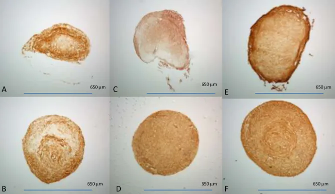
collagen type II (DSHB, II-II6B). Based on IHC and histological results (Figure 1), Formula 6 was chosen
for all further experiments.
Materials
•Six different treatment medias
Group Components
Group 1 DMEM, ITS+, Pen/Strep, Dexamethasone, Ascorbic acid
Group 2 Group 3 Group 4 Group 5 Group 6
TGF -1 : 10ng/ml BMP-6 : 10ng/ml
DMEM, ITS+, Pen/Strep, Dexamethasone, Ascorbic Acid, TGF-1
DMEM, FBS, ITS+, Pen/Strep, Dexamethasone, Ascorbic acid
DMEM, ITS+, Pen/Strep, Dexamethasone, Ascorbic Acid, TGF-1, BMP-6
DMEM, FBS, ITS+, Pen/Strep, Dexamethasone, Ascorbic acidTGF-1, BMP-6
DMEM, FBS, ITS+, Pen/Strep, Dexamethasone, Ascorbic acid, TGF-1
Alcian Blue Staining of Pellets
Control TGF-B1 TGF-B1 + BMP6
Figure 1. Alcian blue stain of pellets
cultured in six different CDM formulas
(A-F). IHC for collagen II in pellets
cultured in six different CDM
formulas (G-L). Media formulations (M)
A
B
C
D
E
F
G
H
I
J
K
L
M
Pellet culture
24
concentration was elevated by dissolving CaCl2 (Sigma, C7902) in the culture medium. Pellets were cultured in the same manner as previously described.
Histological Analysis
Pellets from each condition were collected on day 21 for histological analysis. Pellets were fixed in 10% formalin, dehydrated, and embedded in paraffin blocks. The paraffin blocks were sectioned and stained with Safranan O/Fastgreen FCF (Fisher Chemical) /Hematoxylin (Sigma) for cartilage tissue, alcian blue for sulfated GAG content, and alizarin red for calcium deposition. Immunohistochemistry was conducted using primary antibodies for collagen type II, as well as types I and X (Abcam, ab90395, Sigma, c7974). The sections were viewed at room temperature with a Leica DM LFSA microscope (Wetzlar, Germany) equipped with a 40x water immersion, high resolution camera (Hamamatsu City, Japan), and SimplePCI image capture and analysis software (Compix, Sewickley, PA).
Real-time reverse transcriptase polymerase chain reaction
25
ABI Prism 7000 system (Applied Biosystems). Signals were normalized to GAPDH expression levels using the –CT method9.
DNA Quantification
DNA quantification of pellets from each condition was conducted utilizing a picogreen dsDNA quantification assay (Invitrogen), and a modified protocol provided by Molecular Probes Protocols (MP07581). Samples were digested in a solution of Papain, phosphate buffered saline (PBS), L-Cysteine, and EDTA overnight at 65o. Digested samples and prepared DNA standards were combined with a Picogreen dye solution in a 96 well microplate, and allowed to incubate at room temperature protected from light. Fluorescence of the samples and standards was obtained by a Tecan GENios with Magellan 5 software (Tecan, Zurich, Switzerland) exciting samples at 480nm and reading emissions at 530nm. Readings were corrected and converted to concentrations of DNA for each sample.
DMB Assay
Previously digested samples were analyzed for sulfated GAG content using a modified spectrophotometric DMB assay. DMB dye was prepared by combining 1,9-dimethlemethylene blue (Sigma-Aldrich, St. Louis, MO) with ethanol and sodium formate (Sigma-Aldrich, St. Louis, MO). Digested samples and prepared standards of chondroitin-4-sulfate were combined with DMB dye in a 96-well microplate. Optical density was measured at 595 nm using a Tecan GENios with Magellan 5 software (Tecan, Zurich, Switzerland). Corrected readings were converted to concentrations of sulfated GAGs.
Hydroxyproline assay
26
standards were then dried for 72 hours, and reconstituted with an acetate-citrate buffer solution (sodium acetate trihydrate, citric acid monohydrate, acetic acid, and sodium hydroxide). Reconstituted samples and standards were centrifuged through activated charcoal and .4 micron filters. Filtered samples were then oxidized with a fresh chloramine-T reagent (.062 M, chloramine-T, propanol, acetate-citrate buffer) in a 96 well microplate, and agitated for 15 minutes at room temperature. Then, p-DMBA reagent (.94 M, p-dimethylaminobenzaldehyde, propanol, perchloric acid) was added to each well, and incubated at 37°C for thirty minutes, allowing chromophore development. Optical density at 550 nm was measured using a Tecan GENios with Magellan 5 software (Tecan, Zurich, Switzerland). Corrected readings were converted to concentrations of hydroxyproline.
3.3 Results
Histological Analyses
All samples showed distinct morphological differences between elevated 8mM Ca2+
27
28
A
B
C
D
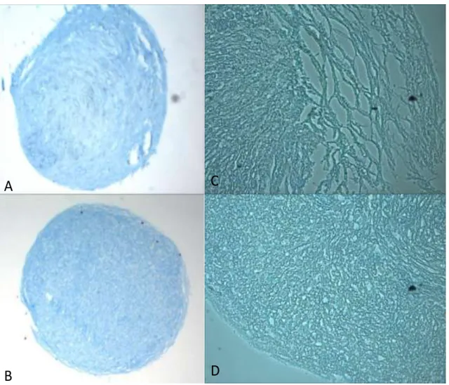
Figure 2. Alcian Blue staining of 8mM Ca
2+pellet sections (A,C) and
29
A
C
B
D
650 mm650 mm
650 mm
650 mm
Figure 3. Safranan O/FCF Fastgreen/Hematoxylin and Alizarin red
stains of 8mM Ca
2+Pellet sections (A,C) and control 1.8mM Ca
2+30
A
B
C
D
E
F
650 mm 650 mm 650 mm
650 mm 650 mm
650 mm
Figure 4. IHC of pellet sections for col I (A-8mM Ca2+, B-control), col II (C-8mM Ca2+,
D-control), and col X (E-8mM Ca2+, F-control) Real time RT-PCR Analyses
No significant differences were found in mRNA expression between the 8 mM Ca2+ samples and 1.8 mM controls. Experimental pellets demonstrated diminished aggrecan mRNA expression at day 7 relative to controls; however this difference was not statistically significant.
Sulfated GAG and Hydroxyproline
31
.
Figure 5. Quantification assays: Sulfated GAG content of pellets normalized to DNA.
Hydroxyproline contents of pellets normalized to DNA.
0 0.001 0.002 0.003 0.004 0.005 0.006
DMB/DNA1
m
g/
n
g
S-GAG/DNA
Control 8mm
0 0.1 0.2 0.3 0.4 0.5 0.6 0.7 0.8 0.9
OH-Pro/DNA
m
g/
n
g
Hydroxyproline/DNA
32 3.4 Discussion
Histological analyses demonstrated clear differences between control (1.8 mM Ca2+) and elevated 8mM Ca2+ experimental pellets, most notably the differences in pellet morphology and IHC collagen staining. The pellets cultured in elevated Ca2+ exhibited loose and stratified layers towards the outer portion of the pellet, as opposed to the compacted uniform appearance of control pellets. Hematoxylin staining of cell nuclei indicated a sparse cell population in the loose outer layers of the 8 mM Ca2+ pellets, with the majority of cells being distributed towards the center of the pellet. This cell distribution was the opposite of the control pellets, where cell number was higher in the pellet periphery, and diminished towards the center. Immunohistochemistry results for collagen X indicated enhanced collagen X staining within the loose peripheral layers of the elevated Ca2+ pellets. Collagen X is generally associated with chondrocyte hypertrophy and ossification and calcified cartilage, where bone and cartilage tissue interact. These results suggest that cells toward the outside of the pellet may have become hypertrophic and possibly apoptotic. This is consistent with previous studies showing the role of PTHrP in inhibiting hypertrophy in chondrocytes10-13. Parathyroid hormone related-protein is down-regulated in environments of high Ca2+ concentration due to signaling through the extracellular calcium sensing receptor4, 11. Increased PTHrP has been associated with increased chondrocyte proliferation and maintenance of cells in a matrix forming phenotype, resulting in increased matrix production10, 11.
33
supported with the results of hydroxyproline quantification. Control pellets produced nearly three times more hydroxyproline than pellets cultured in 8 mM Ca2+ when normalized to DNA (p=.09), corresponding to nearly three times the collagen II present within the pellet. At the level of gene expression, aggrecan mRNA expression at day 7 was diminished in hASC cultured in 8mM Ca2+, although the difference was not significant.
In addition to histological and IHC analyses for chondrogenic markers, further analyses were performed to determine the effects of elevated Ca2+ on osteogenic differentiation. Pellets in 8 mM Ca2+ exhibited positive alizarin red staining, especially towards the center of the pellet, while the 1.8 mM Ca2+ controls did not exhibit any positive staining. IHC for collagen I showed both control and experimental pellets stained positively, but in different locations. In elevated Ca2+ sections, positive IHC staining was mostly localized to the loose outer layers of the pellet, whereas the control pellets demonstrated positive collagen I staining in a swirled pattern in the center of the pellet. The mechanisms responsible for these positive results are not clear and require further study.
34
intermediate and deep zones, a sparse community of chondrocytes are surrounded by a network of sulfated GAGs and proteoglycans, with larger collagen II fibrils running perpendicular to the joint
through the tide mark into the zone of calcified cartilage. The zone of calcified cartilage is found beneath the tidemark, as the tissue transitions to subchondral bone with the function of anchoring the cartilage tissue to the bone14-17. The observed hypertrophy in this study may provide key insights for engineering appropriately layered articular cartilage, with integration of that cartilage to the underlying bone.
3.5 Conclusion
35 Chapter 4:
Targeting the Extracellular Calcium Sensing Receptor in Human Adipose Derived Stem Cell Osteogenic Differentiation
The previous experiment further demonstrated the ability of elevated extracellular Ca2+ to impact hASC differentiation, however the mechanism by which elevated calcium directs cells during differentiation is still not clear. This study aims to modulate the effects of elevated calcium by targeting two different proteins. PC2 is a permeable ion channel associated with the primary cilia, in a
mechanosensing role. It utilizes Ca2+ as a secondary messenger for cell signaling. We will use siRNA to knock down its expression. The extracellular calcium sensing receptor is a G-protein coupled receptor which allosterically binds with Ca2+, allowing it to act as a first messenger. We will modulate it using allosteric binding ligands to activate (calcimimetics) and inactivate (calcilytics) the signaling process.
4.1 Introduction
Calcium plays a key role in cell metabolism and can act as an extracellular signaling molecule, directing a number of cellular processes through various mechanisms. One mechanism is through intra- and extracellular transport of calcium ions through voltage-gated calcium channels, while others require the presence of a ligand to allow ions to travel across the cell membrane1, 2. This ion transport can result in cell secretion or contraction activities, directing specific cell activities. A number of ion transport channels are associated with mechanotransduction, allowing the cell to interpret mechanical loads through the movement of calcium ions. Polycystin 2, a sensory protein associated with
36
Calcium can also act as an external first messenger without being transported into the cell. The extracellular calcium sensing receptor (CaR) is a membrane bound G-protein coupled receptor that binds to calcium ions externally, initiating a signaling mechanism internally4, 5. This receptor plays a key role in maintaining calcium ion homeostasis in that it signals to the cell the concentration of extracellular calcium, allowing the cell to respond appropriately. The CaR is often associated with the secretion of PTHrP, and has been targeted in pharmacological treatments for a variety of diseases, including
osteoporosis. Various ligands have been allosterically bound to the CaR to either activate (calcimimetics) or inactivate (calcilytics) this signaling mechanism4, 6-10.
Our lab has found that elevating the concentration of extracellular calcium in the culture medium can impact both osteogenic 11 and chondrogenic (unpublished data) differentiation of human adipose derived stem cells (hASC). We have shown that a Ca2+ concentration of 8mM is effective in directing hASC osteogenic lineage specification in the absence of other osteogenic factors, while the same concentration appears to inhibit chondrogenic lineage specification when hASC are cultured in chondrogenic differentiation media including soluble chondrogenic growth factors transforming growth factor beta-1 (TGF-β1) and bone morphogenetic protein 6 (BMP6) (unpublished data). The mechanism by which elevated Ca2+ influences hASC lineage specification is unclear.
The aim of this study was to determine if the PC2 channel and/or the CaR play a significant role in regulating hASC differentiation in response to elevated extracellular calcium. Understanding the mechanism by which hASC lineage specification is directed by extracellular Ca2+ may lead to significant advances in use of these stem cells for tissue engineering and regenerative medicine applications.
37
Excess adipose tissue was obtained from five donors (24 to 37 year old females, multiple ethnicities) in accordance with an approved IRB protocol at UNC Chapel Hill (IRB 04-1622). Human ASC were isolated from the tissue using a method described by Zuk et al12 as previously described by our lab 13, 14. At passage 2, 100K cells from each donor were seeded in a single flask in complete growth media (CGM) comprised of alpha-modified minimal essential medium (α-MEM)(Invitrogen) supplemented with 10% fetal bovine serum (FBS) (Premium Select, Atlanta Biologicals, Lawrenceville, GA), 200 mM L-glutamine, and 100 I.U. penicillin/ 100 µg streptomycin per ml (Mediatech, Inc.) The cells were allowed to proliferate at 37°C in 5% carbon dioxide until reaching 70% confluency, and then trypsinized. The amassed cells were characterized for multipotent lineage capability, ensuring the amassed cells differentiated representative of an average of the five cell lines.
PC2 siRNA Knockdown
A single cell line of hASC was plated in a 12 well tissue culture plate and allowed to reach 50% confluency. Polycystin 2 was transiently knocked down in the cells using the Invitrogen siRNA protocols. Small interfering RNA specific to PC2 was used for experimental cells, while nonspecific scramble siRNA was used as a knockdown control. Lipofectamine 2000 was used as a transfection reagent to introduce the siRNA into the cells during the knock-down process. Knockdown cells were cultured in complete growth media with elevated (8mM) Ca2+. Calcium levels were elevated to a concentration of 8mM by dissolving CaCl2 into the media. Control cells were cultured in complete growth media with and without elevated Ca2+, and all cells were incubated at 37o in 5% carbon dioxide. At day 7 the knockdown
38
(Pierce) and the same microplate reader. Calcium values were normalized to protein concentrations for all samples.
Ligand Preparation for Pharmacologic Regulation of CaR
Allosteric binding ligand solutions were prepared by dissolving samples of each chemical (Cinacalcet HCL (Santa Cruz Biotech, calcimimetic), NPS 2143 (Santa Cruz Biotech, calcilytic), Calhex 231 (Santa Cruz Biotech, calcilytic), and AMG 568 (Santa Cruz Biotech, calcimimetic)) in absolute ethanol at a final concentration of 10-3 M. Solutions were aliquoted and stored at -20oC.
Ligand Selection
Preliminary experiments were utilized to determine which ligands modulated the effects of elevated extracellular Ca2+. A single cell line of hASC was plated in tissue culture treated plates and allowed to reach 50% confluency. Ligands were combined with media at a final concentration of 10-5 M and added to cells. Control cells were treated with 10ml of pure ethanol. Experimental cells and
untreated controls were cultured in complete growth media with and without elevated extracellular Ca2+ for 14 days receiving media changes treated with ligands every 48-72 hours. At day 14, Cells from each well were scraped and analyzed for Calcium and protein as previously described.
Cell Culture
39
media changes. Cinacalcet HCL (calcimimetic) was added at a final concentration 10-6 M and NPS2143 was added at 5x 10⁻⁷ M. Control cells were cultured with 1ml of ethanol at each media change. Cells were cultured at 37°C in 5% carbon dioxide for up to 14 days, receiving media changes every 48-72 hours.
Real time PCR
Cells from each experimental condition were collected on days 3, 9, and 12 of culture for real-time quantitative reverse transcriptase polymerase chain reaction (RT-PCR) analysis. Each construct was dissolved in RNA lysis buffer and total RNA was isolated using a Perfect RNA Eukaryotic Mini kit (Eppendorf, Westbury, NY). Complementary DNA was synthesized using a SuperScript III First-Strand Synthesis System for RT-PCR (Invitrogen Life Technologies, Carlsbad, CA) with oligo (dt) 20 primers. Primers and probes for human Runx2 (Assay HS00231692 M1), BMP2 (Assay, HS00154192 M1), Osteopontin (SPPI, Assay HS2408577 M1), Alkaline Phosphatase (ALP, Assay HS01029144 M1), and glyceraldehydes-3-phosphate dehydrogenase (GAPDH, Assay HS99999905 M1) were purchased from Assays-on-demand (Applied Biosystems, Foster City, CA). Real-time RT-PCR was performed using TaqMan PCR Master Mix (Applied Biosystems) in an ABI Prism 7000 system (Applied Biosystems). Signals were normalized to GAPDH expression levels using the –CT method 15.
4.3 Results
Knockdown of PC2 did not result in significantly different cell mediated calcium accretion from scramble siRNA controls (Fig 1a). Cells treated with CaR binding ligands in CGM at basal calcium levels (1.8 mM) did not demonstrate significant differences in proliferation or calcium to protein ratios (not shown). However, hASC treated with allosteric binding ligands and cultured in elevated 8 mM Ca2+ demonstrated significantly different calcium to protein ratios. Cells treated with either calcilytic
40
either calcimimetic (R568, Cinacalcet HCL, Santa Cruz Bio) exhibited higher calcium to protein ratios. (Fig 1b)
0.0000 0.0500 0.1000 0.1500 0.2000 0.2500
PC-2 Scramble siRNA None None
8mm cgm
Calcium/Protein
m
g/
m
g
Avg Ca/Protein
Avg Ca/Protein
41
Figure1. Calcium to protein ratios: A) Transient knockdown of PC2 B) Allosteric binding of CaR in elevated calcium
Assays
All cells cultured in CGM with 8mm Ca2+ demonstrated significantly higher calcium to protein ratios when compared to untreated controls cultured in CGM with basal levels (1.8 mM) of Ca2+(Fig 2). Human ASC cultured in ODM or in the presence of 8 mM Ca2+ and combined with the calcimimetic Cinacalcet exhibited significantly higher calcium to protein ratios when compared to untreated controls cultured in CGM with 8mM Ca2+ without Cinacalcet (Fig 2). However, culture with the calcilytic NPS 2143 produced inconsistent results at various stages and often resulted in cell death.
*
*
* *
0 0.0001 0.0002 0.0003 0.0004 0.0005 0.0006
Control NPS 2143 Calhex 231 NPS 568 Cincalcet
m
g/
m
g
Avg Ca/Protein
CALCIUM/ PROTEIN
* P<.02
42 Figure 2.Calcium to protein ratio at Day 12
RT-PCR
43
day 12 cells cultured in CGM with 8 mM Ca2+ expressed alkaline phosphatase significantly greater than that of hASC cultured in ODM and hASC treated with Cinacalcet (Fig 3c). Additionally, hASC cultured in CGM with 8mM Ca2+ expressed significantly higher levels of osteopontin than cells cultured in ODM, yet lower than hASC in CGM with 8mM Ca2+ and further treated with Cinacalcet (Fig 3c).
44
45 Figure 3. qPCR results C) Day 12 ALP and SPPI expression
4.4 Discussion
46
negative effects. We believe the inconsistency in hASC response to the ligand was potentially a result of inconsistencies in the chemical as received from the manufacturer as cell responses from same donors varied with different samples of the same compound purchased at different dates. Due to these variations, we will not conclusively state that addition of a calcilytic that inhibits CaR activity results in diminished hASC osteogenic response, although our preliminary analyses indicate that it might.
Our study demonstrates that elevating extracellular Ca2+ in complete growth medium to a concentration of 8mM is effective in directing hASC osteogenic differentiation similar to culture in traditional osteogenic differentiation medium as demonstrated by cell mediated calcium accretion and the expression of osteogenic markers. At day 3 the two conditions are not significantly different in mRNA expression of Runx 2. At Day 9, hASC cultured in CGM with 8 mM Ca2+ express BMP2 and
osteopontin mRNA at significantly greater levels than cells cultured in ODM, but ALP mRNA expression is significantly less. By day 12 however, hASC cultured in 8mM Ca2+ exhibited significantly higher levels of ALP mRNA expression compared to hASC cultured in ODM, with little change in the difference of SPP1 expression. BMP2 is often associated with the early phases of osteogenesis, while osteopontin,
47
This is consistent with other studies that have investigated the role of elevated extracellular Ca2+ on osteogenic differentiation of other cells18, 19. An et al19 recently showed that elevated Ca2+ increased mineralized matrix nodule formation in human dental pulp cells. They also reported increased expression of osteopontin and diminished alkaline phosphatase expression, consistent with findings from this study.
While the calcilytic NPS 2143 was inconsistent in effectively modulating cell activity, the calcimimetic Cinacalcet performed as expected by enhancing hASC osteogenic differentiation. The calcium to protein ratio observed in treated cells at day 12 was similar to that observed in cells cultured in ODM, however, as discussed earlier, the various media types resulted in differentiation at different rates, with different time points for gene expression. These findings suggest that a longer culture period may produce significant differences in mineralized matrix production, especially considering the
significantly upregulated SPPI expression at day 12 in Cinacalcet treated cells. Overall, cells cultured with Cinacalcet in 8mM Ca2+ behaved similarly to cells cultured in ODM throughout the experiment, with the exception of the increased osteopontin expression at day twelve. These data suggest that the addition of Cinacalcet to CGM with 8mM Ca2+ causes hASC osteogenic lineage specification similar to ODM, with an enhanced mineralization potential.
48
differentiation21. CaR was also shown as a key mechanism with elevated Ca2+ in human keratinocyte differentiation. Tu et al demonstrated how inhibiting CaR negatively affected cell differentiation, and proliferation of human keratinocytes20. That study utilized antisense RNA to inhibit CaR similar to our early use of siRNA to knock down PC2, whereas our study used allosteric ligand binding to inactivate CaR signaling. Future studies may compare the effectiveness of modulating CaR activity using transient knock down via siRNA, and allosteric binding, with the ultimate goal of optimizing stem cell differentiation. Additional studies must determine the unintended impacts of each treatment on cell culture and
differentiation. Specifically, we must determine how transfection impacts proliferation, and cell viability, as well as ensuring ligands are in appropriate concentration to be effective without becoming toxic. Both may affect gene expression during the osteogenic differentiation process.
4.5 Conclusion
49 Chapter 5:
Conclusions
5.1 Conclusions
This body of work has focused on various factors affecting chondrogenic differentiation of hASC with the ultimate goal of optimizing conditions for engineering functional cartilage tissue. The use of hASC in novel treatments for cartilage defects is inevitable, and the application of the work presented here should greatly affect that emerging field.
The first focus of this work was on the conditions needed to optimize chondrogenic
differentiation of hASC, specifically, the appropriate mechanical loading environment. Like hMSC, hASC best differentiate when cultured in a three dimensional environment, under hypoxic conditions, cultured in media containing insulin, high glucose, dexamethasone, ascorbic acid, and soluble growth factors TGF-3 and BMP6. Additionally, compression and hydrostatic pressure at magnitudes
encountered in vivo seem best suited to induce chondrogenic differentiation, though fluid shear and tensile strain each play a role in cartilage tissue homeostasis and could be incorporated in conditioning engineered tissue.
50
pathways in directing intracellular Ca2+ concentration, and ultimately stem cell differentiation 1. This demonstrates the importance of Ca2+ in mechanical signaling. Additionally, elevating extracellular Ca2+ concentration has been shown to increase osteogenic differentiation in hASC, as well as decreased adipogenesis in preadipocytes 2,4. In this study, we observed the impact of elevated Ca2+ on
chondrogenic differentiation of hASC in pellet culture. Pellets cultured in elevated 8mM Ca2+ produced less cartilage products than control pellets suggesting the elevated Ca2+ inhibited chondrogenic
differentiation. Histological analyses demonstrated the outer layers of experimental pellets experienced terminal differentiation of chondrogenic cells, resulting in hypertrophic calcified cartilage. The zone of calcified cartilage plays a key role in anchoring cartilage tissue into place. Engineering cartilage tissue with a functional anchoring layer may introduce new methods of implantation and improved integration into native tissue. The observed results are significant in that they may provide insight on engineering cartilage tissue with each layer seen in vivo, improving the functionality of engineered tissue.
51
of elevated Ca2+ on hASC differentiation may have more to do with signaling pathways, than ion transport. Additionally, it demonstrates that the CaR is an effective target to modulate hASC differentiation, and can be used to optimize tissue engineering.
5.2 Future Work.
Additional work with hASC pellets cultured in chondrogenic media with elevated Ca2+ may be in order to determine the material properties of the matrix products created. Further, these pellets should be analyzed to determine if the layers observed are cohesive in a manner similar to that of articular cartilage. This may allow for the development of engineered cartilage tissue designed to be anchored on the subchondral bone, allowing for better assimilation.