ANALYSIS OF TELOMERE MAINTENANCE MECHANISMS IN THE ROUNDWORM CAENORHABDITIS ELEGANS
Ludmila Shtessel
A dissertation submitted to the faculty of the University of North Carolina at Chapel Hill in partial fulfillment of the requirements for the degree of Doctor of Philosophy in the Curriculum
of Genetics and Molecular Biology.
Chapel Hill 2012
Approved by:
Shawn Ahmed, Ph.D.
Christopher Counter, Ph.D.
Jeff Sekelsky, Ph.D.
Michael Jarstfer, Ph.D.
ii
ABSTRACTLUDMILA SHTESSEL: Analysis of telomere maintenance mechanisms in the roundworm Caenorhabditis elegans
(Under the direction of Dr. Shawn Ahmed)
The ends of linear chromosomes are composed of repetitive hexameric sequences called
telomeres. Because canonical DNA polymerases cannot fully replicate the ends of linear
chromosomes, they shorten with each cycle of replication. Once telomeres become critically
short, cells stop replicating and enter a state of replicative arrest—senescence. In this way,
telomere length limits the replicative potential of all cells. The enzyme telomerase can maintain
telomere length by adding de novo telomeric repeats via reverse transcription from an associated RNA template. Although telomerase expression is exclusive to human somatic and germ stem
cells, its expression in somatic stem cells is not sufficient to prevent telomere erosion. Therefore,
telomeres shorten with age, effectively serving as a mitotic clock. As in mammals, components of
the C. elegans 9-1-1 (HPR-9/MRT-2/HUS-1) DNA damage response complex and its clamp loader, HPR-17, are necessary for telomerase-mediated telomere repeat addition in vivo. Here we present the mapping and characterization of three new telomerase-defective alleles of mrt-2, hpr-17, and hpr-9, and a mutation in a novel gene, all of which possess defects in repairing ionizing radiation- and interstrand crosslink-induced DNA damage.In addition to telomerase, several
canonical telomere-binding proteins and other associated proteins maintain telomere length
homeostasis and protect chromosome ends from exacerbated erosion and erroneous DNA damage
iii
POT-2 as negative regulators of telomerase-mediated telomere repeat addition in vivo. I demonstrate that POT-1, but not POT-2, protects telomeres from exacerbated erosion. I employ
several biochemical strategies to assess whether POT-1 or POT-2 interact with telomerase in vivo. Although an epitope-tagged pot-1 transgene rescued the telomere elongation phenotype of the pot-1 mutant, it exacerbated the onset of senescence in mutants defective for telomerase-mediated telomere repeat addition. We suggest that alterations in telomere capping proteins may drive
telomere dysfunction in telomerase-negative cells, which may help to define causal mutations for
iv
DEDICATIONGiven that I am now not only a self-proclaimed geneticist but also a UNC-proclaimed
one, I cannot ignore the reason for who I am, why I am, and where I am today: my genes. And
until 3 decades ago, my parents were the sole protectors of those genes, which were clearly well
maintained until my arrival. During the pursuit of my doctorate, I became even more enamored
with the genome and its simplicity and self-preservation. During that time I also entered into a
compromise, with the help of Matt Ridley’s Nature via Nurture, with the rest of the world— acknowledging that, yes, you too have a hand in my destiny. However, as it turns out, my parents
also raised me, pushing me towards positive experiences and protecting me from nasty ones. So,
without a doubt, I attribute most of my talents, successes, and personal proclivities to my
v
PREFACEThe successes of my efforts presented in this thesis have greatly benefited from
several collaborators. A technician, Yan Liu, and a graduate student, Dr. Julie Hall,
participated in the
Mortal germline
screens, including passaging animals for sterility and
assessing for the presence of end-to-end fusions, that yielded the mutants discussed in
Chapter 1. Yan Liu mapped the initial, approximate locations of
yp7
and
222g
, and Dr.
Hall mapped the initial, approximate location of
279
as well as analyzed telomeres of
279
mutants, shown in Figure 1.2C. In the later stages of
279
mapping, an extremely
hard-working and persevering undergraduate-turned-PharmD UNC student, Ashley Hedges,
participated in the SNP mapping that narrowed the ultimate
279
candidate list to 29
genes. The mapping of
yp8
appears in the following publication and is reproduced with
the permission of the publisher:
Chen Cheng, Ludmila Shtessel, Megan M. Brady, and Shawn Ahmed. Caenorhabditis
elegans POT-2 telomere protein represses a mode of alternative lengthening of telomeres
with normal telomere lengths. Proc Natl Acad Sci USA. 2012
I employed many transgenic animals in the biochemistry and mutant analysis
presented in Chapters 2 and 3. A graduate student, Matt Simon, and a technician, Kyle
Wang, participated in the construction of all the transgenes and their insertion into
animals.
Chapter 3 describes the analysis of several mutants, which was initially performed
vi
mutants to confirm the presence of their respective mutations and analyzed their
telomeres, shown in Figure 3.1B & C. The first five figures of this chapter have been
published and are reproduced with the permission of the publisher:
Ludmila Shtessel, Mia Rochelle Lowden, Chen Cheng, Matt Simon, Kyle Wang, and
Shawn Ahmed. Caenorhabditis elegans POT-1 and POT-2 Repress Telomere
vii
TABLE OF CONTENTS
LIST OF TABLES ... ix
LIST OF FIGURES ... x
INTRODUCTION ... 1
Telomeres and Telomerase ... 1
Human diseases of telomere dysfunction ... 4
Human diseases of accelerated aging ... 7
Two disease states separated by telomerase status ... 10
CHAPTER 1: Genetic mapping and characterization of C. elegans telomere maintenance-defective mutants ... 12
Mapping and characterization of hpr-17(yp7) ... 14
Mapping and characterization of mrt-2(yp8) ... 18
Mapping and characterization of hpr-9(222g)... 22
Mapping and characterization of 279 ... 25
Discussion ... 28
CHAPTER 2: Biochemical analysis of proteins that participate in telomere length homeostasis ... 32
Creation of transgenic animals ... 34
Analysis of protein expression by western blot ... 35
Direct telomerase activity assays ... 44
Discussion ... 49
CHAPTER 3: Telomere length regulation by C. elegans POT-1 and POT-2 ... 52
viii
POT-1 localizes as punctate foci to C. elegans telomeres in vivo ... 58
POT-1 and POT-2 repress telomerase activity at telomeres ... 61
POT-1::mCherry perturbs telomere stability in the absence of telomerase ... 65
Discussion ... 74
MATERIALS AND METHODS ... 83
ix
LIST OF TABLES
Table
x
LIST OF FIGURES
Figure
1.1 A novel mutation that displayed hallmarks of telomerase
deficiency, yp7, was mapped to the hpr-17 gene on Chromosome II. ... 16
1.2 Characterization of novel telomere maintenance-defective mutants. ... 19
1.3 A novel mutation that displayed hallmarks of telomerase deficiency, yp8, was mapped to the hpr-17 gene on Chromosome III. ... 21
1.4 A novel mutation that displayed hallmarks of telomerase deficiency, 222g, was mapped to the hpr-9 gene on Chromosome III. ... 24
1.5 A novel mutation that displayed hallmarks of telomerase deficiency, 279, was mapped to a candidate region of 29 genes on Chromosome I. ... 26
2.1 Analysis of phenotype rescue for FLAG::POT-1 and TRT-1::FLAG constructs. ... 36
2.2 Immunodetection of proteins from whole worm extracts via western blot. ... 39
2.3 Immunodetection of proteins from whole worm extracts via western blot. ... 43
2.4 Direct telomerase assays with immunoprecipitated proteins... 47
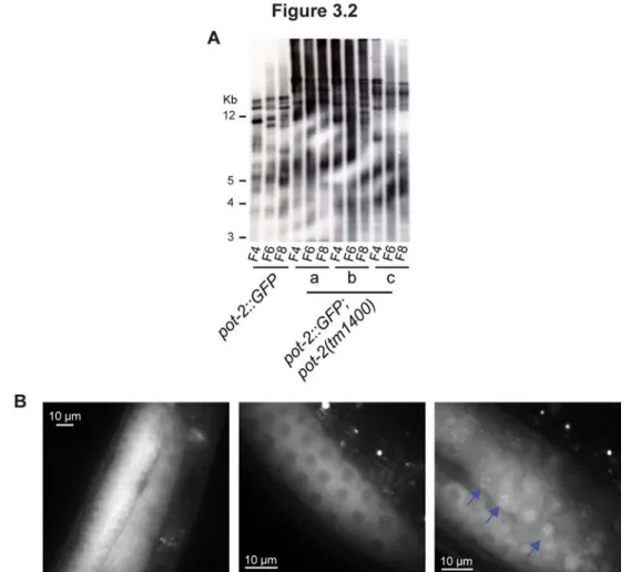
3.1. POT-1 and POT-2 are negative regulators of telomere replication. ... 57
3.2. The POT-2::GFP construct does not rescue mutant pot-2 phenotypes. ... 57
3.3. POT-1:mCherry localizes to telomeres as punctate foci independent of POT-2. ... 59
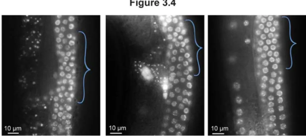
3.4. POT-1::mCherry localization in transition zone germline nuclei. ... 60
3.5. POT-1 and POT-2 negatively regulate telomerase-mediated telomere repeat addition. ... 63
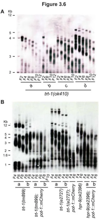
3.6. Telomerase reverse transcriptase mutants exhibit progressive telomere erosion, which is exacerbated by the presence of a POT-1::mCherry fusion protein... 64
3.7. POT-1::mCherry exacerbates telomere loss in telomere maintenance-defective mutants. ... 68
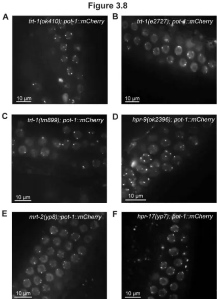
3.8. POT-1::mCherry localization in several telomere maintenance-defective mutants. ... 71
3.9. Schematic representation of the transgenes constructed. ... 72
3.10. Telomerase-deficient pot-1::mCherry mutants do not exhibit higher levelsof telomeric circles than telomerase mutants alone... 73
INTRODUCTION
Telomeres and Telomerase
The ends of linear chromosomes, called telomeres, are comprised of repetitive sequence
tracts that function in various ways to maintain chromosome fidelity. In the 1930s, forty years
before telomeres were molecularly defined, Herman Muller noticed that the ends of chromosomes
were resistant to the detrimental effects of X-rays. He concluded that they must be special,
coining the term “telomere” from the Greek words “telos” and “meros”, meaning “end part”, in
the process. Concurrently, Barbara McClintock’s work with the variable color patterns in maize
led her to define the functional importance of telomeres. She described a “breakage-fusion-bridge
cycle” in maize as a continuous loop initiated fusion of two chromosomes, followed by
segregation-mediated breakage of dicentric chromosomes, resulting finally in a new fusion of
improperly capped chromosome ends that re-enter the breakage-fusion-bridge cycle (McClintock
1939). These kinds of chromosomal dynamics illustrated the crucial function telomeres play in
preventing genomic instability.
Elizabeth Blackburn, as a post-doctoral associate in John Gall’s laboratory, was the first
to molecularly define the ends of chromosomes in 1978, revealing mysterious repetitive
hexameric sequences in Tetrahymena, a single-celled ciliate (Blackburn & Gall 1978). Four years later, in collaboration with Jack Szostak, Blackburn cloned telomeres, demonstrating that terminal
Tetrahymena repeats (TTGGGG) retained their chromosome capping function when placed onto the ends of a linearized yeast chromosome (Szostak & Blackburn 1982). Since then, telomeres
from a myriad of organisms have been cloned, comprising different repeats and tract sizes.
2
in mice and humans span up to 80 kb and 5-15 kb, respectively (Moyzis et al. 1988; Harley et al. 1990; Kipling & Cooke 1990; Prowse & Greider 1995; Zhu et al. 1998), Arabidopsis plants exhibit 2-4 kb-long TTTAGGG tracts (Richards & Ausubel 1988; Riha et al. 1998), TTAGGC tracts span 4-9 kb in the roundworm C. elegans (Wicky et al. 1996), and more complicated TTAC(A)(C)G(1-8) or G(2-3)(TG)(1-6)T repeats comprise telomeric tracts of approximately 300
base pairs (bp) in fission and budding yeasts, respectively (Shampay et al. 1984; Murray et al. 1986).
Organisms that package their DNA into linear chromosomes are burdened by incomplete
replication of chromosome termini. Because conventional DNA polymerases cannot initiate
synthesis de novo and instead rely on small RNA primers, an obligate gap remains at the 5’ ends once the primers are removed (reviewed in Levy et al. 1992). This “end-replication problem” results in loss of telomeric DNA with each cell division, along with additional nuclease-mediated
telomere processing, until a critically short telomere triggers senescence—a non-dividing state of
arrest (Bodnar et al. 1998; Hemann et al. 2001; Sfeir et al. 2005). The enzyme telomerase is tasked with maintaining telomeres by adding telomeric repeats de novo via reverse transcription using an RNA template (Greider & Blackburn 1985; Greider & Blackburn 1989). Most human
somatic cells do not express telomerase and thus exhibit telomere shortening during cellular
replication, which limits their replicative lifespan in vitro (Sharma et al. 1995; Hayflick & Moorhead 1961; Harley et al. 1990). Telomere length is not only indicative of in vitro replicative lifespan but also decreases as a function of age in vivo (Harley et al. 1990; Allsopp et al. 1992; Aubert & Lansdorp 2008). Therefore, human lifespans are not actually defined by a temporal
clock but rather a “mitotic clock” based on the replicative potential of cells.
Telomere maintenance is the lynchpin in the delicate battle between cancer and
organismal aging. While limiting amounts of telomerase result in telomere attrition and eventual
cellular senescence, a hallmark of aging tissues, the uncontrolled expression of telomerase that
3
Shay & Wright 2011; Kim et al. 1994). Proper telomere length homeostasis is maintained in part by canonical telomere-binding proteins and other associated components.
In mammals, the shelterin complex, composed of the six canonical telomere proteins
TRF1, TRF2, POT1, TIN2, TPP1, and RAP1, maintains the integrity and length of telomeres
(reviewed in Palm & de Lange 2008 and in Diotti & Loayza 2011). TRF1 and TRF2 both bind
the duplex telomeric DNA and negatively regulate telomere length (van Steensel & de Lange
1997, Smogorzewska et al. 2000). While over-expression of TRF2 drives telomere attrition in culture, it also protects telomeres from fusion, underscoring both critical roles of TRF2 in
telomere biology (Karlseder et al. 2002; Richter et al. 2007). TRF2 conditional and dominant negative alleles in mammals induce end-to-end chromosomal fusions, extrachromosomal circles,
telomere rapid deletion events, and an increase in sister chromosome exchange (reviewed in
Longhese 2008; Wang et al. 2004; Li et al. 2008). A TRF2 binding partner, RAP1 also exerts a negative force on telomere length but does not bind DNA directly (Li et al. 2003; O’Connor et al. 2004). Curiously, over-expression of wildtype RAP1, or RAP1 truncation mutants that do not
localize to telomeres, prompted telomere elongation in cultured human cells (Li et al. 2000; Li et al. 2003), which the authors suggest as an indication that RAP1 may titrate a negative regulator of telomere length away from the telomere when present in excess.
Although TRF1 and TRF2 do not directly bind each other, they are both bound by TIN2,
which connects the single-stranded telomeric binding protein POT1 to the telomeric duplex via an
interaction with TPP1 (Ye et al. 2004; Kim et al. 2004). TIN2 also negatively regulates telomere length, and can dramatically affect the stability of the shelterin complex on telomeres via its
direct interactions with multiple components (Kim et al. 1999; Kim et al. 2004). TPP1, which bridges POT1 and TIN2, exerts regulation on telomere length through its interaction with POT1
4
inhibit elongation, which it does so via POT1. POT1 binds single stranded telomeric DNA and
TIN2 directly and exerts both positive and negative regulation on telomere length (Liu et al. 2004; Lei et al. 2005). Critically, POT1 also protects telomeres from eliciting a DNA damage response, which requires TIN2-mediated recruitment (Takai et al. 2011).
C. elegans has four proteins with homology to human POT1: POT-1, POT-2, POT-3, and MRT-1. MRT-1 positively regulates telomere length, while POT-3 appears dispensable. My work
in Chapter 3 describes the roles of C. elegans POT-1 and POT-2, which are negative regulators of telomere length, in telomere maintenance. Surprisingly, I show that an epitope-tagged pot-1 transgene rescues pot-1 mutant defects but exacerbates the onset of senescence in telomerase mutants. This work is the basis for the remainder of this introduction, where we propose the
existence of two telomere dysfunction-associated disease states—one that is directly caused by
mutations in proteins that promote telomerase activity, including telomerase itself and one that is
accompanied by telomere shortening that remains to be directly attributable to dysfunctional
telomere biology, the pathologies of which we hypothesize may occur due to dysfunction
specifically in telomerase-deficient cells.
Human diseases of telomere dysfunction
Dyskerin was the first telomerase component to be causally identified in human disease
(Heiss et al. 1998). Dyskerin participates in the stability and maturation of small nucleolar RNAs (snoRNAs) by binding to an H/ACA motif, and its binding to this motif at the 3’ end of human
telomerase RNA (hTR) is required for hTR’s biogenesis and stability (Mitchell et al. 1999a; Mitchell et al. 1999b). This disease, dyskeratosis congenita (DC), was the first disease with a molecularly defined cause seeded in telomere biology (Heiss et al. 1998). DC is diagnosed predominantly within the first 10-20 years of life by the presence of two of three classical
5
failure (reviewed in Dokal 2011). The vast majority of patients die prematurely from bone
marrow failure, followed by pulmonary disease and cancer.
DC patients without mutations in dyskerin harbor mutations in other proteins that
function directly in telomere biology, including heterozygous, homozygous and biallelic
mutations in the telomerase reverse transcriptase (TERT) (Vulliamy et al. 2005; Marrone et al. 2007), heterozygous mutations in the telomerase RNA (encoded by TERC)(Vulliamy et al. 2001), homozygous mutations in both NOP and NHP2, which are similar to dyskerin in their
RNA-binding and telomerase stabilization functions (Walne et al. 2007; Vulliamy et al. 2008), heterozygous mutations in TINF2, which encodes the shelterin component TIN2 (Savage et al. 2008; Walne et al. 2008), biallelic mutations in a trafficking protein, TCAB1, which transports the telomerase holoenzyme to sites of RNA processing called Cajal bodies (Zhong et al. 2011; Venteicher et al. 2009), and homozygous mutations in the gene C16orf57, which remains poorly characterized (Walne et al. 2010).
Several other syndromes, including Hoyeraal-Hreidarsson (HH), Revesz,
Rothmund-Thomson (RTS), PN (poikiloderma with neutropenia), Coats’ plus, and myelodysplastic
syndrome (MDS) are characterized by a subset of pathologies seen in DC patients. HH is
considered a severe form of DC, where patients exhibit DC-like symptoms, such as bone marrow
failure, at an earlier age accompanied by intrauterine growth retardation, microcephaly, and
cerebellar hypoplasia, resulting in premature mortality within the first decade of life (Touzot et al. 2011; reviewed in Dokal 2011). RTS patients characteristically exhibit poikiloderma (a skin rash)
and short stature that typically present within the first 2 years of life (reviewed in Larizza et al. 2010). A myriad of other heterogeneous features, including thin hair, cataracts and retinal defects,
dental abnormalities, nail dystrophy, and skeletal abnormalities, also can appear at a prematurely
young age. Strikingly, RTS patients do not live shorter lives if cancer is avoided. Coats’ plus
disease is marked by telangectasias (small blood vessels) in the eye, cataracts and even eventual
6
RTS. Some patients also exhibit coarse and sparse hair with eventual premature hair graying,
dystrophic nails, aplastic anemia, and ataxia (reviewed in Crow et al. 2004; Savage et al. 2012). Identification of dyskerin, TINF2, and TERT mutations in Hoyeraal-Hreidarsson (HH) patients underlines the molecular connection among these complex and heterogeneous
pathologies (Knight et al. 1999; Marrone et al. 2007; Walne et al. 2008). Additionally,
heterozygous mutations in TINF2 have been observed in Revesz syndrome patients (Walne et al. 2008; Sasa et al. 2012). Causal heterozygous mutations in TERC and TERT have been identified in patients with AA and MDS (Vulliamy et al. 2002; Yamaguchi et al. 2003; Yamaguchi et al. 2005) as well as in patients with idiopathic pulmonary fibrosis, liver cirrhosis, and leukemia
(Tsakiri et al. 2005; Armanios et al. 2007; Diaz et al. 2010; Calado et al. 2009; Dokal 2011). RTS and PN patients also harbor homozygous mutations in C16orf57 (Volpi et al. 2010; Walne et al. 2010; Clericuzio et al. 2011). Most recently, biallelic mutations in CTC1, a single-stranded telomeric DNA-binding protein,were identified in 16 Coats’ plus patients (Anderson et al. 2012). Mammalian CTC1 forms a heterotrimeric complex with STN1 and TEN1, which localizes to
telomeres in mice and human cells and binds ssDNA telomeric DNA in vitro (Miyake et al. 2009).
While these diseases are separable, they exhibit vast overlaps in pathologies. Moreover,
although these diseases can be distinguished molecularly, overlap there exists as well. This
overlap in etiology suggests an underlying pathology that may be caused from directly perturbing
telomere maintenance, whether by mutation of telomerase holoenzyme components or by
mutations in telomere-binding proteins. In particular, telomere dysfunction appears to affect a
specific subset of tissues, such as blood cells (red and white), the lungs, liver, skin, bones and
hair. These tissues may represent somatic stem cells that harbor the highest levels of telomerase
activity or the highest rates of cell turnover (Chiu et al. 1996; Kilian et al. 1997; Kolquist et al. 1998) and thus are most sensitive to perturbations to telomere maintenance. In fact, families
7
an earlier and more severe onset of symptoms due to inheriting both the TERC or TERT mutation and short telomeres, indicating that telomere maintenance is affected in germ cells (Vulliamy et al. 2004; Armanios et al. 2005; Goldman et al. 2005; Basel-Vanagaite et al. 2008). Given the molecular and pathological overlaps between DC and these other diseases, the 40% of DC cases
that remain molecularly undefined are likely attributable to components with major functions in
telomere biology (reviewed in Dokal 2011).
Human diseases of accelerated aging
Several human diseases of premature aging, such as Werner syndrome (WS) and
Hutchinson-Gilford progeria, are accompanied by short telomeres, but proteins with canonical
functions in telomere biology have yet to be implicated in their etiology (Ishikawa et al. 2011; Decker et al. 2009; Allsopp et al. 1992). Cultured fibroblasts from Werner and HGPS patients exhibit telomere shortening and senesce prematurely (Faragher et al. 2003; Huang et al. 2008), which can be overcome with telomerase expression (Wyllie et al. 2000; Choi et al. 2001; Benson et al. 2010), providing a compelling argument for telomere involvement in the pathology of these syndromes. Moreover, telomerase-positive cells from WS patients do not senesce prematurely
(James et al. 2000), again indicating that telomerase activity may participate in mechanisms that regulate the premature cellular senescence observed in WS patients.
Homozygous mutations in WRN, a homolog of the RecQ family of DNA helicases, are responsible for the majority of WS cases (Yu et al. 1996; Oshima et al. 1996; Sogabe et al. 2004; Huang et al. 2006). WRN is a DNA helicase, one of several human homologs of the E. coli RecQ DNA helicase, that also harbors exonuclease activity (Gray et al. 1997; Huang etal. 1998). WRN has a myriad of roles in mediating the repair of and response to DNA damage, and it does so
through acting at several targets, such as stalled replication forks, Holliday junctions, and
8
known telomeric proteins, such as Ku, TRF2, and POT1 (Cooper et al. 2000; Li & Comai 2000; Opresko et al. 2002; Ghosh et al. 2009; Machwe et al. 2004) and participates in some aspects of telomere biology, such as excision of loops of telomeric DNA (t-loops) from chromosome ends
and suppressing telomeric sister chromatid exchanges (t-sce) (Opresko et al. 2004; Opresko et al. 2005; Machwe et al. 2004; Hagelstrom et al. 2010; Li et al. 2008). WS patients are characterized by short stature in their teens and typically begin to manifest other features, including thinning
and graying hair, skin atrophy, cataracts, osteoporosis, diabetes, atherosclerosis and mesenchymal
or epithelial cancer after the third decade of life (reviewed in Muftuoglu et al. 2008). Cancer and heart attack are the predominant causes of premature death in patients in their 50s.
Although numerous studies have shown short telomeres in WS cells and patients,
whether telomere length or shortening rate is causal to WS pathologies is still contentious (Schulz
et al. 1996; Baird et al. 2004). Work in Wrn-deficient mice supports a role for telomeres in WS pathology. Mice lacking Wrn not display overt premature aging phenotypes (Lebel & Leder 1998; Lombard et al. 2000), but telomerase mutants do after multiple generations (Lee et al. 1998). Strikingly, introducing deficiencies for Wrn into mice lacking the telomerase RNA, Terc,
dramatically exacerbated the pathologies of Terc-/- mice, including early-onset sterility, reduction in lifespan, growth retardation, gastrointestinal defects, wound healing defects, and increased
cancer incidence (Du et al. 2004; Chang et al.).Moreover, splenocytes cultured from Wrn-/-mice do not prematurely senesce in culture, whereas Wrn-/-fibroblasts do, perhaps indicating a
telomerase-dependent effect (Lebel & Leder 1998; Lombard et al. 2000). Thus, pathologies of human WS patients are likely affected by telomere shortening, but current models suggest that
this is not the sole form of age-related stress that is repressed by WRN.
Approximately 20% of WS patients do not carry any identifiable mutations in WRN and
instead are said to exhibit atypical Werner syndrome. Of these atypical WS cases, 15% harbor
mutations in LMNA (Chen et al. 2003; Doh et al. 2009; Renard et al. 2009; reviewed in
9
structural components of the nuclear envelope. Lamin A is initially translated into the precursor
prelamin A, which is modified and consequently processed by the metalloprotease ZMPSTE24
into mature lamin A (reviewed in Bertrand et al. 2011). Most mutations interfere with its processing, resulting in a persistent and toxic prelamin that dominantly interferes with wildtype
lamin A. Expression of known LMNA mutations in human fibroblasts has been shown to drive premature senesce in culture (Huang et al. 2008).
To date, there are 458 different mutations in LMNA cause nine clinically distinct diseases in addition to WS, including HGPS, Emery–Dreifuss
muscular dystrophy (EDMD), limb-girdle muscular dystrophy type 1B (LGMD1B), Charcot–
Marie–Tooth axonal neuropathy (CMT2B1), familial partial lipodystrophy of Dunnigan type
(FPLD), mandibuloacral dysplasia (MAD), LIRLLC, restrictive dermopathy (RD), and fetal
akinesia (reviewed in Bertrand et al. 2011). While these diseases exhibit distinct pathologies, several are shared among two or more diagnoses, including cardiac defects, muscle degeneration,
abnormal fat distribution, insulin resistance, skin defects, and osteolysis (reviewed in Bertrand et
al. 2011).
The vast majority of HGPS patients harbor mutations in LMNA (De Sandre-Giovannoli et al. 2003; Eriksson et al. 2003; reviewed in Bertrand et al. 2011). One patient, displaying
symptoms of HGPS as well as MAD and RD, harbored biallelic mutations in ZMPSTE24, which effectively resulted in the lack of prelamin A maturation (Shackleton et al. 2005). Patients are diagnosed with HGPS as toddlers predominantly due to their failure to thrive and the presence of
hair loss, skin defects, lipodystrophy, and characteristic bird-like facial features (reviewed in
Hennekam 2006). Additionally, height and weight is dramatically stunted, nails are poorly
formed (dystrophic), joint mobility erodes, and teeth decay. Although overt cardiac defects are
not apparent in most patients, heart attacks are the primary cause of death at about 13 years.
10
2003). Although MAD patients exhibit similar pathologies to HGPS patients, these are
alleviated—MAD patients live longer lives, are bigger, and display less severe hair loss,
lipodystrophy and osteolysis (bone breakdown) later in life (reviewed in Hennekam 2006).
Curiously, osteolysis is more severe in these patients than classical HGPS.
Two disease states separated by telomerase status
My work in Chapter 3 describes a premature onset of senescence in worms deficient for
telomere maintenance in the presence of an epitope tagged telomere-binding protein, POT-1.
However, in the context of telomerase proficiency, pot-1::mCherry animals appear and behave as wildtype. As C. elegans POT-1 is homologous to the bona fide telomere-capping protein POT1 in mammals, and perturbing the telomere cap has clinical relevance, we were prompted to consider
two scenarios of human disease that may be dictated by the presence or absence of telomere
maintenance.
In the first scenario, as illustrated by defects in telomerase holoenzyme components and
telomere-binding proteins, it appears that tissues that typically express telomerase or turn over
rapidly are affected. In this context, telomerase deficiencies manifest as defects in hematopoietic,
pulmonary, and hepatic lineages, and most patients die of bone marrow failure. Moreover,
diseases caused by telomerase defects can display genetic anticipation, where the next generation
is sicker earlier, indicating that germ cells are also affected. However, instances of severe defects
observed suddenly in one generation also exist, where patients that harbor homozygous mutations
in telomerase or heterozygous mutations in TINF2 display extremely short telomeres and other pathologies within the first decade of life (Marrone et al. 2007; Walne et al. 2008; Sarper et al. 2010; Sasa et al. 2012). Studies assessing how TINF2 mutations promote such severe dysfunction suggest that they do so, in part, through defects in telomerase-mediated telomere maintenance
11
case (Touzot et al. 2010), clinical diagnoses of DC-related defects are due to mutations in proteins that directly affect telomerase activity at telomeres.
In contrast, pathologies in the second scenario, as illustrated by mutations in WRN and LMNA, manifest in distinct tissues, such as the skin, eyes, bones, and vasculature, and most patients die of a heart attack. Because the presence of telomerase alleviated cellular senescence of
telomerase-negative cells from patients in vitro, the tissues that exhibit defects in these patients may represent telomerase-deficient environments that are sensitized to the additional stresses
caused by losing WRN. Defects caused by a WRN deficiency, such as poorly replicated telomeres or recombination-mediated telomere loss, perhaps could be healed by the presence of telomerase
in somatic and germ stem cells but would persist in cells that naturally lack telomerase or express
it at low levels. Since WS patients do not display the more severe and life-threatening pathologies
until their 30s or 40s, extensive information about familial inheritance of WS-causing mutations
is available. However, we were not able to find any description of genetic anticipation,
highlighting the possibility that WS pathologies may be due to defects in telomerase-negative
cells, where germ cells, which express extremely high levels of telomerase, would be immune.
Biallelic mutations in RECQL4, another member of the RecQ DNA helicase family with some roles in telomere biology, have been identified in RTS patients (Ghosh et al. 2012; Kitao et al. 1998; Kitao et al. 1999), who display a subset of DC-like symptoms, indicating that distinct molecular pathways could manifest as similar defects in humans. As approximately 20% of WS
patients remain molecularly undefined, we propose that mutations in proteins with major
functions in telomere biology represent ideal candidates and thus could definitively illustrate the
role of telomere biology in WS and other diseases marked by accelerated aging. In particular,
because WRN has cooperative functions at telomeres with both TRF2 and POT1, mutations in
neither of which have been identified in any disorders, these canonical telomere proteins could be
CHAPTER 1:
Genetic mapping and characterization of C. elegans telomere maintenance-defective mutants
To understand the mechanisms governing telomerase-mediated telomere maintenance in
multicellular organisms, we employed the roundworm Caenorhabditis elegans. Genetic
approaches bypass temporal and physical limitations of biochemical purification schemes, which
may be limited by transient interactions as well as small concentrations of proteins or accessory
factors that might be required for telomerase activity in vitro. While the in vivo and in vitro data from budding yeast lend a nice model of telomerase action, the level of analogy with metazoan
telomerase is unclear. A forward mutagenesis genetic screen to identify genes required for
maintaining germline immortality was performed in the laboratory prior to my arrival. Numerous
animals were mutagenized with ethyl methanesulfonate (EMS), and approximately 7,000 F2
animals, likely homozygous for hundreds of mutations, were passaged once weekly for 15 weeks,
where one week accounted for two generations of growth. Animals that harbored mutations in
genes necessary for germline maintenance, termed mortal germline (mrt) mutants, eventually became sterile. Because starved stage one larvae (L1) animals can survive freezing and thawing,
all strains were frozen as stocks prior to passaging for sterility. To determine which animals
became sterile due to defects in telomere maintenance, all mutants were assayed for the presence
of chromosomal fusions, a hallmark of dysfunctional telomeres. Of over 400 mrt mutants, six displayed late-onset chromosomal fusions, assayed by DAPI staining and fluorescence
microscopy.
13
that, when mutated, yield relatively easily discernible phenotypes, such as short and fat worms,
“dumpy” or “Dpy”, immobile or slow moving worms, “uncoordinated” or “Unc”, and worms that
roll in circles, “roller” or “Rol”. For example, if a mutant animal is suspected to harbor a mutation
in a gene that lies on the right arm of chromosome I, those animals can be crossed to hermaphrodites that harbor mutations in genes that flank that region, such as dpy-5 unc-29 animals. dpy-5 unc-29 animals harbor a mutation in dpy-5, which isat the center of chromosome I, also referred to as genetic position 0,and unc-29, which isat genetic map distance +3.3 on chromosome I (all six chromosomes are defined as roughly 50 genetic map units in length). Progeny from these crosses, referred to as the F1 generation, will be heterozygous for the mutant
allele on one chromosome and the dpy-5, unc-29 alleles on the other, and selection for Dpy-non-Unc or Dpy-non-Unc-non-Dpy F2 progeny indicates that a recombination event has occurred necessarily
between these two markers. The number of recombinants that harbor the mutant phenotype being
mapped indicates where the mutation lies relative to dpy-5 or unc-29.
Using such genetic mapping techniques, two of the six mrt mutants with chromosomal fusions were identified as mutations in the telomerase reverse transcriptase, trt-1(yp1), and in a nuclease, mrt-1(yp2) (Meier et al. 2006; Meier et al. 2009). This chapter describes the efforts taken to map and characterize the remaining four telomere maintenance-defective mrt mutants, three of which are novel alleles of components of the Rad9/Rad1/Hus1 (9-1-1) DNA damage
response (DDR) complex and its clamp loader, Rad17.
The structure of the 9-1-1 complex resembles the proliferating nuclear cell antigen
(PCNA) sliding clamp of DNA polymerase and mediates the DNA damage checkpoint by
inducing cycle arrest when chromosomes are damaged or improperly replicated (Weinert &
Hartwell 1988; St Onge et al. 1999; Caspari et al. 2000; reviewed in Parilla-Castellar et al. 2004). Fidelity of this checkpoint is vital for maintaining chromosomal integrity. Sites of stalled
replication or DNA damage harbor single-stranded DNA coated with RPA, which is necessary for
14
is loaded onto junctions of single-stranded and double-stranded DNA adjacent to these sites by
Rad17, initiating a signaling cascade that primarily hinges on CHK1, a kinase with a myriad of
targets (reviewed in Cimprich & Cortez 2008). Moreover, 9-1-1 participates in establishing a
DNA double strand break-induced signaling cascade via ATM and CHK2 (Chen et al. 2001). Work in yeast has also suggested that 9-1-1 components may function directly in repair of DNA
damage (Lydall & Weinert 1995).
While several canonical telomere proteins function to protect telomeres from
DDR-mediated repair processes, effective telomere maintenance relies on DNA damage repair proteins,
including the 9-1-1 complex. The mammalian 9-1-1 complex has been shown to localize to
telomeres as well as associate with catalytically active telomerase (Francia et al. 2006). Moreover, human and mouse cells deficient for Rad9 display an increase in end-to-end
chromosome fusions and loss of fluorescent telomere-specific signals (Pandita et al. 2006). Fission yeast rad1, rad3 (ATR in humans), rad17, and rad26 (ATRIP in humans) mutants and budding yeast ddc1 (Rad9 in humans) and rad17 (Rad1 in humans) mutants exhibit short telomeres after prolonged growth (Dahlen et al. 1998; Longhese et al. 2000; Nakamura et al. 2002).
C. elegans homologs of Rad1, Hus1 and Rad17 have been previously shown to be necessary for telomere maintenance in vivo (Ahmed & Hodgkin 2000; Hofmann et al. 2002; Boerckel et al. 2007). In addition to novel alleles of mrt-2 (hRad1) and hpr-17 (hRad17), here I describe the identification and characterization of the Rad9 homolog, hpr-9, which I reveal is also required for in vivo telomere maintenance.
Mapping and characterization of hpr-17(yp7)
15
placed onto new plates (“singled”) and allowed to self-fertilize. Next, non-Dpy F2 progeny were
singled and allowed to self-fertilize, and F2 lines that did not segregate Dpy F3 progeny were
propagated weekly to assay for progressive sterility. If the yp7 mutation is in a gene close to dpy-5, then most of the F2 lines should become sterile. However, if the yp7 mutation is on a different chromosome or close to the ends of chromosome I, then approximately 25% of the F2 lines should become sterile. In this way, an approximately left, middle, or right chromosomal location
is assigned to each mutation.
I employed three-factor mapping to refine its position. yp7 males were crossed to rol-6 unc-52 or rol-1 unc-52 hermaphrodites, F1 progeny were singled and allowed to self-fertilize, Rol-non-Unc F2 recombinant progeny were singled and allowed to self-fertilize, Rol-non-Unc F3
progeny were singled and allowed to self-fertilize, and F3 lines that did not segregate Unc F4
progeny were propagated weekly to assay for progressive sterility (Figure 1.1A). Seven out of ten
16
Figure 1.1 A novel mutation that displayed hallmarks of telomerase deficiency, yp7, was
mapped to the hpr-17 gene on Chromosome II. A) yp7 males were crossed to rol-6 unc-52 or rol-1 unc-52 hermaphrodites, F1 progeny were singled and allowed to self-fertilize, Rol-non-Unc F2 and F3 recombinant progeny were singled and allowed to self-fertilize, and F3 lines that did
not segregate Unc F4 progeny were propagated weekly to assay for progressive sterility. B)
Seven out of ten rol-6 non-unc-52 and 24/67 rol-1 non-unc-52 became sterile, indicating a genetic map position between +13 and +16 on chromosome II. C) N2 Bristol males, homozygous
for yp7, were crossed to rol-1 unc-52 with intervening Hawaiian sequence, and F1 heterozygotes were singled and allowed to self-fertilize. D) Nine SNPs were genotyped in 50 Rol-non-Unc
recombinants, revealing a candidate region between +14.2 and +14.8 on chromosome II,
including hpr-17, a gene previously identified to be required for in vivo telomere maintenance. E) PCR amplification and sequencing of hpr-17 using genomic DNA from yp7 as a template
revealed a G to A mutation that changes the predicted methionine start codon to an isoleucine,
likely resulting in either a complete absence of the HPR-17 protein or a truncated mutant that is
17
To more precisely identify the position of yp7, single nucleotide polymorphism (SNP) mapping was employed. SNP mapping utilizes the frequent single nucleotide differences that
occur between the genomes of two C. elegans wildtype strains isolated from Bristol, UK, called N2 Bristol, and from Hawaii, USA, called CB4856 Hawaiian. A SNP occurs approximately once
every 1,000 base pairs between these two strains (Fay & Bender 2008). Thousands of these SNPs
yield restriction fragment length polymorphisms (RFLPs). These RFLPs cause a change in a
restriction enzyme recognition site, thereby removing or creating a restriction site at that locus
(Wicks et al., 2001). Instead of genotyping via sequencing, these enzyme digestion products can easily be visualized by gel electrophoresis. A CB4856 Hawaiian strain for mapping was
constructed by crossing Hawaiian males to N2 Bristol rol-1 unc-52 hermaphrodites, and F3 Rol-non-Unc plates that did not segregate Unc F4 progeny, selected analogously as previously
described (Figure 1.1A), were analyzed for the presence of Hawaiian DNA between rol-1 and unc-52. N2 Bristol males, homozygous for yp7, were crossed to rol-1 unc-52 hermaphrodites that harbored CB4856 Hawaiian sequence in between the two genes, and analogous F1, F2, and F3
progeny were selected as described above (Figure 1.1C). Fifty rol-1 non-unc-52 F3 lines were genotyped for the presence of the N2 Bristol vs. CB4856 Hawaiian SNP at each indicated locus
(Figure 1.1D).
The genotyping revealed that yp7 was located between +14.2 and +14.8, a region that harbors approximately 15 genes, including hpr-17, a gene we have previously shown to be required for telomerase-mediated telomere repeat addition in vivo (Figure 1.1D) (Boerckel et al. 2007). PCR amplification and sequencing of hpr-17, using genomic DNA from yp7 mutants as the template, revealed a G to A mutation that changes the predicted methionine start codon to an
isoleucine, likely resulting in either a complete absence of the HPR-17 protein or a truncated
mutant that is transcribed from a secondary ATG, leading to a shorter, incomplete transcript
18
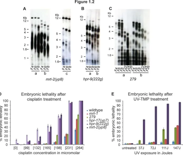
I crossed yp7 males to rol-1 unc-52 hermaphrodites and maintained yp7 animals as heterozygotes for several generationsto remove additional mutations that may lie in the yp7 background. I re-isolated several yp7 homozygote lines and analyzed them for characteristic features of hpr-17 mutants. yp7 animals exhibited defects in repairing DNA damage, illustrated as embryonic lethality, induced by interstrand crosslink (ICL) agents (Figure 1.2D) and ionizing
radiation (IR), consistent with the other previously identified mutant allele of hpr-17(tm1579) and mutant 9-1-1 complex components (Boerckel et al. 2007; Meier et al. 2009). Although I did not analyze the telomere dynamics of this mutant, it likely exhibits progressive telomere attrition
based on the similarities with the other mutant allele of hpr-17, including late onset end-to-end fusions, progressive drops in brood size, a Mortal Germline phenotype, a High incidence of males
(Him) phenotype (due to end-to-end fusion between the X chromosome and an autosome and the
resulting high frequency of mis-segregation of the X chromosome), and hypersensitivity to DNA
damaging agents.
Mapping and characterization of mrt-2(yp8)
Genome-wide SNP mapping was undertaken to obtain an approximate, initial position for
yp8. N2 Bristol males, homozygous for yp8, were crossed to CB4856 Hawaiian hermaphrodites, F1 heterozygotes were singled and allowed to self-fertilize, and 100 F2 progeny were passaged
for sterility (Figure 1.3A). All F2 lines were genotyped for the presence of the N2 Bristol vs.
19
Figure 1.2 Characterization of novel telomere maintenance-defective mutants. A-C)
21
Figure 1.3 A novel mutation that displayed hallmarks of telomerase deficiency, yp8, was
mapped to the hpr-17 gene on Chromosome III. A) N2 Bristol males, homozygous for yp8, were crossed to CB4856 Hawaiian hermaphrodites, F1 heterozygotes were singled and allowed to
self-fertilize, and 100 F2 lines were propagated weekly to assay for sterility. B) F2 lines were
genotyped for the presence of the N2 Bristol vs. CB4856 Hawaiian SNP at several loci on each
chromosome, as described in Davis et al. 2005. Genotyping of F2 lines that became progressively sterile, indicative of a homozygous yp8 genotype, placed yp8 in the middle of Chromosome III. C) yp8 males were crossed to unc-32 vab-7 hermaphrodites, and F1s were singled and allowed to self-fertilize. Twenty F2 Vab-non-Unc recombinants were propagated for sterility, which was
never attained. Therefore, yp8 must be to the right of vab-7. D) Because mrt-2, a gene previously identified to be required for telomere replication, is located at +12 map units on chromosome III,
we sequenced this gene in yp8 animals. A 365-nucleotidedeletion, starting with the fifth base pair of the first exon and ending after the second exon, was identified, likely precluding a functional
22
Because mrt-2, a gene previously identified to be required for telomerase-mediated telomere repeat addition (Ahmed & Hodgkin 2000), is located to the right of vab-7 at +12 map units on chromosome III, I sequenced this gene in yp8 animals. A 365-nucleotide-deletion, starting with the fifth base pair of the first exon and ending after the second exon, was identified,
likely precluding a functional protein product (Figure 1.3D).
We crossed yp8 males to dpy-18 unc-64 hermaphrodites and maintained yp8 animals as heterozygotes for several generationsto remove additional mutations that may lie in the yp8 background. I re-isolated several yp8 homozygote lines and analyzed them for characteristic features of mrt-2 mutants. To assess telomere length in yp8 animals, DNA was collected from several yp8 mutant lines during several weeks of growth and subjected to terminal restriction fragment analysis. Southern blotting revealed progressive telomere attrition for all yp8 lines (Figure 1.2A). Additionally, yp8 animals exhibited defects in repairing DNA damage, illustrated by scoring embryonic lethality after exposure to IR or ICL agents, consistent with the other
previously identified mutant allele of mrt-2(e2663) (Figure 1.2D & E) (Ahmed & Hodgkin 2000; Meier et al. 2009).
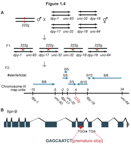
Mapping and characterization of hpr-9(222g)
Prior to my arrival to the laboratory, 222g was roughly mapped to the right arm of chromosome III using genome-wide linkage analysis as described for yp7. To refine this position, I constructed the following double mutants for three-factor mapping: dpy-1 unc-93,dpy-17 unc-32,unc-32 dpy-18, and dpy-18 unc-64. I crossed 222g males to dpy-1 unc-93 hermaphrodites, and Dpy-non-Unc F3 lines, generated analogously as previously described (Figure 1.1A), that did not
segregate Unc F4 progeny were propagated weekly to assay for progressive sterility.
23
and Unc-non-Dpy F3 lines were established analogously from crosses between 222g males and dpy-18 unc-64 hermaphrodites, and all were propagated for sterility (Figure 1.4A). The location of the 222g mutation, which was approximately at +4, was determined by calculating percent sterility. Because hpr-9, the Rad9 homolog component of the 9-1-1 complex, is located at +4 map units on chromosome III, I sequenced this gene in 222g animals. A premature opal stop (TGG to TGA) was identified in the 8th exon, which likely results in either a truncated or non-functional
protein product (Figure 1.4B).
I crossed 222g males to unc-32 vab-7 hermaphrodites and maintained 222g animals as heterozygotes for several generationsto remove additional mutations that may lie in the 222g background. I re-isolated several 222g homozygote lines and analyzed them for characteristic features of other 9-1-1 mutantcomponents. To assess whether telomeres in 222g also eroded progressively, DNA was collected from several 222g mutant lines during several weeks of growth and subjected to terminal restriction fragment length analysis. Southern blotting revealed
progressive telomere attrition for both 222g lines (Figure 1.2B). Additionally, 222g animals exhibited defects in repairing DNA damage, illustrated as embryonic lethality, after exposure to
IR or ICL agents, consistent with the phenotypes of mrt-2 and hpr-17 mutants (Figure 1.2D). Lastly, to confirm that the 222g mutation specifically was driving the onset of sterility, and not another mutation in a nearby gene, I performed complementation analysis with an independent
allele of hpr-9(ok2396). Both hpr-9(222g)/hpr-9(ok2396) trans-heterozygotes and 222g and ok2396 homozygous mutants were hyper-sensitive to IR. Because hpr-9(ok2396) did not complement 222g for IR hyper-sensitivity, we conclude that the 222g mutation, as opposed to independent mutations in adjacent genes, drives the telomere attrition and the resulting sterility.
24
Figure 1.4 A novel mutation that displayed hallmarks of telomerase deficiency, 222g, was
mapped to the hpr-9 gene on Chromosome III. A) 222g males were crossed to dpy-1 unc-93, unc-32 dpy-18, dpy-17 unc-32, and dpy-18 unc-64 hermaphrodites, F1 heterozygotes were singled and allowed to self-fertilize, and F2 lines were propagated weekly to assay for sterility. The
numbers of sterile animals from each cross indicate that 222g is located at approximately +4. map units. B) Because hpr-9, the Rad9 homolog component of the 9-1-1 complex, is located at +4 map units on chromosome III, we sequenced this gene in 222g animals. A premature opal stop (TGG to TGA) was identified in the 8th exon, which likely results in either a truncated or
25
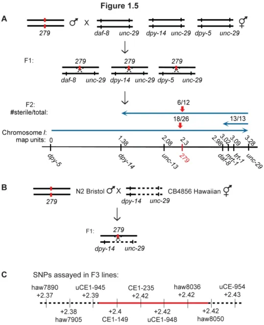
Mapping and characterization of 279Prior to my arrival to the laboratory, the 279 mutation was mapped to the right arm of chromosome I using genome-wide linkage analysis as previously described. To refine this position, I constructed the following double mutants for three-factor mapping: daf-8 unc-29, dpy-14 unc-29, and dpy-5 unc-29. Unc-non-Daf F3 lines were established analogously as previously described (Figure 1.1A) from crosses between 279 males and daf-8 unc-29 hermaphrodites, Unc-non-Dpy F3 lines were established analogously from crosses between 279 males and dpy-14 unc-29 hermaphrodites, Dpy-non-Unc F3 lines were established analogously from crosses between 279 males and dpy-5 unc-29 hermaphrodites, and all lines were propagated for sterility. The cumulative map positions, based on percent sterility, placed 279 at approximately +2.3 on chromosome I, which is close to but likely to the left of the mrt-1 and trt-1 genes (Figure 1.5A).
Although the Unc-non-Daf recombinant data suggested that 279 lies in a gene to the left of daf-8, it is possible that 279 may also reside in a gene extremely close to the right of daf-8. Additionally, because genetic mapping relies on recombination frequency, it may not always be
accurate if recombination is perturbed in the mapping area, thus distorting the analogy between
the genetic and physical map. Therefore, I wanted to assess whether 279 may be an allele of mrt-1 or trt-1, socomplementation analysis was performed with trt-1(ok410) and mrt-1(e2661). While 279, trt-1(ok410), and mrt-1(e2661) homozygous mutants became sterile after multiple
26
Figure 1.5 A novel mutation that displayed hallmarks of telomerase deficiency, 279, was
mapped to a candidate region of 29 genes on Chromosome I. A) 279 males were crossed to daf-8 unc-29, dpy-14 unc-29, and dpy-5 unc-29 hermaphrodites, F1 heterozygotes were singled and allowed to self-fertilize, and F2 lines were propagated weekly to assay for sterility. The
numbers of sterile animals from each cross indicate that 279 is located at approximately +2.3 map units. B) N2 Bristol males, homozygous for 279, were crossed to dpy-14 unc-29 hermaphrodites with intervening Hawaiian sequence, F1 heterozygotes were singled and allowed to self-fertilize,
and 30 Dpy-non-Unc recombinant lines were propagated weekly to assay for sterility. C) Nine
SNPs were genotyped in 30 lines, revealing a candidate region between +2.39 and +2.42 on
27
To further refine the position of 279, I employed three-factor mapping in combination with SNP mapping. Attempts at selecting Daf-non-Unc or Unc-non-Daf recombinants from
crosses between CB4856 Hawaiian males and unc-13 daf-8 hermaphrodites failed, perhaps because introduction of the Hawaiian sequence at that locus somehow interfered with the viability
of these strains or crossovers in this region. Instead, I crossed Hawaiian males with dpy-14 unc-13 hermaphrodites, and selected Dpy-non-Unc F2 recombinant progeny as previously described. Next, dpy-14 hermaphrodites (obligately harboring Hawaiian sequence to the right of unc-13) were crossed with unc-29/+ males, F1 progeny were singled and allowed to self-fertilize, and dpy-14 unc-29 recombinant progeny were selected analogously, as previously described, for genotyping to assess for the presence of Hawaiian sequence in between the two genes. N2 Bristol
males, homozygous for 279, were crossed to dpy-14 unc-29 hermaphrodites that harbored intervening CB4856 Hawaiian sequence, F1 progeny were singled and allowed to self-fertilize,
and the eventual Dpy F3 lines, selected for as previously described, that did not segregate Unc F4
progeny were propagated weekly to assay for progressive sterility (Figure 1.5B). Thirty F3 lines
were genotyped for the presence of the N2 Bristol vs. CB4856 Hawaiian SNP at each indicated
locus (Figure 1.5C). The genotyping revealed that 279 was located between the SNPs uCE1-945 (+2.39) and haw8050 (+2.42), a region that harbors the following 29 protein-coding and
non-coding genes: F55D12.1, F55D12.8 (ncRNA), F55D12.7 (miRNA), F55D12.2, F55D12.9
(ncRNA), F55D12.3 (nhr-191), F55D12.4 (unc-55), F55D12.6, F55D12.5, F20G4.2, F20G4.1 (smgl-1), F20G4.3 (nmy-2), T22C1.1, T22C1.2 (glb-26), T22C1.3, T22C1.4, T22C1.5, T22C1.6, T22C1.7 (jph-1), T22C1.15 (ncRNA), T22C1.14 (ncRNA), T22C1.13 (ncRNA), T22C1.8, T22C1.9, T22C1.12, T22C1.10 (rgb-2), T22C1.11, H05L14.1, and H05L14.2.
28
dynamics in 279 animals, DNA was collected from several 279 mutant lines during several weeks of growth and subjected to terminal restriction fragment length analysis. Southern blotting
revealed progressive telomere attrition for both 279 lines (Figure 1.2C). Additionally, 279 animals exhibited defects in repairing DNA damage, illustrated as embryonic lethality, after
exposure to ICL agents (Figure 1.2D & E). Curiously, 279 mutants were moderately
hypersensitive to ICL agents, similar to mrt-1 mutants that are more sensitive than wildtype but less sensitive than 9-1-1 components.
Discussion
I identify novel alleles of hpr-17(yp7) and mrt-2(yp8), which both display telomere defects that result in late onset end-to-end chromosomal fusions and sterility. Additionally, I
demonstrate for the first time that the C. elegans Rad9 homolog, hpr-9, is required for telomere repeat addition in vivo. Abrogating 9-1-1 complex components in mice is incompatible with life (Weiss et al. 2000; Hopkins et al. 2004), and their loss is not tolerated in culture (Francia et al. 2006; Hopkins et al. 2004), precluding analysis of telomere length dynamics. However, the progressive nature of telomere length defects in C. elegans 9-1-1 complex mutants allows for examination of the roles that these components play in telomere length maintenance over time. In
fact, TRF analysis of human cells deficient for Rad9 revealed normal-length telomeres (Pandita et al. 2006), whereas analysis of progressive growth of 9-1-1-deficient C. elegans clearly establishes a role for 9-1-1 components in promoting telomere maintenance.
Work with mammalian 9-1-1 suggests that its components may promote telomere
elongation through direct effects on telomerase. Mouse Hus1, Rad1, and Rad17 have been shown
29
identified 9-1-1 components in an exclusively human telomerase-negative cell line that maintains
telomeres via a telomerase-independent mechanism, termed alternative lengthening of telomeres
(ALT)
with telomeres only when they were associated with promyelocytic leukemia (PML) bodies,
which occurs exclusively in ALT cells (ALT-associated PML bodies or APBs) (Nabetani et al. 2004). Consistently, we have recently shown that 9-1-1 components are necessary for survival of
telomerase-deficient C. elegans lines (Cheng et al. 2012). These results suggest that while 9-1-1 components may be required for the recombination processes that drive ALT-mediated telomere
maintenance, similar 9-1-1-governed processes may be necessary to stimulate
telomerase-mediated telomere repeat addition in C. elegans and other multi-cellular organisms. More recently, 9-1-1 was shown to physically interact with WRN, a helicase with some roles in
telomere maintenance (Pichierri et al. 2011). Although the authors suggest that 9-1-1 cooperates with WRN at stalled replication forks, which have been proposed to occur at telomeres naturally
(reviewed in Maizels 2006), 9-1-1’s mechanism of action at telomeres remains unclear.
All of the new mrt mutants that displayed chromosomal fusions also experienced progressive telomere erosion, thus we can be confident that assaying for these fusions is an
accurate method for identifying telomere maintenance defects in mrt lines. However, this kind of selection excludes mutations that may result in short, but stable telomeres or progressively
shortening telomeres that are protected from end-to-end fusions. In fact, it is likely that additional
genes required for telomere maintenance in C. elegans remain unidentified. For example, the telomerase RNA component has yet to be identified.
A candidate gene approach is one way to tackle uncovering the identity of 279. Of the five predicted non-coding RNAs present in the candidate region, one (T22C1.13) harbors
complementary base pairs to the telomeric template: 3’-cggatt-5’, representing an excellent
candidate for the telomerase RNA. The length of the RNA, 186 nucleotides (nt), is within a
30
159 nt in Tetrahymena species, and 189-191 nt in Euplotes species. However, telomerase RNAs identified to date harbor more than one telomere repeat of complementary sequence, which
allows for primer re-alignment. A minimum of eight nucleotides are conserved throughout all
vertebrates, and more complementary sequences promote telomerase processivity, where one
telomere is elongated for more than one round of alignment (Chen & Greider 2003). Thus, this
non-coding RNA is not likely to code for the C. elegans telomerase RNA subunit. However, if sequencing this non-coding RNA reveals a mutation in 279 animals, expressing this gene with mutations in the predicted template would be an appropriate next step in assessing whether it
represents the telomerase RNA. Sequencing the telomeres from animals expressing a mutated
construct should reveal the aberrant sequence if it does indeed serve as the telomeric template.
Because we did not recover several mutations in the same gene, the conditions used in
this screen were likely not saturating. For example, approximately 7,000 F2 EMS-mutagenized
animals were passaged for up to 30 generations, where an EMS concentration that yields a per
gene mutation frequency of approximately 1 in 4,000 F2 mutagenized animals was used (Ahmed
& Hodgkin 2000). Passaging 7,000 animals provided almost a two-fold gene coverage, predicting
that a mutation in each gene would be recovered more than once in this screen. However, all of
the six telomere maintenance-defective mutants we identified represent distinct genes.
Additionally, mutations in genes that resulted in slower telomere attrition may have been missed
if those animals became sterile at much later generations (>30). Furthermore, while the aim of
this screen was to identify mutations that cause late-onset sterility, which is great for studying
telomere dynamics, genes that cause drastic telomere defects when mutant, resulting in more
sudden and rapid sterility, would have been missed using such an identification scheme.
In conclusion, identification of additional alleles of 9-1-1 components and its clamp
loader underscores the major role this complex plays in telomere maintenance. Moreover, our
31
mechanism(s) by which 9-1-1 components regulate telomere repeat addition might include the
following: 1) identification of binding partners using affinity purification of epitope-tagged 9-1-1
components, 2) analysis of in vivo localization via fluorescence microscopy using fluorescent protein-tagged 9-1-1 components, 3) analysis of changes in telomerase in vivo localization in 9-1-1 mutant vs. wildtype animals using fluorescent protein-tagged telomerase, and 4) analysis of in vitro telomerase activity from 9-1-1 mutant vs. wildtype animals. Additionally, if any of our mrt-1 or 9-1-1 mutant alleles cause telomere dysfunction by interfering with their binding or
localization to telomeres, fusing mutant MRT-1 or 9-1-1 proteins to wildtype POT-1 or POT-2,
known telomere-binding proteins, may rescue their mutant phenotypes. Similarly, if any of our
mrt-1 or 9-1-1 mutant alleles cause defects in recruitment of telomerase to telomeres, expressing a TRT-1::POT-1/2 fusion protein in mrt-1 or 9-1-1 mutant animals may overcome such a defect. In contrast, identification of additional genes that exacerbate or rescue the phenotypes of mutant
9-1-1 animals using forward genetic screens may be more effective or yield results distinct from
biochemistry. The following chapter describes biochemical efforts taken to understand how
CHAPTER 2:
Biochemical analysis of proteins that participate in telomere length homeostasis
In addition to proteins that participate in telomere length homeostasis through their
interaction with chromosome termini, the activity of the telomerase enzyme itself is regulated by
several factors. Analysis of the mechanisms involved in the formation, stabilization, and activity
of the telomerase holoenzyme is vital to dissecting telomerase function. A number of biochemical
approaches have been employed to identify components of the telomerase holoenzyme. In 1995, a
cofractionation approach identified the first two proteins to co-purify with telomerase activity in
Tetrahymena extracts—p80 and p95 (Collins et al. 1995). However, these proteins were later shown to be dispensable for telomere maintenance in vivo and could not precipitate telomerase or telomerase activity in vitro or in vivo (Miller & Collins 2000; Mason et al. 2001). In fact p80/p95 mutants exhibit telomere elongation instead (Miller & Collins 2000).
An oligonucleotide-based affinity purification of telomerase from Euplotes extracts successfully identified a 43-kilodalton protein, p43 (Lingner & Cech 1996). p43 directly interacts
with telomerase RNA in vivo and in vitro and has been suggested to promote the RNA stability based on its role in stabilizing other SNP complexes (Aigner et al. 2000). Additionally, affinity purification of epitope-tagged telomerase in Tetrahymena effectively identified three telomerase holoenzyme subunits, p45, p65, and p75 (Witkin & Collins 2004; Witkin et al. 2007). These proteins are required for viability, like the telomerase catalytic and RNA subunits in
33
Euplotes—both proteins harbor the La motif necessary for RNA binding (Witkin & Collins 2004; Aigner et al. 2004). In contrast, p20, which initially co-purified with p45, p65, and p75, did not co-immunoprecipitate telomerase activity or the RNA in later experiments, and p20 mutants actually exhibit long telomeres, a phenotype strikingly similar to the p80 and p95 proteins
purified a decade prior (Witkin et al. 2007; Collins et al. 1995).
In vertebrates, telomerase-mediated telomere extension in vivo requires not only the catalytic and RNA telomerase subunits, hTERT and hTR, but also several RNP assembly
proteins. Because hTR harbors an H/ACA domain observed in small nucleolar RNAs (snoRNAs),
dyskerin, one of several proteins that interacts with snoRNAs and is necessary for their
maturation, was assessed for interactions with hTR. Indeed, immunoprecipitation of dyskerin also
recovered co-transfected hTR from human cells (Mitchell et al. 1999). Furthermore, telomere repeat addition was completely abolished in cells from patients with mutated dyskerin, which had
been implicated in the disease Dyskeratosis Congenita a year prior (Heiss et al. 1998), revealing dyskerin’s crucial role in stabilizing telomerase (Mitchell et al. 1999).
Several previously known and novel proteins were identified through tandem affinity
purification of telomerase. However, the Sm proteins, which participate in stabilizing small
nuclear RNPs (snRNPs), and heterogeneous nuclear RNP (hnRNP) family RNA-binding proteins
were shown to be dispensable for telomere elongation (Fu & Collins 2007). These proteins, and
other identified chaperones, are proposed to perhaps bridge the interaction between a stable,
active telomerase and telomeres (reviewed in Collins 2006). Ultimately, the telomerase RNP is
comprised of hTERT, hTR, three hTR-stabilizing proteins, dyskerin, NHP2, and NOP10, and one
additional less abundant participant GAR1, which is required for RNP function but not its
stabilization (Fu & Collins 2007).
A strategy combining gradient sedimentation and dual-affinity purification of
epitope-tagged telomerase identified two novel players, ATPases pontin and reptin, which interact with