See discussions, stats, and author profiles for this publication at: https://www.researchgate.net/publication/316177386
Molecular Study of Cryptosporidium spp. in
Dogs from Southwest of Iran
Article
in
Jundishapur Journal of Microbiology · April 2017
DOI: 10.5812/jjm.43412 CITATIONS0
READS40
4 authors
, including:
Rahman Abdizadeh
Islamic Azad University, Shahrekord Branch
19PUBLICATIONS
147CITATIONS
SEE PROFILE
All content following this page was uploaded by
Rahman Abdizadeh
on 12 September 2017.
Published online 2017 April 17. Research Article
Molecular Study of
Cryptosporidium
spp. in Dogs from Southwest of
Iran
Mehdi Tavalla,
1,2,*Elham Kord,
2Rahman Abdizadeh,
3and Fatemeh Asgarian
21Infectious and Tropical Diseases Research Center, Ahvaz Jundishapur University of Medical Sciences, Ahvaz, IR Iran 2Department of Parasitology, School of Medicine, Ahvaz Jundishapur University of Medical Sciences, Ahvaz, IR Iran
3Department of Medical Parasitology and Mycology, Faculty of Medicine, Shahrekord University of Medical Sciences, Shahrekord, IR Iran
*Corresponding author: Mehdi Tavalla, Department of Medical Parasitology, Ahvaz Jundishapur University of Medical Sciences, P. O. Box: 61357-33118, Ahvaz, IR Iran. Tel: +98-6113367543, Fax: +98-6113367545, E-mail: [email protected]
Received2016 October 29;Revised2017 March 11;Accepted2017 April 05.
Abstract
Background:Cryptosporidiumis a protozoan parasite that effects rodents, dogs, calves, humans, and cats. Infection with this par-asite is known as cryptosporidiosis.Cryptosporidiumspp. may induce clinical or subclinical signs in infected hosts. In the life cycle of this parasite infected dogs freely living in urban and rural areas of Khuzestan province are the definitive hosts that should be considered as a real problem in public health for humans.
Objectives:This study aimed at determining the frequency of cryptosporidiosis in dogs in southwest of Iran.
Methods:Overall, 350 fresh fecal samples were collected from domestic dogs living in 43 villages, from June 2012 to September 2013. All samples were investigated by Sheather’s concentration method and fecal smears were stained with modified Ziehl-Neelsen followed by light microscope examination, and polymerase chain reaction (PCR).
Results: The results revealed that frequency ofCryptosporidiuminfection was 8% and 12.3%, using direct smear and molecular method, respectively.
Conclusions:The present findings indicated that domestic dog feces from southwest of Iran may contain zoonotic parasites such as Cryptosporidiumspp. and may be a potential risk for humans and other animals, especially when they contaminate the environment. The role of dogs as source of human infection should be investigated by further studies.
Keywords:Sheather’s, Modified Ziehl-Neelsen, PCR, Dogs, Iran,Cryptosporidiumspp
1. Background
Cryptosporidium is a widespread zoonotic intestinal protozoan parasite belonging to the phylum Apicomplexa that contains 30 species with more than 50 genotypes and infects a wide range of vertebrate animals, includ-ing mammalians, avians, amphibians, reptiles, and fish species as well as humans by the fecal-oral route via inges-tion of sporulated oocysts (1-3). Cryptosporidiumspp. in-fects the epithelial cells of the gastrointestinal tract (pri-marily small intestine and colon) of hosts and may induce clinical or subclinical signs, including vomiting, diarrhea, abdominal pain, fever, anemia, anorexia, dermatitis, and loss of weight, yet, occasionally, some infected hosts may present no symptoms (4,5).
Human cryptosporidiosis in immunocompetent indi-viduals usually causes acute infection of the digestive sys-tem and self-limiting diarrhea, yet, in immunocompro-mised patients, such as people infected with Human Im-munodeficiency Virus (HIV), people with malignancies, solid-organ transplants, and those on hemodialysis may suffer from severe diarrhea and dissemination to extra-intestinal sites, particularly the gall bladder, biliary tract,
pancreas, and respiratory tract (6,7).
Domestic dogs (Canis lupus familiaris) are generally considered as the first domesticated mammal from very early in human history (about 12,000 years ago) and are the most abundant species of carnivore around the world today, whoch are the definitive or reservoir hosts of more than 60 zoonotic parasites, such asCryptosporidiumspp. (8-10). Therefore, they are a real problem in public health for humans, particularly in villages and poorly marginal-ized communities of towns. There are 2 types of dogs in Iran including stray and owned dogs. Stray dogs often live freely in urban and rural areas, and the growing number of these animals in urban and rural residential areas of Iran and their easy access to public environments in order to obtain their nutritional needs from garbage may contami-nate soil, food, and water with discharge of helminths eggs and protozoan oocysts, and consequently an increase in parasitic infections in humans and animals. Furthermore, if owned dogs, including shepherd dogs, police dogs, gar-dener dogs, and pet dogs, are infected by parasites, they can infect occupational groups, such as shepherds, po-lice, gardeners and veterinarians or physicians. In 1983,
Tavalla M et al.
canine cryptosporidiosis was first reported by Fukushima and Helman (11) in a 3-month-old puppy, which was in-fected by Distemper disease.
Dogs become infected with the most common species/genotypes of Cryptosporidium spp., which are
responsible for human cryptosporidiosis, including
Cryptosporidium canis,C. parvum,C. muris,C. felis, andC. meleagridisby direct contact with infected animals (in-fected dogs and other animals such as ruminant, rodent, and/or ingestion of contaminated food or water from the environment) (12,13). Therefore, these animals are 1 of the major sources of cryptosporidiosis, which causes the spread of this protozoan in the environment.
Different methods are used for detection of cryp-tosporidiosis, which are generally based on analysis of stool samples for identification of oocysts using mi-croscopy with tinctorial and fluorescent stains (modi-fied acid-fast, safranin methylene blue, and auramine-rhodamine), antigen detection (immunofluorescence and enzyme-linked immunosorbent assay (ELISA) or genome detection (Polymerase chain reaction (PCR) amplification) in stool samples (14). In addition, serological assays are used for epidemiological studies because specific antibody responses develop after both symptomatic and asymp-tomatic infection, especially for immunocompromised in-dividuals (15), also considering the importance of zoonotic
Cryptosporidiumand the possibility of contamination of water and food with this parasite by infected animals.
2. Objectives
Due to the possible roles of dogs in parasite spreading rate, determination of frequency is necessary. Thus, the current study aimed at determining the frequency of cryp-tosporidiosis in dogs in southwest of Iran.
3. Methods 3.1. Study Area
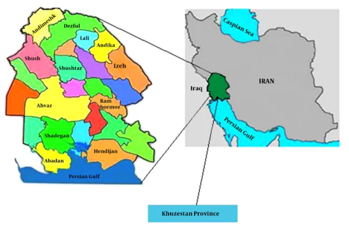
The study was undertaken in the city of Ahvaz, the cap-ital of Khuzestan Province, which covers approximately 63 238 km2and is located at 31° 3’ longitude north and 48° 7’
longitude east in southwest of Iran, bordering Iraq and the Persian Gulf (Figure 1). The climate of this area is generally hot and occasionally humid. Summer time temperatures exceed 52°C. This province is known to master the hottest temperatures on record for a populated city anywhere in the world (16).
Khuzestan is highlighted with green. Cities of this province are distinguished by colors. The map of Khuzes-tan province by Uwe Dering was highlighted by Dr. Blofeld.
3.2. Fecal Samples Collection
Villages of Khuzestan province were classified to 5 ar-eas: east, west, north, south, and center. Then, 350 fresh fecal samples were collected from domestic dogs with dif-ferent ages, according to their teeth, and were grouped in 3 groups, including puppies (< 1-year-old), young dogs (1 to 5 year-old), and old dogs (> 5-year-old), who were living in 43 villages (eas t = 8, west = 9, north = 9, south = 8, and center = 9 villages) from June 2012 to September 2013. In each geo-graphical area, 70 fresh samples were collected from house dogs (owned dogs) and then they were placed in polyethy-lene bags, marked according to area and were separately carried to the laboratory and kept at 4°C, until processing
3.3. Fecal Examination
All samples were concentrated by sucrose flotation procedure (Sheather’s method, with a specific gravity of 1.21), and thin smears of the concentrated layer of sam-ples were then prepared on glass slides, air-dried, and fixed with methanol, and stained by modified Ziehl-Neelsen and investigated by a light microscope. Each slide was accessed at 1000×magnification under oil emersion, and Cryp-tosporidiumspp. was confirmed using morphological char-acteristic of oocysts. The positive samples were preserved in 2.5% potassium dichromate (K2Cr2O7) and stored at 4°C
until DNA extraction.
3.4. DNA Extraction
Approximately 200µL of concentrated oocysts of each sample was added to a 2.0-mL eppendorf tube. The sam-ples were pretreated by the freeze and thaw method by liq-uid nitrogen to break down the oocyst walls. Briefly, tubes were placed in liquid nitrogen for 15 minutes, and were then transferred to 100°C water bath for another 5 min-utes. These steps were repeated for a total of 5 times. Next, the genomic DNA was purified using the AccuPrep® Ge-nomic DNA Extraction Kit (Bioneer, Korea), according to the manufacturer’s instruction. DNA was eluted in 100µL of elution buffer and stored at -20°C.
3.5. Polymerase Chain Reaction Amplification
The PCR protocol, based on the
amplifica-tion of a specific sequence of the SSU rRNA gene,
was used to detect Cryptosporidium by primers
CryF: (50-CTGACCTATCAGCTTTAGA- 30) and CryR: (50 -GCTGAAGGAGTAAGGAACA- 30), which produced a piece of DNA with a molecular weight of 720 bp (17). In order to per-form the PCR reaction, AccuPower® PCR PreMix(Bioneer, Korea) was used, including Taq polymerase enzyme, dNTP, MgCL2, reaction buffer, and tracking dye. In this step, 15µL
of deionized distilled water, 2.20µL of extracted genomic
Figure 1.Map of Iran and Khuzestan Province
DNA (100 ng), and 1µL of forward and reverse primers at 25 pmol were applied in a total volume of 25µL. The PCR condition was as follows: predenaturation at 94°C for 4 minutes; denaturation at 94°C for 1 minute, annealing at 52°C for 1 minute and extension at 72°C for 1 minute, followed by 30 cycles; final extension at 72°C for 5 minutes in a thermal cycler (Bio-Rad, Hercules, CA, US). The PCR product was analyzed by electrophoresis on 2% agarose gel in 1X TBE buffer and visualized using ethidium bromide staining on UV transilluminator.
4. Results
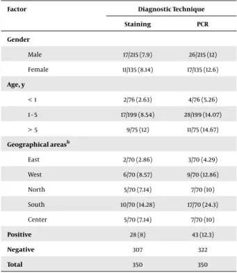
The frequency ofCryptosporidiuminfection in dogs by PCR was 12.3% (95% CI: 8.86% to 15.75%). Also the frequency ofCryptosporidiuminfection in dogs using staining and molecular methods were 8% (28/350) and 12.3% (43/350), respectively (Figure 2). In this study a comparison was made between gender, age, and geographical areas of dogs to assess the existence ofCryptosporidiumspp. Investiga-tion results indicated that the frequency of Cryptosporid-iumspp. infection in stools collected from villages in dif-ferent geographical areas of Khuzestan province
includ-ing east, west, north, south, and center were 4.28%, 12.85%, 11.42%, 24.28%, and 10%, respectively. The statistical analy-sis showed a significant relationship between geographi-cal areas and frequency ofCryptosporidiumspp. in dogs (P < 0.05). The results showed that there was no statistically significant difference among gender and age groups as in-dicated by staining and PCR methods for diagnosis of Cryp-tosporidiumin females and males (P > 0.05) (Table 1).
5. Discussion
Cryptosporidiosis is a zoonotic protozoal disease, which is reported in animals and humans with world-wide distribution in more than 106 countries, especially in developing countries (18,19), and may cause gastroin-testinal problems such as diarrhea in immunocompro-mised and immunocompetent people and even the en-vironment (5). Molecular epidemiological investigations strongly suggest that zoonotic species and genotypes of
Cryptosporidiumplay an important role in cryptosporidio-sis and were mentioned as a risk factor for human cryp-tosporidiosis (20). A single oocyst is sufficient to produce
Tavalla M et al.
Figure 2.Polymerase Chain Reaction ofCryptosporidiumspp. in Dog Feces Samples
M, 100 bp molecular marker; C.P, positive control; C.N, negative control; 1 - 7 lines, positive samples; line 8, negative sample.
Table 1. Frequency ofCryptosporidiumspp. in Different Regions of Khuzestan Provincea
Factor Diagnostic Technique
Staining PCR Gender Male 17/215 (7.9) 26/215 (12) Female 11/135 (8.14) 17/135 (12.6) Age, y < 1 2/76 (2.63) 4/76 (5.26) 1 - 5 17/199 (8.54) 28/199 (14.07) > 5 9/75 (12) 11/75 (14.67) Geographical areasb East 2/70 (2.86) 3/70 (4.29) West 6/70 (8.57) 9/70 (12.86) North 5/70 (7.14) 7/70 (10) South 10/70 (14.28) 17/70 (24.3) Center 5/70 (7.14) 7/70 (10) Positive 28 (8) 43 (12.3) Negative 307 322 Total 350 350
aValues are expressed as No. (%).
bThe frequency ofCryptosporidiumspp. in dogs indicated a statistically signifi-cant relationship with geographical areas, according to diagnostic techniques (P < 0.05).
infection and disease in susceptible hosts (21). Dogs are of-ten considered faithful friends and intimate companions of humans from very early in human history that can act as definitive or reservoir hosts for a large number of zoono-sis parasitic diseases of parasitic zoonoses, such asTaenia
sp.,Echinococcussp.,Toxocara canis,Giardiaspp., and
Cryp-tosporidiumspp. (22). Dogs are vertebrate animals that are infected withCryptosporidiumspp. in the wildlife and represent a potentially significant source of environmen-tal contamination and reservoir of the disease for domes-tic livestock and humans, due to transmission of the infec-tion through close contact with infected dogs (21).
Epidemiological studies onCryptosporidiuminfection indicated that the prevalence ofCryptosporidiumspp. in dogs is very different in various countries (from 0% to 52.7%) (23-26); a prevalence of 1.4% in the Czech Republic (27), 2.1% in Thailand (28), 2.4% in Brazil (29), 3.9% in Japan (30), 4.1% in Northern Spain (31), 18.5% in Nigeria (32), and 52.7% in Romania. This difference depends on factors, such as geographical location, the number of dogs, status of an-imals ownership, existence and number of other hosts cor-related with dogs, including domestic animals (such as cat-tle, horses, sheep, goats, and pigs), species of Cryptosporid-ium, sampling protocols, anthelmintic use, and diagnostic techniques (33,34).
Infected dogs with cryptosporidiosis shed oocysts with their feces, which can contaminate environment. Cryp-tosporidiumspp. oocysts are resistant to harsh environ-mental conditions and can be well preserved under cold and wet environments. In addition, these are very resistant to the most common disinfectants, therefore, can contam-inate water, and there is a potential risk for areas with a large dog population (35-37). Prevalence ofCryptosporidium
spp. in geographical regions of Khuzestan province, south-west of Iran, is variable and it seems that the prevalence of this protozoa in south of the province is higher than other areas (38-40).
The current findings may be due to the following rea-sons; southern provinces have a high temperature and hu-mid weather conditions, life style of people regarding con-sumption of seafood more than other regions, and birds immigration to south of Khuzestan province, which may be carriers of infection. In Iran, people who live in villages because of lifestyle and closely related agricultural and an-imal husbandry sources are exposed to zoonotic pathogen-esis microorganisms, such as parasitic zoonoses. There-fore, the potential for zoonotic transmission from domes-tic animals such as dogs, that are reservoirs via environ-mental contamination, is of increasing concern.
In the current study,Cryptosporidiumspp. oocysts were identified in 8% (28/350) and 12.3% (43/350) of samples ex-amined using staining and molecular methods, respec-tively. This rate is more than the results of some previous studies, which were carried out about cryptosporidiosis in dogs of different areas of Iran. Bahrami et al. (41) re-ported 7.04% infection in stray dogs of Ilam using the Ziehl-Neelsen staining method. In another study conducted in the Southeast of Iran (Kerman), prevalence of
iumspp. was 2% using the formalin ether sedimentation and modified Ziehl-Neelsen staining technique. In addi-tion, Gharekhani (42) reported that 3.8% of the infections among pet dogs in Hamedan in Western Iran were infected withCryptosporidiumspp. by formalin-ether and modified Ziehl-Neelsen technique.
The study of prevalence of gastrointestinal parasites of pet dogs in Tehran (Central Iran) and Urmia (Northwest of Iran) indicated that 1.6% and 2.9% of animals were infected withCryptosporidiumspp., respectively (43,44). In another study, Mosallanejad et al. (45) investigated the prevalence ofC. parvumin urban and rural dogs of Ahvaz district by us-ing antigenic detection and modified Ziehl-Neelsen stain-ing methods, the results of which indicated 4.3% and 2.5% of dogs were infected, as indicated by ELISA and staining methods. Also, the infection had greater prevalence in ru-ral dogs (6.4%) in comparison with urban dogs (2.17%). The results were in agreement with other studies in different parts of the world, such as Bahía Blanca of Argentina that indicated 14.7% of dogs were infected withCryptosporidium
spp. (9). The results of the current study showed that the frequency ofCryptosporidiumspp. in feces of dogs in vil-lages of Ahvaz was high.
Despite the results of the current study, in some re-searches the frequency ofCryptosporidiuminfection was higher in young dogs (21, 31, 33, 46). Some studies in comparison to the current study indicated that infection rates in female dogs were higher than male dogs, which may be due to reduced immunity at certain periods in fe-male physiologic cycle (32). Other investigations in Iran indicated that female dogs had more infection than male dogs. Bahrami et al. and Gharekhani reported that the prevalence ofCryptosporidiumin female dogs of Ilam and Hamedan were more than male dogs (41,42). Also, Mirzaei showed that the prevalence ofCryptosporidiumin female dogs was higher than male dogs in Kerman (34). Although the role of infected dogs in transmission of Cryptosporid-iuminfection to humans is not exactly clear, yet,C. canis
can be infect immunocompromised patients. Also, a small number of zoonoticCryptosporidiumincludingC. parvum,
C. muris, andC. meleagridiscan infect dogs (12).
The free entrance of stray dogs to public places of vil-lages and existence of owned dogs, such as shepherd dogs in houses has caused defecation in different areas of vil-lages and may contaminate soil, food or water. Therefore, it can be a potential hazard for humans and domestic an-imals and this phenomenon is important in public health and livestock husbandry.
In conclusion, control programs including, public ed-ucational activities, regarding risk of parasite transmis-sion, and role of animals such as dogs in parasite distribu-tion should be considered. It is suggested to conduct
con-trol programs, including education for people about cryp-tosporidiosis and the potential transmission of this proto-zoan to humans and animals, prevention of free entrance of stray dogs in public places and houses, also collection and hygienic disposal of dogs feces. In addition, determin-ing the frequency and treatdetermin-ing cryptosporidiosis in owner dogs should be done by veterinarians or physicians.
References
1. Ryan U, Fayer R, Xiao L. Cryptosporidium species in humans and animals: current understanding and research needs. Parasitol-ogy. 2014;141(13):1667–85. doi:10.1017/S0031182014001085. [PubMed:
25111501].
2. Slapeta J. Cryptosporidiosis and Cryptosporidium species in animals and humans: a thirty colour rainbow?.Int J Parasitol. 2013;43 (12-13):957–70. doi:10.1016/j.ijpara.2013.07.005. [PubMed:23973380]. 3. Xiao L, Feng Y. Zoonotic cryptosporidiosis.FEMS Immunol Med
Micro-biol.2008;52(3):309–23. doi:10.1111/j.1574-695X.2008.00377.x. 4. Guerrant RL. Cryptosporidiosis: an emerging, highly infectious
threat.Emerg Infect Dis. 1997;3(1):51–7. doi: 10.3201/eid0301.970106. [PubMed:9126444].
5. Rossle NF, Latif B. Cryptosporidiosis as threatening health problem: A review.Asian Pac J Trop Biomed.2013;3(11):916–24. doi: 10.1016/s2221-1691(13)60179-3.
6. Current WL, Garcia LS. Cryptosporidiosis. Clin Microbiol Rev.
1991;4(3):325–58. [PubMed:1889046].
7. Hunter PR, Hadfield SJ, Wilkinson D, Lake IR, Harrison FC, Chalmers RM. Subtypes of Cryptosporidium parvum in humans and disease risk.Emerg Infect Dis. 2007;13(1):82–8. doi: 10.3201/eid1301.060481. [PubMed:17370519].
8. Awoke E, Bogale B, Chanie M. Intestinal nematode parasites of dogs: Prevalence and associated risk factors.Int J Anim Vet Adv.2011;3(5):374– 8.
9. La Sala LF, Leiboff A, Burgos JM, Costamagna SR. Spatial distribu-tion of canine zoonotic enteroparasites in Bahia Blanca, Argentina.
Rev Argent Microbiol. 2015;47(1):17–24. doi: 10.1016/j.ram.2014.12.006. [PubMed:25705047].
10. Perera PK, Rajapakse R, Rajakaruna RS. Gastrointestinal parasites of dogs in Hantana area in the Kandy District.J Nat Sci Foundat Sri Lanka.
2013;41(2) doi:10.4038/jnsfsr.v41i2.5703.
11. Fukushima K, Helman RG. Cryptosporidiosis in a pup with distem-per.Vet Pathol. 1984;21(2):247–8. doi: 10.1177/030098588402100218. [PubMed:6730208].
12. Caccio SM, Widmer G. Cryptosporidium: parasite and disease. Springer Science & Business Media; 2013.
13. Xiao L. Molecular epidemiology of cryptosporidiosis: an update.
Exp Parasitol. 2010;124(1):80–9. doi: 10.1016/j.exppara.2009.03.018. [PubMed:19358845].
14. Fayer R, Morgan U, Upton SJ. Epidemiology of Cryptosporidium: transmission, detection and identification.Int J Parasitol.2000;30 (12-13):1305–22. [PubMed:11113257].
15. Lopez-Urbina MT, Gonzalez AE, Gomez-Puerta LA, Romero-Arbizu MA, Perales-Camacho RA, Rojo-Vazquez FA, et al. Prevalence of Neonatal Cryptosporidiosis in Andean Alpacas (Vicugna pacos) in Peru.Open Parasitol J.2009;3(1):9–13. doi:10.2174/1874421400903010009. 16. Wikipedia . Khuzestan Province 2014. Available from: http://en.
wikipedia.org/wiki.
17. Rai AK, Chakravorty R, Paul J. Detection of Giardia, Entamoeba, and Cryptosporidium in unprocessed food items from northern India.
World J Microbiol Biotechnol. 2008;24(12):2879–87. doi: 10.1007/s11274-008-9824-1.
Tavalla M et al.
18. Dillingham RA, Lima AA, Guerrant RL. Cryptosporidiosis: epidemi-ology and impact.Microbes Infect. 2002;4(10):1059–66. [PubMed:
12191656].
19. Thompson A. Review of "Cryptosporidium and cryptosporidiosis" by Ronald Fayer and Lihua Xiao (eds.).Parasit Vectors. 2008;1(1):47. doi:
10.1186/1756-3305-1-47.
20. Xiao L, Fayer R. Molecular characterisation of species and geno-types of Cryptosporidium and Giardia and assessment of zoonotic transmission. International J Parasitol. 2008;38(11):1239–55. doi:
10.1016/j.ijpara.2008.03.006.
21. Ramirez NE, Ward LA, Sreevatsan S. A review of the biology and epi-demiology of cryptosporidiosis in humans and animals.Microb Infect.
2004;6(8):773–85. doi:10.1016/j.micinf.2004.02.021.
22. Jian F, Qi M, He X, Wang R, Zhang S, Dong H, et al. Occurrence and molecular characterization of Cryptosporidium in dogs in Henan Province, China.BMC Vet Res.2014;10:26. doi:10.1186/1746-6148-10-26. [PubMed:24433398].
23. Batchelor DJ, Tzannes S, Graham PA, Wastling JM, Pinchbeck GL, German AJ. Detection of endoparasites with zoonotic potential in dogs with gastrointestinal disease in the UK. Transbound Emerg Dis.2008;55(2):99–104. doi:10.1111/j.1865-1682.2007.01005.x. [PubMed:
18397497].
24. Hackett T, Lappin MR. Prevalence of enteric pathogens in dogs of north-central Colorado.J Am Anim Hosp Assoc. 2003;39(1):52–6. doi:
10.5326/0390052. [PubMed:12549614].
25. Katagiri S, Oliveira-Sequeira TC. Prevalence of dog intestinal para-sites and risk perception of zoonotic infection by dog owners in Sao Paulo State, Brazil.Zoonoses Public Health.2008;55(8-10):406–13. doi:
10.1111/j.1863-2378.2008.01163.x. [PubMed:18811905].
26. Simpson JW, Burnie AG, Miles RS, Scott JL, Lindsay DI. Prevalence of Giardia and Cryptosporidium infection in dogs in Edinburgh.Vet Rec.
1988;123(17):445. [PubMed:3201688].
27. Dubna S, Langrova I, Napravnik J, Jankovska I, Vadlejch J, Pekar S, et al. The prevalence of intestinal parasites in dogs from Prague, rural areas, and shelters of the Czech Republic.Vet Parasitol. 2007;145 (1-2):120–8. doi:10.1016/j.vetpar.2006.11.006. [PubMed:17169492]. 28. Koompapong K, Mori H, Thammasonthijarern N, Prasertbun R,
Pin-tong AR, Popruk S, et al. Molecular identification of Cryptosporid-ium spp. in seagulls, pigeons, dogs, and cats in Thailand.Parasite.
2014;21:52. doi:10.1051/parasite/2014053. [PubMed:25297887]. 29. Huber F, Bomfim TCB, Gomes RS. Comparison between natural
infec-tion by Cryptosporidium sp., Giardia sp. in dogs in two living situa-tions in the West Zone of the municipality of Rio de Janeiro.Vet Para-sitol.2005;130(1-2):69–72. doi:10.1016/j.vetpar.2005.03.012.
30. Yoshiuchi R, Matsubayashi M, Kimata I, Furuya M, Tani H, Sasai K. Sur-vey and molecular characterization of Cryptosporidium and Giardia spp. in owned companion animal, dogs and cats, in Japan.Vet Para-sitol.2010;174(3-4):313–6. doi:10.1016/j.vetpar.2010.09.004.
31. Gil H, Cano L, de Lucio A, Bailo B, de Mingo MH, Cardona GA, et al. Detection and molecular diversity of Giardia duodenalis and
Cryp-tosporidium spp. in sheltered dogs and cats in Northern Spain.Infect Genet Evol.2017;50:62–9. doi:10.1016/j.meegid.2017.02.013. [PubMed:
28219812].
32. Olabanji GM, Maikai BV, Otolorin GR. Prevalence and Risk Factors As-sociated with Faecal Shedding ofCryptosporidiumOocysts in Dogs in the Federal Capital Territory, Abuja, Nigeria.Vet Med Int.2016;2016:1– 6. doi:10.1155/2016/4591238.
33. Titilincu A, Mircean V, Achelaritei D, Cozma V. Prevalence of Cryp-tosporidium spp. in asymptomatic dogs by ELISA and risk fac-tors associated with infection.Lucrari Stiinłifice Medicina Veterinara.
2010;43(1).
34. Mirzaei M. Epidemiological survey of Cryptosporidium spp. in com-panion and stray dogs in Kerman, Iran.Vet Ital. 2012;48(3):291–6. [PubMed:23038075].
35. Campbell I, Tzipori A, Hutchison G, Angus K. Effect of disinfectants on survival of cryptosporidium oocysts.Vet Record.1982;111(18):414–5. doi:10.1136/vr.111.18.414.
36. Dorny P, Praet N, Deckers N, Gabriel S. Emerging food-borne parasites.
Vet Parasitol.2009;163(3):196–206. doi:10.1016/j.vetpar.2009.05.026. 37. Fayer R. Cryptosporidium: a water-borne zoonotic parasite.Vet
Para-sitol.2004;126(1-2):37–56. doi:10.1016/j.vetpar.2004.09.004. 38. Hoghooghi-Rad N. Some epidemiological aspects of
cryptosporidio-sis in ahwaz, capital of khoozestan province, islamic republic of Iran.
Med J Islam Repub Iran.1994;8(1):17–22.
39. Rafiei A, Rahdar M, Valipour Nourozi R. Isolation and Identification of Parasitic Protozoa in Sampled Water From the Southwest of Iran.
Jundishapur J Health Sci.2014;6(4) doi:10.5812/jjhs.23462.
40. Rafiei A, Rashno Z, Samarbafzadeh A, Khademvatan S. Molecu-lar Characterization of Cryptosporidium spp. Isolated From Im-munocompromised Patients and Children.Jundishapur J Microbiol.
2014;7(4) doi:10.5812/jjm.9183.
41. Bahrami A, Doosti A, Nahravanian H, Noorian AM, Asbchin SA. Epi-demiological survey of gastro-intestinal parasites in stray dogs and cats.Australian J Basic App Sci.2011;5(9).
42. Gharekhani J. Study on Gastrointestinal Zoonotic Parasites in Pet Dogs in Western Iran.Turk J Parasitol. 2014;38(3):172–6. doi:
10.5152/tpd.2014.3546.
43. Dalimiasl A, Mojarad KS, Jamshidi S. Intestinal parasites of pet dogs in Tehran and evaluation of knowledge of dog owners about zoonotic risk of parasites of dog.J Vet Res.2001;56:6–13.
44. Tavassoli M, Javadi S, Soltanalinejad F, Rosouli S, Etminanfar R. Gas-trointestinal parasites of pet dogs in Urmia city.Vet J.2010. 45. Mosallanejad B, Hamidinejat H, Avizeh R, Ghorbanpoor Najafabadi
M, Razi Jalali MH. Antigenic detection of Cryptosporidium parvum in urban and rural dogs in Ahvaz district, outhwestern Iran.Iran J Vet Res.
2010;11(3):273–8.
46. Hamnes IS, Gjerde BK, Robertson LJ. A longitudinal study on the occurrence of Cryptosporidium and Giardia in dogs during their first year of life.Acta Veterinaria Scandinavica. 2007;49(1):22. doi:
10.1186/1751-0147-49-22.
6 Jundishapur J Microbiol. 2017; 10(4):e43412.
View publication stats View publication stats