MINI-REVIEW
The Boggarts of Biology: How non-genetic changes influence the genotype
Laasya Samhita
National Centre for Biological Sciences, Tata Institute of Fundamental Research, Bangalore, India
Correspondence:
Laasya Samhita ([email protected], [email protected])
Abstract
The notion that there is a one to one mapping from genotype to phenotype was overturned a long time ago. It is now well established that besides the genetic background, environmental inputs guide the development of phenotype. In addition, altered RNA and protein molecules also influence phenotype in conjunction with the external environment, leading to ‘non-genetic’ changes. The phenotypic variation we see across all living organisms therefore results from a combination of genetic and non-genetic changes. In spite of the prevalent idea that variability arising from non-genetic changes is transient and does not influence
evolution, recent work has shown that it can impact both short and long-term adaptation. In this review, I propose that one way in which non-genetic inputs can affect evolution is by indirectly influencing genetic change. I classify and review the many ways in which non-genetic changes influence genotype and impact cellular fitness across generations, with an emphasis on the enticing idea that non-genetic changes act as stepping stones for genetic adaptation. Overall, I review how non-genetic changes impact phenotype via their influence on the genotype, and thus can play a role in evolutionary change.
Keywords
Non-genetic change, translation errors, phenotypic variability, adaptation, evolution
Introduction
The saying ‘The more things change, the more they stay the same’ aptly describes the constant activity required to maintain the status quo within biological systems. However, living systems are complex, and function with the help of multiple error prone chemical processes. They also innovate, adapt and respond to environmental stimuli, creating a foundation for phenotypic variation to emerge. In fact, heritable phenotypic variability, i.e, phenotypic changes that are faithfully passed on from generation to generation, lies at the heart of Darwinian evolution. Typically, heritable variation stems from differences in the genetic material (usually DNA) of the organism. In turn, these differences manifest as phenotypic variation via RNA and protein transactions within the cell. However, unlike DNA, for the most part, RNA and protein molecules cannot be used as templates to generate more copies of themselves (For exceptions, see True and Lindquist 2000; de Farias et al. 2017). Changes in these molecules are therefore typically short lived, often dictated by changes in the external environment; and thought to have a limited impact across generations (reviewed in Bonduriansky and Day 2009; Jablonka and Raz 2009; Charlesworth et al. 2017). Thus, any phenotypic variation that alters cellular fitness in the long term might be expected to stem only from changes in the genetic material.
In contrast to this thought, a large body of work has shown that non-DNA based phenotypic alterations can both impact fitness, and be inherited (reviewed in Bonduriansky and Day 2009; Ribas de Pouplana et al. 2014; Ackermann 2015). Many terms have been coined to refer to non-DNA based changes, and ‘epigenetic changes’ such as DNA and histone modifications and RNA editing in particular have been extensively reviewed (Gott and Emeson 2000; Jablonka and Raz 2009; Charlesworth et al. 2017). For the purpose of this article and to avoid confusion, I define ‘non-genetic changes’ as changes that impact phenotype without altering the primary DNA sequence.
often considered separately, and their impact on phenotypic variation is also viewed through this lens. In this review, I discuss the various ways in which non-genetic changes can influence the genotype and therefore evolution, with a focus on molecular mechanisms. I begin with a brief background, go on to classify examples, and finally highlight some open questions.
Background
Non-genetic changes can impact phenotype in multiple ways
Boggarts are mythical creatures popularized by J.K Rowling. They are shape-shifters with a temporary existence. Like them, non-genetic changes too come in various flavours, and are characterized by rapid response to a new stimulus (reviewed in Fox et al. 2019) (Tyedmers et al. 2008; Tadrowski et al. 2018). They can also help organisms navigate changing
environments via ‘phenotypic switching’, i.e, by switching to one of two or more possible stable phenotypes within the same genotype (reviewed in Fox et al. 2019; Stajic and Bank 2020). From a molecular point of view, quantitative or qualitative differences in RNA and protein molecules (leading to altered transcription and translation) underlie nearly all non-genetic changes that alter phenotype. In general, such changes could either be triggered by variation in the external environment or be independent of the external environment (reviewed in Vogt 2020).
and stop codon readthrough can affect expression of the genotype (Farabaugh 1996; Ivanova et al. 2014; Fan et al. 2017). Recently, a rare +1 frameshift mutation in the essential rpoB
gene (encoding RNA polymerase) in E. coli was identified in a screen for resistance to the antibiotic rifampicin (Huseby et al. 2020). Surprisingly, although they showed no additional mutations, mutant cells continued to retain viability (for which functional RNA polymerase is essential) as well as rifampicin resistance. Further investigation revealed that theframeshift mutation was suppressed by a second phenotypic frameshift downstream ~5% of the time. This non-genetic change restored the original reading frame with just one amino acid change. In addition, translation was upregulated in response to RpoB depletion, resulting in ~70% functional protein being formed (Huseby et al. 2020), and minimising the cost of antibiotic resistance.
Therefore, non-genetic changes can be either induced by the environment or independent of the environment; they can arise from stochastic processes or be part of a stable gene
regulatory network. Overall, non-genetic changes can influence phenotype via multiple routes (reviewed in Ackermann 2015; Ling et al. 2015; Stajic and Bank 2020).
Limits to the impact of non-genetic changes: Heritability and Penetrance
(i) Lack of heritability: As discussed already, many non-genetic changes occur in conjunction with an environmental change, and may not last once the environment changes again. This is often seen as a limit to their long-term impact. However, experiments over the years have uncovered heritable non-genetic changes across model systems, reducing the strength of this argument (See Box 1).
(ii) Low penetrance of the phenotype: Non-genetic changes may mediate phenotypic change in a sub-population of cells, or alter a sub-population of intracellular RNA or protein in all cells. For instance, stable transcriptional regulation (guided by environmental cues) ensures 100% penetrance of one of two phenotypes in clonal cells. A Bacillus subtilis cell is either a dormant endospore or an actively growing cell (Tan and Ramamurthi 2014), Caulobacter crescentus cells are either stalked or swarmers (Tsokos and Laub 2012). On the other hand, phenotypic changes that arise from errors in transcription, translation and protein folding are unpredictable, and the altered molecules form only a small percentage of the total population (Drummond and Wilke 2009). In turn, any phenotypic change associated with such an altered molecule will have much less visibility than a mutation that leads to the same change.
Although this remains a factor in considering the impact of non-genetic changes, experiments have shown that even seemingly small contributions from altered tRNAs and proteins can impact phenotype and short-term adaptation. For example, 5% of altered RNA polymerase molecules are sufficient to confer resistance to low amounts of rifampicin (Javid et al. 2014); a ~1% misacylation of tRNAs is sufficient for E. coli cells to show a growth advantage under oxidative stress as compared with unmodified cells (Schwartz et al. 2016). Therefore, experimental evidence suggests that low penetrance need not be a barrier for non-genetic changes to impact phenotype and fitness. In addition, the many examples discussed later in this article strengthen this view.
Ways in which non-genetic changes can lead to genetic change
2). In the section below, I categorise three ways in which non-genetic changes can impact the genotype (Fig. 3), and discuss evidence for each with a focus on the molecular mechanisms. I review specific examples within each category where non-genetic change either precedes or succeeds genetic change in shaping phenotype. Finally, I discuss the limited experimental evidence available for phenotypic changes that act as stepping stones for genetic change. I review this in light of our recent finding that mistranslation increases early survival under DNA damage in E. coli cells (non-genetic change) followed by beneficial mutations in the gene gyrA (genetic change) (Samhita et al. 2020a). Overall, I present evidence suggesting that non-genetic changes can have a significant impact on evolution via their impact on the
genotype.
Box 1
Direct inheritance: The simplest way for non-genetic change to be perpetuated is to directly pass on from one generation to the next, much like DNA does (Fig. 1). There are two ways in which this can happen. One, where the non-genetic change is generated or established afresh every generation from a template (Fig. 1a). Second, where the same factor is propagated directly and diluted over time (Fig. 1b).
recognition between fungal hyphae (Glass and Kaneko 2003), where genetically different nuclei are prevented from co-existing in one cytoplasm). The prion ensures that vegetative mixing cannot happen, a result that is also thought to keep the spread of cytoplasmic
infections at bay (Debets et al. 2012). There is no genetic basis for this change, it is passed on as a dominant trait through the cytoplasm.
A second way to carry over a non-genetic change into the next generation is through direct transfer without regeneration (Fig. 1b). This occurs when the factor causing the non-genetic change (often altered protein) remains stable across generations. Such a factor could either be diluted at every cell division and be inherited by both daughter cells, or be asymmetrically inherited by only one of two daughter cells. By tracking fluorescently tagged LacY proteins, Lambert and Kussell found that E. coli cells retain a ‘phenotypic memory’ of past exposure to lactose by retaining stable LacY from cytoplasmically inherited protein for ~10 generations (Lambert and Kussell 2014). In turn, this reduces the time taken to re-start exponential growth (lag phase) when cells are moved from glucose to lactose as the carbon source. In contrast to even distribution among daughter cells, protein aggregates that arise from heat shock are asymmetrically inherited by one daughter cell for several generations in E. coli
Figure 1
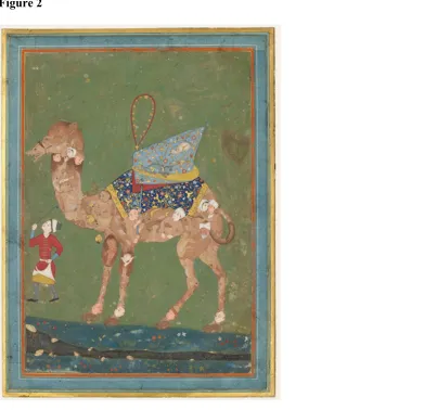
Figure 2
.
Phenotype is a composite of genetic and non-genetic changes: The figure shows a Persian watercolour sketch of a composite camel (~1500 A.D). Several animals (analogous to many non-genetic changes) form the body of the camel and co-exist within it, each with their own thoughts and impulses. The camel’s body provides a broad shape and structure within which these exist (analogous to the genotype). External factors such as the man (environment) guide and can change the direction in which the overall body moves. Image source: Online
Figure 3
Non-genetic changes influence genetic change: (Summary) Altered phenotypes can lead to altered genotypes in various ways (a-d)Four ways in which non-genetic changes can impact the genotype are shown here. A non-genetic change is represented by a change in phenotype while the genotype remains the same (a) Non-genetic changes can expose pre-existing
Non-genetic change influences the manifestation of genetic change
Non-genetic changes can expose, suppress or enhance already existing genetic variation in a population (Fig. 3a). Variation in the untranslated 3’ region of genes can be exposed by stop codon read-through during translation and also impact the evolution of protein coding genes (Kosinski and Masel 2020). The yeast prion [PSI +] provides a good example of an altered protein that uncovers already existing genetic variation. [PSI +] is an alternately folded version of the translation termination protein eRF3. eRF3 normally promotes the disassembly of translation termination complexes after a protein chain is terminated at a stop codon. In prion plus cells, eRF3 is sequestered into prion aggregates, thereby leading to a loss of function phenotype and increased stop codon read-through (Chernoff et al. 1993; True and Lindquist 2000) . Extended proteins are made across cellular mRNAs, exposing cryptic genetic variation in normally untranslated regions and leading to diverse phenotypes across different carbon sources and antibiotics (True et al. 2004) .
Classic experiments by Waddington demonstrated the power of non-genetic change in controlling the penetrance of genetic variation (Waddington 1942). Waddington exposed
Drosophila melanogaster to heat shock or ether (neither is mutagenic), and showed that exposure to these stimuli led to two effects: (a) They generated new phenotypes
(crossveinless and bithorax-like, respectively) and (b) These phenotypes became
independent of the stimulus after ~20 generations of repeated exposure to heat or ether and selection for crossveinless and bithorax-like flies, respectively (reviewed in Crispo 2007). Many years later, experiments with heterozygous D. melanogaster mutants of the gene HSP90 encoding a chaperone recapitulated the same findings under heat shock, leading to the hypothesis that differences in HSP90 may underlie Waddington’s observations (reviewed in Zabinsky et al. 2019).
client proteins (signal transducers, transcription factors and members of multi protein complexes) that are degraded, misfolded or alternately folded (Rutherford and Lindquist 1998; Queitsch et al. 2002), and expose phenotypes associated with previously buffered mutations. All of these outcomes can lead to the manifestation of otherwise hidden (cryptic) genetic variation.
For example, the influence of Hsp90 on the transcription factor Ste12 was studied using S. cerevisiae as a model system. Ste12 regulates the choice between mating and invasion of host tissue in many fungi (Dorrity et al. 2018); it is not an Hsp90 client protein. However, several mutant variants of this factor are chaperoned by Hsp90 under normal conditions in S. cerevisiae, masking the mutant phenotype. When exposed to high temperature, Hsp90 is unable to maintain the buffering. Cells carrying the Ste12 variants lose the ability to mate and demonstrate a dominant hyperinvasive phenotype, unmasking the cryptic variation (Dorrity et al. 2018). Conversely, Hsp90 can also allow new mutations to have immediate phenotypic consequences. The tyrosine kinase c-Src interacts poorly with Hsp90. However, a mutated oncogenic variant of this protein called v-Src is stabilized by Hsp90, activating its
promiscuous kinase activity and capacity for oncogenesis (Boczek et al. 2015).
Other chaperones such as GroEL in bacteria have also shown similar though not quite such far ranging effects (Sabater-Munoz et al. 2015), suggesting that chaperones in general are good candidates to act as a conduit between genotype, environment and phenotype. Non-genetic change impacts mutation rate and identity
Altered transcription and translation can indirectly impact mutation rates via the generation of novel transcripts and proteins, and interestingly, even by altering the degree and timing of gene expression. In particular, several examples of translation errors affecting the nature of mutations have surfaced in the past decade. In one example, genes evolved in a translation error prone background showed different sets of mutations as compared with those evolved in a wild type background. Bratulic and co-workers propagated a plasmid borne antibiotic resistance gene (TEM-1 beta lactamase) through multiple rounds of mutation and selection in the bacterium E. coli, selecting for resistance against a different class of antibiotic
increased cost of such mutations in an error prone background, and selection on destabilizing protein sequences (Bratulic et al. 2015).
Translation errors can also impact global mutation rate, both via an overall increase in mistranslation (Krisko and Radman 2013) and by specific amino acid changes (Humayun 1998; Bacher and Schimmel 2007). In general, mistranslated proteins tend to misfold and aggregate (Drummond and Wilke 2008), and so are typically unable to carry out their normal cellular functions efficiently. When such dysfunctional proteins are involved in DNA
replication and repair, they can introduce mutations. Studies carried out over 20 years ago isolated an E. coli mutant carrying a mutation in the anticodon of a glycine tRNA gene, resulting in increased substitutions of glycine in place of aspartate in cellular proteins. Such cells also showed an increase in mutagenesis (Slupska et al. 1996). Recently, it was found that DNA polymerase III (Pol III) isolated from these cells showed significantly higher error prone replication in vitro as compared with those isolated from WT cells (Al Mamun et al. 2006). Given that Pol III is the chief replicating polymerase in bacteria and also responsible for correction of mismatches post replication (reviewed in Sutton and Walker 2001), it is reasonable to hypothesize that heterogeneity in DNA Pol III sequence contributed to the increased mutagenesis.
In addition to acting via altered proteins, mistranslation can trigger stress responses and in turn briefly elevate the basal mutation rate. For example, global mistranslation (Samhita et al. 2020a) as well as mistranslation induced by a defective alanyl tRNA synthetase (Bacher and Schimmel 2007) trigger a DNA damage response (SOS response) which can be mutagenic (Baharoglu and Mazel 2014). Overall, both mutation rate and identity can be impacted in multiple ways by non-genetic changes (Fig. 3b) such as translation errors.
fastest, with the rapid appearance and spread of severalmutants where URA3 expression was abrogated. In addition, both M and L lines showed mutations in genes other than URA3, whereas the H lines only showed resistant mutations in URA3. While the mutation rates remained unchanged across the three lines, the kinds of mutations sampled and the rate of adaptation were clearly influenced by the degree of gene expression. This work also helped to disentangle the genetic from non-genetic contributions in a given phenotype, something that has been a challenge for experimentalists.
Therefore, both mutation rates and specific mutational paths can be influenced by non-genetic changes (Fig. 3b).
Non-genetic changes as stepping stones to genetic adaptation
Perhaps the most exciting aspect of non-genetic changes has been the speculation that when beneficial, they may ‘buy time’ for genetic change, thus linking short term adaptation to long term evolutionary change (Fig. 3c and Fig. 4). The hypothesis has been laid out in several forms over the years, but experimental evidence remains extremely limited. A specific case of this situation where the beneficial non-genetic change is induced by the environment, was proposed by Baldwin (Baldwin 1896). He postulated that an organism acquired adaptive phenotypic changes as a consequence of its interaction with the environment. With time, ‘heritable characters’ that produced the same changes would be favoured by natural selection and spread in the population (reviewed in Crispo 2007). Multiple theoretical models have shown that this can happen, both with environmentally induced change and with environment independent changes such as alterations in RNA and protein sequence or protein folding (Hinton and Nowlan 1987; Whitehead et al. 2008; Bonduriansky and Day 2009; Klironomos et al. 2013).
evidence for the second step (second beneficial mutation) has been elusive for some time. However, recent work with antibiotic tolerant cells offers one instance where a beneficial phenotype is achieved through two independently beneficial mutations; where the first can potentially be substituted for, or enhanced by, a phenotypic change. Liu and co-workers examined clinical samples of Staphylococcus aureus from patients under treatment in a multi drug regime (Liu et al. 2020). They found that, as per previous predictions and observations (Levin-Reisman et al. 2017) , mutations leading to antibiotic tolerance (slower killing of the population) preceded the appearance of resistant mutants. In addition, the presence of mutations that conferred tolerance (first beneficial mutation) enhanced resistance in the multi-drug regime, when compared with cells that only carried resistance mutations (second beneficial mutation). Tolerance can also be achieved by non-genetic means (Levin and Rozen 2006; Cohen et al. 2013); however, the current study mapped all tolerant phenotypes to mutations. The pattern of antibiotic resistance evolution was also duplicated in laboratory experiments (Liu et al. 2020), suggesting that the evolution of antibiotic resistance in S. aureus and other bacteria might occur through this two-step process.
Klironomos et al modelled a population where both genetic and non-genetic changes occur independently (Klironomos et al. 2013). Non-genetic changes were assigned a higher rate of appearance than genetic change (mutations). In addition, non-genetic changes could revert to the original state at a given probability. The authors found that populations with options for both kinds of change adapted faster than those that relied only on mutations, a finding that is broadly supported by both earlier (Hinton and Nowlan 1987; Behera and Nanjundiah 2004) and later work (Kronholm et al. 2017). In addition, because non-genetic changes are fast, they rapidly lead to a fitness peak. In such populations, non-genetic changes do ‘buy time’ for adaptive genetic change, however, mutations can accumulate neutrally in the meantime. This leads to increased standing genetic variation; another way in which non-genetic changes can indirectly impact the supply of genetic variants. The model also highlights the fact that current mutations observed in populations could well have been preceded at one time by non-genetic change, creating a stepping stone to the current phenotype.
yeast strains such as Ashbya gossypii carries possible signals for peroxisomal targeting beyond the stop codon, but they are in a +1 translational reading frame, and therefore cryptic. When the A. gossypii IDP2 was expressed in S. cerevisiae, ~30% of the total protein product could be detected in the peroxisome. Further, a protein with size corresponding to the frameshifted product was detected by mass spectrometry, showing that frameshifting must have taken place and made the peroxisomal targeting signal effective. The authors also mutagenized another ancestral IDP2 (from Kluyveromyces waltii) in the region around the stop codon, followed by selection on petroselinate containing medium (peroxisomal IDP is essential for growth on petroselinate). This led to the rapid selection of mutant proteins carrying genetic single base deletions (one per mutant) that brought the peroxisomal targeting signal in frame. Therefore, the ability to generate an alternate protein product may have served as a stepping stone for mutations that fixed novel cellular localization for this protein (c). However, although this is a novel finding that highlights the role of translational errors in adaptation, it still involves some speculation about past evolution. That is, it falls short of a demonstration that adaptive non-genetic change can be a precursor to genetic change. Recently, we showed that generalized (non-directed) mistranslation can increase survival when E. coli cells are treated with the DNA damaging antibiotic ciprofloxacin (Samhita et al. 2020a). Irrespective of the non-genetic route by which basal mistranslation in E. coli is elevated, there is a common consequence: the level of the protease Lon, a key player in degrading misfolded proteins, also goes up. Increased Lon leads to an increase in the levels of RecA, a protein that is essential for DNA repair and recombination. As a consequence, when faced with DNA damage from ciprofloxacin (cip), mistranslating cells are already closer to the threshold for activation of the DNA repair response (the SOS response) than wild-type (WT) cells that retain a basal (high) translational fidelity and constitute the control
(resistance to ciprofloxacin), and the same phenotype is subsequently fixed by genetic change (Fig. 4c). Interestingly, reducing the basal level of mistranslation in the wild-type
concomitantly lowered its resistance to ciprofloxacin, suggesting that translation error and DNA damage repair remain relevant even in the wild-type under normal conditions, and perhaps giving a broader context to the stepping stone effect.
Among the ways in which a non-genetic change induced by stress can act as a stepping stone, two broad mechanisms deserve attention. They are (a) an elevated population size, which increases the number of cells within which a favourable mutation can take place, and (b) an elevated mutation rate, which increases the probability per cell of a favourable mutation taking place. Experiments and modelling work with persister cells (metabolically inactive non-dividing cells that survive antibiotic treatment) in E. coli suggests that both methods may be at play in moving from persistence to antibiotic resistance (Windels et al. 2019). Persisters could be a pre-existing part of the population (Fig. 4c) (Cohen et al. 2013), or be generated by exposure to the antibiotic itself (Fig. 4b) (Dorr et al. 2010). Persister numbers are strongly correlated with the number of resistant cells that arise later; and genetic mutants that show a constitutively high level of persistence also show a higher mutation rate (Windels et al. 2019), which may help in the generation and establishment of resistant mutations. However, more work is needed to establish a direct link here from an adaptive phenotype, to genotypic change.
The idea that short lived phenotypic changes can pave the way for specific genetic change is conceptually different from the other ways in which non-genetic changes can impact the genotype (discussed above: uncovering cryptic change and altering mutation supply or identity). In some ways, it is the simplest category, merely requiring a phenotypic change that can provide a short-term survival benefit. Mutations will then occur over time irrespective of the nature of the change as long as the population has a reservoir of viable cells; and some of these mutations will be beneficial. Why then do we not see more examples of such
case can be made for non-genetic changes to act as stepping stones for genetic adaptation (Fig. 4).
Figure 4
Perspective
From the examples reviewed above, it is clear that non-genetic changes can alter mutational trajectories, uncover cryptic genetic diversity, and influence the direction of future genetic change. By doing so, they gain the power to affect long term adaptation and evolution. The categories discussed here are by no means comprehensive. For instance, some
microorganisms show deviations from the universal genetic code (Ling et al. 2015), and decode the same codon differently depending on environmental cues (Prat et al. 2012). Altered decoding changes the codon to amino acid mapping, but can also go on to influence genotypic change. Jing Ma and Isaacs (Ma and Isaacs 2016) found that a ‘recoded’ E. coli
strain in which the standard stop codon UAG was re-assigned to UAA (Lajoie et al. 2013), was resistant to several viruses. A reversal of this genome wide recoding from UAA back to UAG restored viral infectivity. Interestingly, after just five days of propagation in recoded E. coli, bacteriophage MS2 regained the ability to infect the strain via mutations in two genes; one mutation altered its own UAG stop codon to UAA, while the other created a new premature stop codon in lieu of UAG (Ma and Isaacs 2016). In addition, the recoded E. coli
strain was unable to take up conjugative plasmids, potentially altering future genotypic change through horizontal gene transfer. Therefore, re-assignment of even one stop codon to another stop codon (UAG to UAA) can have consequences on host genotype as well as the genotype of co-evolving organisms.
in this article, in recent years, translation errors have emerged as important contributors to altered phenotype and adaptation. DNA remains the primary code of life. Like with all codes though, the key that decodes it can conceal or reveal it to different degrees. How much of the genetic change that we measure today was preceded by non-genetic changes? Do cells employ errors in translation and transcription as strategies to generate variation? Have cells evolved to rely more on genetic change in some environments and more on non-genetic changes in others? These and several other open questions remain, as experiments broaden our knowledge of non-genetic changes.
ACKNOWLEDGEMENTS
I thank Vidyanand Nanjundiah, Deepa Agashe, Sunil Laxman, Asha Joseph and members of Deepa’s lab for critical comments and suggestions on this review. I acknowledge Ipsa Jain for figures 1, 3 and 4. I acknowledge funding and support from the DBT/Wellcome trust India Alliance (grant IA/E/14/1/501771), and support from the National Centre for Biological Sciences.
REFERENCES
Ackermann M. 2015. A functional perspective on phenotypic heterogeneity in microorganisms. Nat Rev Microbiol 13:497-508.
Al Mamun AAM, Gautam S, Humayun MZ. 2006. Hypermutagenesis in mutA cells is mediated by mistranslational corruption of polymerase, and is accompanied by replication fork collapse. Molecular Microbiology 62:1752-1763.
Bacher JM, Schimmel P. 2007. An editing-defective aminoacyl-tRNA synthetase is
mutagenic in aging bacteria via the SOS response. Proc Natl Acad Sci U S A 104:1907-1912. Baharoglu Z, Mazel D. 2014. SOS, the formidable strategy of bacteria against aggressions. FEMS Microbiol Rev 38:1126-1145.
Baldwin MJ. 1896. A New Factor in Evolution. The American Naturalist 30:441-451. Behera N, Nanjundiah V. 2004. Phenotypic plasticity can potentiate rapid evolutionary change. J Theor Biol 226:177-184.
Boczek EE, Reefschlager LG, Dehling M, Struller TJ, Hausler E, Seidl A, Kaila VR, Buchner J. 2015. Conformational processing of oncogenic v-Src kinase by the molecular chaperone Hsp90. Proc Natl Acad Sci U S A 112:E3189-3198.
Bodi Z, Farkas Z, Nevozhay D, Kalapis D, Lazar V, Csorgo B, Nyerges A, Szamecz B, Fekete G, Papp B, et al. 2017. Phenotypic heterogeneity promotes adaptive evolution. PLoS Biol 15:e2000644.
Bonduriansky R, Day T. 2009. Nongenetic Inheritance and Its Evolutionary Implications. Annual Review of Ecology, Evolution, and Systematics 40:103-125.
Bratulic S, Gerber F, Wagner A. 2015. Mistranslation drives the evolution of robustness in TEM-1 beta-lactamase. Proc Natl Acad Sci U S A 112:12758-12763.
Bullwinkle TJ, Reynolds NM, Raina M, Moghal A, Matsa E, Rajkovic A, Kayadibi H, Fazlollahi F, Ryan C, Howitz N, et al. 2014. Oxidation of cellular amino acid pools leads to cytotoxic mistranslation of the genetic code. Elife 3.
Carey JN, Mettert EL, Roggiani M, Myers KS, Kiley PJ, Goulian M. 2018. Regulated Stochasticity in a Bacterial Signaling Network Permits Tolerance to a Rapid Environmental Change. Cell 173:196-207 e114.
Charlesworth D, Barton NH, Charlesworth B. 2017. The sources of adaptive variation. Proc Biol Sci 284.
Chernoff YO, Derkach IL, Inge-Vechtomov SG. 1993. Multicopy SUP35 gene induces de-novo appearance of psi-like factors in the yeast Saccharomyces cerevisiae. Curr Genet 24:268-270.
Cohen NR, Lobritz MA, Collins JJ. 2013. Microbial persistence and the road to drug resistance. Cell Host Microbe 13:632-642.
Crispo E. 2007. The Baldwin effect and genetic assimilation: revisiting two mechanisms of evolutionary change mediated by phenotypic plasticity. Evolution 61:2469-2479.
de Farias ST, Dos Santos Junior AP, Rego TG, Jose MV. 2017. Origin and Evolution of RNA-Dependent RNA Polymerase. Front Genet 8:125.
Debets AJ, Dalstra HJ, Slakhorst M, Koopmanschap B, Hoekstra RF, Saupe SJ. 2012. High natural prevalence of a fungal prion. Proc Natl Acad Sci U S A 109:10432-10437.
Dorr T, Vulic M, Lewis K. 2010. Ciprofloxacin causes persister formation by inducing the TisB toxin in Escherichia coli. PLoS Biol 8:e1000317.
Dorrity MW, Cuperus JT, Carlisle JA, Fields S, Queitsch C. 2018. Preferences in a trait decision determined by transcription factor variants. Proc Natl Acad Sci U S A 115:E7997-E8006.
Drummond DA, Wilke CO. 2009. The evolutionary consequences of erroneous protein synthesis. Nat Rev Genet 10:715-724.
Drummond DA, Wilke CO. 2008. Mistranslation-induced protein misfolding as a dominant constraint on coding-sequence evolution. Cell 134:341-352.
Fan Y, Evans CR, Barber KW, Banerjee K, Weiss KJ, Margolin W, Igoshin OA, Rinehart J, Ling J. 2017. Heterogeneity of Stop Codon Readthrough in Single Bacterial Cells and Implications for Population Fitness. Mol Cell 67:826-836 e825.
Fan Y, Wu J, Ung MH, De Lay N, Cheng C, Ling J. 2015. Protein mistranslation protects bacteria against oxidative stress. Nucleic Acids Res 43:1740-1748.
Farabaugh PJ. 1996. Programmed translational frameshifting. Microbiol Rev 60:103-134. Fox RJ, Donelson JM, Schunter C, Ravasi T, Gaitan-Espitia JD. 2019. Beyond buying time: the role of plasticity in phenotypic adaptation to rapid environmental change. Philos Trans R Soc Lond B Biol Sci 374:20180174.
Glass NL, Kaneko I. 2003. Fatal attraction: nonself recognition and heterokaryon incompatibility in filamentous fungi. Eukaryot Cell 2:1-8.
Goldsmith M, Tawfik DS. 2009. Potential role of phenotypic mutations in the evolution of protein expression and stability. Proc Natl Acad Sci U S A 106:6197-6202.
Gott JM, Emeson RB. 2000. Functions and mechanisms of RNA editing. Annu Rev Genet 34:499-531.
Gout JF, Thomas WK, Smith Z, Okamoto K, Lynch M. 2013. Large-scale detection of in vivo transcription errors. Proc Natl Acad Sci U S A 110:18584-18589.
Hinton GE, Nowlan SJ. 1987. How Learning Can Guide Evolution. Complex Systems 1:495-502.
Humayun MZ. 1998. SOS and Mayday: multiple inducible mutagenic pathways in Escherichia coli. Mol Microbiol 30:905-910.
Huseby DL, Brandis G, Praski Alzrigat L, Hughes D. 2020. Antibiotic resistance by high-level intrinsic suppression of a frameshift mutation in an essential gene. Proc Natl Acad Sci U S A 117:3185-3191.
Ivanova NN, Schwientek P, Tripp HJ, Rinke C, Pati A, Huntemann M, Visel A, Woyke T, Kyrpides NC, Rubin EM. 2014. Stop codon reassignments in the wild. Science 344:909-913. Jablonka E, Raz G. 2009. Transgenerational epigenetic inheritance: prevalence, mechanisms, and implications for the study of heredity and evolution. Q Rev Biol 84:131-176.
Javid B, Sorrentino F, Toosky M, Zheng W, Pinkham JT, Jain N, Pan M, Deighan P, Rubin EJ. 2014. Mycobacterial mistranslation is necessary and sufficient for rifampicin phenotypic resistance. Proc Natl Acad Sci U S A 111:1132-1137.
Klironomos FD, Berg J, Collins S. 2013. How epigenetic mutations can affect genetic evolution: model and mechanism. Bioessays 35:571-578.
Klosin A, Casas E, Hidalgo-Carcedo C, Vavouri T, Lehner B. 2017. Transgenerational transmission of environmental information in C. elegans. Science 356:320-323. Kosinski LJ, Masel J. 2020. Readthrough Errors Purge Deleterious Cryptic Sequences, Facilitating the Birth of Coding Sequences. Mol Biol Evol 37:1761-1774.
Kramer EB, Farabaugh PJ. 2007. The frequency of translational misreading errors in E. coli is largely determined by tRNA competition. RNA 13:87-96.
Krishna S, Laxman S. 2020. Phenotypic switching: Implications in Biology and Medicine. In: Levine H, Jolly MK, Kulkarni P, Nanjundiah V, editors: Academic Press. p. 335-358.
Krisko A, Radman M. 2013. Phenotypic and genetic consequences of protein damage. PLoS Genet 9:e1003810.
Kronholm I, Bassett A, Baulcombe D, Collins S. 2017. Epigenetic and Genetic Contributions to Adaptation in Chlamydomonas. Mol Biol Evol 34:2285-2306.
Lajoie MJ, Rovner AJ, Goodman DB, Aerni HR, Haimovich AD, Kuznetsov G, Mercer JA, Wang HH, Carr PA, Mosberg JA, et al. 2013. Genomically recoded organisms expand biological functions. Science 342:357-360.
Lambert G, Kussell E. 2014. Memory and fitness optimization of bacteria under fluctuating environments. PLoS Genet 10:e1004556.
Levin-Reisman I, Ronin I, Gefen O, Braniss I, Shoresh N, Balaban NQ. 2017. Antibiotic tolerance facilitates the evolution of resistance. Science 355:826-830.
Levin BR, Rozen DE. 2006. Non-inherited antibiotic resistance. Nat Rev Microbiol 4:556-562.
Ling J, O'Donoghue P, Soll D. 2015. Genetic code flexibility in microorganisms: novel mechanisms and impact on physiology. Nat Rev Microbiol 13:707-721.
Liu J, Gefen O, Ronin I, Bar-Meir M, Balaban NQ. 2020. Effect of tolerance on the evolution of antibiotic resistance under drug combinations. Science 367:200-204.
Ma NJ, Isaacs FJ. 2016. Genomic Recoding Broadly Obstructs the Propagation of Horizontally Transferred Genetic Elements. Cell Syst 3:199-207.
Moazed D. 2011. Mechanisms for the inheritance of chromatin states. Cell 146:510-518. Mordret E, Yehonadav A, Barnabas GD, Cox J, Dahan O, Geiger T, Lindner AB, Pilpel Y. 2018. Systematic detection of amino acid substitutions in proteome reveals a mechanistic basis of ribosome errors. bioRxiv.
Prat L, Heinemann IU, Aerni HR, Rinehart J, O'Donoghue P, Soll D. 2012. Carbon source-dependent expansion of the genetic code in bacteria. Proc Natl Acad Sci U S A 109:21070-21075.
Prusiner SB. 1982. Novel proteinaceous infectious particles cause scrapie. Science 216:136-144.
Queitsch C, Sangster TA, Lindquist S. 2002. Hsp90 as a capacitor of phenotypic variation. Nature 417:618-624.
Ribas de Pouplana L, Santos MA, Zhu JH, Farabaugh PJ, Javid B. 2014. Protein mistranslation: friend or foe? Trends Biochem Sci 39:355-362.
Rutherford SL, Lindquist S. 1998. Hsp90 as a capacitor for morphological evolution. Nature 396:336-342.
Sabater-Munoz B, Prats-Escriche M, Montagud-Martinez R, Lopez-Cerdan A, Toft C, Aguilar-Rodriguez J, Wagner A, Fares MA. 2015. Fitness Trade-Offs Determine the Role of the Molecular Chaperonin GroEL in Buffering Mutations. Mol Biol Evol 32:2681-2693. Samhita L, Raval PK, Agashe D. 2020a. Global mistranslation increases cell survival under stress in Escherichia coli. PLoS Genet 16:e1008654.
Samhita L, Raval PK, Stephenson G, Thutupalli S, Agashe D. 2020b. The impact of mistranslation on phenotypic variability and fitness. bioRxiv.
Schwartz MH, Waldbauer JR, Zhang L, Pan T. 2016. Global tRNA misacylation induced by anaerobiosis and antibiotic exposure broadly increases stress resistance in Escherichia coli. Nucleic Acids Res.
Slupska MM, Baikalov C, Lloyd R, Miller JH. 1996. Mutator tRNAs are encoded by the Escherichia coli mutator genes mutA and mutC: a novel pathway for mutagenesis. Proc Natl Acad Sci U S A 93:4380-4385.
Stajic D, Bank C. 2020. Phenotypic switching: Implications in Biology and Medicine. In: Levine H, Jolly MK, Kulkarni P, Nanjundiah V, editors: Academic Press. p. 281-301. Stajic D, Perfeito L, Jansen LET. 2019. Epigenetic gene silencing alters the mechanisms and rate of evolutionary adaptation. Nat Ecol Evol 3:491-498.
Sutton MD, Walker GC. 2001. Managing DNA polymerases: coordinating DNA replication, DNA repair, and DNA recombination. Proc Natl Acad Sci U S A 98:8342-8349.
Tadrowski AC, Evans MR, Waclaw B. 2018. Phenotypic Switching Can Speed up Microbial Evolution. Sci Rep 8:8941.
Tan IS, Ramamurthi KS. 2014. Spore formation in Bacillus subtilis. Environ Microbiol Rep 6:212-225.
True HL, Berlin I, Lindquist SL. 2004. Epigenetic regulation of translation reveals hidden genetic variation to produce complex traits. Nature 431:184-187.
True HL, Lindquist SL. 2000. A yeast prion provides a mechanism for genetic variation and phenotypic diversity. Nature 407:477-483.
Tsokos CG, Laub MT. 2012. Polarity and cell fate asymmetry in Caulobacter crescentus. Curr Opin Microbiol 15:744-750.
Tuite F, Mick. 2020. Phenotypic switching: Implications in Biology and Medicine. In: Levine H, Jolly MK, Kulkarni P, Nanjundiah V, editors: Academic Press. p. 105-123.
Tyedmers J, Madariaga ML, Lindquist S. 2008. Prion switching in response to environmental stress. PLoS Biol 6:e294.
Vogt G. 2020. Phenotypic switching: Implications in Biology and Medicine. In: Levine H, Jolly MK, Kulkarni P, Nanjundiah V, editors: Academic Press. p. 207-242.
Waddington CH. 1942. Canalization of development and the inheritance of acquired characters. Nature 160:563-565.
Windels EM, Michiels JE, Fauvart M, Wenseleers T, Van den Bergh B, Michiels J. 2019. Bacterial persistence promotes the evolution of antibiotic resistance by increasing survival and mutation rates. ISME J 13:1239-1251.
Yanagida H, Gispan A, Kadouri N, Rozen S, Sharon M, Barkai N, Tawfik DS. 2015. The Evolutionary Potential of Phenotypic Mutations. PLoS Genet 11:e1005445.