R E V I E W
Open Access
Regulation of urinary bladder function by
protein kinase C in physiology and
pathophysiology
Joseph A. Hypolite and Anna P. Malykhina
*Abstract
Background:Protein kinase C (PKC) is expressed in many tissues and organs including the urinary bladder,
however, its role in bladder physiology and pathophysiology is still evolving. The aim of this review was to evaluate available evidence on the involvement of PKC in regulation of detrusor contractility, muscle tone of the bladder wall, spontaneous contractile activity and bladder function under physiological and pathophysiological conditions. Methods:This is a non-systematic review of the published literature which summarizes the available animal and human data on the role of PKC signaling in the urinary bladder under different physiological and
pathophysiological conditions. A wide PubMed search was performed including the combination of the following keywords:“urinary bladder”,“PKC”,“detrusor contractility”,“bladder smooth muscle”,“detrusor relaxation”,“peak force”,“detrusor underactivity”,“partial bladder outlet obstruction”,“voltage-gated channels”,“bladder nerves”,“PKC inhibitors”,“PKC activators”. Retrieved articles were individually screened for the relevance to the topic of this review with 91 citations being selected and included in the data analysis.
Discussion:Urinary bladder function includes the ability to store urine at low intravesical pressure followed by a subsequent release of bladder contents due to a rapid phasic contraction that is maintained long enough to ensure complete emptying. This review summarizes the current concepts regarding the potential contribution of PKC to contractility, physiological voiding, and related signaling mechanisms involved in the control of both the storage and emptying phases of the micturition cycle, and in dysfunctional voiding. Previous studies linked PKC activation exclusively with an increase in generation of the peak force of smooth muscle contraction, and
maximum force generation in the lower urinary tract. More recent data suggests that PKC presents a broader range of effects on urinary bladder function including regulation of storage, emptying, excitability of the detrusor, and bladder innervation.
Summary:In this review, we evaluated the mechanisms of peripheral and local regulation of PKC signaling in the urinary bladder, and their impact on different phases of the micturition cycle under physiological and
pathophysiological conditions.
Keywords:Detrusor smooth muscle, Micturition, Nerves, Contractility, Signaling pathways, Bladder pathophysiology
* Correspondence:[email protected]
Division of Urology, Department of Surgery, University of Colorado Denver, Anschutz Medical Campus, 12700 E 19th Ave. Mail Stop C317, Aurora, CO 80045, USA
Background
Physiological contractions of the detrusor smooth muscle (DSM) involve the ability to generate and maintain contractile force during the emptying phase of the micturition cycle [1–13]. Initial animal studies suggested that PKC plays a minimal role in DSM contractility [14–19]. This conclusion was based on the failure of PKC to make a significant contribution to maximal force generation in response to muscarinic receptor agonists [14, 16, 18]. Subsequent investigations provided evidence that PKC likely has dual effects on bladder function regulating both contractility and relaxation of the detrusor [3, 7, 8, 20, 21]. For instance, it was determined that low levels of PKC stimulation could inhibit spontaneous contractions and lower basal resting tone of DSMin vitro, thereby, likely contribut-ing to bladder storage [8], while high levels of PKC activity contributed to an increase and maintenance of total DSM force (integral force) required for bladder emptying [22].
Stimulation of muscarinic receptors triggers activation of phospholipase C (PLC)/PKC downstream signaling associated with smooth muscle contractility via G-protein-coupled receptors [15, 16, 23, 24]. Animal studies confirmed that the same pathway is present in the urinary bladder [15], and that PKC is involved in a variety of cellu-lar events that regulate DSM contractility. Among these are the effects on the smooth muscle itself [3, 7, 8, 17, 25–30], expression and function of ion channels [8, 26, 31–33], and intrinsic bladder nerves assessed by in vitro contractility studies on isolated DSM strips during electric field stimula-tion (EFS) [22, 34, 35]. In this review, we summarized current data on the involvement of PKC signaling in modulation of bladder function with focus on the contractile force of DSM, relaxation of the muscle during filling phase, as well as the ability of the detrusor to develop and maintain muscle tone throughout the micturition cycle under physiological and pathophysiological conditions.
Methods
This is a non-systematic review of the published literature which summarizes the available animal and human data on the role of PKC signaling in the urinary bladder under different physiological and pathophysiological conditions. A wide PubMed search was performed including the com-bination of the following keywords: “urinary bladder”,
“PKC”, “detrusor contractility”, “bladder smooth muscle”,
“detrusor relaxation”, “peak force”, “detrusor underactiv-ity”, “partial bladder outlet obstruction”, “voltage-gated channels”, “bladder nerves”, “PKC inhibitors”, “PKC activators”. Retrieved articles were individually screened for the relevance to the topic of this review with 91 citations being selected and included in the analysis.
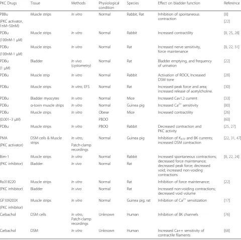
Table 1 summarizes the effects of a variety of PKC in-hibitors, activators, and cholinergic agonists on PKC sig-naling and contractility of DSM.
Discussion
The aim of this review was to evaluate the available evidence on the involvement of PKC in regulation of detrusor contractility, muscle tone of the bladder wall, spontaneous contractile activity and bladder function under physiological and pathophysiological conditions.
Modulation of DSM excitability and ion channel activity by PKC
Bladder smooth muscle cells express a variety of ion channels controlling cell excitability. Voltage-gated calcium channels (VGCC), large- (BK), and small- (SK) conductance Ca2+-activated potassium channels, and ATP-sensitive potassium channels (KATP) are the main
channels involved in the control of detrusor excitability and excitation-contraction coupling [4, 15, 26, 32, 36].
Voltage gated calcium channels
phasic contraction, and, likely, underlie the absence of spontaneous contractile activity during relaxation phase after muscarinic stimulation (Fig. 1). This action may be mediated by carbachol-induced activation of PKC [24] at low or resting calcium concentration, since PKC stimulation by PDBu was reported to activate BK chan-nels resulting in inhibition of spontaneous contractions [8, 22]. Interestingly, pre-incubation of the DSM strips
in vitrowith the PKC inhibitor, Bim-1, before carbachol stimulation preserved spontaneous contractility during relaxation phase (Fig. 1).
Effects of PKC activation on BK channels
The storage phase of the micturition cycle requires a quiescent smooth muscle that can accommodate increasing volumes at low intravesical pressure. Potassium channels contribute to this process by helping maintain the resting membrane potential via regulation of both the intracellular calcium concentration, and calcium entry into the cell from extracellular sources [32, 36, 45]. Several types of potassium channels are expressed in DSM, and are known to be involved in this regulation. Among them, BK and KATP
channels were shown to be regulated by PKC. PKC was
Table 1Effects of PKC signaling on bladder function
PKC Drugs Tissue Methods Physiological
condition
Species Effect on bladder function Reference
PBBu Muscle strips In vitro Normal Rabbit, Rat Inhibition of spontaneous
contraction
[8]
(PKC activator, 1nM–50nM)
[22]
PDBu Muscle strips In vitro Normal Rabbit Increased contractility [8,25,28]
(100nM-1μM)
PDBu Muscle strips In vitro Normal Rat Increased nerve sensitivity,
force maintenance
[8,22,31]
(100nM-1μM)
PDBu Bladder In vivo
(cystometry)
Normal Rat Bladder emptying, and frequency
of urination
[22]
(1μM)
PDBu Muscle strip In vitro Normal Rabbit Activation of ROCK, Increased
DSM tone
[28]
PDBu Muscle strips In vitro, EFS Normal Rat Increased peak force and area;
increased release of acetylcholine.
[30]
PDBu Bladder myocytes In vitro Normal Mice Increased Cav1.2 current [33]
PDBu α-toxin muscle strips In vitro Normal Guinea pig Increased Ca2+sensitivity [80]
PDBu Muscle strips In vitro Obese Mice Increased contractility [26]
(0.001–3μM) PBOO [60]
PDBu Muscle strips In vitro PBOO Rabbit Decreased contraction and
PKC activity
[25,27]
PMA DSM cells & Muscle strips
In vitro, Normal Guinea pig Inhibition of KATPand BK currents;
increased DSM contraction
[22,31,47]
(PKC activator) Patch-clamp
recordings
Bim-1 Muscle strips In vitro Normal Rabbit Increased spontaneous contractions;
decreased force maintenance; decreased peak force; decreased void; increased non-voiding contractions
[8,22,24]
(PKC inhibitor) Bladder In vivo Normal Rat
Ro318220 Muscle strips In vitro Normal Rat Inhibition of force maintenance; [22]
(PKC inhibitor) Bladder In vivo Normal Rat Increased non-voiding contractions;
decreased void volume
GF109203X Muscle strips In vitro Normal Guinea pig; rat Inhibition of Ca2+sensitization [17]
(PKC inhibitor)
Carbachol DSM cells In vitro,
Patch-clamp recordings
Unknown Human Inhibition of BK channels [76]
Carbachol DSM In vitro Unknown Human Increased Ca++ sensitivity of
contractile filaments
shown to be involved in the regulation of BK channels expressed in DSM tissue of rabbit [8] and guinea pig [31] bladders. Hypolite et al. (2013) reported that low levels of PKC stimulation by the PKC activator, PDBu, inhibited spontaneous myogenic contractions, and reduced basal DSM tone in rabbit DSM, which was dependent on activa-tion of BK channels [8]. Addiactiva-tionally, PDBu was unable to inhibit spontaneous contractions in the presence of the BK channel blocker, iberiotoxin, suggesting that the mech-anism of PDBu-induced inhibition of spontaneous con-tractions was via activation of BK channels. Utilizing whole-cell patch clamp recordings in guinea pig DSM cells, Hristov et al. (2014) confirmed that PKC does regu-late BK channel activity, however, these authors reported that PKC stimulation with PMA increased muscle force along with amplitude of spontaneous contractions [31]. The variability in the responses to PKC stimulation re-ported by the abovementioned studies could be attributed to different species (guinea pigsvsrabbits), or the use of distinct PKC activators (PDBu versus PMA), as well as slightly different methodological approaches.
Modulation of KATPchannels by PKC
Similar to BK channels, KATPchannels also participate in
the control of detrusor excitability across a broad spectrum of mammalian species including guinea pigs [46, 47], humans [48], and rats [49]. Activation of these channels induces DSM relaxation in response to KATP channel
openers pinacidil [50], and cromakalim [51]. Activation of
M3receptors can induce inhibition of these channels in
smooth muscle cells via PKC signaling pathways [52]. Stimulation of muscarinic receptors by carbachol was shown to inhibit KATP currents by 60.7 %, while
acti-vators of PKC inhibited KATP channels by 74 % [52].
Additionally, PKC blockers used before stimulation with muscarinic receptor agonists, significantly reduced carbachol-induced inhibition of KATP currents confirming
that muscarinic-dependent inhibition of KATP currents is
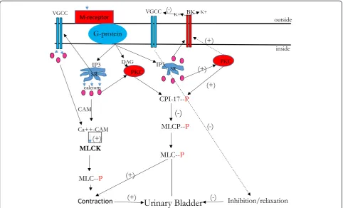
mediated via PKC pathways [52]. This speculation is consistent with in vitro studies showing that the PKC inhibitor, Bim-1, reduced both intrinsic basal tone, and maintained force [8], while awake cystometry performed in rats revealed that inhibition of PKC resulted in increased frequency of urination, and decreased void volume [22]. Schematic presentation of the ion channels involved in regulation of BSM excitability and contractility as well as downstream signaling including PKC pathways is depicted in Fig. 2.
The link between muscarinic receptors and PKC signaling in the control of detrusor contractility
Muscarinic receptor type 3 (M3) signaling via Gq/11
G-protein-coupled receptors includes activation of PLC/PKC as determined for several types of smooth muscle [23] including the DSM of the bladder [16, 53–55]. Initial exper-iments in the urinary bladder established that inhibition of PLC/PKC pathway by a number of specific inhibitors had no significant effect on maximal contractility of rat bladder
Fig. 1Effects of carbachol (CCh) on the contractile force and spontaneous contractions in DSMin vitro.Isolated muscle strips from rabbit bladders were mounted in organ baths with Tyrode’s buffer (equilibrated with 95%O2/5%CO2) and allowed to develop spontaneous contractions.
muscle strips in response to carbachol stimulation leading to the conclusion that PLC/PKC-dependent signaling via M3 receptors does not significantly contribute to DSM
contractility [16]. This data was further buttressed by the studies showing that direct inhibition of PKC had no significant effect on the peak contractile force in response to both carbachol and EFS stimulation [8, 14].
An exception to the above findings was the observation that one of the PKC inhibitors, Bim-1, did reduce maximal force in response to carbachol upon application of a higher concentration of the inhibitor [24]. Another PKC inhibitor, GF 109203X (1μM), was also reported to reduce peak force in control bladders by 29 % [56]. Subsequent studies clarified that inhibition of PKC with Bim-1, at lower concentrations, did not significantly affect electric field stimulation (EFS)-induced maximal amplitude of DSM contractions, however, significantly reduced the integral, or total force [8, 22]. The integral (total) force is calculated as the area under the curve of a single recorded contraction, and represents the ability of DSM to maintain muscle force required for physiological bladder emptying [8]. The effects of Bim-1 observedin vitrowere further confirmedin vivo by cystometric recordings in unanesthetized rats [22]. During urodynamic recordings (cystometry), the urinary bladder was slowly infused with a solution containing
Bim-1, causing the voided urine volumes to decline progressively in comparison with infusion of saline in the control group. Thesein vivo results supported the importance of PKC for muscle force maintenance associated with bladder emptying.
Additional studies confirmed the involvement of PKC in the maintenance of muscle force at the molecular and biochemical level [24]. Stimulation of muscarinic recep-tors in the smooth muscle results in inhibition of myosin light chain (MLC) phosphatase activity, an increase in MLC phosphorylation and, therefore, contractile force. One of the underlying pathways for inhibition of MLC phosphatase activity is protein kinase C (PKC)-catalyzed phosphorylation of CPI-17 protein [57, 58]. Wang and co-authors evaluated phosphorylation of protein com-plex Thr(38)-CPI-17, the downstream target of PKC signaling, and established that PKC activation increased Thr(38)-CPI-17 phosphorylation throughout the phasic and tonic portions of carbachol stimulation confirming that PKC is involved in maintaining the contractile force in the detrusor [24].
Limited contribution of the PLC/PKC pathway to the peak amplitude of the contractile response in DSM seems to be physiologically justified as the maximal contractile force of smooth muscle is predominantly controlled by
calcium/calmodulin-dependent activation of myosin light chain kinase (MLCK) followed by subsequent phosphoryl-ation of the myosin light chain (MLC) [59]. Since stimula-tion of DSM by muscarinic receptor agonists leads to a rapid rise in intracellular calcium followed by an equally rapid decline of the calcium to almost basal levels [44], it is highly likely that PKC-induced calcium sensitization via Gq11 receptors [3] plays a major role
in the maintenance phase of the contraction that is critical for bladder emptying. This mechanism may partly explain why inhibition of PKC significantly reduces DSM force maintenance, and bladder emptying, without significantly affecting peak force generation [14, 22].
Role of PKC in regulation of spontaneous contractions in the urinary bladder
Protein kinase C is also involved in other aspects of DSM contractility, including the inhibition of basal DSM tone, and spontaneous myogenic contractions [8, 22]. For instance, PDBu, a PKC activator, inhibits spontan-eous contractions, and basal myogenic tone at low levels of application (1–50 nM) whereas high concentrations of the drug increase the sensitivity to EFS stimulationin
vitro, and also trigger micturition contractions in vivo confirming a dual, concentration-dependent activation profile of PKC effects in DSM [22]. The ability of PKC to modulate basal DSM tone, and inhibit spontaneous contractions at low levels of stimulation may have physiological implications for bladder storage function, while its contribution to the maintenance of DSM force and nerve-mediated contractions may be important for bladder emptying.
Spontaneous myogenic contractions of DSM have been recorded in vivo andin vitro in several species including rabbit [8, 11], rat [60], guinea pig [31], pig [40], and humans [61]. The mechanism of these contractions involves calcium entry into the smooth muscle cells via VGCC [40, 43, 62, 63] to trigger action potential gener-ation, while the repolarization phase is thought to be mediated mainly by opening of calcium-activated potas-sium channels and exit of potaspotas-sium ions from the smooth muscle cells leading to muscle relaxation [64]. Early reports suggested that spontaneous large-amplitude contractions recordedin vitroin isolated DSM strips were likely an artifact with no significant functional role [11]. Subsequent studies suggested that these contractions participate in the maintenance of an intrinsic DSM tone by helping the smooth muscle adjust its length and shape in response to bladder filling [4]. They have also been reported to be associated with peripheral sensory processing that informs the need to void as the bladder approaches capacity [6].
The inhibitory effect of the PKC activator, PDBu, on spontaneous contractions was linked to activation of BK
channels, subsequent hyperpolarization of the membrane, and restriction of calcium entry via VGCC [8, 65]. Freshly isolated DSM strips from the rabbit bladder exhibit high amplitude spontaneous contractions which decline over several hours, however, the amplitude of contractions remains at a high level in the presence of PKC inhibitor Bim-1 [8]. This observation suggests that endogenous PKC signaling likely maintains the amplitude of spontaneous contractions at a low, physiological level under normal physiological conditions. This assumption is supported by the fact that commercially available PKC activator, PDBu, at low levels of stimulation (1–50 nM), accelerated the decline in amplitude of spontaneous contractions over timein vitro [8]. PDBu was also reported to inhibit carbachol-induced phasic contractions in neonatal bladders associated with the effects on T-type channels [29], and was also observed to inhibit spontaneous contractions in rat DSM [22].
Effects of calcium on PKC activity
Protein kinase C conveys both calcium-dependent [66–68], and calcium-independent [3, 68, 69] effects on DSM contractilityin vitromediated via inhibition of MLCP. It is still unknown if these separate effects are mediated by different PKC isoforms. However, the ability of PKC to mediate both of these actions in DSM may be well suited to urinary bladder storage and emptying function. The storage phase requires the maintenance of basal intrinsic DSM tone at low calcium concentrations which can be mediated, in part, by calcium-independent contractile effects of PKC involved in an increase in tone during the storage phase. This process may be aided by the stretch of the bladder wall associated with an up-regulation of the proteins involved in calcium sensitization [70] and contributing to the basal tone during the early phase of the micturition cycle. However, as the bladder continues to expand, it is likely that enhanced stretch-induced calcium release [71], and calcium-dependent PKC activity increase wall tension further as the bladder approaches capacity. This dual, calcium-independent and dependent, flexibility of PKC may be vital in helping ensure that wall tension rises in a controlled fashion so as to prevent steep increases in bladder pressure during the storage phase of the micturition cycle. PKC-dependent effects have been observed in DSM in response to EFS, which activates the intramural nerves, suggesting that PKC-dependent regulation of neuronal function in DSM is also a distinct possibility [65, 72–74].
PKC signaling in the human bladder
[14]. However, only the peak amplitude of contraction (maximum force) was evaluated in the presence of various PKC inhibitors, and no difference was found in peak force generation in comparison to the absence of inhibitors. Since bladder emptying requires both, the generation of peak force and the ability to maintain force, it is possible that PKC could play a more significant role in force maintenance in the human DSM in addition to force generation.
A recent study [75] provided evidence that carbachol, a muscarinic receptor agonist, could indirectly inhibit large conductance Ca2+-activated potassium (BK) channels in human DSM cells leading to increased excitability [76]. This is an interesting finding as the ongoing studies in our laboratory using human DSM indicate that carbachol can induce a significant increase in phasic contractions in some human DSM isolated strips, but not in all of them (unpub-lished data). It is possible that PKC could be an important secondary messenger between muscarinic receptors and BK channels in the human detrusor. Therefore, further studies are warranted to comprehensively characterize the role of PKC in the regulation of cholinergic activity in the human DSM under normal and pathologic conditions.
Regulation of bladder function by PKC under pathophysiological conditions
The role of PKC in bladder pathophysiology is largely related to DSM contractile dysfunction and dysfunctional voiding associated with partial bladder outlet obstruction (PBOO), and detrusor overactivity (DO). Initial studies, using a PBOO model in rabbits, reported that PKC signaling under pathophysiological conditions was uncoupled from its downstream targets and produced little or no force in response to PKC activator, PDBu [27]. Chang et al. [25] also determined that decreased force generation in decompensated PBOO bladders in response to PKC activator, PDBu, was associated with reduced expression of PKC and increased frequency of urination in the obstructed bladders. It should be noted, however, that a broad range of contractile and metabolic dysfunctions were linked to decompensated bladder func-tion in PBOO models [77, 78]. Further studies confirmed a deficit in the PKC pathway in guinea pig DSM after PBOO [79]. Partial bladder outlet obstruction causes two phenotypes of bladder dysfunction referred to as compen-sated and decompencompen-sated bladders [25, 77]. Compencompen-sated bladders are characterized mainly by increased smooth muscle hypertrophy, however, residual urine and frequency of micturition have been reported in com-pensated bladders associated with PKC dysfunction [25, 80, 81]. Decompensated bladders develop an increase in extracellular matrix including collagen and connective tissue resulting in a loss of bladder compliance, decreased contractility, and a substantial loss of bladder
emptying function [25, 77, 78, 80–83]. Changes in PKC expression and activity in a PBOO model seem to be dependent on the species and degree of obstruction. Thus, in the rabbit model of PBOO, decompensated bladders revealed a reduction in PKC expression, activity and force generation associated with reduced bladder emptying and frequency of urination, while compensated bladders exhibited increased PKC expression [25]. However, in the rat bladders with PBOO, PKC expression was reduced in both compensated, and decompensated bladders [81]. Additionally, recent in vivo studies established that inhibition of PKC during cystometric recordings in awake rats induced bladder decompensation char-acterized by an increase in non-voiding contractions (DO), and a significant decrease in void volume [22].
Increased PKC-mediated frequency of urination along with DO have also been reported in the absence of PBOO. In a model of obesity associated with insulin resistance, PKC expression was significantly higher in the bladders of obese mice [26] which showed increased contractility to PDBu, increased frequency, and non-voiding contractions. The changes in the voiding cycle were reversed by metfor-min treatment which restored high PKC expression in the urinary bladders. High levels of PKC stimulation in rat bladders [22], and high level of expression in compensated rabbit bladders [25] were associated with enhanced nerve-mediated contractions, and frequency of urination, respectively. Thus, a high level of PKC activity and expres-sion may lower the threshold for activation of intramural nerves leading to neurogenic frequency of urination in both diabetic, and compensated bladders. It was estab-lished that the protein adiponectin, expressed in adipose tissue, contributed to the increased expression of calcium-dependent PKC-αand enhanced contractile force of DSM in adiponectin-sense transgenic mice [67]. Additionally, carbachol-induced phosphorylation of PKC-α was also elevated in these animals in comparison with WT mice suggesting that adiponectin increases calcium dependency of DSM contractions mediated by PKC-αexpression.
sensitization mechanisms mediated by PKC, and Rho-kinase in a calcium-independent manner [3, 8, 24, 68]. Therefore, the frequency of micturition induced by reduced expression of PKC in decompensated rabbit bladders, and pharmaco-logic inhibition during cystometry in rat bladders are largely myogenic. The inability to maintain muscle force due to sig-nificantly low levels of PKC activity results in reduced bladder emptying, increased residual volumes, and an overall shorten-ing of the micturition cycle leadshorten-ing to voidshorten-ing frequency [22].
Partial bladder outlet obstruction, well known for its prevalence in aging males and often secondary to benign prostatic hyperplasia, has also been shown to alter struc-tural proteins within the bladder wall leading to reduced compliance, DO, and frequency of urination. Among the structural proteins altered in PBOO are smooth muscle myosin (SMM) [84] collagen [85], caldesmon [80] and con-nective tissue [86]. A change in the ratio and organization of smooth muscle to non-smooth muscle elements is gen-erally believed to lead to stiffening of the bladder wall, and a reduction in compliance resulting in a reduced storage capacity, and frequency of urination [77, 80, 85, 87]. How-ever, significant changes in structural elements such as con-nective tissue, and collagen may represent some of more severe consequences of acute bladder outlet obstruction, associated with bladder decompensation, and may not
represent some of the more subtle changes in bladder func-tion that occur due to changes in regulatory proteins and associated with bladder compensation [3, 25, 88, 89].
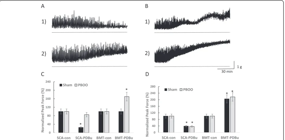
Figure 3 highlights the changes which characterize a milder form of dysfunction in compensated bladders such as decreased relaxation and increased DSM tone in response to the PKC activator, PDBu. Low levels of PKC stimulation which induce inhibition in normal DSM (panel A1) resulted in increased force in com-pensated DSM (panel A2). Additionally, moderate to high levels of PKC stimulation caused a modulated rise in tension in normal bladder DSM (panel B1), but a linear increase in the compensated DSM (panel B2). A failure to relax sufficiently and a linear, instead of a modular, increase in DSM tone are the contributing factors to a higher bladder pressure and bladder dysfunction observed in compensated bladders in comparison with normal and sham-operated controls.
Study limitations
We acknowledge that our study has several limitations. First, we were not able to find any direct data on the role of PKC signaling in detrusor underactivity nor whether PKC pathways could be pharmacologically targeted to improve this condition. Second, we could
not find sufficient information on the role of PKC pathways in sensory and efferent innervation of the lower urinary tract. This could be especially important for understanding the role of PKC in other pathological urologic conditions such as chronic pelvic pain (CPP) and bladder pain syndrome (BPS) [90, 91]. For example, a high level of PKC expression and/or activity has been suggested to play a role in feline interstitial cystitis [73], a naturally occurring bladder pain and dysfunctional voiding in cats. It would be of interest to determine whether or not PKC is overexpressed in these bladders, since high PKC stimulation was shown to increase bladder wall tension, and enhance neuronal sensitivity and contractility [8, 22, 25, 28]. Third, future studies focused on the role of PKC in the bladder urothelium are required to clarify the role of epithelial PKC in micturition.
Conclusions
PKC function in the urinary bladder extends far beyond its contribution, or the lack thereof, to maximum force generation in response to agonists, either directly, or in response to EFS. Data indicate that PKC is involved in the regulation of normal bladder function, and that PKC dysfunction is associated with DO, reduced contractility, and decreased void volume. Relaxation and modulation of spontaneous myogenic, and NVC via PKC-dependent activation of BK and KATPchannels may also be one of
its primary functions in support of bladder storage. Both
in vitro, and in vivo studies reveal that PKC may be
involved in regulating neuronal activity at the peripheral level in a concentration-dependent manner that may function in a complementary way to facilitate voiding. Finally, the demonstration that PKC dysfunction coexists with pathological changes such as PBOO, DO and is associated with reduced contractility and bladder emptying is further evidence of a significant and important role for PKC in the regulation of urinary bladder function.
Abbreviations
Bim-1:Bisindilylmaleimide1; BK: Large conductance calcium-activated potassium channel; BSM: Bladder smooth muscle; BPS: Bladder pain syndrome; Ca++: Calcium; CPP: Chronic pelvic pain; CNS: Central nervous
system; DSM: Detrusor smooth muscle; DO: Detrusor overactivity; EFS: Electrical field stimulation; IF: Integral force; M3: Muscarinic;
MLCP: Myosin light chain phosphorylation; NVC: Non-voiding contractions; OAB: Overactive bladder; PBOO: Partial bladder outlet obstruction; PDBu: Phorbol-12,13-dibutyrate; PF: Peak force; PLC: Phospholipase C; PMA: Phorbol-12,13-myristate; PKC: Protein kinase C; ROK: Rho-associated kinase; VGCC: Voltage gated calcium channels; SK: Small conductance ca++-activated potassium channels; SMM: Smooth muscle myosin;
THR: Threonine.
Competing interests
The authors declare that they have no competing interests.
Authors’contributions
JAH - conception and design, analysis and interpretation of the data, drafting of the manuscript. APM - general supervision, critical revision for intellectual
content. All authors have made a significant contribution to the paper, and have read and approved the final draft.
Acknowledgments
The study has been supported by the NIH/NIDDK grant DK095817.
Received: 21 July 2015 Accepted: 22 October 2015
References
1. Andersson KE. Changes in bladder tone during filling: pharmacological aspects. Scand J Urol Nephrol Suppl. 1999;201:67–72. discussion 76–99.
2. Andersson KE. Detrusor myocyte activity and afferent signaling. Neurourol Urodyn. 2010;29(1):97–106.
3. Boopathi E, Hypolite JA, Zderic SA, Gomes CM, Malkowicz B, Liou HC, et al. GATA-6 and NF-kappaB activate CPI-17 gene transcription and regulate Ca2+ sensitization of smooth muscle contraction. Mol Cell Biol. 2013;33(5):1085–102.
4. Brading AF. Spontaneous activity of lower urinary tract smooth muscles: correlation between ion channels and tissue function. J Physiol. 2006;570(Pt 1):13–22.
5. Drake MJ, Harvey IJ, Gillespie JI. Autonomous activity in the isolated guinea pig bladder. Exp Physiol. 2003;88(1):19–30.
6. Gillespie JI. Modulation of autonomous contractile activity in the isolated whole bladder of the guinea pig. BJU Int. 2004;93(3):393–400.
7. Hypolite JA, Chang S, LaBelle E, Babu GJ, Periasamy M, Wein AJ, et al. Deletion of SM-B, the high ATPase isoform of myosin, upregulates the PKC-mediated signal transduction pathway in murine urinary bladder smooth muscle. Am J Physiol Renal Physiol. 2009;296(3):F658–665.
8. Hypolite JA, Lei Q, Chang S, Zderic SA, Butler S, Wein AJ, et al. Spontaneous and evoked contractions are regulated by PKC-mediated signaling in detrusor smooth muscle: involvement of BK channels. Am J Physiol Renal Physiol. 2013;304(5):F451–462.
9. Hypolite JA, Wein AJ, Haugaard N, Levin RM. Role of substrates in the maintenance of contractility of the rabbit urinary bladder. Pharmacology. 1991;42(4):202–10.
10. Levin RM, Hypolite J, Longhurst PA, Wein AJ. Comparison of the contractile and metabolic effects of muscarinic stimulation with those of KCl. Pharmacology. 1991;42(3):142–50.
11. Levin RM, Ruggieri MR, Velagapudi S, Gordon D, Altman B, Wein AJ. Relevance of spontaneous activity to urinary bladder function: an in vitro and in vivo study. J Urol. 1986;136(2):517–21.
12. Wein AJ. Physiology of micturition. Clin Geriatr Med. 1986;2(4):689–99. 13. Zhao Y, Wein AJ, Levin RM. Role of calcium in mediating the biphasic
contraction of the rabbit urinary bladder. Gen Pharmacol. 1993;24(3):727–31. 14. Schneider T, Fetscher C, Krege S, Michel MC. Signal transduction underlying carbachol-induced contraction of human urinary bladder. J Pharmacol Exp Ther. 2004;309(3):1148–53.
15. Frazier EP, Peters SL, Braverman AS, Ruggieri Sr MR, Michel MC. Signal transduction underlying the control of urinary bladder smooth muscle tone by muscarinic receptors and beta-adrenoceptors. Naunyn Schmiedebergs Arch Pharmacol. 2008;377(4–6):449–62.
16. Frazier EP, Braverman AS, Peters SL, Michel MC, Ruggieri Sr MR. Does phospholipase C mediate muscarinic receptor-induced rat urinary bladder contraction? J Pharmacol Exp Ther. 2007;322(3):998–1002.
17. Durlu-Kandilci NT, Brading AF. Involvement of Rho kinase and protein kinase C in carbachol-induced calcium sensitization in beta-escin skinned rat and guinea-pig bladders. Br J Pharmacol. 2006;148(3):376–84.
18. Fleichman M, Schneider T, Fetscher C, Michel MC. Signal transduction underlying carbachol-induced contraction of rat urinary bladder. II. Protein kinases. J Pharmacol Exp Ther. 2004;308(1):54–8.
19. An JY, Yun HS, Lee YP, Yang SJ, Shim JO, Jeong JH, et al. The intracellular pathway of the acetylcholine-induced contraction in cat detrusor muscle cells. Br J Pharmacol. 2002;137(7):1001–10.
20. Aburto T, Jinsi A, Zhu Q, Deth RC. Involvement of protein kinase C activation in alpha 2-adrenoceptor-mediated contractions of rabbit saphenous vein. Eur J Pharmacol. 1995;277(1):35–44.
22. Hypolite JA, Chang S, Wein AJ, Chacko S, Malykhina AP. Protein kinase C modulates frequency of micturition and non-voiding contractions in the urinary bladder via neuronal and myogenic mechanisms. BMC Urol. 2015;15(1):34.
23. Caulfield MP, Birdsall NJ. International Union of Pharmacology. XVII. Classification of muscarinic acetylcholine receptors. Pharmacol Rev. 1998;50(2):279–90.
24. Wang T, Kendig DM, Smolock EM, Moreland RS. Carbachol-induced rabbit bladder smooth muscle contraction: roles of protein kinase C and Rho kinase. Am J Physiol Renal Physiol. 2009;297(6):F1534–1542. 25. Chang S, Hypolite JA, Mohanan S, Zderic SA, Wein AJ, Chacko S.
Alteration of the PKC-mediated signaling pathway for smooth muscle contraction in obstruction-induced hypertrophy of the urinary bladder. Lab Invest. 2009;89(7):823–32.
26. Leiria LO, Sollon C, Calixto MC, Lintomen L, Monica FZ, Anhe GF, et al. Role of PKC and CaV1.2 in detrusor overactivity in a model of obesity associated with insulin resistance in mice. PLoS One. 2012;7(11):e48507.
27. Stanton MC, Austin JC, Delaney DP, Gosfield A, Marx JO, Zderic SA, et al. Partial bladder outlet obstruction selectively abolishes protein kinase C induced contraction of rabbit detrusor smooth muscle. J Urol. 2006;176(6 Pt 1):2716–21.
28. Wang T, Kendig DM, Trappanese DM, Smolock EM, Moreland RS. Phorbol 12,13-dibutyrate-induced, protein kinase C-mediated contraction of rabbit bladder smooth muscle. Front Pharmacol. 2012;2:83.
29. Ekman M, Andersson KE, Arner A. Receptor-induced phasic activity of newborn mouse bladders is inhibited by protein kinase C and involves T-type Ca2+ channels. BJU Int. 2009;104(5):690–7.
30. Somogyi GT, Tanowitz M, Zernova G, de Groat WC. M1 muscarinic receptor-induced facilitation of ACh and noradrenaline release in the rat bladder is mediated by protein kinase C. J Physiol. 1996;496(Pt 1):245–54. 31. Hristov KL, Smith AC, Parajuli SP, Malysz J, Petkov GV. Large-conductance
voltage- and Ca2 +−activated K+ channel regulation by protein kinase C in guinea pig urinary bladder smooth muscle. Am J Physiol Cell Physiol. 2014;306(5):C460–470.
32. Petkov GV. Central role of the BK channel in urinary bladder smooth muscle physiology and pathophysiology. Am J Physiol Regul Integr Comp Physiol. 2014;307(6):R571–584.
33. Huster M, Frei E, Hofmann F, Wegener JW. A complex of Ca(V)1.2/PKC is involved in muscarinic signaling in smooth muscle. FASEB J. 2010;24(8):2651–9. 34. Liu SH, Lin-Shiau SY. Protein kinase c regulates purinergic component of
neurogenic contractions in mouse bladder. J Urol. 2000;164(5):1764–7. 35. Weng TI, Chen WJ, Liu SH. Bladder instillation of Escherichia coli
lipopolysaccharide alters the muscle contractions in rat urinary bladder via a protein kinase C-related pathway. Toxicol Appl Pharmacol. 2005;208(2):163–9.
36. Petkov GV. Role of potassium ion channels in detrusor smooth muscle function and dysfunction. Nat Rev Urol. 2012;9(1):30–40.
37. Schneider T, Hein P, Michel MC. Signal transduction underlying carbachol-induced contraction of rat urinary bladder. I. Phospholipases and Ca2+ sources. J Pharmacol Exp Ther. 2004;308(1):47–53.
38. Wuest M, Hiller N, Braeter M, Hakenberg OW, Wirth MP, Ravens U. Contribution of Ca2+ influx to carbachol-induced detrusor contraction is different in human urinary bladder compared to pig and mouse. Eur J Pharmacol. 2007;565(1–3):180–9.
39. Zderic SA, Sillen U, Liu GH, Snyder 3rd MC, Duckett JW, Gong C, et al. Developmental aspects of excitation contraction coupling of rabbit bladder smooth muscle. J Urol. 1994;152(2 Pt 2):679–81.
40. Buckner SA, Milicic I, Daza AV, Coghlan MJ, Gopalakrishnan M. Spontaneous phasic activity of the pig urinary bladder smooth muscle: characteristics and sensitivity to potassium channel modulators. Br J Pharmacol.
2002;135(3):639–48.
41. Uchida W, Masuda N, Shirai Y, Shibasaki K, Satoh N, Takenada T. The role of extracellular Ca2+ in carbachol-induced tonic contraction of the pig detrusor smooth muscle. Naunyn Schmiedebergs Arch Pharmacol. 1994;350(4):398–402.
42. Masters JG, Neal DE, Gillespie JI. The contribution of intracellular Ca2+ release to contraction in human bladder smooth muscle. Br J Pharmacol. 1999;127(4):996–1002.
43. Kajioka S, Nakayama S, McMurray G, Abe K, Brading AF. Ca(2+) channel properties in smooth muscle cells of the urinary bladder from pig and human. Eur J Pharmacol. 2002;443(1–3):19–29.
44. Levin RM, Hypolite J, Ruggieri MR, Longhurst PA, Wein AJ. Effects of muscarinic stimulation on intracellular calcium in the rabbit bladder: comparison with metabolic response. Pharmacology. 1989;39(2):69–77. 45. Herrera GM, Heppner TJ, Nelson MT. Regulation of urinary bladder smooth
muscle contractions by ryanodine receptors and BK and SK channels. Am J Physiol Regul Integr Comp Physiol. 2000;279(1):R60–68.
46. Bonev AD, Nelson MT. ATP-sensitive potassium channels in smooth muscle cells from guinea pig urinary bladder. Am J Physiol. 1993;264(5 Pt 1):C1190–1200. 47. Gopalakrishnan M, Whiteaker KL, Molinari EJ, Davis-Taber R, Scott VE, Shieh
CC, et al. Characterization of the ATP-sensitive potassium channels (KATP) expressed in guinea pig bladder smooth muscle cells. J Pharmacol Exp Ther. 1999;289(1):551–8.
48. Buckner SA, Milicic I, Daza A, Davis-Taber R, Scott VE, Sullivan JP, et al. Pharmacological and molecular analysis of ATP-sensitive K(+) channels in the pig and human detrusor. Eur J Pharmacol. 2000;400(2–3):287–95. 49. Ha JH, Lee KY, Kim WJ. Actions of potassium channel openers in rat
detrusor urinae. J Korean Med Sci. 1993;8(1):53–9.
50. Edwards G, Henshaw M, Miller M, Weston AH. Comparison of the effects of several potassium-channel openers on rat bladder and rat portal vein in vitro. Br J Pharmacol. 1991;102(3):679–86.
51. Malmgren A, Andersson KE, Sjogren C, Andersson PO. Effects of pinacidil and cromakalim (BRL 34915) on bladder function in rats with detrusor instability. J Urol. 1989;142(4):1134–8.
52. Bonev AD, Nelson MT. Muscarinic inhibition of ATP-sensitive K+ channels by protein kinase C in urinary bladder smooth muscle. Am J Physiol. 1993;265(6 Pt 1):C1723–1728.
53. Frayer SM, Barber LA, Vasko MR. Activation of protein kinase C enhances peptide release from rat spinal cord slices. Neurosci Lett. 1999;265(1):17–20. 54. Braverman AS, Doumanian LR, Ruggieri Sr MR. M2 and M3 muscarinic
receptor activation of urinary bladder contractile signal transduction. II. Denervated rat bladder. J Pharmacol Exp Ther. 2006;316(2):875–80. 55. Longhurst PA, Leggett RE, Briscoe JA. Characterization of the functional
muscarinic receptors in the rat urinary bladder. Br J Pharmacol. 1995;116(4):2279–85.
56. Boberg L, Poljakovic M, Rahman A, Eccles R, Arner A. Role of Rho-kinase and protein kinase C during contraction of hypertrophic detrusor in mice with partial urinary bladder outlet obstruction. BJU Int. 2012;109(1):132–40. 57. Eto M, Ohmori T, Suzuki M, Furuya K, Morita F. A novel protein
phosphatase-1 inhibitory protein potentiated by protein kinase C. Isolation from porcine aorta media and characterization. J Biochem.
1995;118(6):1104–7.
58. Eto M, Senba S, Morita F, Yazawa M. Molecular cloning of a novel phosphorylation-dependent inhibitory protein of protein phosphatase-1 (CPI17) in smooth muscle: its specific localization in smooth muscle. FEBS Lett. 1997;410(2–3):356–60.
59. Adelstein RS, Eisenberg E. Regulation and kinetics of the actin-myosin-ATP interaction. Annu Rev Biochem. 1980;49:921–56.
60. Ng YK, de Groat WC, Wu HY. Muscarinic regulation of neonatal rat bladder spontaneous contractions. Am J Physiol Regul Integr Comp Physiol. 2006;291(4):R1049–1059.
61. Hristov KL, Parajuli SP, Soder RP, Cheng Q, Rovner ES, Petkov GV. Suppression of human detrusor smooth muscle excitability and contractility via pharmacological activation of large conductance Ca2 +−activated K+ channels. Am J Physiol Cell Physiol. 2012;302(11):C1632–1641.
62. Brading AF. Ion channels and control of contractile activity in urinary bladder smooth muscle. Jpn J Pharmacol. 1992;58 Suppl 2:120P–7P. 63. Rivera L, Brading AF. The role of Ca2+ influx and intracellular Ca2+ release
in the muscarinic-mediated contraction of mammalian urinary bladder smooth muscle. BJU Int. 2006;98(4):868–75.
64. Klockner U, Isenberg G. Action potentials and net membrane currents of isolated smooth muscle cells (urinary bladder of the guinea-pig). Pflugers Arch. 1985;405(4):329–39.
65. Nakamura Y, Ishiura Y, Yokoyama O, Namiki M, De Groat WC. Role of protein kinase C in central muscarinic inhibitory mechanisms regulating voiding in rats. Neuroscience. 2003;116(2):477–84.
68. Takahashi R, Nishimura J, Hirano K, Seki N, Naito S, Kanaide H. Ca2+ sensitization in contraction of human bladder smooth muscle. J Urol. 2004;172(2):748–52.
69. Yoshimura Y, Yamaguchi O. Calcium independent contraction of bladder smooth muscle. Int J Urol. 1997;4(1):62–7.
70. Boopathi E, Gomes C, Zderic SA, Malkowicz B, Chakrabarti R, Patel DP, et al. Mechanical stretch upregulates proteins involved in Ca2+ sensitization in urinary bladder smooth muscle hypertrophy. Am J Physiol Cell Physiol. 2014;307(6):C542–553.
71. Ji G, Barsotti RJ, Feldman ME, Kotlikoff MI. Stretch-induced calcium release in smooth muscle. J Gen Physiol. 2002;119(6):533–44.
72. Downing JE, Role LW. Activators of protein kinase C enhance acetylcholine receptor desensitization in sympathetic ganglion neurons. Proc Natl Acad Sci U S A. 1987;84(21):7739–43.
73. Sculptoreanu A, de Groat WC, Buffington CA, Birder LA. Protein kinase C contributes to abnormal capsaicin responses in DRG neurons from cats with feline interstitial cystitis. Neurosci Lett. 2005;381(1–2):42–6.
74. Zhou Y, Zhou ZS, Zhao ZQ. PKC regulates capsaicin-induced currents of dorsal root ganglion neurons in rats. Neuropharmacology. 2001;41(5):601–8. 75. Parajuli SP, Hristov KL, Cheng Q, Malysz J, Rovner ES, Petkov GV. Functional
link between muscarinic receptors and large-conductance Ca-activated K channels in freshly isolated human detrusor smooth muscle cells.Pflugers Archiv.2015;467(4):665-75. doi:10.1007/s00424-014-1537-8. Epub 2014 May 28. 76. Parajuli SP, Hristov KL, Cheng Q, Malysz J, Rovner ES, Petkov GV. Functional link between muscarinic receptors and large-conductance Ca2 +−activated K+ channels in freshly isolated human detrusor smooth muscle cells. Pflugers Arch. 2015;467(4):665–75.
77. Levin RM, Haugaard N, Hypolite JA, Wein AJ, Buttyan R. Metabolic factors influencing lower urinary tract function. Exp Physiol. 1999;84(1):171–94. 78. Levin RM, Monson FC, Haugaard N, Buttyan R, Hudson A, Roelofs M, et al.
Genetic and cellular characteristics of bladder outlet obstruction. Urol Clin North Am. 1995;22(2):263–83.
79. Shahab N, Kajioka S, Takahashi-Yanaga F, Onimaru M, Matsuda M, Seki N, et al. Obstruction enhances rho-kinase pathway and diminishes protein kinase C pathway in carbachol-induced calcium sensitization in contraction of alpha-toxin permeabilized guinea pig detrusor smooth muscle. Neurourol Urodyn. 2012;31(4):593–9.
80. Zhang EY, Stein R, Chang S, Zheng Y, Zderic SA, Wein AJ, et al. Smooth muscle hypertrophy following partial bladder outlet obstruction is associated with overexpression of non-muscle caldesmon. Am J Pathol. 2004;164(2):601–12.
81. Choi BH, Jin LH, Kim KH, Kang SA, Kang JH, Yoon SM, et al. Cystometric parameters and the activity of signaling proteins in association with the compensation or decompensation of bladder function in an animal experimental model of partial bladder outlet obstruction. Int J Mol Med. 2013;32(6):1435–41.
82. Levin RM, Longhurst PA, Monson FC, Kato K, Wein AJ. Effect of bladder outlet obstruction on the morphology, physiology, and pharmacology of the bladder. Prostate Suppl. 1990;3:9–26.
83. Stein R, Gong C, Hutcheson JC, Canning DA, Zderic SA. The decompensated detrusor III: impact of bladder outlet obstruction on sarcoplasmic endoplasmic reticulum protein and gene expression. J Urol. 2000;164(3 Pt 2):1026–30.
84. DiSanto ME, Stein R, Chang S, Hypolite JA, Zheng Y, Zderic S, et al. Alteration in expression of myosin isoforms in detrusor smooth muscle following bladder outlet obstruction. Am J Physiol Cell Physiol. 2003;285(6):C1397–1410.
85. Macarak EJ, Schulz J, Zderic SA, Sado Y, Ninomiya Y, Polyak E, et al. Smooth muscle trans-membrane sarcoglycan complex in partial bladder outlet obstruction. Histochem Cell Biol. 2006;126(1):71–82.
86. Sjuve R, Haase H, Ekblad E, Malmqvist U, Morano I, Arner A. Increased expression of non-muscle myosin heavy chain-B in connective tissue cells of hypertrophic rat urinary bladder. Cell Tissue Res. 2001;304(2):271–8. 87. Iguchi N, Hou A, Koul HK, Wilcox DT. Partial bladder outlet obstruction in
mice may cause E-cadherin repression through hypoxia induced pathway. J Urol. 2014;192(3):964–72.
88. Chacko S, Chang S, Hypolite J, Disanto M, Wein A. Alteration of contractile and regulatory proteins following partial bladder outlet obstruction. Scand J Urol Nephrol Suppl. 2004;215:26–36.
89. Chang S, Gomes CM, Hypolite JA, Marx J, Alanzi J, Zderic SA, et al. Detrusor overactivity is associated with downregulation of large-conductance
calcium- and voltage-activated potassium channel protein. Am J Physiol Renal Physiol. 2010;298(6):F1416–1423.
90. Malykhina A, Hanno P. How are we going to make progress treating bladder pain syndrome? ICI-RS 2013. Neurourol Urodyn. 2014;33(5):625–9. 91. Sadler KE, Stratton JM, Kolber BJ. Urinary bladder distention evoked visceromotor
responses as a model for bladder pain in mice.J Vis Exp.2014:(86).
Submit your next manuscript to BioMed Central and take full advantage of:
• Convenient online submission
• Thorough peer review
• No space constraints or color figure charges
• Immediate publication on acceptance
• Inclusion in PubMed, CAS, Scopus and Google Scholar
• Research which is freely available for redistribution