Sejal Patel
Submitted for the degree of Doctor of Philosophy (PhD)
All rights reserved
INFORMATION TO ALL USERS
The quality of this reproduction is dependent upon the quality of the copy submitted.
In the unlikely event that the author did not send a complete manuscript and there are missing pages, these will be noted. Also, if material had to be removed,
a note will indicate the deletion.
uest.
ProQuest 10016747
Published by ProQuest LLC(2016). Copyright of the Dissertation is held by the Author.
All rights reserved.
This work is protected against unauthorized copying under Title 17, United States Code. Microform Edition © ProQuest LLC.
ProQuest LLC
789 East Eisenhower Parkway P.O. Box 1346
bacterium. The isolate designated Rot34Al was thought to be the extreme thermophile
T. filiformis. The strain grows optimally at 70°C and was isolated from a thermal site in Rotorua, New Zealand. The endonuclease named Tfll^ in accordance to the rules proposed by Smith and Nathans (1973), recognises the sequence GAA/^TC which is a hitherto unknown specificity and has not been reported from either mesophilic or thermophilic sources.
The endonuclease was purified and characterised. The molecular weight (M^) of Tfll
endonuclease, estimated under denaturing conditions was 37 000. The M,. of the native form of TjfL endonuclease, as estimated by gel filtration was approximately 75 000 i.e., Tj^I endonuclease is a dimer in its active form.
The optimal pH for Tfil endonuclease activity was determined to be pH 8.0 although Tfil
endonuclease exhibits activity over a broad pH range. The enzyme is remarkably thermostable, surviving at room temperature for several weeks and having a half life of greater than one hour at 65°C. Other characteristics of Tfil endonuclease have been determined such as ability to cleave single stranded DNA and it’s salt requirement.
Two kinds of “star activity” were observed for TfH endonuclease; the indiscriminate endonucleolytic activity exhibited in certain buffers and the relaxed specificity exhibited by buffers containing Mn^^.
The Tfil methylase was partially purified and a single step method was developed to separate the methylase from the endonuclease. The site at which the TfU methylase incorporates methyl groups into DNA was determined.
throughout this project. Their guidance and support have been invaluable.
Thanks to Dr Hsuan for proving the internal sequencing data.
To Sian Shore (my mentor) and Carol French thanks for all the advice over the past few years and thanks also Paul Shadbolt for all his help.
Thanks to Neil Broadway and Mike Orford for making FPLC so much more fun than System Gold and for introducing me to Huntley Street
Apologies to all on the Ground Floor (past and present) for not mentioning you all individually -thanks for having to put up with the mood swings and the mess.
A big thanks to Majib ur Rehman for all his support and special thanks to Ian Collins (with sincerity!) and Richard Braley (for the proof reading and encouragement).
Title page 1
Abstract 2
Acknowledgem ents 3
Table o f Contents 4
List o f Figures 9
List o f Tables 12
A bbreviations 13
1
Introduction
1.1 Restriction and Modification 16
1.2 Nomenclature 16
1.3 Classification of Restriction Modification Systems 17
1.4 Class n Restriction Endonucleases 19
1.5 Class n Modification Methylases 24
1.6 Occurrence of Restriction Modification Systems 25 1.6.1 Restriction Modification Systems in Non Bacterial Systems 25 1.6.2 Restriction Modification Systems in Bacterial Systems 25 1.6.2.1 Restriction Modification Systems in Thermophiles 26 1.7 Protein Structure - Amino Acid Homology
1.7.1 Amino Acid Homology between Restriction Endonucleases 27 1.7.2 Amino Acid Homology between Methylases 29 1.7.3 Amino Acid Homology between Restriction Endonucleases and
Methylases 31
1.8 Protein-Nucleic Acid Interactions
1.9 Gene Organisation of Restriction Modification Systems 44 1.9.1 Regulation of Restriction Modification Systems 44 1.9.2 Base Composition and Flanking Regions of Restriction Modification
Systems 47
1.10 Cloning Restriction Modification Systems
1.10.1 Introduction 49
1.10.2 Plasmids Coding for Restriction Modification Systems 49 1.10.3 Selection Based on Susceptibility to Phage Infection 50
1.10.4 Selection Based on Modification 50
1.11 Thermophilic Bacteria 53
1.11.1 The genus Thermus 54
1.11.2 Cloned Thermophilic Enzymes 56
1.12 Aims 60
2
Materials and Methods
2.1 Cultures
2.1.1 Growth and Maintenance of Rot34A 1 61 2.1.2 Growth and Maintenance of E. Co/f strains 61
2.2 Preparation of Media and Buffers 61
2.2.1 Media 63
2.2.1.1 Growth Media for Rot34Al 63
2.2.1.2 Growth Medium for E. Co/; and Phage Strains 63
2.2.2 Buffers 64
2.3 Agarose Gel Electrophoresis 67
2.4 Radioactivity 67
2.5 Preparation of Dialysis Tubing 68
2.6 Restriction Digestions and Ligations 68 2.7 DNA Preparation
2.7.1 Large Scale Preparation of Rot34Al Chromosomal DNA 68 2.7.2 Large Scale Preparation of Plasmid DNA 69 2.7.3 Small Scale Preparation of Plasmid DNA 70 2.7.4 Size Fractionation of Rot34Al Chromosomal DNA 71
2.9 Transformation and Plating of E. Coli Strains
2.9.1 Transformations 72
2.9.2 Replica Plating 73
2.10 Packaging DNA 73
2.11 Transfection of E. Coli 73
2.12 Amplification of Libraries
2.12.1 Amplification of the Plasmid Library 74 2.12.2 Amplification of the Phage Library 74 2.13 Preparation of Rot 34A 1 Cell Extracts 74 2.14 Enzyme Assays
2.14.1 Tfil Endonuclease Detection Assay 75 2.14.2 Tfil Endonuclease Activity Assay 75
2.14.3 Tfil Methylase Detection Assay 75
2.14.4 jyîl Methylase Activity Assay 76
2.14.5 GAPDH Activity Assay 76
2.15 Determination Of Protein Concentration 77 2.16 Chromatography
2.16.1 Phosphocellulose Chromatography
2.16.1.1 Preparation Of Phosphocellulose 77 2.16.1.2 Low Pressure Phosphocellulose Chromatography 77 2.16.2 Fast Protein Liquid Gel Filtration Chromatography
2.16.2.1 Hiload Superdex 200 Prep Grade”™ Chromatography 78
2.16.2.2 Superose 6™ Chromatography 78
2.16.2.3 Superdex 75™ Chromatography 79
2.16.3 Adsorption Chromatography
2.16.3.1 Low Pressure Ion Exchange Chromatography 79 2.16.3.2 Fast Protein Liquid Ion Exchange Chromatography 79 2.16.3.3 Affinity Chromatography (Heparin) 80 2.17 Protein Concentration
2.17.1 Protein Precipitation 81
2.17.2 Protein Concentration by Ultrafiltration 81 2.18 SDS Polyacrylamide Gel Electrophoresis 81
2.19 Silver Staining 81
2.21 Thin Layer Chromatography 82 2.22 Effect of Temperature on Endonuclease Activity 83
2.23 pH Profile 83
2.24 Effect of Salt 83
2.25 EDTA and Metal Ion Study 83
2.26 Star Activity Studies 84
3
Purification of
Tfil
Restriction Endonuclease
3.1 Introduction 85
3.2 Production of Cell Biomass 85
3.3 Preparation of Cell Extract 85
3.4 Phosphocellulose Chromatography 86
3.5 Ion Exchange Chromatography 89
3.6 Gel Filtration Chromatography 92
3.7 Mono Q Chromatography 95
3.8 Heparin-Affi-Prep™ Affinity Chromatography 97 3.9 SDS-Page Analysis of Endonuclease Fractions 100
3.10 Internal Sequence Determination 105
4
Characterisation Of
Tfil
Restriction Endonuclease
4.1 Introduction 108
4.2 Gel Filtration Chromatography
4.2.1 Estimation of Molecular Weight of Tfil Endonuclease 109 4.2.2 Separation of the Tfil Endonuclease and Rot34Al GAPDH Enzyme 111
4.3 pH Profile of Tfil Endonuclease 115
4.3 Thermostability of Tfil Endonuclease 117
4.4 Effect of Detergents 119
4.5 Effect of Salt on TjfH Endonuclease 121
4.6 Star Activity Studies 122
4.7 EDTA and Metal Ion Study 126
5
Partial Purification and Characterisation of the
Tfil
Methylase
5.1 Introduction 129
5.2 Partial Purification of the TjU. Methylase
5.2.1 Phosphocellulose Chromatography 129
5.2.2 Heparin-Affi-Prep™ Chromatography 131 5.3 Determination of Méthylation Site of the Tfil Methylase 136
6
Cloning Strategies for theT/fl Restriction
Modification System
6.1 Introduction 139
6.2 Creating and Screening Libraries in Plasmid Vectors 140 6.2.1 Creating Libraries in pUC 18 And pUC 19 142 6.2.2 Strategy for Screening the pUC 18 And pUC 19 Libraries 143
6.2.3 Creating Libraries in pBR322 147
6.2.4 Strategy For Screening pBR322 Libraries 151 6.3 Creating a Library in Lambda DASH™ II 154
7
General Discussion
7.1 Introduction 158
7.2 Purification and Characterisation of Tj^I Endonuclease 158 7.3 Characterisation of Tfil Methylase 165 7.4 Cloning Stratagies for Isolation of the Tfil Restriction Modification Genes 166
7.5 Conclusions 171
8
References
173
Appendix
Appendix I Digestion of X, pUC18 and pBR322 with Tfil Endonuclease I
Appendix n Map of pUC18 II
Appendix in Map of pBR322 IE
List of Figures
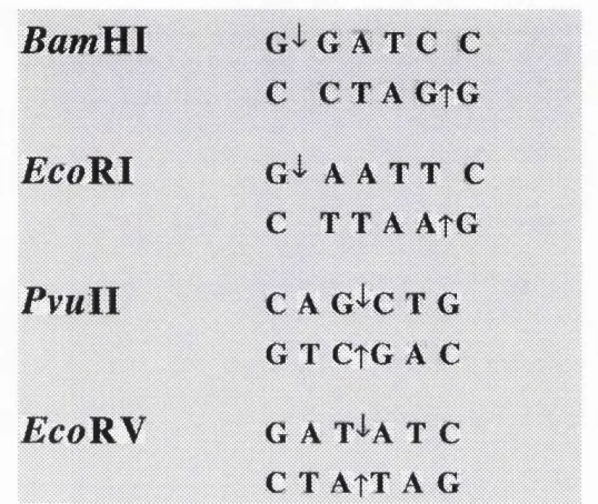
Figure 1.1 DNA recognition sequences of FcoRI, EcoRV, RamHI and PvuTL 20
Figure 1.2 Class II Isoschizomers 21
Figure 1.3 Recognition Sites of FcoRI,/tsri and Afwnl 27 Figure 1.4 Alignment of EcoRI, Rsrl and Muni 28 Figure 1.5 Schematic diagram of the alignment of MTase sequences 30 Figure 1.6 Structures of RamHI and £coRI 34 Figure 1.7 Structures Of RvwII and EcoRV 35 Figure 1.8 Secondary structures of RamHI and EcoRI 39 Figure 1.9 Secondary structures of RvwII and EcoRN 40 Figure 1.10 Ribbon diagrams showing the domain organisation of M-Hhal 42 Figure 1.11 Schematic representation showing the specific base and phosphate
contacts between M'Ehdi and DNA 43 Figure 1.12 Gene organisation of restriction modification systems 45 Figure 1.13 Consensus promoter sequence for Thermus spp. cloned genes 56
Figure 3.1 Elution profile of Rot34AI crude extract from phosphocellulose
chromatography 87
Figure 3.2 Elution profile from Q chromatography 91 Figure 3.3 Calibration curve for Superdex 200 chromatography 93 Figure 3.4 Elution profile for dialysed active Q chromatography fractions
from HiLoad 16/60 Superdex 200 chromatography 94 Figure 3.5 Elution profile from Mono Q chromatography 96 Figure 3.6 Elution profile from Heparin-Affi-Prep™ affinity
chromatography 99
Figure 3.7 Silver stained SDS-PAGE gel showing the initial stages of
purification 102
Figure 3.8 Silver stained SDS PAGE gel showing the final stages of
purification 103
Figure 3.9 Computer generated alignment of the T. aquaticus D
glyceraldehyde 3-phosphate dehydrogenase and the protein
Figure 4.1 Calibration curve for Superdex 75™ chromatography 109 Figure 4.2 Calibration curve for SDS PAGE analysis of Tfll endonuclease 110 Figure 4.3 Elution profile for dialysed Mono Q fractions from Superose 6
chromatography 112
Figure 4.4 Calibration curve for Superose 6 chromatography 113 Figure 4.5 Silver stained SDS PAGE gel showing fractions from Superose-6
chromatography 114
Figure 4.6 Digestion of pUC 18 at different pHs for Tfll endonuclease 116 Figure 4.7 pH profile of Tfll endonuclease 116 Figure 4.8 Thermostability of Tfll endonuclease 118 Figure 4.9 Activity assay of Tfll endonuclease in the presence of 1% (v/v)
Triton XlOO 120
Figure 4.10 Digestion of pUC18 and X DNA in the presence of 30% (v/v)
glycerol 123
Figure 4.11 Tfil activity on pUC 18 and 1 DNA in low ionic strength buffers 124 Figure 4.12 Tfll endonuclease activity in high pH buffer 125 Figure 4.13 Activity of Tfll endonuclease with Mn^+ ion replacing Mg^+ ions 127 Figure 4.14 Activity of Tfll on single stranded substrates 128
Figure 5.1 Elution profile of Rot34AI crude extract from phosphocellulose 130 chromatography
Figure 5.2 Elution profile for PI 1 methylase fraction from Heparin-Affi- 132 Prep™ chromatography
Figure 5.3 Photograph of the silver stained SDS PAGE gel showing the
initial steps of purification of the methylase 134 Figure 5.4 The separation of nucleosides with the 80:20 ethanol:water (v/v)
solvent system 137
Figure 5.5 The separation of nucleosides with the 66:33:1 isobutyric
Figure 6.1 Sau3Aî fractionated chromosomal Rot34Al DNA 149 Figure 6.2. Analysis of pBR322 Sau3Al partial digested generated colonies 153 Figure 6.3 Size fractionation of chromosomal Rot34Al DNA 154 Figure 6.4 Lambda DASH™n ligation with size fractionated chromosomal
Rot34Al DNA 155
Figure 7.1 Tfil restriction modification system cleavage and méthylation
sites 158
List of Tables
Table 1.1 Characteristics of classes of restriction modification systems 18 Table 1.2 Comparison between Bcgl and other classes of restriction
endonucleases 23
Table 1.3. Méthylation systems in E. coli 52 Table 1.4 Comparison of Bacteria and Archaea 54 Table 1.5 Commercial Sources of Restriction Enzymes 59
Table 2.1 Bacterial Strain Genotypes 62
Table 3.1 Purification of the Tfil endonuclease by phosphocellulose
chromatography 88
Table 3.2 Purification of the Tfil endonuclease by Q chromatography
and S chromatography 89
Table 3.3 Purification of the T^zl endonuclease by Q Sepharose chromatography 90 Table 3.4 Purification of the Tfil endonuclease by gel filtration chromatography 93 Table 3.5 Purification of the Tfil endonuclease by Mono Q chromatography 95 Table 3.6 Purification of the Tfil endonuclease by Heparin-Affi-Prep™
affinity chromatography 98
Table 3.7 Purification of Tfil endonuclease 104 Table 4.1 Thermostability of Tfil endonuclease 119 Table 4.2 The effect of salt on Tfil endonuclease 121 Table 5.1 Partial purification of the Tfil methylase 133 Table 5.2 Cellulose TLC of deoxynucleosides 138 Table 5.3 Incorporation of [3H] S-adenosyl-L-methionine in
TJU. methylase methylated DNA 138
Table 6.1 Restriction endonuclease cleavage sites in Rot34AI
chromosomal DNA 141
Table 6.2 Creation of libraries using pUC-derived vectors 142 Table 6.3 pUC-derived libraries at different stages of the screening
protocol 146
Abbrevations
A adenosine
AdoMet S-adenosyl-L- methionine
Amp ampicillin
ATP adenosine triphosphate Pgal P-galactosidase
bp base pair
BSA bovine serum albumin
G cytosine
C controlling element
cpm counts per minutes
ds double stranded
Dam DNA-adenine methyltransferase
Dcm DNA-cytosine methyltransferase
DIT dithiothreitol
EDTA ethylene diamine tetraacetic acid ENase restriction endonuclease
EtBr ethidium bromide
G guanine
kb kilobase
M (MTase) methylase
NAD+ nicotinamide adenine dinucleotide (oxidised form) NADH nicotinamide adenine dinucleotide (reduced form) ORF open reading frame
PGR polymerase chain reaction PGA 3-phosphoglyceric acid
PGK 3-phosphoglyceric phosphokinase R restriction endonuclease
RM restriction modification system
SAM S-adenosyl-L- [methyl-^H] methionine SDS sodium dodecyl sulphate
ss single stranded
T thymine
Tc tetracyline
TCA Trichloroacetic acid Topt Temperature optimum TVR . terminal variable region
U uracil
v/v volume per volume w/v weight per volume w/w weight per weight
X-gal 5-bromo-4-chloro-3-indolyl-p-D-galactoside
ve elution volume
Abbreviation of amino acids
A (Ala) Alanine
C (Cys) Cysteine
D (Asp) Aspartate
I
E (Glu)
1
Glutamic acid
F (Phe) Phenylalanine
G (Gly) Glycine
H (His) Histidine
I (He) Isoleucine
K (Lys) Lysine
L (Leu) Leucine
M (Met) Methionine
N (Asn) Asparagine
P (Pro) Proline
Q (Gin) Glutamine
R (Arg) Arginine
S (Ser) Serine
T (Thr) Threonine
V (Val) Valine
w (Trp) Tryptophan
1 Introduction
1 .1 RESTRICTION AND MODIFICATION
Present day DNA technology is largely dependent upon the ability to cut DNA at specific sites with restriction endonucleases. Prior to 1970 there was no method available for cutting DNA into discrete fragments. A solution to this problem eventually grew from research into the phenomenon of host controlled restriction and modification. Arber and Linn (1968) demonstrated host controlled restriction and modification. The first
endonuclease was isolated by Messelson and Yuan (1968).
Restriction describes the ability to reduce the biological activity of infecting DNA by enzymatic cleavage. It is a hydrolysis reaction in which phosphodiester bonds within the DNA backbone are cleaved by the restriction endonuclease. The restricting host also possesses the abihty to protect it's own DNA from restriction, by modification. Modification is the result of DNA méthylation. In bacteria, which have a restriction modification system, the modification enzyme methylates bases within the restriction endonuclease target site. Méthylation of certain bases in the host DNA alters the
endonuclease recognition sites and serves to protect the organism's own DNA from being digested; i.e., the organism is immune. Hence a restriction system always consists of two enzymatic elements, namely a nuclease and a methylase.
1 .2 NOMENCLATURE
Smith and Nathans (1973) proposed a system of nomenclature for restriction
number of different enzymes isolated from the same strain (For example Hin(X IQ means the endonuclease was isolated from the strain Haemophilus influenzae strain d and was the third endonuclease to be isolated from this organism). To differentiate between the restriction endonuclease and the modification methyltransferase it is generally accepted that the methyltransferase is distinguished by the capital letter M with a raised dot in front of the enzyme name. Recently the endonucleases and methylases are referred to collectively as restriction modification systems (RM). The endonucleases are referred to as the — ENases and the methyltransferases as the —MTases (for example the EcoRl
system would be referred to as the EcoRL ENase and the EcoRl MTase (Kessler and Manta, 1990).
1 .3 CLASSIFICATION OF RESTRICTION MODIFICATION SYSTEMS
Based on their subunit structures, reaction mechanisms and genetic organisation, restriction enzymes fall into three general classes, namely 1, Q and IQ (Table 1.1 shows the different characteristics of the classes).
Class 1 enzymes exhibit both restriction and DNA modification activities located on different subunits of multifunctional enzyme complexes. They require magnesium ions, ATP and S-adenosyl methionine (AdoMet) as cofactors. These enzymes cleave DNA at unspecified sites usually 100 to 1000 base pairs downstream of the specific recognition sequences.
Table 1.1 Characteristics of classes of restriction modification systems.
[The different properties of the classes of restriction modification systems are shown
below. N= any nucleotide, bp = base pair]
CLASS I CLASS II CLASS III
Restriction modification activities
single multifunctional enzyme
separate endonuclease
& methylase
separate enzymes with a subunit in common
Protein structure of endonuclease
3 different subunits Homodimers 2 different subunits
Requirement for restriction
ATP, Mg2+, AdoMet ATP, Mg2+.
AdoMet
Sequence of host specificity sites
non rotational symmetry rotational symmetry non rotational
symmetry
e.g. EcoB e>g. E c o M e.g. EcoPl
T G A N 2 T C G C G A A T T C A G A C C
Cleavage Site Possibly random A t o r n ear host 24-26 bp 3' of host
at least 1000 bp from host specificity site
All class n systems are comprised of two different proteins, i.e., the restriction enzymes are separate from the methylases, which are relatively small compared to their Class I and m counterparts and their cofactor requirements are simple. In vitro restriction requires only magnesium ions and unmodified DNA while the methylase is dependent only on the presence of S-adenosyl methionine and unmodified or partially modified DNA.
It is the class n enzymes which have received the most intense attention over the years as they are capable of reproducibly cutting DNA in a fixed position with respect to the sequence they recognise.
1.4 CLASS II RESTRICTION ENDONUCLEASES
Endonucleases cleave phosphodiester bonds at defined positions in or adjacent to the recognition site, resulting in 5' or 3' self-complementary terminal extensions (e.g.,
BamRl and EcoRl ) or fully base paired termini [e.g., EcoBN and PvuR, (Figure 1.1) see Kessler and Manta (1990) for review].
The recognition sequences usually exhibit two fold symmetry with the cleavage sites in each strand arranged around the axis of symmetry. Some sequences are degenerate (e.g.,
HhaR where the recognition site is GANTC where N is variable).
Figure 1.1 DNA recognition sequences of EcoRl, E coR V, BarnHl and Pv mI I
[The site of cleavage within the recognition sequence is indicated by the arrows.]
BamVLl
G A T C C
C C T A G t G
E c o R I
G'i' A A T T C
C T T A A t G
P v u l l
C A G^C T G
G T CtG A C
E c o B y
G A T'l'A T C
C T ATT A G
Class 11 endonucleases usually act as homodimers; i.e., interact with their substrate as a
dimer (Modrich, 1982). The endonucleases can be a range of sizes from 157 amino acids
(Pvull) to 576 amino acids (BsuRl) [Wilson and Murray, 1991]. Some isoschizomers can cleave DNA at different positions within the same recognition sequence. Where the
cleavage sites are identical, isoschizomers can have different sensitivity to méthylation
(Figure 1.2).
Canonical site specific méthylation refers to the méthylation exhibited by the methylase of
a specific restriction modification system. This méthylation always inhibits DNA cleavage
by it's corresponding (or cognate) restriction endonuclease. For example M BamHl methylase modifies GGAT^4(^C, and BamUl endonuclease cannot cut this methylated sequence. However non canonical site méthylation will inhibit the rate of DNA cleavage.
effect on restriction cleavage. For example BarnRl cuts DNA which has been modified at GGATC"^4c or GGATC^^C but cannot cut DNA methylated at GGAT^^CC.
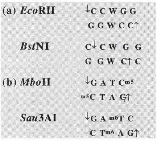
Figure 1.2 Class 11 isoschizomers
[Two examples of isoschizomers, (a) isoschizomers with the same recognition sequence
but different cleavage sites, (b) isoschizomers with identical cleavage sites but different
méthylation sites (W is either A or T).]
( a ) JScoRII
'I'C C W G G
G G w
c
e t
B s t N l
C^ C w G G
G G W e t c
<b)
M boll
'l'G A T C“ S
T A <^t
Sau
3
A l
{ g
a »»«t c
c
A G t
Endonucleases can show preferential cleavage; i.e., within a given DNA molecule some
recognition sites are cleaved more easily than other sites. This phenomenon is partly
explained by the influence of adjacent sequences. PvmII is inhibited by méthylation
outside the Pvwll recognition site. Experiments showed that with methylated DNA
containing two cleavage sites for Pvull, one site being digested four to eight fold more slowly than the other. With unmethylated DNA, the two sites were cleaved at the same
As more and more restriction enzymes are being isolated, the classification system definitions have been found to be insufficient. Class nS enzymes (ENases-llS) are a subclass of Class n restriction modification systems. The enzymes interact with two discrete sites: the recognition site, which is 4-7 bps long, and the cleavage site usually 1-20 bp away from the recognition site. A total of 35 ENase-llS have been isolated (80 if isoschizomers are included).The recognition sequences are totally asymmetric and all of the characterised ENase-llS are monomeric (Bellemare and Potvin, 1990; Szybaski etal
1988; Degtyarev et al, 1990).The Class HS endonucleases are normally twice the size of Class 11 endonucleases and appear to act as monomers (Wilson and Murray, 1991).
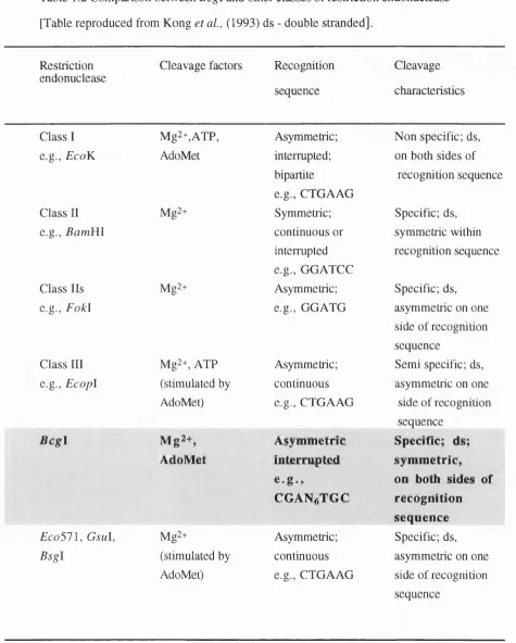
The Bcg\ endonuclease isolated from Bacillus coagulons may be a member of yet another subgroup of restriction enzymes. This restriction endonuclease differs from the other groups as summarised in Table 1.2 (Kong et al , 1993).
Class n restriction endonucleases require Mg^+ as a cofactor for cleavage activity. Other divalent metals replace Mg^+ but with reduced specificity (Hsu and Berg, 1978). This relaxation of activity is termed as “star activity” (Polisky et al , 1975). BarriRl is among the endonucleases that shows a similar relaxation of specificity in buffers containing Mn^+ and also in high concentrations of compounds such as glycerol, ethylene glycol, ethanol or dioxane. It is assumed that this is due to altered DNA-protein interactions (Jack
e t a l , 1991).
Although some restriction endonucleases cleave single stranded DNA specifically, most are unable to or do so with low activity and specificity.
Class n endonucleases which are not accompanied by a class n methylase have
Table 1.2 Comparison between Bcgl and other classes of restriction endonuclease [Table reproduced from Kong et a l, (1993) ds - double stranded].
Restriction endonuclease
Cleavage factors Recognition
sequence
Cleavage
characteristics
Class 1 Mg2+,ATP, Asymmetric; Non specific; ds. e.g., EcoK AdoMet interrupted;
bipartite
e.g., CTGAAG
on both sides of recognition sequence
Class 11 Mg2+ Symmetric; Specific; ds.
e.g., Bam Yll continuous or
interrupted e.g., G GATCC
symmetric within recognition sequence
Class 11s Mg2+ Asymmetric; Specific; ds. e.g., F o kl e.g., GGATG asymmetric on one
side of recognition sequence
Class 111 Mg2+, ATP Asymmetric; Semi specific; ds. e.g., E copl (stimulated by continuous asymmetric on one
AdoMet) e.g., CTGAAG side of recognition sequence
Asymmetric Specific; ds; AdoMet interrupted
COAN^TGC
sym m etric, on both sides of recognition sequence E co 5 1 \, G sul, Mg2+ Asymmetric; Specific; ds. B sgl (stimulated by continuous asymmetric on one
Dpn\ is the best characterised of unaccompanied restriction endonucleases, cleaves the sequence G“ ^ATC. However this sequence does not occur in the hosts genome as this bacterium does not have specific cytosine méthylation (Lacks and Greenberg, 1977).
1 .5 CLASS II MODIFICATION METHYLASES
DNA methylases transfer methyl groups from the donor S-adenosylmethioine onto the adenine or cytosine residues within a specific DNA sequence. Most class II methylases recognise double stranded DNA (which exhibits two fold rotational symmetry) and methylate a single residue on both strands of the recognition sequence. They produce one of three products, N^-methyladenine, modifying the exocyclic amino group of adenine, 5-methylcytosine and N"* methylcytosine, modifying the exocyclic group of cytosine. The latter type of modification was only discovered in the early 1980s (Janulaitis et al., 1983; Ehrlich etal., 1985, see Section 1.6.3).
No class II methylase has been reported to be less specific than its endonuclease counterpart (Wilson and Murray, 1991).
A number of methyltransferases occur separately and are involved in repair mechanisms, unaccompanied by endonucleases. Escherichia coli possesses two such unaccompanied methylases, DNA-adenine methyltransferase (dam) associated with methyl directed mismatch repair and DNA-cytosine methyltransferase (dcm) associated with short patch repair (Marinus, 1987).
1.6 OCCURRENCE OF RESTRICTION MODIFICATION SYSTEMS
There were 1284 endonucleases and 130 methylases of known specificities isolated from 1117 different organisms by January 1990 (Kessler and Manta, 1990). One year later another 8(X) endonucleases had been characterised, including 179 different class-II specificities (Roberts and Macehs, 1992). Some specificities are common: isoschizomers of HaeUi, EcoRB. and Pst\, have been found 50 times over, while others are rare [Xba\
has only been found once (Wilson and Murray, 1991)].
1.6.1 RESTRICTION MODIFICATION SYSTEMS IN NON BACTERIAL SYSTEM S
The vast majority of restriction enzymes come from bacteria, although a few non bacterial systems have been reported. Virulent viruses of the unicellular alga. Chlore lia , carry restriction modification systems (Xia et a l, 1986; Zhang et a l, 1992). Other non bacterial systems which have site specific endonucleases include self sphcing introns from yeast. Though not strictly “true” restriction enzymes, they are used in vitro for cutting DNA (Watabe et al, 1981; Shibata et al, 1984). Hsal from man and an endonuclease from Chlamydomonas reinhardtii (Sklar et a l, 1986) have also been identified (see Kessler and Holtke, 1986, for review and primary sources). There are endonucleases encoded by group I introns in both eukaryotes and bacteriophages which cleave intronless alleles.
1.6.2 RESTRICTION MODIFICATION SYSTEMS IN BACTERIAL SYSTEM S
eluded detection. Among the bacteria that do possess restriction modification systems, approximately half have multiple systems: usually two or three, but sometimes more (Laue et a l, 1991).
1.6.2.1 RESTRICTION MODIFICATION SYSTEMS IN THERMOPHILES
Restriction modification cA/cfmrns have been isolated from many thermophilic sources (e.g., Shaw e t a l , 1991 Zebela gf a/. 1990; Slatko e t a l , 1987; Kiss e t a l , 1985). The thermophihc restriction modification systems are mostly thermostable isoschizomers of mesophilic counterparts such as the B stlN l restriction endonuclease isolated from
Bacillus stearothermophilus LV, an isoschizomer of CM (Lobos and Vâsquez, 1993) although others cleave at novel sites; e.g., Tfil (this study). Within the Archaea, various plasmids have been found to encode restriction modification systems including the
Methanobacterium thermoformicicum | strain (Nolhng et a i, 1992). The
hyperthermophile Desulfurococcus | rnobiis (Dalgaard et a i, 1993) has been found to have a site specific endonuclease encoded by an archaeal intron. This enzyme, l-Dmo I is unusual among the intron endonucleases in that it is thermostable and is expressed only from linear and cychsed intron species and not from its precursor RNA.
°^^C has been identified as a “hot spot” for mutation (Cowlondre et a i, 1978). At
1.7 PROTEIN STRUCTURE - AMINO ACID HOMOLOGY
1.7.1 AMINO ACID HOMOLOGY BETWEEN RESTRICTION ENDONUCLEASES
W ith the exception of some isoschizomers, class II endonucleases exhibit no common
primary sequence homology. Isoschizomers that do show amino acid sequence
homology include the EcoRl and Rsrl endonucleases (Stephenson et a i, 1989), B suBl and Pstl endonucleases (Xu et a i, 1992), BsuFl and M spl (Kapfer et a i, 1991), Tr/iHBSl and Taql (Barany et a l, 1992a), FnuDl and N ^oPll, C fr9l and Xm al
(Wilson, 1991). Comparison of the isoschizomers EcoRl and Rsrl showed their amino acid sequence 50% identity within a 266 amino acid overlap between the
deduced amino acid sequences of Rsrl and EcoRl. The sequence alignment showed six regions 8-17 amino acid in length with 75-100% sequence identity (Stephenson et a l,
1989). Recently the Muni restriction modification system (Siksnys et a i , 1994) has been cloned. The central part of the recognition sequence, including the position of the scissile
bond ('^AATT), are common for M uni, EcoR l and Rsrl (Figure 1.3).
Comparisons between the R M unl deduced amino acid sequence, EcoRl and Rsrl revealed five of the six conserved regions were present in R M unl (Figure 1.4). These structural and functional significances of these conserved regions are discussed in greater
detail in Section 1.8.2.
Figure 1.3 Recognition Sites of EcoRl, Rsrl and M uni
E coV il
A T T C
(Rsrl)
G
T T A A î G
Muni
C^A A T T G
Figure 1.4 Alignment of EcoRl, R srl and M uni
[Columns with open boxes indicate amino acid residues that are identical between aligned
proteins. Columns with grey background indicate similar amino acids (Taylor, 1986).
Roman numerals I-IV above aligned sequence correspond to functionally and structurally
important elements of the EcoRl and Rsrl enzymes, according to Stephenson et a l (1989). Numbers to the left of the alignment refer to the positions in the amino acid
sequences of individual proteins. Reproduced from Siksnys et a l (1994).]
(Mk y v l y e k p i
Ia&v l t k l a e i
KLVKKALSHI
QlTDYNVFirojElI MAGETEFHSI^HMJRIJ&n FGA^œXHDI
F& D \H K A H D I
--nnscnrQWlH
T L — PDD"VniKEIE--- iFTEi T tH&RW )YGEW (LRSlg
RTSLTKKAIH I rNDVEK
RDSIKKTEIHE WmeKDIIN
m
EKF
PP&LLI
-ELRm
5EIA1ÏI
tEKL^
^ i m m
IPIN S
ŒKKYL
(FATES-
(FLTEN-R • MlUll
1.7.2 AMINO ACID HOMOLOGY BETWEEN METHYLASES
In contrast to the endonucleases there are extensive similarities among the primary amino acid sequence of methylases. Approximately ninety methylase genes have been
sequenced and approximately seven architectural classes have been distinguished (Klimasauskas e t a l , 1989).
Figure 1.5 Schematic diagram of the alignment of MTase sequences
[Each line represents one sequence. Gaps were introduced in the alignment where the lines are interrupted. Boxes indicate where the ten blocks of conserved
residues occur. Filled boxes indicate the five highly conserved blocks; the open boxes represents the less conserved blocks. The variable region lies between blocks VIII and IX. Reproduced from Pôsfai e ta l (1989).]
VI VII V III
P h i3T
R h o lI s
Haelll
EcoRl II
The remaining classes have the enzymes that form N4-methylcytosine (m^^C-MTases) and N6-methyladenine (m^A-MTases). The m"KZ-MTases and the m^A-MTases share some
similarity at primary amino acid level. The méthylation of the exocyclic amino group of adenine and cytosine may be similar (Tao et a l, 1989), although the mechanism has yet to be elucidated. There are two common motifs shared by these two types of methylases, DPF-GSGT and TSPPY, which were found to be similar to those of the m^C-MTases.
1.7.3 AMINO ACID HOMOLOGY BETWEEN RESTRICTION ENDONUCLEASES AND METHYLASES
Endonucleases and their cognate methylases share DNA sequence specificity but not primary amino acid sequence similarity. Comparisons of primary amino acid sequences of methylase specificity (target recognition) domains and their cognate endonucleases for nine different restriction modification systems showed no similarity (Chandrasegaran and Smith, 1988). One exception is the EcoRW restriction modification system in which a short motif similar to one found in several heterospecific endonucleases and methylases as well as in the cognate M EcoRV has been identified in R EcoRV (Thielking et a l,
1991). The lack of similarity between endonucleases and their cognate methylases suggests that restriction and modification enzymes are unrelated and that they recognise their targets by different strategies. It is hypothesised that the MTase and ENase genes may have evolved separately, only becoming adjacent to one another on the chromosome when their combined activity provided a selective advantage (Kaszubska et a l, 1989; Wilson and Murray, 1991).
sequence motifs that were partially conserved (Kossykh et a l, 1993). The authors suggested that the motifs were important for sequence recognition by R EcoRU and the isospecific methylases. Whether this evidence suggests that endonucleases and
methylases arose from a common ancestor is yet to be determined. However EcoRU has been shown to require at least two recognition sites for endonucleolytic cleavage (Kroger and Hobom, 1984). The enzyme must bind to an activator site before it wül cut a
cleavage site. EcoRSi also has been found to have similarity between a 29 amino acid sequence in the carboxyl end of EcoRU and the motif defining the integrase family of recombinases (Topal and Conrad, 1993).
1.8 PROTEIN-NUCLEIC ACID INTERACTIONS
1.8.1 PROTEIN-NUCLEIC ACID INTERACTIONS OF
ENDONUCLEASES
Structural studies of DNA binding proteins and their complexes with DNA have
proceeded at an accelerating pace in recent years, due to technical advances in molecular genetics, DNA synthesis, protein crystallography and nuclear magnetic resonance.
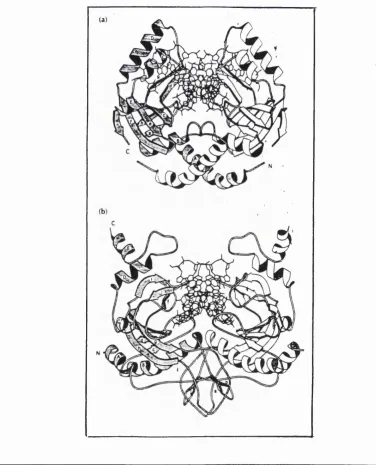
The elucidation of the crystal structures of a number of endonucleases {EcoRl, EcoRY, BamHl and PvmII) has been a significant advance in the understanding of the structural aspects of protein-nucleic acid interactions (Kim et al, 1990; Winkler et a l, 1993; Newman era/., 1994; Cheng gra/., 1994).
Though restriction endonucleases show no primary amino acid homology (except in certain examples of isoschizomers) they have been shown to have certain structural homologies. EcoRL, EcoRY, BamHl and PvmII have all been found to display an a|3 architecture since all contain a 5-stranded p sheet surrounded on either side by a helices (Figures 1.6,1.7, 1.8 and 1.9). The central core of the endonucleases consists of a mixed P-sheet, which carries the active site at one end, facing the reactive phosphate group. In their functional dimeric form, the endonucleases are U-shaped, with a prominent cleft for DNA binding (Aggarwal, 1995). EcoRl and BamHl are structurally related and bind DNA from the major groove side whereas EcoRY and PvmII, which are structurally homologous (Figures 1.7 and 1.8), approach DNA from the minor groove side.
The first structure to be determined with the cognate DNA was EcoRl endonuclease. McClarin et a l (1986) determined the crystal structure of the EcoRl endonuclease complexed with a 12 base pair DNA duplex including the enzyme recognition sequence.
Figure 1.6 Structures of BamWl and EcoKl
[(a) BamWl dimer (originally published by Newman et a i, 1994), (b) EcoRl- DNA complex. The proteins are represented as ribbons and the DNA as a stick model, using the program MOLSCRIPT. The twofold axis runs vertically in the plane of the paper. One of the subunits is shown shaded and with labels for the secondary structural
Figure 1.7 Structures of PvmII and EcoRV
[Structures of (a) PvmI I - D N A (originally published by Cheng et al, 1 9 9 4 ) and (b)
EcoRV-DNA (originally published by Winkler et ai, 1 9 9 3 ) complexes. The proteins are
represented as ribbons and the DNA as a stick models. One of the subunits is shown shaded and with labels for the secondary structural elements Reproduced from Aggarwal
EcoRi endonuclease induces DNA kinking (McClarin et a l, 1986; Fredrick et a l, 1984). Two types of DNA “kinks” are produced by the binding of the DNA to EcoRl. The first is termed a “neoF’ kink and occur at the central dyad axis, the second (the “neoll” kink) is observed 3 base pairs from the dyad axis on both sides of the dyad giving rise to a bend between the DNA axis on either side of the kink. This allows the DNA sequence to be in the correct orientation for cleavage to occur.
Twelve hydrogen bond donors and acceptors from protein side chains provide the sequence specificity exhibited by this enzyme. The side chains are complementary to the donors and acceptors presented by the exposed edge of the base pair in the recognition sequences. Two arginines (Arg^^s^ Arg^^^) and one glutamic acid (Glu^"^) each make two hydrogen bonds to the DNA bases in both subunits of the dimer. These three side chains are able to penetrate the deep major groove by emanating from the ends of the a helices that protrude from the protein into the DNA major groove. Four parallel P sheets (two from each dimer) are “pushed” into the groove for the purpose of direct DNA-base sequence recognition. Each side chain makes two hydrogen bonds with bases Arg^^o iq
the N7 and Og of guanine; the other two side chain interactions serve to cross link the
adenine of the two adjacent base pairs. It has been suggested that a catalytically active form of the enzyme is formed once the requisite sequence specific DNA protein interactions have formed (McClarin et al, 1986; Kim et a l, 1990).
Comparison of the amino acid sequence of the Rsrl, (an isoschizomer of EcoRl) with
EcoRl showed six regions of homology which correspond to key structural and functional regions of EcoRl (Figure 1.4; Stephenson et a l, 1989). There are six conserved amino acid regions in the sequence alignment of EcoRl and Rsrl.
a l, 1990), the residues from conserved regions I and n form a P-meander that contains the active centre. The sequence similarities between Muni and EcoRl {Rsrl) suggest the endonucleases share common structural organisation of the catalytic site (Siksnys e ta l,
1994).
Region HI (Figure 1.4) forms an extended polypeptide chain motif and a part of the inner recognition heUx (Kim et a l, 1990). It is at this motif that the set of hydrogen bonds and Van der Waal contacts occur with the inner tetranucleotide sequence (AATT). The same amino acids are found within the Muni endonuclease, namely Gly*''°, Asn"", Ala’'*^ Arg'^^ from region III and Gln”^ from region II (Siksnys et a l, 1994).
Regions IV and V correspond to parts of structural motifs of EcoRl ensuring correct spatial orientation of the main sequence recognition elements (McClarin et al, 1986; Kim
et a l, 1990). The regions are identical in EcoRl and Muni suggesting a similar spacial orientation between Muni and DNA as for the EcoRI-DNA interaction (Siksnys et a l,
1994).
Region VI forms a part of a loop and outer recognition helix in EcoRl providing a structural basis for discriminating the external nucleotide pairs of the hexanucleotide sequence (Kim et a l, 1990). There is no similarity in this region (Siksnys et a l, 1994), probably due to the different external nucleotide pairs recognised by EcoRl and Muni
(CG for Muni and GC for EcoRI). Kim et a l (1990) showed that EcoRl discrimination for GC is by both hydrogen bond and Van der Waal contacts between Met*^^ and Ala^^ and the C base. These amino acids are not conserved in Muni, which instead has a Arg residue located in the region suggesting this residue has interactions with the G base.
overhang. Withers and Dunbar ( 1 9 9 3 ) revealed that each of the endonucleases induces
bending of the DNA. The bending of the helix axis appears to be in opposite orientations. The orientation of the Smal induced bend appeared to be towards the major groove and is similar to the bend induced by EcoRV (also produces blunt end scissions). The Xmal
appeared to bend the DNA towards the minor groove. These studies provide further examples of endonuclease induced DNA bending to facilitate restriction by enhancing local protein-DNA interactions. EcoRV has been crystallised both with it's specific DNA sequence and also with non specific DNA (Winkler et a i, 1 9 9 3 ) . The conformation of the two DNAs are different. The non specific DNA adopts a p DNA conformation and does not exhibit the bending and unwinding observed with the specific DNA. The major consequence of this is a high affinity Mg"^ binding site formed only with the specific DNA. Recently two groups have shown that DNA distortion is not universal for DNA binding to endonucleases (see Aggarwal, 1 9 9 5 for review). The EvmI I complex reveals
specific DNA that retains a P DNA like conformation without the kinking and untwisting seen in the EcoRI and EcoRV complexes (Cheng et al, 1 9 9 4 ) . This has also been
observed with the BamHl complex.
The endonucleases seem to show structural homology which is not detectable at primary level. The structural homology appears to be related to the cleavage patterns of the enzymes (Figure 1.1). EcoRI and BamHl share cleavage recognition similarity and also structural similarity; i.e., cleave DNA to produce a 5' overhang. Pvull and EcoRV differ from the EcoRI and BamHl both structurally and with respect to the sites they recognise; i.e., cleave DNA to produce blunt ends and are structurally similar to one another. The active sites for the endonucleases are similar in all four endonucleases. However the DNA recognition structural elements are diverse: EcoRV uses a loop, EvmII, a antiparallel P sheet and both BamHl and EcoRI recognise by the same mechanism, though BamHl
Figure 1.8 Secondary structures of BamHl and EcoBl
[Secondary structures of (a) BamHl, (b) EcoKl (originally published by Newman et a l,
1994). The shaded regions correspond to the common core motif. Reproduced from Aggarwal (1995).]
coo*
n h:
Figure 1.9 Secondary structures of PvuU and EcoRW
[Secondary structures of (a) PvuU and (b) EcoRV (originally published by Athanasiadis
et a i, (1994). The shaded regions correspond to topologically equivalent regions. Reproduced from Aggarwal (1995).]
1.8.2 PROTEIN-NUCLEIC ACID INTERACTIONS OF METHYLASES
The Hhal MTase from Haemophilus haemolyticus is a m^C-MTase which recognises the GCGC duplex sequence and catalytically transfers a methyl group from AdoMet to the first cytosine of the sequence. The catalytic transfer from AdoMet involves a covalent intermediate. This is the result of nucleophüic attack at the cytosine C6 position by the
conserved Cys*' residue on the Hhal MTase (Figure 1.10; Cheng et al , 1993).
Replacement of the proton by a fluorine at the cytosine C5 position chemically traps the covalent reaction intermediate. The crystal structure for the chemicaUy-trapped Hhal
methyltransferase, S-adenosylmethionine and a duplex 13-mer DNA ohgonucleotide containing methylated 5-fluorocytosine at it's target, has been determined at 2.8 Â resolution (Khmasaukas e t a l , 1994).
This crystal structure has shown that the enzyme remodels DNA so as to present a particular base to the active site (Klimasaukas et ai, 1994). Hhal MTase binds DNA as a monomer (a binary complex), the cleft providing the binding site for DNA (Cheng et al,
1993). The active site lies in a 20 residue loop (amino acids 80-99) in the large domain. This loop has six of the residues that are conserved (Figure 1.5) including the
catalytically active Cys^\ On binding, the loop undergoes a conformational change bringing the sulphydryl group of Cys*’ into close proximity with the target cytosine, allowing the formation of the covalent link (Klimasaukas et a l, 1994).
Figure 1.10 Ribbon diagrams showing the domain organisation of M Hhal
[The large (amino acids 1-193 and amino acids 304- 327) and small (amino acids 194-275) domains are marked. The AdoMet is omitted from (a). The cleft is large enough to accommodate DNA. Side view showing the DNA binding cleft. Shown in (b) is the view into the cleft. Reproduced from Cheng et al. 1993)]
large domain
Cy»81
cleft
hinge
small domain
AdoM et
Figure 1.11 Schematic representation showing the specific base and phosphate contacts between M Hhal and DNA
[The DNA is represented as a cylindrical projection. The recognition bases and contacted phosphates are shaded. Base contacts are shown with a thick line, phosphate contacts with a thin line, and contacts through main chain atoms with a dashed line. The symbol (W) indicates the water-mediated contacts. Reproduced from Klimasaukas etal. (1994)]
Patel (1994) describes the intermolecular interactions as “a reaching out between extended segments on the protein and the D N A a handshake at the molecular level”. This is the first structure where it has been shown that a protein distorts the DNA by causing a base to flip out of the DNA helix.
1.9 GENE ORGANISATION OF RESTRICTION MODIFICATION
SYSTEMS
The restriction and modifications genes are generally closely linked. In some systems the genes have opposite orientations; (Figure 1.12; see Wilson and Murray 1991, for
review). The organisation may be the result of natural selection (Section 1.7.3) and has been exploited in the cloning of restriction modification systems (Section 1.10).
1.9.1 REGULATION OF RESTRICTION MODIFICATION SYSTEMS
Restriction modification systems are regulated in different ways. Analysis of the BamYÜ
restriction modification system sequences showed two large open reading frames
(ORFs). These corresponded to the ENase and MTase respectively. The two genes were divergently orientated. In the intergenic region between the two genes, a third small ORF aligned in the same orientation and overlapping the open reading frame of the
endonuclease gene was observed (Figure 1.12). This was 306 nucleotides long and could code for a protein of 133511 Daltons It had been postulated that this ORF may
play a regulatory role (Brooks et a l, 1991).
This small ORF of BamYil was designated bamHIC for BamHl controlling element.
bamHIC can act on both bamHIR and bamHIM via a protein product, C BarriHl. Since
binding proteins, the authors hypothesised that C BamHl acted as a transcriptional activator of bamHIR and a repressor of bamHIM (Ives et ai, 1992).
Figure 1.12 Gene organisation of some restriction modification systems.
[The ORF thought to be involved in regulation is shown for PvwII, BamHl, EcoRY, Smal. The figure was adapted from Tao et al (1991). These observations are discussed in greater detail in Section 1.9.1.]
M e th y la s e
E n d o n u q le a s e C o n tr o llin g E le m e n t
M e th y la s e
E n d o n u c le a s e
C o n tr o llin g E le m e n t
M e th y la s e
E n d o n u c l ^ O ConliDlBng Hement
C o n tr o llin g E le m e n t E n d o n u c le a s e
M e th y la s e
M e th y la s e
E n d o n u c leas e
Pvu U
Bam H I
Eco R V
Snw I
A similar mode of action has been proposed for C PvmII (Tao et a l, 1991; Tao and Blumenthal, 1992). Sequence ahgnments of the BamYil small ORF with deduced amino acids sequences of the third ORFs identified in other restriction modification systems including EcoRV, Smal and Pvi/11 (Tao et a l, 1991; Siksnys et a l, 1994) revealed a family of related C-proteins (Figure 1.12; Tao et a l, 1991).
The nucleotide sequence of the genes encoding M Taql and R Taql have been analysed (Barney et al, 1992b). The C terminus of the taqlM gene overlapped the N terminus of the taqlR gene by thirteen codons. This also occurs with the Taql isoschizomer TthUBSl
(Barney et al, 1992a). It is thought that this overlap plays a role in regulating taqlR
expression. The authors speculated that the overlap region in the Taql and 7YAHB81 systems permits the formation of a hairpin structure which would promote termination of transcription.
Similarly Cfr9l has both endonuclease and methylase gene in the same transcriptional orientation, but differing in their translational phases (Lubys et a l, 1994). The ORF of the MTase and the ORF of the ENase overlap by 4 bases (Figure 1.11). In the case of the endonuclease gene, 16 bases upstream from the ATG start codon is the sequence
TAAGGA, predicted to be Shine-Dalgamo sequence. A complementary nucleotide sequence is located within the methylase gene. Formation of secondary mRNA structure involving these complementary sequences cannot be excluded. Since initiation of R C ^ l translation may be hindered by mRNA secondary structure, its synthesis could be
1.9.2 BASE COMPOSITION AND FLANKING REGIONS OF
RESTRICTION MODIFICATION SYSTEMS
Examination of the ORFs of the restriction modification systems has shown the systems to have a preference for rare codons. The overall frequency of optimal codon selection for each of the proteins in the Pstl restriction modification system is less than that predicted even on a random basis (Walder et a i, 1984). There are 8 6 leucine residues in the two
proteins. Of these none utihse the optimal codon CUG but instead are coded for by UUA.
The pattern of codon usage in the Pstl genes is very similar to that observed for the EcoRl genes. The overall frequency of optimal codon selection for the restriction endonuclease is 38% and the methylase 40%. The optimal codon for leucine CUG is rarely used and again UUA is predominant. The coding systems are AT rich for Pstl
(62% and 6 6% for the endonuclease and methylase respectively) and simüarily for the
EcoRl genes (65%). However the flanking regions of both systems are also highly AT rich.
For the BamHl restriction-modification system the base composition within the
sequenced region is 31% GC which is substantially lower than the 47% GC reported for
average of Moraxella bovis which is 41-44.5% (Ueno et a i, 1993). This has also been seen for the MboR restriction modification system (Bockage et a l, 1991). It has been suggested by the authors that this reflects a relatively recent evolutionary origin of restriction modification systems from a progenitor organism with low GC content. However as the restriction modification systems show no homology this argument seems flawed.
Thermus aquaticus is a thermophüe and might be expected to be GC rich. For the RM genes the disproportion is slight; 59% GC and 52% GC for the R and M respectively. In non coding sequences, there is substantially more GC content (6 6%) [Slatko et a l,
1987]. However, codon usage reflects a preference for GC in the third position. This system also differs in that it has seven Taql site in the endonuclease gene and none in the methylase gene. This is unusual and not widespread. It is postulated that this asymmetric distribution of sites could be important in the regulation of the endonuclease gene
although the actual mechanism is unknown. It is thought that in an unmodified cell, interaction between the endonuclease with the Taql site on the restriction endonuclease gene might interrupt transcription of the gene and lead to a deletion or death of the cell. If the DNA was hemimethylated the expressed endonuclease gene would not be able to prevent further endonuclease synthesis until the cell was fully modified.
Surrounding the ORFs of restriction modification systems, other ORFs have been identified. The HpaTl restriction modification system revealed two adjacent genes
Kulakauskas et al, 1994). Upstream and partly overlapping hpallM is the coding sequence for a protein that resembles the very-short-patch -repair endonuclease {Vsr) of
1.10 CLONING RESTRICTION MODIFICATION SYSTEMS
1.10.1 INTRODUCTION
Sixteen years ago, the first paper appeared describing the cloning of a complete restriction modification system in E. coli (Mann et al , 1978). The cloned restriction modification systems provide material for investigating the biology of DNA modification and
restriction and the biochemistry of DNA-protein interactions. They also constitute new, often more convenient or prolific sources from which restriction and modification enzymes can be purified. An example of over-expression of a restriction endonuclease is the Kpnl restriction-modification system, cloned and expressed in E. coli (Hammond et a l, 1990). The genes for restriction and méthylation were isolated sequentially (Section 1.10.3). Using a two step method, the strain was constructed with two compatible plasmids, an inducible plasmid with the Kpnl restriction gene which is activated at elevated temperatures and a pBR322 derivative expressing the methylase. The new strain produced 10 million units of Kpnl restriction endonuclease activity/g of wet weight cells. This was several 1000 fold higher than the level of Kpnl produced by the source bacteria
K pneumoniae (Hammond er a/., 1990).
There are many different methods of cloning restriction modification systems. The cloning methods use the functional properties of the restriction modification system, i.e., the ability to restrict foreign DNA and the ability to methylate DNA to isolate the clones.
1.10.2 PLASMIDS CODING FOR RESTRICTION MODIFICATION
SYSTEMS