Available online on 15.06.2019 at http://jddtonline.info
Journal of Drug Delivery and Therapeutics
Open Access to Pharmaceutical and Medical Research© 2011-18, publisher and licensee JDDT, This is an Open Access article which permits unrestricted non-commercial use, provided the original work is properly cited
Open Access
Research Article
Formulation and Evaluation of Clindamycin phosphate Niosomes by using
Reverse Phase Evaporation Method
Sharma Rajni
1*, Dua Jagdeep Singh
1, Prasad D.N.
2, Kaushal Sahil
1, Puri Anchal
11. Department of Pharmaceutics, Shivalik College of pharmacy, Naya Nangal, Punjab, India
2. Department of Pharmaceutical Chemistry, Shivalik College of pharmacy, Naya Nangal, Punjab, India
ABSTRACT
The formulate and evaluate Niosome drug delivery system for Clindamycin phosphate to increase its effectiveness by increasing penetration through skin and reducing its side effects Sorbitan esters which are Non-ionic surfactants was the key ingredient which forms vesicles upon hydration with aqueous media. Cholesterol was used to make vesicle stable and rigid. Different formulations were preparing by using different sorbitan ester and changing the ratio of surfactant and Cholesterol. Clindamycin Phosphate is an antibiotic widely used for the treatment of acne. The pseudomonas colitis occurs with oral dosage form while in topical dosage forms it has side effects like irritation, skin rash, itching etc. its topical bioavailability is also less. An attempt has been made to overcome these limitations for the preparation to prepare niosomes of clindamycin phosphate as well as for the enhanced delivery through skin by the variation in cholesterol level. Niosome were prepared by reverse phase evaporation method using span 60 as polymer. The compatibility of drug and polymer is analyzed by using FTIR and DSC method. There was no interaction detected by FTIR, DSC study. Further the prepared niosomes wer e evaluated for drug entrapment efficiency, drug content, and in vitro drug release. Amongst all the formulation batch 3 shows the best release when compared to other batch. SEM (Scanning electron microscopy) revealed that niosomes were spherical and porous. Finally it was concluded that clindamycin phosphate have been found suitable for controlled release formulation due to its bioavailability and biodegradability and thus lead to improved patient compliance.
Keywords: Niosomes, Clindamycin Phosphate, Reverse phase evaporation method, Span60.
Article Info:Received 07 May 2019; Review Completed 06 June 2019; Accepted 09 June 2019; Available online 15 June 2019 Cite this article as:
Sharma R, Dua JS, Prasad DN, Kaushal S, Puri A, Formulation and Evaluation of Clindamycin phosphate Niosomes by using Reverse Phase Evaporation Method, Journal of Drug Delivery and Therapeutics. 2019; 9(3-s):515-523
http://dx.doi.org/10.22270/jddt.v9i3-s.2895 *Address for Correspondence:
Sharma Rajni, Department of Pharmaceutics, Shivalik College of pharmacy, Naya Nangal, Punjab, India
INTRODUCTION
In the past few decades, considerable attention has been focused on the development of drug delivery system (NDDS). The NDDS should ideally fulfill two prerequisites. Firstly, it should deliver the drug at a rate directed by the needs of the body, over the periods of treatment. Secondly, it should channel the active entity to action. Conventional dosage forms including prolonged release dosage forms are unable to meet desire action of product. At present, no available drug delivery system behaves ideally, but sincere attempts have made to achieve them through various types of novel approaches in drug delivery. Niosomes non-ionic surfactant vesicles are microscopic lamellar vesicles formed when non-ionic surfactants (mainly of alkyl or dialkyl polyglycerol ether class) are added to cholesterol with subsequent hydration in aqueous media.Vesicular systems such as liposomes and niosomes are also widely used in cosmetic and skin care applications because of their ability to increase
the stability of entrapped drugs, improved bioavailability of poorly absorbed ingredients and enhanced skin penetration Bacterial infection is one of the most continual and significant issue around the world especially Indians are genetically more prone to it.1
MATERIAL AND METHODS
Clindamycin phosphate was granted from M/S Glen-mark pharmaceuticals Pvt.Ltd, Baddi. Span 60 was obtained from Central drug house Pvt. Ltd., New Delhi Chloroform was obtained from Thermo Fisher Scientific India Pvt. Ltd., Mumbai. Methanol was obtained from Merck Speciliality Ltd., Mumbai. n-propanolol, Avantor Performance Materials India Ltd., Gujarat.
Preformulation Studies:
Preformulation study is the initial step in the development of both active pharmaceutical ingredient (API) and drug product. A preformulation study is an important mechanism for determination of physical and chemical properties of the drug before including it in formulation advancement. Preformulation studies are essential step for progress of safe, effective, and stable dosage form. Thus, in order to establish supreme condition for clinically useful delivery system, preformulation studies were executed. Valuable pharmaceutical dosage form development requires various preformulation guidelines. Preformulation studies include solubility, melting point, and partition coefficient.4, 5 the drug and excipient interaction studies were carried out by FT-IR and DSC.
Determination of λmax:
UV visible spectrophotometric method was used to gather structural information about chromophoric part of Clindamycin phosphate. 10 mg of the drug was dissolved in 10 ml and further volume made up to 100 ml with distilled water, suitable dilutions were made. The spectra were reported in the range of 200-400 nm to establish absorption maxima of Clindamycin phosphate.6
Fourier Transform Infra-Red (FT-IR) Spectroscopy: IR spectroscopy is most effective system for qualitative identification of compound. The necessary information regarding the group present in specific compound has been given by IR spectroscopy. IR study was executed by using Perkin Elmer Fourier transformed infrared spectrophotometer. The potassium bromide (KBr) disk technique was applied using 100 mg of spectroscopic grade dried KBr. KBr was ground into fine powder using a mortar/pestle and coagulate into disc under a hydraulic pressure at 10,000psi. Clindamycin phosphate and drug excipient mixture were deposited on the KBr disc with the help of a capillary tube. Each KBr disc was examined at a resolution of 400 cm-1 over the wavelength region of 4000 – 400 cm-1 and characteristics bands were reported.7
Differential Scanning Calorimetry (DSC)
To analyze any possible correlation between the drug and the utilized additives, DSC thermo grams of pure drug (Clindamycin phosphate) and drug excipient mixture with additives were accomplished. The samples of 10 mg were taken and heated in open aluminum pans at a heating rate of 10ºC/min in a 30ºC to 300ºC temperature range.8
Preparation of standard curves of Clindamycin phosphate:
By reading the instrument (UV-visible spectrophotometer) at 210 nm Clindamycin phosphate concentration in the solution was determined. The standard curves of Clindamycin phosphate were prepared in distilled water.
Preparation of niosomes by reverse phase evaporation method:
The niosomal formulations were prepared by Reverse phase evaporation method. Accurately weighed quantities of drug, non- ionic surfactant (Span60) and cholesterol were dissolved in sufficient quantity of solvent mixture (Chloroform: Methanol 2:1) to give a clear solution. The formulation containing the amount in ratios i.e. Drug clindamycin phosphate (100 mg), Span-60 (15mg), Cholesterol (10mg), Chloroform (20ml), Methanol (5ml). The resulting solution is poured into a 1000 ml rotary flask and evaporated under vacuum (20- 25mm Hg) at 60°±2°C with the rotation speed of 100 rpm to form a uniform thin dry film. The rotary flask was removed from the bath and allowed to return to room temperature. The thin film formed was hydrated with 20 ml of distilled water while rotating the flask at 50 rpm (gentle agitation) at a temperature 60°±2°C. The resulting niosomal suspension was stored in a tightly closed container in a refrigerator.9, 10
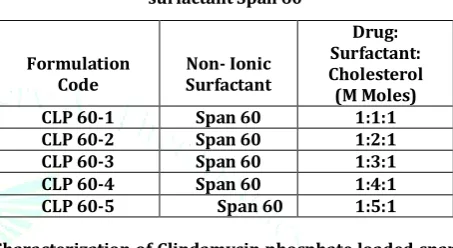
Table 1: Formulation Code of Niosomes containing surfactant Span 60
Characterization of Clindamycin phosphate loaded span 60 niosomes:
Determination of drug content: The amount of drug (100mg) in the formulation was determined by lysing the niosomes using50% n-propanol. 50 ml of the niosomal preparation was pipetted out; sufficient quantity of 50% n-propanol was added and shaken well for the complete lysis of the vesicles. After suitable dilution with water containing 10% Methanol, and drug used in the formulation 100 mg used, the absorbance of the solution was measured at 210nm in the UV- Visible Spectrophotometer. The excipients mixture without the drug treated in the similar manner as the niosomal suspension was used as blank. The drug content was calculated.11
Determination of Entrapment Efficiency: The entrapment efficiency of the formulations was determined by centrifuging 1 ml of the suspension diluted to 10 ml with distilled water at 15,000 rpm for 60 minutes at 4°C using high speed cooling centrifuge in order to separate niosomes from unentrapped drug. The free drug concentration in the supernatant was determined at 210nm using UV- Visible Spectrophotometer after suitable dilution, and drug used in the formulation 100 mg used. The percentage of drug entrapment in niosomes was calculated using the following formula.12
% drug entrapment
In- vitro dissolution studies: The release of Clindamycin phosphate form niosome was investigated in distilled water as dissolution medium (900ml) USP type1 apparatus .A sample of niosomes equivalent to100mg of clindamycin phosphate was taken in the basket. A speed 52 rpm and temperature of 37±0.5℃ was maintained throughout the experiment. At fixed intervals, aliquots (5ml) were withdrawn and replaced with fresh dissolution media. The
Formulation
Code Non- IonicSurfactant
Drug: Surfactant: Cholesterol (M Moles)
CLP 60-1 Span 60 1:1:1
CLP 60-2 Span 60 1:2:1
CLP 60-3 Span 60 1:3:1
CLP 60-4 Span 60 1:4:1
concentration of drug released at different time intervals to determine the absorbance of collected samples were analyzed spectrophotometrically at 210 nm using UV- Visible spectrophotometer, and drug used in the formulation 100 mg used.13
Kinetics Modeling of Drug Dissolution Profiles
To analysis the in vitro release data various kinetic models were used to describe the release kinetic. The dissolution profile of the all formulations was fitted to higuchi and Korsmeyer – Peppa’s model to ascertain the kinetic modeling of the drug release and mechanism of drug release. Zero Oder kinetics (Constant rate process)
Drug dissolution from pharmaceutical dosage forms that do not disaggregate and release the drug slowly can be represented by following equation.
Ft= Kot
Where ft is the fraction of drug dissolved in time t and Ko is zero order release constant.
Pharmaceutical dosage form following this profile release the same amount of drug by unit of time and this is ideal method of drug release in order to achieve a pharmacological prolonged action. The following relation can in a simple way express this model.
Qt= Qo + Kot
Where, Qt is the amount o drug dissolved in time t, Qo is initial amount of drug in solution and Ko is zero order release rate constant. Most of the time Qo = 0.
Application: This relationship can be used to describe the drug dissolution of several types of modified release pharmaceutical dosage forms, as in the case of some transdermal systems, as well as matrix tablets with low soluble drugs in coated forms, osmotic systems, etc.
First order kinetics (Linear kinetics):
Rate is directly proportional to the concentration, of drug undergoing reaction i.e. greater the concentration, faster the reaction. The drug dissolution study was first proposed by Gibaldi and Feldman in 1967. The following relation in a simple way can be expressed as
In (Qt/Qo) = K1t or in decimal logarithm.
Log Qt= log Qo- K1t/2.303
Where, Q1 is amount of drug dissolved in the time t, Qo is initial amount of drug in solution and K1 is first order release rate constant. In this way a graphic of decimal logarithm of the release amount of drug versus time will be linear.
Application: This relationship can be used to describe the drug dissolution in pharmaceutical dosage forms such as those containing water-soluble drugs in porous matrices. Higuchi model:
Higuchi described the release of drug insoluble matrix as a square root of time dependent processes based on Fickian diffusion. A large number of modified release dosage form contain some sort of matrix system. In such instance, the drug dissolved from the matrix. The dissolution pattern of the drug is dictated by water penetration rate (diffusion controlled). In higuchi model, a plot of % drug release versus square root of time is linear.
Q= Kh t1/2 Where, Kh is constant, t is time.
Application: This relationship can be used to describe the drug dissolution from several types of modified release pharmaceutical dosage forms, as in the case of some transdermal systems and matrix tablets with water soluble drugs.
Korsmeyer- Peppas model:
To find the mechanism of drug release, drug release data was fitted in Korsmeyer –Peppas model, log of % cumulative drug release versus log of time.
Mt/M∞= Ktn
Where, Mt/M∞ is fraction of drug released at time t and K is rate constant, n is release exponent.
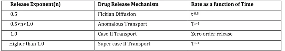
Table 2: Interpretation of diffusion release mechanism from polymeric film
Release Exponent(n) Drug Release Mechanism Rate as a function of Time
0.5 Fickian Diffusion t-0.5
0.5<n<1.0 Anomalous Transport Tn-1
1.0 Case II Transport Zero order release
Higher than 1.0 Super case II Transport Tn-1
Scanning Electron Microscope (SEM):
Particle size of niosomes is very important characteristic. The surface morphology (roundness, smoothness, and formation of aggregates) and the size distribution of niosomes were studied by Scanning Electron Microscopy
(SEM). One drop of niosomal system was mounted on clear
glass stub, air dried and sputter-coated with gold palladium (Au/Pd) using a vacuum evaporator and examined using a scanning electron microscope equipped with a digital camera, at 15 or 20 kV accelerating voltage. The sizes of the
vesicles were measured by scanning electron microscope.14
Optimization Of Process- Related Variables Effect of hydration time:
The niosomal formulations containing Span 60 at different ratios and a fixed amount of cholesterol (1:1:1, 1:2:1,1:3:1,1:4:1,1:5:1) were hydrated with 10 ml of distilled water for 30 minutes, 60minutes and 90 minutes. The vesicle formation and entrapment efficiency of the formulations were calculated by centrifugation method.
Effect of Sonication time
The Niosomal formulations containing Span 60 at different ratios and a fixed amount of Cholesterol (1:1, 2:1, 3:1, 4:1, 5:1) were subjected to ultrasonic vibration using Ultrasonicator. To study the effect of sonication time, the formulations were subjected to sonication for various time intervals (1 min, 2 mins, 3 mins, 4 mins and 5 mins). The entrapment efficiency of the formulations was measured.
RESULT AND DISCUSSION
Preformulation Studies: Description:
Clindamycin phosphate was found to be white or almost white, crystalline powder.
Solubility:
Freely soluble in water; very slightly soluble in ethanol (~750 g/L) TS and acetone R, practically insoluble in dichloromethane R.
Category: Antibacterial drug. Melting Point:
The calculated melting point of Clindamycin phosphate was found to be 114℃. This result is the same as reported in reference and helps in the identification and purity of the drug powder used in the study.
Determination of absorption maxima:
Absorption maxima of Clindamycin phosphate in distilled water were found to be 210nm which is similar to the pharmacopoeia standards.
Fourier Transform Infra-Red Spectroscopy:
The FT-IR spectrum of Clindamycin phosphate, span60 and their physical mixture evidenced various characteristics peaks which are showed in Fig 1, Fig 2.The spectra showed that the free Clindamycin phosphate and formulated Clindamycin phosphate were in resemblance that ratified that the obtained sample of Clindamycin phosphate was pure.
Figure 1: FT-IR spectrum of Clindamycin phosphate
Figure 2: FT-IR spectrum of Clindamycin phosphate, Span60.
Differential Scanning Calorimetry (DSC) studies
Thermal characteristics of pure drug, polymer, and chitosan unloaded microspheres and chitosan drug loaded
Figure 3: DSC thermogram of Clindamycin phosphate
Figure 4: DSC thermogram of drug loaded span60 niosomes. (CLP-3)
Standard curve of Clindamycin phosphate in distilled water.
Standard stock solution and standard working solutions were prepared in distilled water and standard curves were obtained by plotting the data. In fig.5
Figure 5: Standard curve of Clindamycin phosphate in distilled water
Formulation and evaluation of Clindamycin phosphate loaded span 60 niosomes.
Niosomes were prepared by using Reverse phase evaporation method. Further the prepared niosomes were evaluated for drug content, and entrapment efficiency.in fig.6
Table3.Drug Content and Entrapment Efficiency of Clindamycin phosphate Niosomes containing Span-60
Formulation Code
% DRUG CONTENT
(% w/w)(DC) % ENTRAPMENT EFFICIENCY (% w/w)(EE)
CLP – 1 55.02 68.25%
CLP – 2 58.25 69.56%
CLP – 3 59.02 70.25%
CLP-4 70.25 72.52%
CLP-5 71.02 74.25%
y = 0.0832x + 0.0429 R² = 0.996
0 0.1 0.2 0.3 0.4 0.5 0.6 0.7 0.8 0.9 1
0 5 10 15
Ab
so
rb
an
ce
a
t
2
1
0
n
m
Concentration(μg/ml)
ab
Figure 6: Drug Content and Entrapment Efficiency of Clindamycin phosphate Niosomes containing Span-60
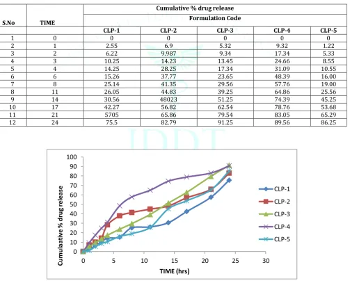
In- vitro dissolution studies
The in- vitro dissolution studies of Clindamycin phosphate from niosomes was examined in distilled water.in fig.7-10 Table 4: Cumulative % drug permeated from optimized formulation
S.No TIME
Cumulative % drug release Formulation Code
CLP-1 CLP-2 CLP-3 CLP-4 CLP-5
1 0 0 0 0 0 0
2 1 2.55 6.9 5.32 9.32 1.22
3 2 6.22 9.987 9.34 17.34 5.33
4 3 10.25 14.23 13.45 24.66 8.55
5 4 14.25 28.25 17.34 31.09 10.55
6 6 15.26 37.77 23.65 48.39 16.00
7 8 25.14 41.35 29.56 57.76 19.00
8 11 26.05 44.83 39.25 64.86 25.56
9 14 30.56 48023 51.25 74.39 45.25
10 17 42.27 56.82 62.54 78.76 53.68
11 21 5705 65.86 79.54 83.05 65.29
12 24 75.5 82.79 91.25 89.56 86.25
Figure 7: In-vitro drug release profile of different Span 60 niosomes
0
10
20
30
40
50
60
70
80
CLP-1
CLP-2
CLP-3
CLP-4
CLP-5
DC
EE
0
10
20
30
40
50
60
70
80
90
100
0
5
10
15
20
25
30
C
u
mu
la
at
iv
e
%
d
ru
g
rel
e
as
e
TIME (hrs)
CLP-1
CLP-2
CLP-3
CLP-4
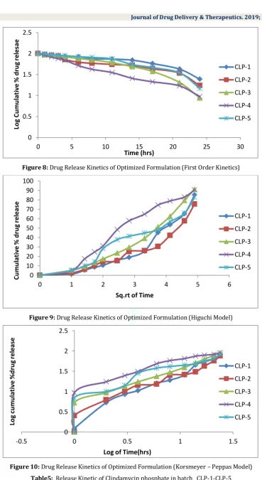
Figure 8: Drug Release Kinetics of Optimized Formulation (First Order Kinetics)
Figure 9: Drug Release Kinetics of Optimized Formulation (Higuchi Model)
Figure 10: Drug Release Kinetics of Optimized Formulation (Korsmeyer – Peppas Model) Table5: Release Kinetic of Clindamycin phosphate in batch CLP-1-CLP-5
0
0.5
1
1.5
2
2.5
0
5
10
15
20
25
30
L
o
g
C
u
m
u
la
ti
ve
%
d
ru
g
rel
es
ae
Time (hrs)
CLP-1
CLP-2
CLP-3
CLP-4
CLP-5
0
10
20
30
40
50
60
70
80
90
100
0
1
2
3
4
5
6
C
u
m
u
la
ti
ve
%
d
ru
g
rel
ea
se
Sq.rt of Time
CLP-1
CLP-2
CLP-3
CLP-4
CLP-5
0
0.5
1
1.5
2
2.5
-0.5
0
0.5
1
1.5
Lo
g
cu
m
u
la
ti
ve
%
d
ru
g
rel
e
as
e
Log of Time(hrs)
CLP-1
CLP-2
CLP-3
CLP-4
CLP-5
F.C. Zero order First Order Higuchi Equation Korsmeyer Peppas
K0(mg/h) R2 K1(h-1) R2 K R2 N R2
CLP – 1 2.78 0.9 -0.02 0.86 14.185 0.867 1.09 0.77
CLP – 2 3.03 0.93 -0.025 0.866 16.55 0.877 1.09 0.82
CLP – 3 3.68 0.96 -0.028 0.88 17.15 0.92 1.011 0.91
CLP-4 3.69 0.97 -0.03 0.91 19.035 0.953 1.012 0.93
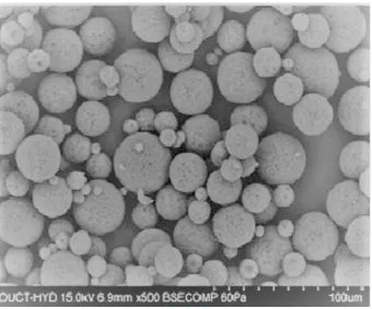
Scanning Electron Microscopy (SEM)
Scanning electron microscopy revealed that the niosomes were spherical and porous. The surface of the was niosomes rough and exhibited the presence of pores in the drug loaded niosomes. The initial burst release of the drug during
dissolution is mainly due to the presence of drug particles on the surface. Surface study of the after niosomes dissolution showed bigger pores indicating that pores are responsible for the drug release and the mechanism of the drug may be diffusion controlled in fig.11
Figure11: Scanning electron microscopy of CLP -3span60 niosomes
Stability studies of Clindamycin phosphate loaded Span 60 niosomes:
Optimum niosomes were exposed to accelerated storage conditions, 40ºC±2ºC/75%RH±5% for six months according to ICH guidelines for stability testing of new drug substances
and products. Niosomes were characterized for physical appearance, drug content and in-vitro drug release at regular intervals. There was no change in the physical appearance of niosomes. Additionally there was no great difference in the drug content and in vitro release characteristics of drug. Thus result implies good stability of the formulation.
Optimization of Process Related Variables Effect of hydration time
Table 6.Effect of Hydration Time on Entrapment Efficiency of CLP-3
S. No. Hydration Time (minutes) Entrapment Efficiency (%w/w)
1. 30 31.06 ±2.52 2. 45 39.5 ±1.15 3. 60 65.2 ±0.16 4. 75 63.53 ±1.5 5. 90 61.27 ±1.11
*Mean± SD (n=3) The niosomal formulations were hydrated with 20 ml of
distilled water for 30, 45, 60, 75 and 90 minutes. The parameters like vesicle formation and entrapment efficiency was studied. The results indicated that vesicles were not formed properly but resulted in aggregates with hydration time of 30 minutes. On increase in hydration time, the
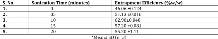
Effect of Sonication time
Table 7.Effect of Sonication Time on Entrapment Efficiency of CLP-3
S. No. Sonication Time (minutes) Entrapment Efficiency (%w/w)
1. 0 46.06 ±0.124
2. 05 51.13 ±0.016
3. 10 62.90±0.040
4. 15 57.20 ±0.081
5. 20 55.20 ±1.11
*Mean± SD (n=3) Formulations were sonicated three times for 5 minutes with
2 sec running time and 1 sec interval. Spherical vesicles were not observed after 10 minutes of sonication. The entrapment efficiency decreased as sonication time increased above 10 minutes. Exposure to Ultrasound for more than 10 minutes damages the vesicles. 10 minutes of sonication resulted in uniform unilamellar vesicles.
CONCLUSION
This research affirmed that the controlled release of Clindamycin phosphate loaded span 60 niosomes can be attained by Reverse phase evaporation method using span 60 as polymer. DSC and FTIR studies exposed that there is no interaction between the drug and polymer which implies that drug is congenial with polymer. The five batches were evaluated for drug content, drug loading and entrapment efficiency. The batch 3 (CLP-3) with the drug and polymer
ratio (1:3.1) was considered to be superior which showed maximum drug content and entrapment efficiency and prolonged release of drug. The drug release from niosomes was affected by various span60 concentrations. Scanning Electron Microscopy (SEM) studies revealed that the niosomes were spherical and porous. The drug release was found to be in controlled manner from batch (CLP-3). Therefore, it was concluded that Clindamycin phosphate loaded span60 niosomes were found to be best in the treatment of acne with reduced the side effect on skin and better diagnosis in a controlled release mode.
ACKNOWLEDGMENTS
The author wishes to acknowledge the Principal, Shivalik college of Pharmacy, Nangal for providing the chemicals and infrastructure and also Registrar, IKG Punjab Technical University, Jalandhar, Punjab.
REFERENCES
1. Keshav J., Niosomes as a potenitial carrier system, Journal of Pharmaceuticals Chemical and Biological science Assistant Professor, Department of Pharmaceutics, Chandigarh University, Gharuan, distt.Mohali, Punjab, India.2015, 947.
2. Goodman and Gilman’s , The pharmacological Basis of therapeutics, 10th edition McGraw Hill Med. Pub. Div. New York, 2001, 777- 779
3. Vyas J, Vyas P, Savant K, “ Formulation and evaluation of Topical Niosomal Gel ofErythromycin”, International journal of Pharmacy and Pharmaceutical Sciences, 2011,3,123-126
4. Government of India Ministry Health and Family Welfare. Indian Pharmacopeia. .
5. British Pharmacopeia. London: The stationery office. 2001; 77(2):22-31.
6. Martindale: The Extra pharmacopeia, 31st ed. London: The royal Pharmaceutical society. 1996. 7. Dubey M, Kesharwani P, Tiwari A, Chandel R, Raja K,
Sivakumar T. Formulation and evaluation of floating microsphere containing anti-diabetic drug. International Journal of pharmaceutical and chemical Sciences. 2012; 1(3):1387-1396.
8. Sawakar S, Dhurvey Y, Sakaekar D. Improvement in micromeritics properties of fenofibrate by spherical crystallization technique. International Journal of Biopharmaceutics. 2012; 3(2):89-95.
9. S.Saumya , “Niosome: A Role in targeted drug delivery” International journal of pharmaceutical science and research, Jan 2013, 550-557
10. Nasir A, SL H and Amanpreet K, “ Niosome: an excellent tool for drug delivery”, international journal of research in pharmacy and chemistry, 2012,481-487
11. Vyas SP, Khar RK. Targeted and controlled drug delivery novel carrier systems. New Delhi: cbs publishers and distributors; 2004:3- 13, 40.
12. Rastogi Aruna. Novel drug delivery system. Pharma tutor- art- 1652.
13. Hillery Anya M, Lloyd Andrew W, James Swarbrick. Drug delivery and targeting for pharmacists and pharmaceutical scientists. Crc press; 2010:63- 82. 14. Azmin MN, Florence AT, Handjani-Vila R.M, Stuart
JFB, Vanlerberghe G and Whittaker JS. The effect of non-ionic surfactant vesicle (niosome) entrapment on the absorption and distribution of methotrexate in mice. J. Pharm.Pharmacol. 1985; 37: 237-242. 15. IF Uchegbu; AT Florenc. Adv. Colloid Interface Sci.,
1995, 58(1), 1-55.
16. Arunothayanum.P, I. F. Uchegbu, D. Q. M. Craig, J. A. Turton and A. T. Florence In vitro/In vivo characterization of polyhedral niosomes International Journal of Pharmaceutics, Volume 183, Issue 1, 10 June 1999, 57-61.
17. Vyas SP, Khar RK. Targeted and controlled drug delivery novel carrier systems. New delhi: cbs publishers and distributors; 2004:3- 13, 40.