Some studies on the
effects of a-chym otrypsin
on mast cells
by
Behzad Emadi Khiav
A thesis presented to the University of London in partial fulfilment of the requirements for the degree of
Doctor of Philosophy in the Faculty of Science
The Christopher Ingold Laboratories,
All rights reserved
INFORMATION TO ALL USERS
The quality of this reproduction is dependent upon the quality of the copy submitted.
In the unlikely event that the author did not send a complete manuscript and there are missing pages, these will be noted. Also, if material had to be removed,
a note will indicate the deletion.
uest.
ProQuest 10018517
Published by ProQuest LLC(2016). Copyright of the Dissertation is held by the Author.
All rights reserved.
This work is protected against unauthorized copying under Title 17, United States Code. Microform Edition © ProQuest LLC.
ProQuest LLC
789 East Eisenhower Parkway P.O. Box 1346
To my mother
Yet Truth syrvives, and only Truth can give The law o f right, and set us free to live In love and peace; and so I wryly smile When falsehood masks in Truth's prerogative
It has been suggested that activation o f one or more proteolytic enzymes might constitute the earliest biochemical change in the sequence o f events that ultimately leads to mediator secretion from the mast cell. This effect may be mimicked by the addition of exogenous serine esterases and, with this in mind, we first characterized the effect o f a-chymotrypsin (a-CT) on isolated mast cells from different sources.
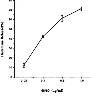
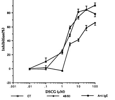
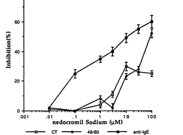
a-C T (10-500 pg/ml) induced a dose-dependent secretion o f histamine (< 80%) from purified and non-purified populations o f rat peritoneal mast cells. The release was non-cytotoxic and was inhibited by metabolic blockers and extremes of temperature. The process was relatively slow, being essentially com plete w ithin 20 min, and was unaffected by phosphatidylserine. A substantial component of the secretion persisted in the absence of extracellular calcium. The release was suppressed by extremes of pH and a variety of anti allergic compound and serine esterase inhibitors.
a -C T (10-300 pg/m l), in addition to the secretion o f preform ed mediators, also induced the metabolism of arachidonic acid, resulting in the release of prostaglandin D% (PGD2) in a dose-related manner from purified rat
peritoneal mast cells.
a-C T exhibited a marked tissue and species selectivity in its action. The protease was a much weaker releaser of histamine from tissue mast cells of the rat. It was effective against human cells from lung, intestine and skin only at cytotoxic concentrations and ineffective against mouse peritoneal mast cells.
The effect of inhibitors of, and substrates for, a -C T in normal and permeabilized rat mast cells were investigated. Rat peritoneal mast cells were recovered by direct lavage and purified by density gradient centrifugation over Percoll. Where appropriate, agents were introduced into the cells after reversible permeabilization with ATP. The seryl enzyme inhibitor phenyl methyl sulphonyl fluoride (PMSF), the suicide inactivator isatoic anhydride, and a number o f chymotryptic substrates all effectively inhibited histamine release from rat mast cells stimulated with anti-IgE but not with compound 48/80. Their potency was strikingly increased in permeabilized cells, indicating their effective incorporation into the cytosol. Activation of a chymotrypic enzyme as evidenced by hydrolysis of the fluorescent substrate, was directly demonstrated following immunologic stimulation of permeabilized mast cells containing N-Succinyl-Leu-Leu-Val-Tyr- 7-amino-4-methylcoumarin. No such activation was observed with compound 48/80. Imm unological stimulation also led to a significant increase in total chymotryptic activity recoverable from rat mast cells.
Acknowledgements
I would like to express my sincere gratitude to my supervisor Professor Frederick L. Pearce for his invaluable advice and support throughout the course of my study. I would also like to thank my colleagues and friends for their assistance and helpful discussions. I would especially like to thank M r Graham A. Mackay for his guidance and many helpful discussions during the writing o f this thesis.
I am deeply grateful to the Department o f Chemistry and Joint Animal House, University College London, for the use of their facilities.
Section
Page
Title
Abstract
A cknow ledgem ents
C ontents
I
III
IV
V
Chapter 1 Introduction 3
1 . 1 Historical Background 4
1 . 2 A Role for the Mast Cell in Physiology and Pathology 5
1.3 Activation of Mast Cells 6
1.3.1 Immunological Activation 7
1.3.2 Non-Immunological Activation 9
1.4 Structural Changes following Mast Cell Activation 9
1.5 Mast Cell Mediators 1 1
1.5.1 Preformed Mediators 1 1
1.5.1.1 The Biogenic Amines 1 1
1.5.1.2 Proteoglycans 1 2
1.5.1.3 Granule Enzymes 13
1.5.2 Newly Synthesised Mediators 18
1.5.2.1 Arachidonic Acid Metabolites 18
1.5.2.2 Platelet Activating Factor 2 0
1.5.2.3 Chemotactic Mediators 2 1
1.5.2.4 The Cytokines 2 1
1 . 6 The Biochemistry Involved in Mast Cell Activation 2 2
1.6 . 1 The Role of Calcium Ions 23
1.6 . 2 Calcium Pools Involved in the Secretory Process 24
1.6.3 The Role of Calmodulin 26
1.6.4 Phospholipid Metabolism 27
1.6.4.1 The Phosphoinositide Cycle 28
1.6.4.2 Phospholipid Méthylation 32
C ontents
1.6.5.1 The Role o f G Proteins 33
1.6.5.2 Activation o f Serine Esterases 36
1.6.6 The Role of cAMP 37
1.7 Mast Cell Heterogeneity 39
1.7.1 The Origin of Mast Cells 41
1.7.2 The Role o f the Microenvironment in the Regulation of
Mast Cell Phenotypic Development 42
1.7.3 Mast Cell Cultures 44
1.7.4 Histochemical and Morphological Differences
between Mast Cells 44
1.7.5 Biochemical Differences between Mast Cells 46 1.7.6 Functional Difference between Mast Cells 47
1.8 General Summary 48
1.9 Aims O f Present Study 49
Chapter 2 Materials and Methods 59
2.1 Introduction 60
2.2 Human Tissue 60
2.3 Materials 60
2.3.1 Full Hepes Buffer (FHB) 60
2.3 .2 2 X Calcium-Tyrode's 61
2.3.3 Calcium-Magnesium-Free Buffer 61
2 .3 .4 Glucose-Free-Tyrode's 61
2.3.5 Heparinized FHB 61
2.3 .6 2 X EDTA-Tyrode's 61
2.3.7 Physiological Saline 61
2.3.8 High Salt-Buffers 62
2.3.9 BSA-Tyrode's 62
2.4 Chemicals and Reagents 62
2.4.1 Histamine Liberators 62
2 .4.2 Inhibitors of Histamine Release 63 2.4.3 Serine Esterase Substrates and Inhibitors 63
2 .4.4 Metabolic Blockers 64
2.4.5 Compounds for Buffers 64
2 .4.6 Chemicals for Histamine Assay 64
2.4.8 Radioactive Materials 65
2.4.9 Miscellaneous 65
2.4.10 Stock Solutions 6 6
2.5 Isolation of Mast Cells 6 6
2.5.1 Peritoneal Mast Cells 6 6
2.5.2 Rat Mesenteric and Lung Mast Cells 67 2.5.3 Guinea-Pig Mesenteric and Lung Mast Cells 6 8
2.5.4 Rat Skin Mast Cells 6 8
2.5.5 Human Lung Mast Cells 6 8
2.5 .6 Human Colonic Mast Cells 69
2.5.7 Human Cutaneous Mast Cells 69
2.5.8 Human Basophils 70
2.6 Mast Cell Characterization 70
2.6.1 Preparation of Cells for Counting 70
2.6.2 Histamine Content 71
2.6.3 Evaluation of Mast Cell Histamine Content 71
2.6.4 Fixation and Staining 71
2.7 Active Sensitisation of Rats 72
2.7.1 Sensitization of Rats with Nippostronglylus brasiliensis 72 2.7.2 Preparation of Third S tage Larvae of Nippostrogylus
brasiliensis 73
2.8 Purification of Peritoneal Mast Cells 73 2.9 Procedure for Histamine Release Experiments 74 2.9.1 Histamine Release from Isolated Mast Cells and Basophils 74
2.9.2 Effect of Metabolic Blockers 75
2.9.3 Effect of Temperature on Histamine Release 75
2.9.4 Kinetic Studies 75
2.9.5 Effect o f pH 75
2 .9.6 Calcium Dependence of the Release Process 76
2.9.7 Synergistic Histamine Release 76
2.9.8 Effects of Inhibitors of Histamine Release 76
2.10 Histamine Assay 77
2.10.1 Manual Assay 77
2.10.2 Automated Assay 78
2.11 Procedure for Prostaglandin D2 Assay 78
2.11.1 Procedure for PGD2 Assay 78
2.11.2 PGD2 Assay Protocol 79
C ontents
2.13 Use of Phosphatidylserine
2.14 Permeabilization of Cells U sing ATP 2.14.1 Preparation of Mast Cells
2.14.2 Introduction of Serine Esterase Substrates into Rat Peritoneal Mast Cells
2.14.3 Measurement of Fluorescence
2.15 Isolation of Chymotrypsin-like Proteinases from Rat Peritoneal Mast Cells
2.16 Spectrophotometric Determination o f Serine Esterase
81 81 81 82 82 83 83
Chapter 3 Some Characteristics of the Effect of a-C T on Rat
Peritoneal Mast Cells
88
3.1 Introduction
3.2 Methods and Materials 3.3 Results
3.3.1 Basic Characteristics of the Effect of a-C T on Histamine Release from Rat Peritoneal Mast Cells
3.3.1.1 Histamine Release by a-C T and Compound 48/80 3.3.1.2 Effect of Metabolic Inhibitors
3.3.1.3 Effect of EDTA and Calcium-Free Buffers 3.3.1.4 Effect of Temperature on the Release Process 3.3.1.5 Kinetics of the Release Process
3.3.1.6 Variation of pH on the Release Process 3.3.1.7 Effect of Phosphatidylserine
3.3.1.8 Effect of a-Chymotrypsinogen on Mast Cells 3.3.2 Prostaglandin D2 (PGD2) Release
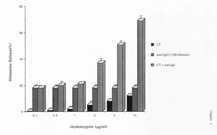
3.3.3 Synergistic Effects of a-C T with Anti-IgE 3.4 Discussion
89 92 92 92 92 92 93 93 93 93 94 94 94 95 95
Chapter 4 The Effect of Anti-Allergic Compounds, Serine Esterase
Substrates and other Inhibitors on Histamine Release from
Rat Peritoneal Mast Cells 115
4.1 4.1.1 4.1.2
Introduction
Anti-Allergic Compounds
Serine Esterase Inhibitors and other Inhibitors
4.2 Methods and Materials 119
4.3 Results 119
4.3.1 Results Pertaining to Part One 119
4.3.1.1 The Effect of Anti-Allergic Compounds 119
4.3.2 Results Pertaining to Part Two 1 2 0
4.4 Discussion 1 2 2
Chapter 5 The Effect of Chymotrypsin and Various Secretagogues
Variety of Histaminocytes Isolated from Different
on
S p ecies 148
5.1 Introduction 149
5.2 Methods and Materials 151
5.3 Results 151
5.3.1 Basic Characteristics of Rat Mast Cells 151
5.3.1.1 Histamine Release by Lectins 151
5.3.1.2 Anaphylactic Histamine Release 151
5.3.1.3 Histamine Release by Polybasic Compounds 152
5.3.1.4 Histamine Release by a-C T 152
5.3.1.5 Histamine Release by Calcium lonophores 152 5.3.2 Basic Characteristics of Guinea Pig Mast Cells 153 5.3.2.1 Histamine Release by Polybasic Compounds 153 5.3.2.2 Histamine Release Induced by a-C T 153 5.3.3 Histamine Release from Mouse Mast Cells by
Compound 48/80 154
5.3.4 Histamine Release from Mouse Mast Cells by a-C T 154 5.3.5 Basic Properties of Human Colonic, Lung and
Skin Mast Cells 154
5.3.5.1 Effect of Anti-Human IgE 155
5.3.5.2 Effect of Calcium lonophores 155
5.3.5.3 Effect of Polybasic Compounds 155
5.3.5.4 Effect of a-Chymotrypsin 156
5.3.5.5 Synergistic Effect of a-C T and Anti-IgE on Histamine
Release from Human Lung Mast Cells 156
Co n ten ts
Chapter 6 Involvement of a Serine Protease in Immunologic Mast
Cell Activation 182
6.1 Introduction 183
6.2 Methods and Materials 185
6.3 Results 185
6.3.1 Characteristics of the ATP-Induced Membrane Lesions in
Mast Cells 185
6.3.2 Inhibition of Histamine Release by
Succinyl-Leu-Leu-Val-Tyr-MCA 186
6.3.3 Inhibition of Histamine Release by S-Pep-MCA
Following Various Incubation Periods 186 6.3.4 Induction of Membrane Permeabilization by ATP and Use of
the Nucleotide as a Tool for Introducing Low Molecular
Weight Substances into Mast Cells 186 6.3.4.1 Detection of Membrane Permeabilization Using Ethidium
Bromide 187
6.3.4.2 Effect of Various Stimuli on Fluorescence Changes
in Rat Peritoneal Mast Cells loaded with S-Pep-MCA 187
6.3.4.3. Effect of Anti-IgE on Fluorescence Changes in Rat
Peritoneal Mast Cells Loaded with B-Arg-MCA 188
6.3.4.4 Fluorescence Photomicroscopy 188 6.3.4.5 Effect of Various Serine Esterase Inhibitors and Substrates
on Histamine Release from Normal and Permeabilized
Mast Cells 189
6.3 .4. 6 Effect o f Various Serine Esterase Inhibitors and Substrates
on Histamine Release from Purified and Non-Purified
Mast Cells 190
6.3.5 Chymotryptic Activity 190
6.4 Discussion 190
Chapter 7 General Conclusions 2 0 9
Structures 23 6
Some studies on the
effects of a-chym otrypsin
Chapter 1
1 .1
Historical Background
The mast cell was originally described by Paul Ehriich as far back as 1878 in his doctoral thesis, in which he named them "mastzellen", or well-fed cells, because of the prominent granules in their cytoplasm (1). Two years later Ehriich also discovered the circulating equivalent o f the mast cell, namely the peripheral blood basophil (2).
Unna in 1894 (3) first reported the association between the mast cell and a pathological condition. He noted that cutaneous lesions o f urticaria pigmentosa consisted almost exclusively o f mast cells. In 1902 Portier and Richet were the first to demonstrate immediate type hypersensitivity reactions in (4). They observed the phenomenon of anaphylaxis (from the Greek phylaxis, meaning guarding) from their work in dogs with sea-anemone toxin. Those dogs that did not die following the initial injection of the toxin, exhibited a dramatic reaction upon a second injection some days later.
It was Dale and Laidlaw (5) in 1919 who put forward the idea that histamine might be one of the chief factors involved in anaphylatic reactions, following their discovery that injection o f histamine into animals induced symptoms similar to those seen during anaphylactic shock. Later work by W ebb (6) implicated histamine in the
anaphylactoid reaction (so called because it resembled anaphylaxis but did not require prior sensitization).
Further work on the concept o f anaphylaxis was carried out by W ilander and co-workers in 1938. They reported that mast cells in the liver o f a dog undergoing anaphylatic shock became grossly damaged, thus liberating their cellular contents into the surrounding tissue (7). Later work by Jorpes and his co-workers (8) in Sweden,
histamine content and mast cell counts in a variety o f tissues (1 0), thus indicating the
mast cell as the primary storage site for histamine.
U n til 1959 the m easurem ent o f h istam in e w as done by classical pharmacological assay using isolated guinea pig ileum. It was then that Shore et a l. (1 1) published work on a fluorometric assay for histamine based on the condensation
product o f the reaction between histamine and o-phthaladehyde (OPT). This enabled the rapid and highly reproducible quantatative determination of histamine concentrations from biological samples.
During the later 1960's Ishizaka and co-workers identified a serum antibody responsible for immediate hypersensitivity reactions as belonging to a novel class, namely immunoglobulin E (IgE) (13,14). Ishizaka and co-workers also discovered that human IgE preferentially bound to basophils via specific receptors (15). Subsequent work identified surface receptors with a high affinity for this particular antibody on tissue mast cells (16).
1 .2
A Role for the Mast Cell in Physiology and Pathology
Mast cells are widely distributed throughout the human body and the bodies of other vertebrate species. They are found essentially in those areas which come into frequent contact with foreign substances, namely in association with nerves and blood vessels and in the loose connective tissue of the bronchi, conjunctiva, skin, lung, ear, nose and gastrointestinal tract (17,18). In short, mast cells are to be found in locations which are frequently exposed to environmental antigens, suggesting they play a role in host defence.
Chapter 1
has been shown that there is a strong correlation between mast cell hyperplasia, eosinophilia and increased IgE antibody level exists after parasitic infestations (19).
Since its reported discovery over 100 years ago, the mast cell and its blood counterpart the basophil have been linked to a number of human disorders especially in the immunopathology o f immediate hypersensitivity reactions. Indeed, histologic and biochemical studies suggest that mast cells, and the products o f their activation, play a role in the pathogenesis of inflammatory conditions such as conjuctivitis (2 0), asthma
(16), rhinitis (21) and urticaria (22). Another factor indicating that mast cells are linked to the pathogenesis of the above allergic conditions is that the number of mast cells in allergic subjects increases compared to normal subjects. M ast cells have also been implicated in a variety of other physiological processes unrelated to allergic disease, such as menstruation (23) and embryo implantation (24).
1 .3
Activation of Mast Cells
Mammalian mast cells are ovoid or irregularly elongated in connective tissues whereas in suspension they are round and have diameter o f 10-30 |im (25-27). The characteristic feature of the mast cell is the presence o f many dense cytoplasmic granules which sometimes occupy the cytoplasm to such an extent as to obscure the nucleus (27). In most mammals, each granule averages 0.2-0.4 jim in diameter and contains, according to the species, a variety of biogenic substances including histamine, 5-hydroxytryptamine, neutrophil and eosinophil chem otactic factors, heparin o r a related glycosaminoglycan and hydrolytic enzymes (27,28). Activation of the mast cell leads to the release of the granules and their associated preformed mediators, and also evokes the de novo generation of further bioactive substances such as leukotrienes, prostaglandins and thromboxanes (29-31).
1 . 3 . 1 Immunological Activation
Allergy is a disorder of the immune system in which entirely innocuous substances such as animal danders, certain grass pollens or house dust mite,
DermatophagoideSy are mistaken as harmful, resulting in the production of IgE against epitopes o f these antigens (32). Individuals w ho produce IgE under these circumstances are said to be atopic.
Immunoglobulins are glycoproteins composed o f 82-96 % polypeptide and 4- 18 % carbohydrate. IgE exhibits the basic four chain structure common to other antibodies and is thus composed o f two light and two heavy chains, linked by disulphide briges. There are two types o f light chains, kappa and lamba, and five heavy chains (y, a , |i, o and e) which distinguish the subclasses of immunoglobulin (33,34). Fig 1.1 shows that immunoglobulin molecules have a Y-shaped configation. The immunoglobulin, when treated with the enzyme papain, breaks into two Fab fragments and one Fc fragment. Fab regions contain the antigen binding sites and each immunoglobulin is divalent. The lower part of the molecule, designated the Fc portion, binds to specific receptors on various cell surfaces. It appears that a certain steric relationship between the two heavy chains and conform ational structure in the Fc portion is required for IgE molecules to combine with target cells with high affinity (34). Since the receptors on mast cells and basophils are specific for the Fc portion of IgE which consists of two characteristic heavy chains (e), these receptors are named Fce receptors to distinguish them from other Fc receptors for other immunoglobulins (35).
Chapter 1
process (36,32). When B-cells mature into plasma cells, IgE antibody possessing the antigenic determinant is released into the circulation. Thus, on primary exposure to a causative allergen, such as grass pollen, susceptible individuals produce specific IgE antibodies to these agents. When IgE is secreted into the circulation, it can bind to the high affinity IgE receptors on mast cells, through its FCg portion, leaving free the recognition site for allergen (Fab). At this point, the cells are said to be sensitized but with no apparent sign o f degranulation. However, subsequent exposure to the same allergen results in the cross-linking of IgE molecules and the consequent release of both preformed and newly synthesized mediators of anaphylaxis (38,39).
Information concerning the high-affinity IgE receptor has mainly been derived from work on rat peritoneal mast cells and rat cultured basophilic leukaemia (RBL) cells (40,41). The receptor was shown to be a glycoprotein containing ca. 13% carbohydrate. It was also demonstrated to consist of three subunits (a , p and y), having an overall molecular weight of 87,000 ( a 45,000, P 33,000 and y 9,000). The a , p and y subunits are further divided into a j , a2, P%, P2; the two y chains being
identical (Fig 1.2). The a subunit of the receptor is directed to the surface of the cell and binds IgE, while the p and y subunits, which could not be labelled using selective antibodies from the cell surface, are thought to be buried in the cell membrane. A utoradiographic and radioassay studies on IgE binding to normal mast cells and basophils dem onstrated that each o f these cells possess approximately 10^ IgE receptors.
cross-links IgE by binding to sugar moieties in the Fc^region) and anti-receptor antibody (IgG antibody prepared against purified Fcg receptors) (44-46).
1.3.2 Non-Im m unological Activation
Secretion from mast cells, according to species, may be induced experimentally by a wide range o f pharmacological stimuli. In general, these agents can be classified into two main categories based on their mode of action.
The first group consists o f agents known collectively as non-selective liberators. They are cytotoxic agents such as Triton X-100 and Tween 20 (47,48). These compounds act by disruption of the mast cell membrane, thereby liberating all of the intracellular contents including histamine.
In contrast, agents belonging to the second group are non-cytotoxic and can cause release without the loss of characteristic cytoplasmic markers such as the enzyme lactate dehydrogenase. These selective liberators include polybasic cations typified by compound 48/80, substance P, polylsine, polymyxin and peptide 401 (49-52), the anaphylatoxins C3a, C4a and C5a (53), the plasma substitute dextran (54), the calcium ionophores ionomycin and ionophore A23187 (55-57), adenosine 5 ’-triphosphate (ATP) (58,59), a-chymotrypsin (60) and a variety o f drugs and miscellaneous organic molecules (61,62). These diverse agonists provide useful pharmacological tools for studying the biochemical processes involved in mast cell activation.
1 .4 Structural Changes Following Mast Cell Activation
Chapter 1
cells show considerable diversity in structure. In general, human skin mast cells were found to contain crystal granules; lung mast cells, scroll granules; and colonic mucosal mast cells, particle granules (63-65).
The sequence o f events leading to exocytosis may be dissim ilar for different mast cell types. In the rat peritoneal mast cell, the initial ultrastructural change involves those granules situated immediately below the cell membrane. These granules become swollen in appearance and the release process begins via openings produced by the fusion o f the two membranes. Following this initial contact, many intergranular fusions are formed which spread towards the interior of the cell and open up extensive labyrinthic cavities within the cell which can com municate with the extracellular medium through multiple openings. As granules are discharged, histamine and other ionically-bound molecule are then released, possibly through a simple exchange process with external sodium ions (66,67).
The mechanism of human mast cell degranulation has been shown to differ somewhat from its rat counterpart. In human mast cells, one minute after stimulation, the granules swell and dissolution of their matrix material can be seen. By three to five minutes after stimulation, the surrounding swollen membrane granules fuse with each other. This leads to the formation of channels through the plasma membrane extending outwards. Thus, the solubilized granular contents are then finally extruded into the surrounding medium (6 8). However, unlike rat peritoneal mast cells, no apparent extracellular
1 .5
Mast Cell Mediators
The role of the mast cell in allergy and inflammation is dictated by the mediators released upon activation and the tissues into which they are secreted. Chemical mediators may either be preformed and stored within the secretory granules of the mast cell or be synthesized de novo upon activation.
1 . 5 . 1 Preformed Mediators
1.5.1.1 The Biogenic Amines
Histamine which is primarily stored in mast cells and basophils, is one of the main contributors to the immediate allergic response. Increased plasma and tissue histamine levels occur during anaphylaxis and allergic disorders of the skin, nose and respiratory airways (70). Histamine (from the Greek word for tissue, histos) is synthesized in nature from histidine by the enzyme histidine decarboxylase (71). Rat serosal mast cells contain 10-30 pg of the amine per cell (27,72), rat mucosal mast cells 1-2 pg (73), human mast cells (74-76) and human basophils 1-3 pg (77). The most im portant effects o f histamine in allergic reactions are mediated through the Hj^-receptors. These include contraction of bronchial and gastrointestinal smooth muscle, vasodilation, increased capillary perm eability and neutrophil and eosinophil chemokinesis (78). In contrast, the effects mediated via the H2-receptor are more anti
inflammatory and include the inhibition of T-lymphocyte cytotoxicity, suppression of lymphocyte proliferation, and mediation o f gastric acid secretion (79). A third histamine receptor, Hg, thought to mediate a negative feed-back action on histamine synthesis, has recently been described in neural tissues (80). Many other biological effects, although predominantly H^ or H2 - mediated, result from the combined effects
Chapter 1
Despite its wide range of activities, histamine is rapidly metabolised, having a plasm a half-life o f 2-3 min. M etabolism occurs via two routes, N-methylation by histamine N-methyltransferase or oxidation by histaminase (diamine oxidase).
In addition to histamine, 5-hydroxytryptam ine (5-HT), which derived its alternative name serotonin from its first isolation from hum an serum and its vasoconstrictor activity (81), is found in connective tissue mast cells o f rodents. Human mast cell granules, however, do not contain 5-HT (82). The spasmogenic and vasoactive amine is also found stored within the granules o f human platelets and is a well-established neurotransmitter of the central nervous system (83).
1.5.1.2 Proteoglycans
Proteoglycans are major components o f tissue ground substance, bone and cartilage. They form the structural basis o f lysosomal granules and, in the mast cell granules, act as storage matrices for preformed mediators such as histamine and, in rat m ast cells, 5HT. There are two major types o f glycosaminoglycans found in mast cells; heparin and chondroitin sulphates. These glycosam inoglycans (GAGs) are attached within the mast cell to small peptide cores, hence the term proteoglycans (84). The exact function of mast cell proteoglycans has yet to be fully established. It is known that proteoglycans bind to histamine within the secretory granules and in so doing may facilitate the uptake and packaging o f histam ine into the granules. Proteoglycans also regulate the stability and activation o f other enzymes present in the mast cell.
Heparin consists of a central peptide core, comprising alternating serine and glycine residues. GAGs are attached to every second or third serine residue by a unique sequence of sugars, comprising glucuronic acid-galactose-galactose-xylose, a linkage that is common to all proteoglycans (85). The GAG is made up of a series of disaccharide units that are a 2 . 4 linked and composed of either glucuronic or iduronic
causes it to shift the colour absorption and emission spectrum of certain basic dyes such as azure A (metachromasia) which results in the ability to stain and hence visualize mast cells by light microscopy.
Chondroitin sulphates are less highly sulphated but structurally similar to heparin. The repeating dissacharide unit of this GAG consists of galactosamine and glucuronic acid joined through as well as p^ . 4 linkages.
As a result o f their acidic properties and capacity to exclude water, proteoglycans are able to inactivate and package other stored mediators in the mast cell granule (8 8). Approximately 10% of granule histamine is bound ionically to the ester
sulphate and carboxyl groups of GAGs. A further small proportion is complexed to zinc but the majority of the histamine is bound via carboxyl groups on highly basic polypeptides which are found complexed with heparin.
.5.1.3 Granule Enzymes
There are three main classes o f enzyme in the mast cell granule; the neutral proteases, the acid hydrolases and the oxidative enzymes.
The neutral proteases are a group of proteolytic enzymes or serine esterases with optimal activity at neutral pH, which cleave peptide and ester bonds on the carboxyl side of basic (tryptase), aromatic (chymase) or terminal aromatic (carboxy- peptidase) amino acids.
IgE-Chapter 1
mediated degranulation and in pathobiological alterations in connective tissue around venules and in bronchial and gastrointestinal mucosa (96,97). However, the actions of these proteases in allergic inflammation are not well understood.
Recently, serine protease inhibitors, substrates, substrate analogs, and antibodies of protease were shown to inhibit histamine release from IgE-activated mast cells, and the protease involved in the process o f degranulation was identified (93,96-101). Pathobiological alterations that occur in allergic disorders may in part be explained by these serine proteases secreted from activated mast cells. Endogenous regulators o f the activities of these proteases, such as trypstatin (1 0 2) for rat tryptase, heparin for human
tryptase (103) and for rat and human chymase (89,103,104), and phosphoglycerides and fatty acids for rat chymase (105,106) have been reported.
The chymotrypsin-like serine protease in connective tissue mast cells, named chymase, was first detected in rat peritoneal mast cells by Benditt and Arase (107). This protease was purified and crystallized from rat skeletal muscle and liver as "group- specific pro tease" (108-110) and was later shown to be localized in connective tissue mast cells in these organs (111). It has also been purified from rat peritoneal mast cells (89,104). Human chymase was found in extracts from human skin (111) and has also been purified (95).
Chymase is localized in m ast cell granules as a com plex with heparin proteoglycan and is solubilized from granule fractions only at high ionic strength (89,103). Although (3-hexosaminidase and histamine are fully soluble on their release from mast cells into physiologic buffer, the com plex o f chym ase and heparin proteoglycan is retained by the cells in an insoluble form exposed to the extracellular milieu (100,112). Therefore, the action of chymase may be restricted to a relatively small protein that can penetrate the chymase-heparin network. The prolonged presence o f heparin proteoglycan with chymase at the cell surface of activated mast cells may be relevant for the chronicity of the host response to mast cell activation.
corresponding to the catalytic triad characteristic o f serine proteases (His 57, Asp 102, and Ser 195 in a-chymotrypsin). The isoelectric point o f the chymase is 9.3, whereas that o f a-chym otrypsin is 8.5 (104). Unlike bovine a-chy m otry psin , which is composed of two protein chains linked by disulphide bonds and with a molecular weight o f 25,000, rat and human mast cell chymases are single chain proteins with molecular weights o f 24,000-29,000 (89,104,108). Like a-chym otrypsin, chymase catalyzes the cleavage o f peptide and ester bonds on the carboxyl end of internal aromatic amino acids at pH 8-9 (11).
Little is known about the function of chymase in vivo, but the rat enzyme can stimulate other mast cells in the microenvironment (113), generates a chemotactic factor for neutrophils from IgG (98), and degrades basement membrane type IV collagen and fibronectin but not type I, II, or III collagen (114) in vitro. The human enzyme has also been shown to convert angiotensin I to angiotensin II (115). M oreover, the proteolytic product(s) of IgG formed by rat chymase has potent chemotactic activity on neutrophil leukocytes in vitro and in vivo, whereas IgG itself has little chemotactic activity (96). In addition, as discussed previously, mast cells have been shown to have specific receptors for both IgE and IgG (116), and therefore it may be that released chymase causes a limited hydrolysis of IgG and the resultant production of chemotactic factor(s). It has also been shown that exposure of peritoneal mast cells to rat chymase or a-chym otrypsin results in degranulation (113, 117), and this degranulation is attenuated by enzyme inhibitors such as diisopropylfluorophosphate, indicating that the release is a function of enzymatic activity (113).
Chapter 1
diverge phenotypically due to the various micro-enviromental factors present at the site o f their final tissue location (1 2 1).
The amino acid sequence of atypical chymase from rat small intestine has been determined (92). The enzyme contains 224 amino acid residues in a single polypeptide chain, has a minimum molecular weight o f 24,768, and contains three di sulphide bonds. Atypical chymase has nine few er lysyl residues than chymase, which may account for the striking difference in the ease o f extraction o f the two enzymes from mast cells. Hence, atypical chymase, which may be associated with proteoglycan, is readily extracted with physiological saline, whereas chymase, which interacts tightly with the more sulphated heparin in the granules, can be extracted only with 1 M salt (89,103). Another structural difference between chymase and atypical chymase is the presence of a seryl residue in place of an alanyl residue at position 176, this being the putative substrate pocket of atypical chymase.
Atypical chymase from rat mucosal mast cells, which is a highly soluble protein, is released into the blood circulation or gut lumen after intestinal anaphylaxis; thus the plasma level of atypical chymase is correlated with, and its a good marker of, events related to mucosal mast cells (122). The enzyme released from mucosal mast cells has been suggested to play a role in the generation o f intestinal epithelial permeability (123) and to be involved at some level in the expulsion o f intestinal parasites (124). A typical chymase has also been shown to degrade basement membrane type IV collagen in vitro, in the same way as chymase.
from rat peritoneal mast cells was also suggested to be localized in the granules, but its placement in rat mucosal mast cells has yet to been reported (91,127).
The enzymes from humans and rats are tetramers, with apparent molecular weights o f 144,000 and 142,000, respectively (91,94). Human tryptase has two subunits of 37,000 daltons and two subunits o f 35,(XX) daltons (94). Rat tryptase has four identical subunits of 35,000 daltons, each having one active site (94). There are several differences between human and rat tryptases. It has been reported that human tryptase is resistant to inhibition by aprotinin, an a^-trypsin inhibitor (94), whereas rat tryptase is inhibited by these agents and by trypstatin (128), an endogenous inhibitor of tryptase. Moreover, human tryptase binds to heparin proteoglycan in physiologic buffer in vitro and may be present as a complex with heparin proteoglycan in vivo,
whereas rat tryptase does not interact with heparin (94).
Tryptase is involved in complement activation by cleavage o f C3 into its component parts C3b and the anaphylatoxin, C3a (129). In the presence of heparin, however, which has a modulatory effect on the enzyme, tryptase further cleaves and inactivates C3a. The protease also has a local anticoagulant action through degradation of fibrinogen and high molecular weight kininogen (129) and is involved in the breakdown of connective tissue (130,131). In addition, tryptase from human lung mast cells may play a role in bronchial hyperresponsiveness in asthma through its ability to degrade and inactivate vasoactive intestinal peptide (VIP), the main bronchorelaxant neurotransmitter o f non-adrenergic, non-cholinergic (NANC) nerves in the lung (BS). This is selective since substance P (SP), a bronchoconstrictor, is not affected by tryptase.
Chapter 1
The oxidative enzymes present in rat peritoneal and human lung mast cell granules include superoxide dismutase (132) and peroxidase (133). Both enzymes function to remove super-oxide anions and, in the case o f peroxidase, inactivate dihydroxy- and sulphidopeptide leukotrienes.
1 . 5 . 2 Newly Synthesised Mediators
1.5.2.1 Arachidonic Acid Metabolites
Mast cells have the capacity to synthesize and release a range of lipid mediators following immunological or non-immunological stimulation, both in vitro and vivo.
All the newly formed lipid mediators, collectively known as eicosanoids, possess inflam matory properties to varying degrees. This process begins when arachidonic acid (5,8,11,14-eicosatetraenoic acid) is released from the membrane phospholipids by the action o f the enzym e phospholipase A2 . A rachidonic acid serves as the
predominant biosynthetic precursor of an array of lipid mediators that are generated and released by mast cells, basophils and other leukocytes in the course of immediate-type hypersensitivity reactions.
A substantial portion of the arachidonic acid that is liberated from the phospholipids of the leukocytes is metabolized by the enzymes 5-lipoxygenase or cyclooxygenase and leads to the production of prostaglandins (PCs) and thromboxanes (TXs), via the cyclooxygenase pathway, or leukotrienes (LTs), via the lipoxygenase pathway (134) (Fig 1.4).
Cycloxygenase is a membrane associated heme protein enzyme complex that catalyzes the incorporation of molecular oxygen into the arachidonic acid molecule, follow ed by ring closure to form the relatively unstable cyclic endoperoxide interm ediates PGG2 and PGH2. These intermediates may then be converted to form
the primary prostaglandins PGD2, PGE2 and PG p2a , in addition to PFI2, TXA2 and
of PGD2 (50-60 ng/cell) after stimulation by calcium ionophores, anti-IgE and antigen
(135). Once liberated, PGD2 causes smooth muscle contraction and increased vascular
permeability. Recent studies have also indicated that PG D2 may cause systemic
flushing and hypotension in humans with mastocytosis (136).
A second enzyme, 5-lipoxygenase, initiates the synthesis of leukotrienes. 5- Lipoxygenase has been purified from several sources and, in each case, activity is dependent on Ca^'*' and ATP, a feature that distinguishes the enzyme from other lipoxygenases. 5-Lipoxygenase is normally present in the cytosol but, on cell activation, the enzyme undergoes Ca^'^'-dependent translocation to the cell membrane. The mechanism o f translocation was elucidated by researchers at Merck-Frost, who dem onstrated that 5-lipoxygenase associates with a novel m embrane protein now known as 5-lipoxygenase activating protein (FLAP) (137). Although cell-free assays for 5-lipoxygenase are norm ally carried out using soluble enzym e fractions, transfection experiments have clearly shown that the presence of both 5-lipoxygenase and FLAP is essential for leukotriene biosynthesis in intact cells (138). The initial product is the unstable intermediate 5-hydroperoxyeicosatetraenoic acid (5-HPETE) and LTA4. The latter is rapidly converted to LTB4 by reaction with water or, alternatively,
is converted into LTC4, by addition o f the tripeptide glutathione. The sequential peptidolytic cleavages of the glutathione generates LTD4 and LTE4. The mixture of L T C 4 , LTD4 and LTE4 com prises the classical slow -reacting substance of anaphylaxis.
Peptidoleukotrienes have powerful spasmogenic actions, particularly in airway smooth muscle and in the vasculature. These mediators are at least 100 times more potent than histam ine or m ethacholine as bronchoconstrictors in man when administered by aerosol (139) and asthmatic patients show enhanced sensitivity to the bronchoconstrictor effects of peptidoleukotrienes (140). LTB4 is a potent mediator of
inflammation via iis chemotactic activity for leukocytes. LTB4 is a potent activator of
Chapter 1
diseases, suggest that 5-lipoxygenase inhibitors may have therapeutic potential in a range o f allergic and inflammatory conditions such as asthm a, allergic rhinitis, rheumatoid arthritis, ulcerative colitis and psoriasis.
Recent studies into whether arachidonic acid metabolism occurs concurrently with histamine release have indicated that, in many instances, the lipoxygenase pathway o f arachidonic acid metabolism fails to commence, although histamine release occurs readily (142,143). Thus, the findings suggest that arachidonic acid metabolism via both o f the two enzymic pathways is not always initiated but depends on the type of stimulation received by the mast cell or basophil.
1.5.2.2 Platelet Activating Factor
There is evidence that mast cells, especially those cultured and differentiated in vitro from mouse bone marrow in the presence of interleukin 2, can synthesize a unique
phospholipid named platelet activating factor (PAF) ( 1 -O-alkyl-2-acetyl-jM-glyceryl- phosphorylcholine) (144). More recently it has been reported that purified preparations o f isolated human lung mast cells (145) can synthesize PAF following challenge of these cells by anti-IgE and calcium ionophore A23187.
Although PAF was originally characterized by its potent aggregatory effects on platelets (146), it has now been dem onstrated that PAF activates a num ber of inflammatory cells. It is one of the most potent eosinophil chemotaxins and activators
in vitro (146) and also induces neutrophil chemotaxis and activation. In the lungs it causes pulmonary oedema, vasoconstriction and bronchoconstriction, pointing towards a possible role in asthma. Systemically, PAF causes hypotension and on injection into human skin it produces a wheal and flare reaction which, on a molar basis, is 1 0 0 - 1 0 0 0
1.5.2.3 Chemotactic Mediators
M ast cells also release a variety o f chem icals which are chemotactic for eosinophils and neutrophils after activation by certain stimuli. M ast cell chemotaxins include EC F-A , which is eosinophil chem otactic factor o f anaphylaxis, and inflammatory factor of anaphylaxis (IF-A) which stimulates neutrophil (2-8 hours) and monocyte (24 hours) infiltration (148). Newly generated mediators LTB4 and PAF,
also have chemotactic activity.
Chemotactic factors are responsible for the second wave of inflammation or late phase reaction (LPR), which occurs 3-6 hours after immediate type hypersensitivity and is characterized by the influx of neutrophils and eosinophils. Such LPRs dominate more chronic allergic reactions such as asthma, eczema and urticaria.
1.5.2.4 The Cytokines
It has become increasingly recognised in recent years that atopic asthma, rhinitis and dermatitis are more than simple allergic conditions involving type I hypersensitivity reactions to allergens. They are now known to involve a number o f inflammatory and other cells, producing not only various chemical mediators but also many cellular products with wide-ranging effects on both target and effector cells. The net result of this is the generation of a chronic inflammatory process which, in the long term, may be more important in the pathogenesis of allergic diseases than the acute type I reaction involving mast cell activation.
Chapter 1
increase immunologbulin secretion, and enhance the production and differentiation of further mast cells (149). Recently, tumour necrosis factor-a (TN F-a) or cachetin was shown to be produced by certain cultured murine mast cell lines (150,151), and mast cell/basophils from human bone m arrow cultures (152). The use o f in situ
hybridization techniques located T N F-a mRNA in the cytoplasm and T N F -a protein in the granules o f individual cells (152), suggesting the cytokine is both inducible and preformed in the mast cell. Mast cells are involved in a variety of processes including tumour cell cytotoxicity (153), bone remodelling (154) and fibroblast growth (155) in which TNF has also been shown to play a role (156).
Murine mast cell lines also produce on impressive array of other polypeptide cytokines following immunologic or Ca^"^ ionophore-induced stimulation. These include IL3 (a mast cell growth factor), IL4 (a switch for IgE), IL5 (eosinophil differentiation factor), IL6 (controls IgG secretion) (156), GM-CSF (157), IL l, IFNy,
M IP a and MIPp (macrophage inflammatory protein) (158). Mast cells may therefore modulate their own growth and functional activity by the secretion o f IL3 and IL4, respectively, and be involved in the asthmatic LPR, characterized by the presence of large numbers of eosinophils, through their production of IL5. These novel findings place the mast cell back into the middle o f the overall allergic reaction, not only in initiating the im m ediate reaction but possibly in controlling the more chronic inflammatory events of the late phase reaction.
1.6
The Biochemistry Involved in Mast Cell Activation
In addition to its clinical importance, the mast cell has also provided a model system for the study of the biochemical processes involved in stimulus-secretion coupling.
It is now known that mast cell activation and subsequent mediator release involves not one but many complex biochemical pathways. However, it is important to bear in mind that most o f the current information has been obtained from studies with murine mast cells. Results generated from this cell type cannot easily be extrapolated to human cells.
1.6.1 The Role o f Calcium Ions
For some time it has been appreciated that calcium ions play an important role in secretion. M ongar and Schild (159) were the first to demonstrate a requirement for extracellular calcium in the anaphylactic release o f histamine from guinea pig lung fragments.
In the early 1970's, it was shown that histamine release could be induced by direct micro-injection o f calcium ions into rat mesenteric m ast cells (160). This observation indicated the necessity of calcium ions to induce, if not initiate, histamine release from mast cells. Thus, calcium ions were thought o f as being a second messenger as they transduced the ligand-receptor signal to the cell interior and, in doing so, triggered processes which led to mediator release.
In view of the apparent association between calcium ions and mediator release from mast cells. Foreman, et al. (161) investigated cation fluxes following cellular activation. Studies using radioisotopic Ca indicated that the time course, and magnitude, of calcium influx correlated to the degree o f histamine liberation.
Chapter 1
Lanthanum ions and to a lesser extent, other members of the lanthanide series, have also been used to demonstrate this calcium dependency (163). Lanthanum has an ionic radius comparable to that o f calcium and is able to displace competitively the divalent cation from superficial sites in the cell membrane. By virtue of its higher valency, lanthanum binds to these sites with a greater affinity than calcium and blocks the transport of the divalent cation across the membrane. Employed in this way, it has been shown th at lanthanum inhibits histam ine release induced by various secretagogues.
Further studies on the role played by calcium have been facilitated by the use of the fluorescent tetracarboxylate calcium chelator, quin-2. After antigen stimulation of rat mast cells, an increase in fluorescence indicated a rise in the concentration of cytosolic calcium ions (164).
1 . 6 . 2 Calcium Pools Involved in the Secretory Process
It is now well established that a rise in the concentration of free calcium ions in the mast cell's cytosol is a necessary and sufficient trigger for histamine secretion. There are two basic mechanisms by which mast cells can increase their cytosolic free calcium ion levels: via release of calcium ions from intracellular stores or by the entry of extracellular calcium ions through membrane calcium channels (165,166).
of calcium ions, displayed a time-dependent decrease in the level of histamine release in response to stimulation (168).
Although optimal histamine secretion requires the presence of extracellular calcium ions, many test secretagogues can release a considerable amount of this amine in the absence o f extracellular calcium ions (169). Such secretagogues include compound 48/80, peptide 401 and polylysine (163,169). The release is attributed to the mobilization of intracellular pools of calcium ions or sequestered calcium ions attached to the inner surfaces of the plasma cell membrane or cellular organelles. P ro lo n g e d e x p o su re o f th e se c ells to th e c a lc iu m -c h e la tin g ag en t ethylenediam inetetraacetic acid (EDTA) (163,169) and preincubation with the ionophore A23187 (170) in the absence of extracellular calcium rendered them unresponsive to compound 48/80. The cellular response was restored, however, follow ing réintroduction o f Ca^"^ to the medium. The im plication from these observations was that such agents mobilized intracellular stores o f the cation which were depleted by the above treatments and replenished by addition of extracellular
Ca2+.
In contrast to the above, brief exposure to EDTA markedly enhances the secretion produced by many inducers (169). This treatment is believed to remove calcium ions from superficial, regulatory sites in the cell membrane, leading to its destabilization and thus facilitating the release of more internal stores o f the cation. Conversely, high concentrations of extracellular calcium have the opposite effect, the result is saturation of these regulatory sites, thus stabilizing the cell membrane and possibly restricting uptake of the cation (169).
Chapter 1
or substance P revealed the existence of a sustained plateau o f elevated [Ca^'‘"]i, which was dependent on external calcium and which followed the initial, large, transient increase in (171). The Ca^"*" plateau, in contrast to the early Ca^'*' rise, was ideally timed to influence the secretory response.
Three ionic mechanisms were identified which explained this calcium influx. A chloride channel, activated by internally applied cAMP, hyperpolarizes the membrane (holds the membrane potential at a negative value) and provides the electrical driving force necessary for the uptake o f Ca^"*". This hyperpolarization facilitates the opening o f Ca^"*"- specific channels in the membrane which are also sensitive to intracellularly applied IP3. Finally, there is a non-specific channel through which divalent cations can
permeate. Such channels are not voltage-gated but are potential-dependent in so much as the Ca^"^ influx depends on an electrical driving force. Thus, it would appear that Ca^"*" influx in the mast cell is regulated by membrane channels sensitive to the two intracellular messengers IP3 and cAMP. Indeed, where depolarization in excitable cells
results in the opening of voltage-gated Ca^"*" channels, hyperpolarization has often been associated with Ca^"*" influx in non-excitable cells (174,175).
These exciting findings have stimulated interest in the use of calcium channel blockers, such as nifedipine and verapamil in clinical inhibition of immunological histamine release (176).
1 . 6 . 3 The Role o f Calmodulin
four calcium ion binding sites. Binding o f Ca^'*' to CaM confers a more stable structure on the protein and the resultant conformational changes reveal active sites on the surface of the molecule which allow interaction with and activation of apoenzymes or effector proteins. Thus, calmodulin regulates the activities o f a large number of key enzymes involved in secretion: adenylate cyclase, methyltransferase, phospholipase A2
and cyclic nucleotide phosphodiesterases (169,177,178).
A number of neuroleptic drugs, particularly those of the phenothiazine family, are able to combine with the calcium-calmodulin complex. These compounds prevent interaction o f the complex with its target apoenzymes and thus block calmodulin- dependent reactions. Importantly, these calmodulin antagonists have been found to inhibit histamine secretion from mast cells and human basophils (179-181), thus indicating that calmodulin may be significantly involved in the release process.
In total, calmodulin controls many key events involved in exocytosis including the regulation o f calcium homeostasis, the synthesis and degradation o f cyclic nucleotides, the organization of the cell cytoskeleton and protein phosphorylation (177,
178).
On the theme of protein phosphorylation, pharmacological activation of rat mast cells has been shown to result in a rapid, calcium-dependent phosphorylation of three specific proteins o f m olecular weight 68,000, 59,000 and 42,000 (182). The phosphorylation accompanies or precedes histamine secretion from the cells. The natural termination o f the release process involves the phosphorylation of a further protein of molecular weight 78,000. Calmodulin, by activating a number of specific kinases, may therefore be involved in both the induction and termination of exocytosis.
1 . 6 . 4 Phospholipid Metabolism
Chapter 1
1.6.4.1 The Phosphoinositide Cycle
It has been shown that activation of cell-surface receptors initiates hydrolysis of a membrane-bound inositol lipids, which produces at least two second messengers diacylglycerol (DAG) and inositol-1,4,5-trisphosphate (IP3). These messengers are
generated by a membrane transduction process comprising three main components: a recep to r, a coupling G p ro tein and p h o sp h o lip ase C (PLC ). In itially , phosphatidylinositol (PI) breakdown to diacylglycerol (DAG) was measured by analyzing the incorporation of radiolablled [^^P] phosphate and [^H] inositol into PI, phosphatidylcholine (PC) and phosphatidic acid (PA). This finding led to many groups reporting that a variety of secretagogues induce accumulation o f [^^P] into rat mast cell PI (183-185). Later work by Ishizaka and co-w orkers (186) confirm ed that an increased uptake of [^^P] into membrane phospholipids occurs after IgE-dependent stimulation of human cultured basophils.
Phosphorylation o f PI by A TP-dependent specific kinases results in the s e q u e n tia l fo rm a tio n o f p h o s p h a tid y lin o s ito l-4 -p h o s p h a te (P IP ) and phosphatidylinositol-4,5-bisphosphate (PIP2) (187) by a specific phospholipase C
enzyme.
Several types of phosphatidylinositol specific phospholipase C enzymes have been identified by either purification or subsequent isolation o f cDNAs. Based primarily on size and characterization of activities, these were initially grouped into five categories (a,p,Y ,6 ,e). The cDNAs for three o f these have been isolated and
sequenced. Comparison of the sequences of several isotypes o f each of these led to the identification of 2 domains (X and Y) which were highly conserved. The regions are hypothesized to contain the catalytic site o f the enzymes. These comparisons also showed that there were structural arrangements that characterized the major groups of phosphatidylinositol specific phopholipase C (PIPLC) (188-191).
receptors, such as PDGF (platelet-derived growth factor) and EGF (epidermal growth factor) and subsequent co-immunoprecipitation o f the enzyme with growth factor receptors has been observed. Thus, this isoform o f PIPLC presumably accounts for the increased production of inositol phosphates caused by these hormones. In contrast, PIPLC a isoforms are regulated by G proteins and are therefore the targets of numerous hormones whose receptors use these pathways (discussed below). At this time there are no known mechanisms for regulation o f the activity o f the 6 isoforms or
the putative e subclass of PIPLC.
The involvement of G proteins in the regulation of phospholipase C activity by hormones had been known for some time. Historically G protein classification, has been divided into two categories, pathways that are either sensitive or insensitive to pertussis toxin (PTX), a toxin derived from B ordatella p ertu ssis which ADP- ribosylates the a subunits of the Go and Gi proteins. The modification o f these proteins prevents their activation by receptors; thus, attenuation by PTX predicts the involvement of Go or Gi in a pathway. However, the G protein involved in PLC coupling, al thought sensitive to PTX, could not be accounted for by the classical description of PTX sensitive G proteins and thus the identity o f the proteins involved remained a mystery for several years. Recently, the Gq family of G proteins has been shown to be inhibited by PTX.
Several laboratories dem onstrated that purified a q or its closely related homologs stimulated the activities of PIPLC p j (192-194). Expression of members of the a q family, and later experiments with purified expressed proteins have indicated that all o f the members of this family regulate PIPLC activity. Moreover, antibodies raised against Oq have been used to attenuate hormonal activation of PIPLC activity by a number of hormones. Together this confirms the role of these proteins in PIPLC modulated functional pathways and readers are referred to a number of reviews for a more detailed description of this (195,196).
Inositol phosphatases may sequentially hydrolyse IP3 to free inositol, thus
Chapter 1
and hence potentially diverse effects on cellular activity. However, IP3 can also be
futher phosphorylated to form inositol-(l,3,4,5)-tetrakisphosphate (IP4) (197). An
isom er o f IP3, inositol-(l,3,4)-triphosphate (198) is generated as a result o f the
dephosphorlylation o f IP4 at the 5 position.
IP3 has been shown to cause mobilisation o f calcium from intracellular stores
(199), more specifically from the endoplasmic reticulum, via a specific receptor. D etailed studies using permeabilised cells have dem onstrated that submicromolar concentrations o f IP3 are required for half-m axim al release o f calcium from its
intracellular stores (2 0 0).
IP4 together with IP3 have, however, recently been implicated in regulation of
the opening of specific channels in the plasma membrane thereby allowing the influx of extracellular Ca^'^ (2 0 1,2 0 2), which would result in an amplification o f the Ca^"^
signal, (see section 1.6.2). According to a model proposed by Putney (203), influx of extracellular Ca^"*" occurs in order to replenish intracellular stores and persists as long as IP3 is being produced. Once production ceases, and stores of the cation are refilled,
entry of Ca^"*" from the cell exterior stops.
The other product of PIP2 hydrolysis, DAG, is now established as an activator
o f protein kinase C (PKC) (204). Studies over the past decade have revealed that PKC may play a role in many signal transduction processes, inducing the receptor-coupling events which preceed mediator release, by interfering with several metabolic pathways including that o f PI (16). PKC, a calcium and phospholipid-dependent enzyme, is endogenously activated by diacylglycerol and can be experimentally stimulated by phorbol esters such as PMA (phorbol myristate acetate).
bone-marrow-derived mast cells following IgE-mediated stimulation. Three proteins of 45,000 dalton were described in these cells (206). Other PKC substrates were reported, namely myosin heavy and light chains and a plasma membrane glycoprotein (207).
The endogenous activator of PKC, diacylglycerol (DAG) is usually recognized as the result o f phosphatidylinositol breakdown catalysed by phospholipase C. However, recent studies have pointed out that the m ajor source o f DAG is not phosphatidylinositol and that the major enzyme responsible for DAG synthesis is not phospholipase C (208).
When the DAG level becomes maximal in IgE-dependent stimulation of mast cells, no more than 25% is derived from phosphatidylinositol whereas as much as 75% can com e from phosphatidylcholine (208). This synthesis o f DAG from phosphatidylcholine results from two pathways. The first (direct) involves PLC with the concomitant release of phosphatidylcholine and the second (indirect) would implicate phospholipase D (PLD) leading to the generation o f phosphatidic acid and subsequently DAG by the action of phosphatidic acid hydrolase (209). IgE-receptor cross-linking resulted in a 3- to 10- fold increase in PLD activity for 10 min following stimulation (209). Further observations allowed Kennerly's group to suggest that the m ajor mechanism of DAG formation during m ast cell activation is through the conversion of phosphatidylcholine to phosphatidic acid and thence to DAG (210).
Similar results were obtained with IgE-dependent stimulation of rat basophilic leukaem ia cells (211). N evertheless, a recen t rep ort (212), showed that phosphatidylcholine hydrolysis in rat basophilic leukaemia cells may be dependent on PKC and calcium. Therefore, phospholipase D activation would be secondary to PIP2
Chapter 1
1.6.4.2 Phospholipid Méthylation
An alternative mechanism for membrane signal transduction was proposed in 1980 by Hirata and Axelrod (214). According to this pathway, two membrane-bound m ethyltransferases (I and II) are responsible for the sequential méthylation of the membrane phospholipid phosphatidylethanolamine (PE) to form phosphatidylcholine (PC) using the methyl donor, S-adenosyl-L-methionine (S-AM), a process termed phospholipid méthylation (Fig. 1.5)
M ethyltransferase I is situated on the cytoplasmic face of the cell membrane and transfers a methyl group from the donor S-adenosyl methionine to give rise to the first intermediate in the pathway, PE. M ethyltransferase II, located in the plasm a cell membrane facing outwards, catalyses two successive N-methylations of phosphatidyl- N-monomethyl-PE to form PC. This results in the simultaneous translocation of methylated phospholipid across the membrane and hence increases membrane fluidity. These changes in microviscosity may be associated with increased permeability to Ca^"^ ions (215) or may facilitate activation of an ecto-ATPase, thought by some to be responsible for Ca^"^ transport into the mast cell (216). Bridging o f IgE receptors results in phospholipid m éthylation in human lung mast cells, cultured human basophils, RBL cells and rat peritoneal mast cells (217-220). In each case this preceeded an increase in '^^Ca^'*’ uptake and histamine release, suggesting that phospholipid méthylation played a vital role in initiating secretion from mast cells. However, it should be noted that phospholipid méthylation was undetectable in rat mast cells stimulated with compound 48/80 and the calcium ionophore A23187 (221). This observation indicates that phospholipid méthylation is only associated with IgE- dependent histamine secretion.
Secondly, use o f the inhibitors o f S-AM-mediated méthylation also inhibit various biochem ical changes associated with cell activation. M oreover, inhibitors o f m ethyltransferases, such as 3-deazaadenosine (3-D ZA ) and L -hom ocysteine thiolactone, were found to block phospholipid m éthylation, calcium influx and histamine release (219,222,223).
The precise mechanism by which phospholipid méthylation leads to histamine release has yet to be fully elucidated. Also, it should be mentioned that more recent studies (224-226) have failed to confirm the original observations of the importance of phopholipid méthylation in mast cell activation.
1 .6 .5 R eg u la to ry C o m p o n en ts o f the S ig n a l T ra n sd u ctio n M e c h a n ism
1.6.5.1 The Role o f G Proteins
Binding of agonists to cell surface receptors provokes the release of a variety of intracellular second messenger molecules. There is now substantial evidence that the generation of many of these second messengers is controlled by a growing family of G proteins (227,228) that direct the flow of signals from the receptor to the rest o f the cell. G proteins are so named because they bind to guanine nucleotides, which like all nucleotides consist o f an organic base (in this case guanine), a sugar and one or more phosphate groups.
Chapter 2
synthesis o f the second messenger, cAMP. G|. (transducin) stimulates a cGMP- dependent phosphodiesterase in response to light. The function of the Gi and Go proteins are less well defined but the inhibition o f adenylyl cyclase and regulation of various K"*" and Ca^"*" currents are counted among their spectrum o f action. Finally, the
Gq subclass o f G proteins has recently been shown to modulate PIPLC and, thus, the stimulation o f production of the two second messengers, IP3 and DAG (reviewed in
section (1.6.4.1)). The G proteins are heterotrimers consisting o f a ,p and y subunits.
Numerous experim ents support a simple model for the activation of G proteins (229,230) (Fig. 1.6). In the basal state, the a subunit contains bound GDP and association o f a and py subunits is highly favoured. Stimulation o f the G protein occurs when it binds GTP rather than GDP. Receptors interact most efficiently with the heterotrimeric form of the G protein and accelerate activation by increasing the rate of dissociation of GDP and potentially enhancing association of GTP. When activated, the affinity between the a and py subunits of the G protein is decreased. This increases the likelihood of dissociation of subunits and the generation o f two potential pathways (a[G TP] and free py subunits) for down stream regulation. Finally, the G protein a subunit has an intrinsic hydrolytic activity that slowly converts GTP to GDP and returns the G Protein to its inactive form.
Although most of the pioneering work on G protein structure and function has had little relevance to the immune system, some o f the most exciting advances in the field o f G protein regulation in recent years have been made using cells involved in various aspects of immunity. It has been shown that a wide variety o f receptors involved in eliciting immune responses, such as chemoattractants, antigens, Fc and lymphokine receptors mediate their effects via G proteins.