rsob.royalsocietypublishing.org
Review
Cite this article:
Alfieri C, Zhang S, Barford D.
2017 Visualizing the complex functions and
mechanisms of the anaphase promoting
complex/cyclosome (APC/C).
Open Biol.
7
:
170204.
http://dx.doi.org/10.1098/rsob.170204
Received: 30 August 2017
Accepted: 10 October 2017
Subject Area:
biochemistry/cellular biology
Keywords:
APC/C, cell cycle, spindle assembly checkpoint,
cryo-EM
Author for correspondence:
David Barford
e-mail: [email protected]
†
These authors contributed equally to this
review.
Visualizing the complex functions and
mechanisms of the anaphase promoting
complex/cyclosome (APC/C)
Claudio Alfieri
†, Suyang Zhang
†and David Barford
MRC Laboratory of Molecular Biology, Francis Crick Avenue, Cambridge CB2 0QH, UK DB, 0000-0001-8810-950X
The anaphase promoting complex or cyclosome (APC/C) is a large multi-subunit E3 ubiquitin ligase that orchestrates cell cycle progression by mediating the degradation of important cell cycle regulators. During the two decades since its discovery, much has been learnt concerning its role in recognizing and ubiquitinating specific proteins in a cell-cycle-dependent manner, the mechanisms governing substrate specificity, the catalytic process of assembling polyubiquitin chains on its target proteins, and its regulation by phos-phorylation and the spindle assembly checkpoint. The past few years have witnessed significant progress in understanding the quantitative mechanisms underlying these varied APC/C functions. This review integrates the overall functions and properties of the APC/C with mechanistic insights gained from recent cryo-electron microscopy (cryo-EM) studies of reconstituted human APC/C complexes.
1. The anaphase promoting complex or cyclosome
regulates cell cycle transitions
The anaphase promoting complex or cyclosome (APC/C) is a multi-subunit cullin-RING E3 ubiquitin ligase that functions to regulate progression through the mitotic phase of the cell cycle and to control entry into S phase [1–4]. The APC/C also plays a role in regulating meiosis, and has been implicated in post-mitotic functions including dendrite formation in neurons, as well as metabolic, learning and memory processes [5–10]. APC/C-mediated coordination of cell cycle progression is achieved through the temporal and spatial regulation of APC/C activity and substrate specificity. The APC/C becomes activated at the onset of mitosis, and ubiquitinates Nek2A and cyclin A (an S- and M-phase cyclin) at prometaphase. At metaphase, the APC/C targets for degradation two inhibitors of the anaphase transition, namely, securin and cyclin B (M-phase cyclin) [11,12]. Securin is a protein inhibitor of separase, a protease that cleaves the cohesin subunit kleisin [13]. Cleavage of kleisin disassembles cohesin to trig-ger sister chromatid segregation and the onset of anaphase [14–16], reviewed in Nasmyth [17]. Reduced cyclin B levels are also required for entry into anaphase, since Cdk1 (cyclin-dependent kinase 1)-cyclin B1 inhibits separase [18–20]. After anaphase, cyclin destruction continues to maintain negligible Cdk activity, necessary for the cell to disassemble the mitotic spindle and exit mitosis [12,21–25]. During G1, the main role of the APC/C is to sustain low levels of mito-tic Cdk activity to allow for resetting of replication origins as a prelude to a new round of DNA replication in S phase [26,27].
The temporal regulation of APC/C activity is achieved through a combination of two structurally related coactivator subunits, Cdc20 and Cdh1 [28–38], coupled to protein phosphorylation, APC/C inhibitors and differential affinity for APC/C substrates. The two APC/C coactivators have opposing activity profiles. Cdc20 activates the APC/C during early mitosis when the APC/C is phosphorylated and Cdh1 activity is low due to its Cdk-dependent phosphorylation, whereas
APC/CCdc20-mediated reduction of Cdk activity stimulates
Cdh1. In turn, APC/CCdh1 ubiquitinates Cdc20, leading to
APC/CCdc20 inactivation (with Cdc20 auto-ubiquitination
also playing a role [39]). Thus, Cdc20 activates Cdh1 that in turn antagonizes Cdc20 activity. The switching between APC/CCdc20and APC/CCdh1fulfils two main functions. First,
APC/CCdc20 and APC/CCdh1 have over-lapping but
never-theless distinct substrate specificities. Therefore, specific cell cycle regulators are degraded during the separate phases of APC/CCdc20 and APC/CCdh1 activity, allowing for ordered
progression through the cell cycle. Second, Cdc20 and Cdh1 are subject to control by different regulatory mechanisms. Cdc20 activates the APC/C that is phosphorylated by Cdk and Plk1 protein kinases during early mitosis, whereas Cdh1 is inhibited by its Cdk-mediated phosphorylation. Importantly, APC/CCdc20activity towards securin and cyclin B is inhibited
by the mitotic checkpoint complex (MCC), a multi-protein complex activated by the spindle assembly checkpoint (SAC), reviewed in Lara-Gonzalezet al.[40] and Musacchio [41]. The SAC ensures that anaphase is delayed until every chromosome is aligned on the mitotic spindle. Emi1 inhibits metazoan APC/ CCdh1 during interphase [42–44], whereas Acm1 inhibits
Saccharomyces cerevisiae APC/CCdh1 [45,46]. The structurally
related protein Emi2 (XErp1) regulates the APC/C in embryonic cells and meiosis [47].
2. The APC/C is a multi-subunit cullin-RING
E3 ligase
The large size and complex architecture of the APC/C is inti-mately linked to its regulatory mechanisms involving control by reversible phosphorylation, the SAC, Emi1 and inter-changeable coactivator subunits. These regulatory mechanisms ensure the APC/C is controlled in a cell-cycle-dependent manner and that its substrate specificity is also modulated throughout the cell cycle.
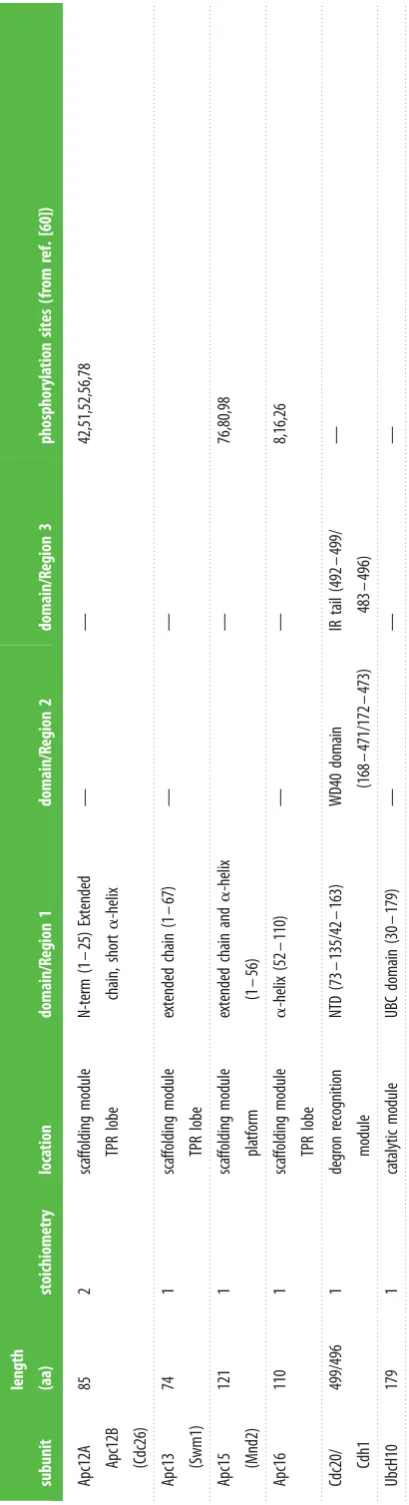
Subunit composition.The APC/C comprises the core complex (14 subunits in metazoans, 13 in yeast) [48–59], together with the interchangeable coactivator subunits (either Cdc20 or Cdh1) [28,29,31] (table 1). APC/C subunits are functionally and structurally organized into three classes: the catalytic module, the substrate recognition module and the scaffolding module (table 1). The catalytic module comprises Apc11, the RING domain subunit [61–63] and Apc2, the cullin subunit [50,51,63]. These two subunits are orthologues of Rbx1 and the cullin subunit of cullin-RING ligases (CRLs), respectively. In both the APC/C and CRLs, an N-terminal b-strand of the RING domain subunit is integrated within the b-sheet of the C-terminal domain (CTD) of the cullin subunit. As discussed below, the catalytic module incorporates two conformationally-variable domains, the RING domain of Apc11 (Apc11RING
) and the WHB domain of Apc2 (Apc2WHB), both attached to the
CTD of Apc2 (Apc2CTD) by flexible linkers. The
conformatio-nal flexibilities of Apc2WHB and Apc11RING have important implications for APC/C catalysis and regulation.
Together, the coactivators and Apc10 form the substrate recognition module, with the coactivator’s WD40b-propeller domain being primarily responsible for mediating degron rec-ognition (D box, KEN box and ABBA motif) [64–71]. Optimal D-box recognition requires the core APC/C subunit Apc10 (Doc1 in S. cerevisiae) [54,72,73]. The substrate recognition and catalytic modules represent the key functional subunits
of the APC/C, reflected in their high degree of conservation. It is striking that these two functional modules represent only 15% of the total mass of the molecule. Most of the APC/C mass is conferred by the seven large scaffolding sub-units, four of which form homo-dimers—further contributing to the high relative mass of the scaffolding module [74]. Remarkably, the majority of APC/C subunits, particularly the scaffolding subunits, are composed of multiple repeat motifs. Five scaffolding proteins are tetratricopeptide repeat (TPR) proteins, being composed of 13–14 TPR motifs arranged in contiguous arrays. TPR proteins, ubiquitous in all three domains of life, were first discovered in what were later ident-ified as yeast APC/C subunits [75–78]. Their presence in multiple protein complexes of diverse functions such as the APC/C indicates a role in mediating protein–protein inter-actions and the assembly of multi-protein complexes [79]. Later, atomic resolution structural analysis of the APC/C pro-vided a mechanistic rationale for many of the previously characterized TPR mutations [80–83].
The four canonical TPR proteins (Apc3, Apc6, Apc7, Apc8) are structurally highly homologous, being composed almost entirely of 14 TPR motifs. These self associate to form homo-dimers [81–83]. Apc1, the largest APC/C subunit, features another type of motif that is only observed in Apc1 and the Rpn1 and Rpn2 subunits of the 19S regulatory subunit of the proteasome (in exactly the same number and arrangement) [84]. These approximately 40-residue motifs are termed the PC (proteasome-cyclosome) repeat [85]. Although not discern-able in sequence, cryo-electron microscopy (cryo-EM) studies revealed that Apc1 contains an N-terminal seven-bladed
b-propeller domain [80,86]. Apc4 also comprises ab-propeller domain [87]. Finally, four small intrinsically disordered subunits (vertebrate Apc12, Apc13, Apc15, Apc16) function as TPR-accessory subunits. These subunits interact with TPR subunits and, as explained later, Apc12, Apc13 and Apc16 stabilize TPR subunits and mediate inter-TPR interactions [51,54,56,80,86,88,89]. Apc15 is not required for APC/C assem-bly. It functions to negatively regulate the SAC by controlling the stability of the Cdc20 subunit of the MCC through APC/ C-dependent auto-ubiquitination [59,90–95].
Structural investigations of the APC/C were initiated some 18 years ago, shortly after its discovery in 1995 [1–3]. Initial efforts focused on a complementary approach of crystallogra-phy of individual APC/C subunits and small sub-complexes and homologous proteins [71,81–84,87,89,96–100], together with single particle cryo-EM studies of the intact complex that represented various functional states of the complex purified from endogenous sources: budding yeast, fission yeast,Xenopus
and human [73,101–107]. A combination of crystallography of individual APC/C subunits, native mass spectrometry [74] and electron microscopy provided information on the subunit stoichiometry of the APC/C (table 1).
The recent progress in understanding the structure and mechanisms of the APC/C through atomic resolution struc-tures of various functional states of the complex resulted from technical developments in reconstituting the recombi-nant APC/C [74,91,108] together with recent advances in single particle cryo-electron microscopy (direct electron detectors and software for image analysis and 3D-reconstruc-tions) [109]. Recent EM studies have focused on reconstituted human APC/C complexes [80,86,92,93,99,110– 112].
In 2014 a 7.4 A˚ resolution structure of the reconstituted APC/CCdh1.substrate complex was published [86]. At 7.4 A˚
rsob.r
oy
alsocietypublishing.org
Open
Biol.
7
:
170204
Table
1.
Subunits
of
the
human
anaphase
pr
omoting
comple
x/cyclosome
(APC/C).
Alterna
tiv
e
S.
cer
evisiae
subunits
in
par
enthesis.
subunit length (aa) stoichiometry loca tion domain/R egion 1 domain/R egion 2 domain/R egion 3 phosphoryla tion sites (f ro m ref. [60]) Apc1 1944 1 sca ffolding module pla tform WD40 domain (1 – 612) mid-N (613 – 986) mid-C (1617 – 1944 ) PC domain (1013 – 1616) 60,65,202,233,286,291,297,298,299,309,313,316,317,341,343,351,355,362,364,372,373,377,386,389,394,416,501,518,520,522,524,530,536,537, 542,547,555,563,564,569,576,582,600,686,688,699,701,703,731,916, 920,921,922,1001,1347,1349
Apc2 822 1 ca talytic module NTD (1 – 432) cullin repea ts CTD (433 – 822) including WHB doma in — 205,218,314,466,470,474,532,534,732,736,738,742 Apc3A Apc3B (Cdc27) 824 2 sca ffolding module TPR lobe TPR dimer interfa ce TPR motifs 1 – 7 (1 – 535) TPR superhelix TPR motifs 8 – 14 (536 – 824) — 183,185,186,192,194,200,203,205,209,219,220,222,225,228,230,231,233,
237,241,244,251,252,255,264,267,276,279,281,289,291,302,304,312, 313,327,329,331,334,336,343,349,351,352,356,357,358,364,366,368, 369,383,384,386,387,388,389,419,426,430,434,435,438,443,444,446, 761,800,803,806,807,809,814,821
resolution the secondary structural architecture can be defined. Alpha-helices are resolved as rod-like structures, whereasb-sheets are visualized as planar structures. The sub-unit assignment of the electron microscopy (EM) density map was determined based on two approaches. One was a sub-unit deletion approach where the structures of reconstituted APC/C complexes lacking defined subunits were compared with the wild-type complex [74]. Difference density due to the deleted subunit could be assigned to a specific subunit. In a related approach, comparing two complexes that share a common subunit allows its identification. However Apc1, an essential subunit required for APC/C stability, which therefore cannot be deleted without disrupting the entire complex, was identified based on a process of elimination and by recognizing architectural features of the PC domain in the EM density map [80,86]. Finally, Apc13 inS. cerevisiae
was identified through locating GFP fused to its C-terminus [74]. Importantly, EM density for Apc2CTDwas weak and
dif-fuse whereas that for Apc11RING and Apc2WHB was not
visible, indicating a high degree of conformational flexibility of the catalytic module. Conformational heterogeneity was also confirmed through 3D classification of the cryo-EM data-set [86]. Altogether, the EM studies revealed a striking degree of structural conservation from yeast to metazoan. The APC/ C of higher eukaryotes differs from yeast because of an additional TPR subunit (Apc7) situated on the top of the TPR lobe that interacts only with Apc3 (table 1). The role of Apc7 has yet to be defined.
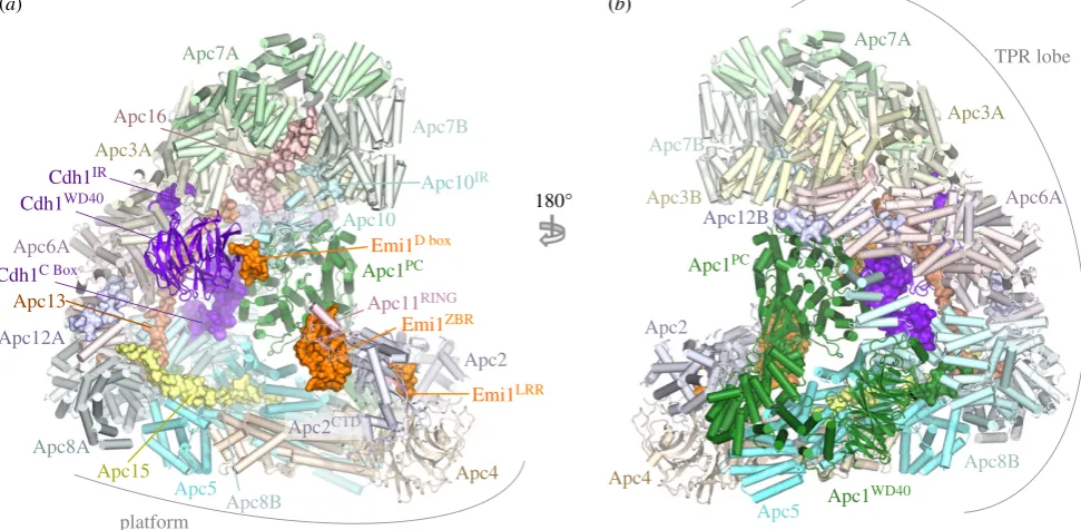
The 7.4 A˚ resolution structure of the APC/C was soon fol-lowed by a near-atomic resolution structure of the complex of APC/CCdh1 with the inhibitor Emi1 (APC/CCdh1.Emi1) [80].
This structure was at 3.6 A˚ resolution overall, but a local resol-ution map showed that the more rigid regions of the map were closer to 3.2 A˚ resolution. Two regions in particular were recov-ered at lower resolution (approx. 5 A˚) due to their higher relative flexibility. These were the catalytic module formed of Apc11 and Apc2CTD, and the coactivator Cdh1.
The 3.6 A˚ resolution cryo-EM map of APC/CCdh1.Emi1
pro-vided the basis for understanding the detailed architecture of the APC/C and served as a template for understanding sub-sequent different functional states, some at lower resolution. Building of the atomic-resolution model was based on fitting of atomic coordinates of X-ray structures of most of the large subunits and close homologues. For Apc1, fitting to the N-terminal WD40 domain and densities adjacent to its central PC domain (Apc1PC) that lack structural homologues was
per-formedab initio. The TPR accessory subunits Apc13, Apc15 and Apc16 were also builtab initio[80].
The APC/C adopts a triangular shape delineated by a lat-tice-like shell organized into two sub-structures (figure 1) [80,86]. The back and top of the complex is formed from a bowl-shaped TPR lobe, an assembly of the four canonical TPR proteins (Apc3, Apc6, Apc7, Apc8) and three TPR acces-sory subunits (table 1). The base of the APC/C comprises the platform subunits Apc4 and Apc5, together with two (non-PC) domains of Apc1. Apc1PCextends from the platform
to contact the TPR lobe. Together, the TPR lobe and platform sub-structures define a central cavity. The degron recognition module of coactivator and Apc10 is located at the top of the cavity with Apc10 interacting extensively with Apc1PC. The
catalytic module of Apc2-Apc11 is positioned at the periphery of the platform such that Apc2CTDand associated Apc11 are at
the front of the cavity situated directly below Apc10 and Cdh1.
Table
1.
(
Continued.
)
subunit
length (aa)
stoichiometry
loca
tion
domain/R
egion
1
domain/R
egion
2
domain/R
egion
3
phosphoryla
tion
sites
(f
ro
m
ref.
[60])
Apc12A
Apc12B (Cdc26)
85
2
sca
ffolding
module
TPR
lobe
N-term
(1
–
25)
Extended
chain,
short
a
-helix
—
—
42,51,52,56,78
Apc13
(Swm1)
74
1
sca
ffolding
module
TPR
lobe
extended
chain
(1
–
67)
—
—
Apc15
(Mnd2)
121
1
sca
ffolding
module
pla
tform
extended
chain
and
a
-helix
(1
–
56)
—
76,80,98
Apc16
110
1
sca
ffolding
module
TPR
lobe
a
-helix
(52
–
110)
—
—
8,16,26
Cdc20/
Cdh1
499/496
1
degr
on
recognition
mo
dule
NTD
(73
–
135/4
2
–
163)
WD40
domain
(168
–
471/172
–
473)
IR
tail
(492
–
49
9/
483
–
496)
—
UbcH10
179
1
ca
talytic
module
UBC
domain
(30
–
179)
—
—
—
rsob.r
oy
alsocietypublishing.org
Open
Biol.
7
:
170204
The canonical TPR proteins form structurally related V-shaped homo-dimers [81–83]. Each subunit comprises an
a-helical solenoid with two turns of TPR helix. Whereas the N-terminal TPR helix forms the homo-dimer interface, the C-terminal TPR helix creates a protein-binding groove. Apc6 binds its accessory subunit Apc12 through this groove (figure 1) [82,89], stabilizing Apc6 [89], whereas the Apc3 and Apc8 homo-dimers use one of their dyad-related C-terminal grooves to engage the coactivator subunits (either Cdc20 or Cdh1) (figures 1 and 2) [60,80,86]. Within the TPR lobe, the four canonical TPR proteins stack in a parallel array generating a left-handed super-helix that adopts pseudo dyad-symmetry. Together the TPR accessory subunits Apc13 and Apc16 (and presumably Apc9 in S. cerevisiae) interact with structurally and symmetry related sites on seven of the eight TPR subunits to stabilize the TPR lobe and contribute to defining the order of TPR protein assembly [80].
Apc10 and both coactivators share structurally related C-terminal Ile-Arg motifs (IR tails) that interact with the C-terminal TPR motifs of Apc3 (figures 1 and 2a,b) [66,88, 96,113,114]. Additionally, coactivators comprise a C-box motif within their N-terminal domain (NTD) [68] that mediates interactions with the APC/C [68,113], dependent on Apc8 [115]. Due to the presence of multiple binding sites on the TPR lobe, the pseudo dyad-symmetry of the TPR lobe has important consequences for mechanisms of interaction with coactivators and substrates. Not only does the dyad symmetry of each TPR protein mean that there is multiplication of protein/ligand binding sites (for example the common IR tails of coactivator and Apc10 interact with separate subunits of the Apc3 homo-dimer (figure 2a,b)), but also the IR-tail bind-ing site on Apc3 is structurally related to the C-box bindbind-ing site on Apc8B, a paralogue of Apc3 (figure 2c). The mechanism of interaction of the IR tail with Apc3 is structurally analo-gous to that of the R[F/Y]I motif of the C box with the C-box
binding site on Apc8B [80]. Because of this, the structurally equivalent C-box binding site on Apc8A is capable of binding the IR tail of Cdc20MCC(in the APC/CMCCcomplex) [92,93].
A conformational transition involving the C-terminal TPR motifs of Apc3A occludes the coactivator IR-tail binding pocket in the absence of the IR-tail ligand [60,80,100]. Finally, regions of the NTD of coactivator also interact with Apc1PC
(figure 3c). Thus the degron-recognition WD40 domains of the coactivators are connected to the APC/C scaffold through three sites, attached through flexible linkers. This allows for conformational flexibility of the WD40 domain.
In the platform, analogous to the Apc6–Apc12 inter-action, the C-terminus of Apc15 inserts into the TPR groove of Apc5 as an extended chain, with its N-terminal a-helix (Apc15NTH) bridging Apc5 and Apc8 [80].
3. Coactivators are primarily responsible for
degron recognition
The APC/C recognizes and ubiquitinates a variety of cell cycle substrates in a cell-cycle-dependent manner. Selection of substrates in a temporal manner is dependent on a variety of factors, but critical among these is the role of coactivators [29]. The APC/C is inactive without coactivator. One func-tion of coactivators is to provide degron recognifunc-tion sites that engage degrons present in most APC/C substrates [66,69–71], thereby recruiting substrates to the APC/C (figures 1, 3a,b, 4 and 5). In a few exceptions, for example Nek2A, the core APC/C recognizes substrates, bypassing degron recognition sites on the coactivator. However, Nek2A ubiquitination still relies upon the coactivator-induced stimulation of UbcH10-binding to the APC/C [86,117].
Due to the critical role coactivators play in defining APC/C activity, regulation of APC/C activity by phosphorylation and
(a) (b)
180° Apc7A
Apc7B
Apc1PC
Apc4
Apc2
Apc5 Apc15 Apc3A
Apc12A Apc6A
Apc13
Apc16
Cdh1WD40
Apc10
Apc8A
Apc7A
Apc7B
Apc1PC
Apc4
Apc2
Apc3A
Apc12B
Apc5
Apc8B Apc3B
Apc1WD40
Apc6A
Cdh1C Box
Emi1D box
Emi1LRR
Emi1ZBR
Cdh1IR
Apc10IR
Apc8B
TPR lobe
platform
Apc11RING
Apc2CTD
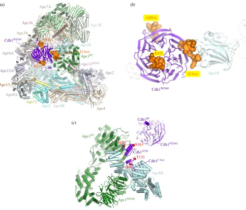
Figure 1.
Overall structure of the human APC/C
Cdh1.Emi1complex. (a) and (b) Two orthogonal views of the APC/C. Large APC/C subunits are represented as cartoons,
whereas small APC/C subunits (Apc12, Apc13, Apc15, Apc16), the IR tails of Cdh1 and Apc10, the Cdh1 NTD and the Emi1 inhibitor are shown as space filling
representations. The TPR and platform sub-structures are labelled. The two subunits of the canonical homo-dimeric TPR subunits (Apc3, Apc6, Apc7 and Apc8) and
Apc12 are labelled with the suffix ‘A’ and ‘B’. Apc2
CTDand Apc11
RINGform the catalytic module, Cdh1 and Apc10 generate the substrate recognition module. PDB
4UI9, from Chang
et al.
[80].
rsob.r
oy
alsocietypublishing.org
Open
Biol.
7
:
170204
inhibitory complexes such as the MCC, Emi1 and Acm1 is exerted primarily at the level of coactivators, either by control-ling their interaction with the APC/C or by controlcontrol-ling coactivator interaction with degrons. Cdc20 activates the APC/C from early mitosis to anaphase after which Cdh1 binds to the APC/C through to late G1. Switching of these two highly structurally conserved and related coactivators at anaphase changes the substrate specificity and regulatory properties of the APC/C. Cdc20 is thought to recognize a restricted set of substrates (specifically cyclin A, cyclin B and securin), whereas Cdh1 is proposed to have a broader substrate specificity, being able to ubiquitinate all Cdc20 substrates, and in addition recognizes the Aurora A and B kinases, which are not substrates of APC/CCdc20[118]. Aurora kinases are
recog-nized by APC/CCdh1through their essential N-terminal A box
motif [119]. The role of the C-terminal D box of Aurora kinases is disputed, as discussed in Davey & Morgan [120]. Both coac-tivators mediate interactions of substrates harbouring D-box and KEN-box motifs to the APC/C. Optimal interactions of the D box also require the Apc10 subunit [54,72,73]. The ABBA motif is recognized by vertebrate Cdc20 [121], and
S. cerevisiae Cdh1 [71] and Cdc20 [120,122]. In S. cerevisiae a
specific coactivator termed Ama1 controls meiosis [123,124] that in turn is antagonized by the Mnd2 (Apc15) subunit [125,126].
To understand structurally how coactivators recognize D-box and KEN-box substrates, advantage was made of the fact that many APC/C inhibitors incorporate pseudo-substrate motifs that mimic D-box and KEN-box degrons in order to block substrate recognition. These inhibitors interact with higher affinity with coactivators than do substrates, thereby facilitating the biochemical isolation and crystallization of these complexes. A structure of the MCC from Schizosaccharo-myces pombe, a complex of Cdc20, Mad2 and BubR1/Mad3, revealed how a KEN box and D box present in BubR1/Mad3 interact with their respective binding sites on theb-propeller domain of Cdc20 [69]. These findings were confirmed and extended in a structure of theb-propeller domain of Cdh1 in complex with Acm1, a Cdh1 specific inhibitor from
S. cerevisiae [71]. The latter structure also revealed how the ABBA motif (A motif in Acm1 terminology [127]) interacts with Cdh1 (figures 3 and 4). A further study in which human Cdc20 was crystallized with a peptide modelled on the BubR1 KEN box also revealed details of Cdc20 interactions with the KEN-box motif [70].
(a) (b)
(c) Apc3A
Cdh1IR
Cdh1C box
Apc10IR Apc3B
Apc8B
E610
L606
H609 Y635
Y639
L671
E610
H609
L606
W544 Y635
Y639
L671
S578 S578
W544
E374
L370
Y399
Y403
Y308 S342
I183
R184
I495 R496
D46 R47 F48
I49 R52
P50 IR tail
IR tail
C box
N575 N575
N339 E410
Figure 2.
The IR-tail and C-box binding sites of Apc3 and Apc8 respectively, are homologous. (a) Cdh1 IR-tail binding site. (b) Apc10 IR-tail binding site. (c) C-box
binding site on Apc8B. The Ile and Arg side chains of the IR tail of both Cdh1 and Apc10 interact with a site on Apc3 that is homologous to the binding sites for
Arg(47) and Ile(49) of the Cdh1 C box on Apc8B. The C box (DR[F/Y]IPxR) forms additional contacts to Apc8B as shown. PDB 4UI9, from Chang
et al.
[80].
rsob.r
oy
alsocietypublishing.org
Open
Biol.
7
:
170204
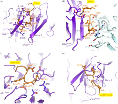
D box.The classical APC/C degron is the destruction box or D box, a ten-residue motif (RxxLx[D/E][Ø]xN[N/S]) (figure 5a,c) first characterized in B-type cyclins as being necessary and sufficient for APC/C mediated ubiquitination [128–130]. Mutation of any of the three most highly con-served residues, Arg (P1), Leu (P4) or Asn (P9), ablated the destruction signal [128]. The D box binds in a mainly extended conformation to a shallow groove at the side of the b-propeller, found between the two b-blades 1 and 7 (figures 3b and 4a). The essential Leu (P4) residue anchors the D box to the channel within a hydrophobic pocket, whereas the N-terminal Arg (P1) residue interacts with an acidic pocket at the N-terminus of the channel (figure 4a). A conserved acidic residue at P6 interacts with an invariant Arg, whereas a hydrophobic residue at P7, conserved in many D-box motifs, interacts with a hydrophobic surface of the b-propeller (figures 4aand 5a,c) [69,71]. The side chain of P3 abuts a conserved Phe of the coactivator, likely account-ing for the high occurrence of residues with small unbranched side chains at this D-box position (figures 4a
and 5a,c). Although Arg and Leu are strongly preferred at P1 and P4, respectively, even these two residues are
not strictly necessary. For example, inDrosophila melanogaster
cyclin A [131] and Homo sapiens cyclin B3 [132], Phe is substituted for Leu.
Significantly, the conserved C-terminal hydrophilic resi-dues (P8 to P10) do not interact with the coactivator, however the cryo-EM structure of APC/CCdh1.Emi1(where the inhibitor
Emi1 incorporates a D box) showed clear EM density extending from the P7 residue of the D box (interacting with the D-box site on Cdh1) to Apc10 [80]. This showed that the C-terminus of the D box interacts with a hydrophilic surface of Apc10 [80,96,97] involving polar and charged residues on two surface-exposed loops (the 80s and 140s loops) (figure 4b). This highly conserved region is required for D-box-dependent APC/C E3 ligase activity, and this potentially dynamic hydrophilic surface may allow for the accommodation of a variety of small polar residues at D-box positions P8 to P10 (figure 5a). Disruption of the 140s loop impairs D-box-dependent substrate recognition [133] and Ala substitutions of Ser88 and Asn147 of Apc10 attenuated APC/CCdh1activity [80]. Although Apc10 primarily
interacts with D-box residues P8 to P10, its 80s loop also contacts N-terminal residues of the D box (figure 4b). For example, the side chain of Glu87 (invariant in Apc10
(b)
(c)
Cdh1WD40
Cdh1WD40
Apc10 Apc7A
Apc7B
Apc1PC
Apc4
Apc2
Apc5 Apc15 Apc3A
Apc12A
Apc13
Apc16
Cdh1WD40
Apc10
Apc8A Apc6A
Apc8B
Apc8B
Apc1PC
Apc1WD40
Cdh1C box Cdh1IR D box
T121 S163
D box ABBA
KEN ABBA
KEN
S151
Apc11RING
S40 Cdh1NTD
Apc2CTD (a)
Figure 3.
Coactivators interact with Apc1 and Apc3 and create a D-box co-receptor with Apc10. (a) Overview of the APC/C with the Cdh1 coactivator subunit. Based
on the APC/C
Cdh1.Emi1coordinates (PDB 4UI9) [80] with the KEN box and ABBA motif modelled on the
S. cerevisiae
Cdh1 – Acm1 complex (PDB: 4BH6) [71]. Except
for the D box, Emi1 coordinates are not shown. (b) Close-up view of the D-box co-receptor formed from Cdh1 and Apc10. (c) Cdk1-dependent phosphorylation of the
NTD of Cdh1 blocks its binding to the APC/C. Red spheres indicate sites of inhibitory phosphorylation.
rsob.r
oy
alsocietypublishing.org
Open
Biol.
7
:
170204
orthologues) is sandwiched between P2, P5 and P7 of the D box, perhaps explaining the occurrence of basic residues at these positions, especially for the non-canonical D-box sequences (discussed below) (figure 5a,c). Notably, Cdk1-phosphorylation at P2 (Pro is common at P3) negatively regulates APC/C-depen-dent substrate ubiquitination, for example Dbf4 [122], possibly due to the electrostatic repulsion between a phosphate group at P2 and Glu87. Thus, the D box is a bipartite degron comprising a coactivator-interacting N-terminal (RxxLx[D/E][Ø]) motif and a hydrophilic C-terminal-Apc10 binding segment. Coactivator and Apc10 create a D-box co-receptor for recognition of the bipartite degron. The atomic resolution structures of D-box motifs engaged by coactivators alone [69,71] and in complex with APC/C-coactivator complexes [60,80] rationalize the resi-due preferences at all 10 positions of the D box. Moreover, the preferences for an acidic residue at P6 and basic residue at P2 are consistent with the promotion of substrate ubiquitination by D-box phosphorylation at P6 [134] and substrate stabilization by phosphorylation at P2 [122].
KEN box.Another APC/C degron, the KEN motif ([DNE]-KENxxP), is commonly present in APC/C substrates usually in addition to the D box [135]. Efficient ubiquitination by either APC/CCdc20or APC/CCdh1of substrates harbouring both D
and KEN boxes is dependent on both degrons [54,64]. By forming a 310 helix, the three consecutive residues of the
KEN box face in the same orientation and engage the top sur-face of theb-propeller (figures 3band 4c) [69–71]. The KEN box is usually immediately C-terminal to acidic residues (figure 5b,d), and the structure of the KEN box–coactivator complex suggested that these would engage a positively charged patch on theb-propeller. A frequently observed Asp or Asn residue at P-1 stabilizes the KEN box conformation by forming a hydrogen bond to the Asn of the KEN box (figure 4c) [71]. Proline residues one to two residues C-terminal of the KEN box would direct the polypeptide chain away from the surface of theb-propeller.
ABBA motif.The A motif was discovered in theS. cerevisiae
Cdh1 inhibitor Acm1 [127,136]. Later bioinformatics studies identified the ABBA motif as a general class that includes the A motif as a six-residue motif (Fx[ILV][FY]x[DE]) common to vertebrate cyclin A (andS. cerevisiaeClb5), BubR1, Bub1 and Acm1 [120–122]. Although the A motif was originally thought to confer specificity forS. cerevisiaeCdh1 [71,127], the situation is more complicated. Cdc20 also binds the ABBA motif— variations in non-consensus residues confer the specificity for
S. cerevisiae Cdh1. Glu65(P5) of the ABBA motif of Acm1
(a) (b)
(c) (d)
K(P5) I(P2)
K(P9) R(P1) A(P3)
S(P6) L(P4)
Q(P8)
Lys(P1)
Glu(P2)
Asn(P3) L(P7)
D(P6) K(P5)
F(P1) E(P6)
L(P3) Y(P4)
Cdh1WD40
Cdh1WD40
Cdh1WD40
Cdh1WD40
Apc10 140s loop
Q473 N407
D box D box
KEN box
ABBA motif Asp(P-1)
R(P1)
I(P2)
L(P4)
V(P7)
blade 7 blade 1
blade 2
blade 3
bD
R253 E537
V254
F286 D256
F210
V178
R517 D263
R338
I328 W352
80s loop E87
S88
D57 D150
N147
V371
Figure 4.
Substrate recognition is mediated by coactivators and Apc10. (a) D-box receptor on Cdh1, (b) D-box co-receptor (Cdh1 and Apc10), (c) KEN-box receptor on
Cdh1, (d) ABBA-motif interactions with Cdh1. Coordinates in (a,c,d) are based on the
S. cerevisiae
Cdh1 – Acm1 complex (PDB: 4BH6) [71]. (b) Based on APC/C
Cdh1.Emi1complex (PDB 4UI9) [80].
rsob.r
oy
alsocietypublishing.org
Open
Biol.
7
:
170204
contacts Lys333 inS. cerevisiaeCdh1 that is a Thr inS. cerevisiae
Cdc20 [121]. Residues of human Cdc20 required for ABBA motif binding are not conserved in human Cdh1 (although are conserved inS. cerevisiaeCdh1), explaining the inability of human Cdh1 to recognize the ABBA motif [121]. A structure of Acm1 in complex withS. cerevisiaeCdh1 revealed that the ABBA motif forms an extended structure and binds to the inter-blade groove betweenb-blades 2 and 3, through a related mechanism to the D box (figure 4d). The side-chains of the three conserved non-polar residues anchor the ABBA motif to the ABBA-motif binding groove, with the Asp at P6 forming a salt-bridge with an Arg of blade 2 [71].
Non-canonical degrons.In addition to the D box, KEN box and ABBA motif, non-canonical degrons have also been identified (figure 5c). However, some of these are likely to be variants of the well-characterized D box and KEN box degrons [71,120]. For example, the conserved Arg (P1) at the N-terminus of the D box can be substituted with Lys, His or Gln although this is often accompanied by a Lys at P7 which can interact with the acidic patch at the N-terminus of the D-box binding channel [71]. The O box identified as an APC/C degron in Orc1 closely matches the D-box consensus [137], suggesting it may interact with the D-box receptor [71], consistent with the ability of a D-box peptide to interfere with O-box recognition by APC/CCdh1[137]. A D-box peptide also
inhibited APC/CCdh1-catalysed ubiquitination of the Spo13
[138] and Cin8p [139], substrates that harbour non-canonical D-box motifs (figure 5c) [71]. Peptides modelled on the non-canonical D-box motifs of Cin8p, the O box and Spo13 inhibited the D-box-dependent ubiquitination of the budding yeast substrate Hsl1, consistent with the idea that these motifs
interact with the D-box receptor of APC/CCdh1[71]. In
mam-mals, the CRY box (CRYxPS) within the NTD of Cdc20 mediates APC/CCdh1-dependent Cdc20 destruction in
oocytes and embryos [140]. Insights into how the CRY box might interact with Cdh1 were provided by cryo-EM struc-tures of the APC/CMCC [92,93] (discussed in §8). These
showed that the CRY box of the MCC Cdc20 subunit interacts with the WD40 domain of Cdc20 of APC/CCdc20in proximity
to the D-box binding site.
In addition to modulation of APC/C–substrate affinities by substrate phosphorylation in or adjacent to the degron, ubiquitination of Lys residues within or in close proximity to degrons may influence APC/C– substrate affinities. One example of this is that the KEN-box Lys residue is one of the most frequently ubiquitinated sitesin vivo[141]. Modifi-cation of the KEN box would be expected to reduce APC/ C–substrate affinities.
Discovery of new APC/C substrates will be facilitated by high-throughput automated approaches based on protein micro-arrays such as the extract-based functional assays [142,143].
4. The APC/C pairs with two E2s to
assemble polyubiquitin chains
The APC/C is a RING domain E3 ligase. RING domains inter-act directly with their canonical E2s and bring these into close proximity with substrates bound to degron recognition sites situated elsewhere on the E3 ligase [144]. Metazoan APC/C assembles atypical Lys11-linked chains to promote proteolysis
0 1 2 3 4
bits
0 1 2 3 4
bits
P
RK
[
AP
]
LG
[
DE
][
VIL
][
SN
][
N
]
N
P
AS[
P
]
L
T[
E
][K ][
N
][A]K
VQ
K
[
P
]
L
Q[
E
][K ][
T
][P]
N
K
KM[
P
]
L
R[L ][S ][
N
][I]
N
P
H
L
[S ]
LK
[
D
][
I
][
T
][
N
]V
Cin8p
E
[
DN
]
KEN
x
PP
(c)(d)
KEN box
D box
O box
Spo13
–1 1 2 3 4 5 6 7 8 9 10
Sgo1 (human)
–2 –1 1 2 3+1 +2 +3
(a) (b)
effect of phosphorylation on substrate ubiquitination
–1 1 2 3 4 5 6 7 8 9 10 –3 –2 –1 1 2 3 +1 +2 +3
Figure 5.
Degron consensus sequences. (a) Sequence motif of D box derived from 68 APC/C substrates [71]. Sequence motif determined using multiple expectation
maximization for motif elicitation (MEME) [116]. (b) Sequence motif of KEN box derived from 46 APC/C substrates [71]. (c) Alignment of consensus D box degron
with non-canonical D box degrons. (d) Consensus KEN box. Adapted from [71].
rsob.r
oy
alsocietypublishing.org
Open
Biol.
7
:
170204
and mitotic exit [145,146], in a process involving two distinct E2 activities. Chain formation is initiated with the E2 UbcH10 (also termed Ube2C) [147,148], whereas Ube2S is pri-marily responsible for chain extension [149–152]. Ube2S interacts with the acceptor ubiquitin to generate Lys11-linked chains through a substrate-assisted catalytic mechanism in which Glu34 on the acceptor ubiquitin activates and orients the target Lys11 to attack the donor ubiquitin conjugated to Ube2S [152]. UbcH10 and Ube2S act in concert to generate branched chains (mixed K11 and K48 linkages). The ubiquitin chain topology determines the efficiency of proteasome-depen-dent proteolysis of the ubiquitinated substrate [153–156]. UbcH10 alone is competent to generate short ubiquitin chains of mixed K11, K48 and K63 linkage [157,158]. Neither UbcH10 nor Ube2S are essential, suggesting an alternative E2 can function in place of UbcH10in vivo, likely to be UbcH5 [159]. However, Ube2S is essential for optimal release from a SAC-dependent arrest, possibly due to its role in reactivating the APC/C on cessation of SAC signalling [149–151]. In
S. cerevisiae the APC/C generates canonical Lys48-linked chains also using two E2s: the initiating E2 Ubc4 and the elongating E2 Ubc1 [160]. A UBA domain in Ubc1 is required for processivity [160] by enhancing Ubc1 association with the APC/C in competition with Ubc4 [161].
4.1. Monoubiquitination catalysed by UbcH10
Cryo-EM studies of human APC/CCdh1 in complex with UbcH10 and Ube2S with and without ubiquitin have provided detailed mechanistic insights into the process of substrate ubi-quitination [80,99,111,112]. UbcH10 is a canonical E2 that interacts with the RING domain of Apc11 [80,111]. In human APC/C, the catalytic module is a region of conformational flexi-bility [60,86]. Binding of UbcH10, but not Ube2S, is dependent on a conformation change mediated by the coactivator subunit (figures 6 and 7) [86,110]. Thus, coactivators are required for both substrate recognition and for stimulating the catalytic activity of the APC/C [117]. This conformational change involves a movement of the catalytic module from a ‘down’ to an ‘up’ position. In the ‘down’ position, Apc11RING
is in con-tact with Apc5 of the platform, blocking the UbcH10-binding site. On conversion to the coactivator-bound state, movement of the catalytic module to an upward position exposes the UbcH10-binding site on Apc11RING-Apc2WHB, resulting in at
least a 10-fold increased affinity for UbcH10 [86]. In this state the catalytic module is flexible with weak density recovered and conformational heterogeneity for a variety of ternary com-plexes [60,86]. Coactivators also increased the catalytic efficiency ofS. cerevisiaeAPC/C (decrease inKmand increase
inVmax) [163], although this may result from a mechanism
other than a coactivator-induced conformational change (D Barford & E Va´zquez Ferna´ndez 2017, unpublished data).
The interaction of the zinc binding region (ZBR) domain of the inhibitor Emi1 with Apc11RINGstabilizes the conformation
of the catalytic module because the ZBR domain bridges Apc1PCwith Apc11RINGand Apc2CTD(figure 1a). This allowed
definition of Apc11RINGand Apc2CTDto a local resolution of
approximately 6 A˚ [80] and it showed for the first time how Apc11RINGinteracts with Apc2CTD. The juxtaposition of
Apc11RING and Apc2CTD is similar to the swung out
con-formation of Rbx1RING in activated Cul5-Rbx1 [164]. Engagement of UbcH10 with Apc11RINGis essentially similar
to other RING domain–E2 interactions (figure 6b) [80,111].
Density for UbcH10 was poorly resolved, probably due to the low stoichiometry of UbcH10–APC/C interactions and conformational flexibility of the catalytic module. The Apc11RING–UbcH10 interface was confirmed by a detailed
mutagenesis study by Schulman and colleagues [111]. On interacting with UbcH10, the catalytic module rotates by 128
relative to its position in the APC/CCdh1.Emi1 complex [80].
Importantly no EM density was visible for ubiquitin in the APC/CCdh1-UbcH10ubiquitin cryo-EM maps [80,111]. This
would indicate that the ubiquitin moiety must be mobile, and only transiently adopts the closed E2ubiquitin confor-mation that primes the E2ubiquitin thioester bond to stimulate the intrinsic catalytic activity of E2ubiquitin [152,165–169]. Formation of the closed E2ubiquitin confor-mation, where the ubiquitin moiety interacts with the RING domain through its Ile36 and E2 through its Ile44, as a require-ment for optimal substrate ubiquitination, is based on the finding that mutating either Ile36 or Ile44 residues in ubiquitin virtually eliminated ubiquitination of APC/C substrates [80]. The APC/C is reminiscent of other single domain RING and U-box E3s that bias the E2ubiquitin conformation from mul-tiple extended states to the closed state [168,170]. As discussed elsewhere [80,111], an interesting possibility is that substrate initiation motifs that promote lysine ubiquitination [158] may induce a closed UbcH10ubiquitin conformation.
The study of Schulman and colleagues revealed that Apc2WHBforms an unusual interaction with the backside of
UbcH10 [111]. This interaction follows a rigidification of the WHB domain (which is mobile in UbcH10-free structures) induced upon UbcH10 binding (figure 7c). Apc2WHBis
essen-tial and specific for APC/C-UbcH10-dependent substrate modification, but is dispensable for UbcH5 activity. The activity of Ube2S, which does not interact with Apc2WHB, is
also independent of Apc2WHB[111]. Apc2WHBboth enhances
APC/C-UbcH10 affinity, but importantly also greatly stimu-lates (by more than 100-fold) the catalytic activity of UbcH10, likely by stabilizing the E2ubiquitin closed conformation through an allosteric mechanism. Since the WHB-binding interface of UbcH10 differs substantially from its counterpart in UbcH5, similar interactions between UbcH5 and Apc2WHB
are not possible, thus explaining how Apc2WHBcontributes to
UbcH10 specificity [111].
4.2. Polyubiquitination catalysed by Ube2S
The processive ubiquitination reaction catalysed by Ube2S involves modification of a constantly changing substrate that is the growing distal ubiquitin moiety of the polyubiquitin chain. Biochemical studies showed that UbcH10 and Ube2S do not compete for the same binding site on the APC/C [150,152], suggesting that Ube2S differs from canonical E2s by not interacting with the RING domain of Apc11, a notion also consistent with the observation that Ube2S catalyses for-mation of unattached K11-linked polyubiquitin chains [171]. APC/C–Ube2S interactions are dependent on the C-terminal LRRL motif of Ube2S [86,154,172,173]. The APC/C dramati-cally improves the catalytic efficiency of Ube2S-mediated Lys11-linked chain assembly [99,173]. This stimulatory effect of the APC/C requires a surface centred on Ala46 of the accep-tor ubiquitin, indicating that APC/C tracks the distal ubiquitin of a growing ubiquitin chain [173]. This finding explains how the APC/C generates ubiquitin chains without altering its interactions with substrates and E2s.
rsob.r
oy
alsocietypublishing.org
Open
Biol.
7
:
170204
Brown and colleagues [99] in agreement with Kellyet al. [173] showed that the APC/C increased Ube2S catalytic effi-ciency to massively increase polyubiquitination. Although this catalytic enhancement requires Apc11RING, two lines of
evidence suggested that this did not involve the canonical E2-binding surface on Apc11RING. Mutagenesis studies identified
a novel surface on Apc11RING (termed the exosite) required
for Ube2S activity, a result complemented by NMR data show-ing chemical shift perturbations in this region of Apc11RINGin
the presence of ubiquitin. Conversely, acceptor ubiquitin mutants with specific defects in APC/C-Ube2S-dependent ubi-quitination [99,152,173] map to a RING-binding surface on ubiquitin identified by NMR [99]. In a subsequent study, the structural basis for Ube2S-catalysed ubiquitin chain extension was defined [112]. A cryo-EM reconstruction of APC/CCdh1
in complex with Ube2S revealed that the Ube2S UBC (ubiquitin
conjugating) domain interacts with Apc2, rationalizing the deleterious effects of mutations of the aC and aD helices (figure 6c) [99,112,173]. Its LRRL C-terminus interacts at a site between Apc2 and Apc4, as previously determined for the Emi1 LRRL tail in the APC/CCdh1.Emi1 structure [80]. The
distal (acceptor) ubiquitin moiety of the ubiquitinated substrate engages the repurposed exosite on Apc11RING, following a
con-formational change of Apc11RING, presenting its K11 residue to
undergo nucleophilic attack onto the donor ubiquitin conju-gated to Ube2S. Thus the Apc11RINGexosite captures the tip
of the growing polyubiquitin chain promoting its reaction with Ube2Subiquitin bound to Apc2 (figure 7e).
The relative locations of the UbcH10 and Ube2S binding sites on the APC/C also fit with their different functions— priming and elongation, respectively (figures 6 and 7). UbcH10 is located closer to the degron binding site on the
(a)
Apc7A
Apc7B
Apc1PC
Apc4 Apc2
Apc5 Apc3A
Apc10
Apc8A
Apc6A
Apc11RING
Apc7A
Apc7B
Apc1PC
Apc4 Apc5
Apc3A
Cdh1WD40 Apc10
Apc8A
Apc6A
UbcH10 Ube2S
Apc7A
Apc7B
Apc1PC
Apc4 Apc5
Apc3A
Cdh1WD40
Apc10
Apc8A
Apc6A
Apc2WHB
KEN ABBA
D box
dUb
aUb
Apc11RING
Apc2CTD
Apc11RING
exosite
Apc11RING D box
Apc2CTD
Apc2CTD
(b) (c)
Figure 6.
APC/C ubiquitination reaction. (a) Apo APC/C. In the absence of coactivator the catalytic module adopts a ‘down’ inactive conformation. UbcH10 binding to
Apc11
RINGis blocked by Apc5, and Apc5 prevents the correct location of Apc2
WHBrequired to engage UbcH10. EM density for Apc11
RINGis weak indicating RING
domain flexibility. PDB 5G05 from Zhang
et al.
[60]. (b) Complex of APC/C
Cdh1.substratewith a UbcH10
ubiquitin conjugate. Apc2
WHBbecomes ordered and engages
UbcH10. dUb: modelled donor ubiquitin conjugated to UbcH10. The C-terminus of dUb is indicated with a red sphere. PDB 5A31, from Chang
et al.
[80]. PDB for
Apc2
WHB4YII Chang
et al.
[111]. (c) APC/C
Cdh1.substrate-Ube2S
Ub complex. Ube2S is partially built. aUb: acceptor ubiquitin bound to the Apc11
RINGexosite. PDB
5L9T, from Brown
et al.
[112]. The figure is based on previous work [60,80,111,112].
rsob.r
oy
alsocietypublishing.org
Open
Biol.
7
:
170204
substrate-recognition module, facing into the central cavity, and this is consistent with the relatively close proximity of the preferred target lysines to APC/C degrons (figures 6b
and 7c). In contrast, Ube2S is sited on the periphery of the molecule, able to accept the distal ubiquitin moiety on the polyubiquitin chain. The growing polyubiquitin chain can then be easily accommodated on the outside of the molecule (figures 6cand 7e).
4.3. Multiubiquitination catalysed by UbcH10
The repurposing of Apc11RINGthat stimulates Ube2S-catalysed
ubiquitin chain extension also plays a role in protein multi-ubiquitination catalysed by UbcH10 through its interaction with the canonical E2-binding site on Apc11RING. A cryo-EM
structure of a monoubiquitinated substrate bound to APC/CCdh1-UbcH10ubiquitin showed that the
substrate-conjugated ubiquitin moiety interacted with the Apc11RING
exosite [112], a finding supported by mutagenesis data reveal-ing that multi-ubiquitination catalysed by UbcH10 was defective in the Apc11RING exosite mutant. The structure
suggests a model for how an interaction between the Apc11 exosite and a substrate conjugated ubiquitin would increase substrate affinity and hence processivity (figure 7d).
Importantly, these data are consistent with the proposal that substrate ubiquitination primes APC/C substrates for further ubiquitination through a mechanism termed processive affinity amplification [174].
The inherent weak affinities between the APC/C–substrate complex and the E2s UbcH10 and Ube2S were overcome by employing artificial reinforcement of these interactions through a three-way chemical linkage involving the substrate, ubiquitin and E2 [111,112]. The interactions between the Apc11RING exo-site and ubiquitin were strengthened by generating a ubiquitin variant (Ubv) with substantially increased affinity for Apc11RING [112]. In another approach to stabilize APC/ CCdh1.substrate interactions with UbcH10, either UbcH10 was directly fused to the C-terminus of Apc11 or the LRRL tail of Ube2S was fused to the C-terminus of UbcH10, enhancing its affinity 10-fold [80].
5. The APC/C controls cell-cycle-dependent
substrate degradation
The capacity of the APC/C to control the degradation of regulatory proteins in a cell-cycle-dependent manner defines the ordered progression through distinct phases of the cell
Cdh1
K K K
D box
KEN
Apc10
Apc2
Apc2WHB Apc11RING
Apc10
Apc11RING
Apc2
Apc5
apo APC/C (inactive state)
APC/C–coactivator–substrate complex (active state)
Cdh1
K
K K
D box
KEN
Apc10
Apc2
Apc2WHB Apc11RING
UbcH10 dU
UbcH10 ~ Ub monoubiquitination
UbcH10 ~ Ub multiubiquitination
Cdh1
K
K K
D box
KEN
Apc10
Apc2
Apc2WHB
Ube2S
U
LRRL tail
aU
K11dU
Apc11RING
Ube2S ~ Ub polyubiquitination Cdh1
K
K K
substrate
Cdh1
K
K K
D box
KEN
Apc10
Apc2
Apc2WHB Apc11RING
UbcH10 U
dU
(a) (b)
(c) (d) (e)
exosite
Apc5
exosite
Figure 7.
Schematic of ubiquitination reaction catalysed by the APC/C. (a) In the apo state, the downward position of the catalytic module would cause a clash between
Apc5 and both UbcH10 and Apc2
WHB(as in the APC/C
Cdh1.substrate– UbcH10
ubiquitin complex). (b) Binding of coactivator shifts the catalytic module (Apc2 and Apc11) to
an upward position. Apc2
CTDtogether with Apc2
WHBand Apc11
RINGare highly flexible. Target lysines on the APC/C substrate are shown as ‘K’. (c) UbcH10-catalysed
monoubiquitination. dU: UbcH10-conjugated donor ubiquitin. Apc2
WHBrigidifies by binding to UbcH10, Apc11
RINGis less flexible. (d) UbcH10-catalysed multiubiquitination.
The substrate-conjugated ubiquitin (U) engages the ubiquitin-binding exosite of Apc11
RING. (e) Ube2S-catalysed polyubiquitination. The distal acceptor ubiquitin (aU) of the
polyubiquitin chain engages the ubiquitin-binding exosite of Apc11
RINGpositioning Lys 11 adjacent to the catalytic site of Ube2S. dU: donor ubiquitin conjugated to Ube2S.
Dashed lines around Apc11
RINGand Apc2
WHBdenote conformational flexibility. Based on schemes from Brown
et al.
[112] and Chang & Barford [162].
rsob.r
oy
alsocietypublishing.org
Open
Biol.
7
:
170204
cycle. The factors that affect differential rates of protein degra-dation during the cell cycle depend upon both changes in the composition and conformation of the APC/C itself as well as direct changes to individual substrates, and their intrinsic processivity. Switching between Cdc20 and Cdh1 contributes to altering APC/C substrate specificity. Cdh1 directs APC/ C-mediated ubiquitination of the Aurora kinases [118], which are not substrates of APC/CCdc20. Nevertheless, apart
from this example, there are relatively few instances known where the timing of substrate degradation can be directly explained by the switch of coactivator. Apart from coactivator switching, the two best-characterized regulatory mechanisms for determining the cell cycle order of APC/C-regulated sub-strate degradation are the spindle assembly checkpoint and substrate phosphorylation.
5.1. Substrate degradation at the spindle assembly
checkpoint
A few APC/C substrates are degraded in early mitosis (prome-taphase), for example Nek2A, cyclin A and Hox10, during an active SAC [175–181]. Thus, ubiquitination of these substrates is not inhibited by the SAC. These substrates differ from the canonical D-box and KEN-box-dependent substrates cyclin B and securin whose ubiquitination is inhibited by the MCC [24,177,178]. This implies that these early substrates would incorporate additional novel APC/C-recognition motifs that do not rely on binding to D-box and KEN-box receptors. Indeed, in the case of Nek2A, its interaction with the APC/C occurs in the absence of coactivators [182,183], through a C-terminal Met-Arg (MR) tail motif that mimics the IR tail of coactivator and Apc10 [182,183]. However, coactivators are required to mediate Nek2A ubiquitination [117,181] by indu-cing a UbcH10-binding site on the APC/C [86]. For Nek2A to be degraded during an active checkpoint it requires both its C-terminal MR tail and the adjacent leucine zipper, imply-ing a requirement for Nek2A dimerization. Deletion of either motif shifts the degradation to anaphase that is KEN-box dependent [181,184]. Nek2A binds to apo APC/C, but not APC/CMCC[181], and its binding required the C-box site of
Apc8, likely through its MR tail (since the IR tail of Cdc20 of the MCC binds to the C-box binding site of Apc8A [92,93]).
Cyclin A is degraded soon after nuclear envelope break-down (NEBD) in prometaphase some 20 min before cyclin B. Importantly cyclin A degradation is not inhibited by an active SAC, although its degradation is affected by the SAC [121,176–178]. When the SAC is repressed by the over-expression of a dominant negative BubR1 mutant, cyclin B1 is degraded shortly after NEBD, similar to cyclin A [177]. In further support that the SAC is a major cause of the difference in timing of cyclin A and cyclin B degradation, inactivating the SAC using the Mps1 kinase inhibitor reversine caused premature cyclin B degradation, with kinetics similar to cyclin A, and importan-tly no longer dependent on Apc15 [59], which is required to reactivate APC/CCdc20when the SAC is switched off.
Both the N-terminal 165 residues of cyclin A and the Cks subunit are necessary and sufficient to confer the SAC-resistant degradation of cyclin A [178,185,186]. Deletion of the cyclin A D box does not stabilize the protein at prometaphase, or affect degradation timing later in mitosis, questioning the importance of this motif in APC/C-dependent recognition [177,178, 182,187]. A region of cyclin A (residues 98–165) C-terminal to
the D box contributes to the degradation timing and this region (which incorporates the ABBA motif [121]) binds directly to Cdc20, competing with BubR1 [186]. An ABBA motif also contributes to the early timing of Clb5 degradation inS. cerevisiaecompared with securin and Dbf4 [122]. However, unlike vertebrate cyclin A2, Clb5 degradation is sensitive to the SAC although there exists a low rate of Clb5 degradation during a SAC that depends on the ABBA motif [122].
The ABBA motif clearly plays a role in determining the early destruction of cyclin A2 and Clb5 relative to cyclin B and securin. However, this may not be entirely due to the abil-ity of the ABBA motif to overcome the SAC-induced inhibition of D-box and KEN-box-dependent substrates. One possibility is that cyclin A2 is a more processive substrate. This could be explained if cyclin A2 has a relatively higher affinity for the APC/C, thus competing effectively for binding sites on the APC/C. The ABBA motif may contribute to the higher affinity. However, against the competition argument is the finding that inS. cerevisiae over-expression of Clb5 did not alter the relative timing of destruction of the later substrate securin [188]. It is also interesting that in inactivatedXenopus
egg extracts (where there is a weak checkpoint), mutation of the ABBA motif (Fx[I/L/V][F/Y]xVD: residues mutated in bold) to Ala had no to little effect on cyclin A degradation [189]. The Cks subunit of the Cdk1–cyclin B1–Cks complex recruits the complex to the checkpoint-inhibited phosphory-lated APC/C at prometaphase, but ubiquitination of cyclin B1 is blocked by the MCC. This prior binding renders cyclin B1 a better APC/C substrate in metaphase [25].
5.2. Phosphorylation can regulate the timing of
substrate ubiquitination
Phosphorylation of D box and KEN box degrons has important consequences for controlling the timing of APC/C-mediated protein degradation. Cdk1-dependent phosphorylation of the P2 site of Dbf4 suppresses its destruction [122], contributing to the timing of its destruction in mitosis. A bulky negatively-charged residue at P2 interferes with D-box binding to the D-box receptor of the coactivator whereas phosphorylation at the P6 position promotes human securin degradation [134]. The structural explanation for this was discussed in §3. In contrast, Cdk1-mediated phosphorylation ofS. cerevisiaesecurin in close proximity to the KEN box (17 residues C-terminal) and D box (14 residues N-terminal) reduces the rate of APC/C-dependent securin ubiquitination some 5–10 fold [190]. Depho-sphorylation of these sites by Cdc14 therefore promotes securin degradation. Interestingly, since active separase (produced as a result of securin degradation) stimulates Cdc14, a positive feedback loop is generated involving Cdc14-mediated depho-sphorylation of securin. Together with the partial inactivation of Cdks at metaphase due to APC/CCdc20-mediated destruction
of mitotic cyclins, it increases the rate of securin degradation and the abruptness of anaphase onset [122,190]. InS. cerevisiae, one factor delaying securin degradation relative to Clb5, even in the absence of the SAC, is Cdk1-dependent phosphorylation of residues proximal to its KEN box.
At S-phase, Cdk-dependent phosphorylation of amino acids in the immediate vicinity of the D box of Cdc6 blocks binding to the APC/C, thereby protecting Cdc6 from ubiquiti-nation, and promoting DNA replication origin licensing [191]. In another example, Aurora A-kinase phosphorylation of the
rsob.r
oy
alsocietypublishing.org
Open
Biol.
7
:
170204
D-box P3 residue stabilizes geminin [192], likely because the P3 position has a preference for non-bulky residues.
5.3. Substrate ubiquitination topology may affect
timing of proteolysis
The pattern of substrate ubiquitination (multi, poly and branched chains) that favours proteasome-dependent proteol-ysis (and possibly inhibition of DUB activity) would also contribute to more effective substrate destruction [153,154]. Processively polyubiquitinated substrates are degraded earlier in the cell cycle [122,155,193,194]. It is possible that the position of degrons relative to target lysines affects the efficiency and type of protein ubiquitination.
Finally, in mitosis, the mitotic spindle regulates the timing of spindle assembly factor (SAF) degradation through the microtubule-mediated protection of SAF ubiquitination [195].
6. Phosphorylation regulates APC/C activity
at multiple levels
6.1. APC/C phosphorylation promotes Cdc20 association
and activation
APC/C activity is entirely dependent on its association with either of the two coactivators Cdc20 and Cdh1, with the APC/C being activated early in mitosis (after NEBD—prometa-phase), remaining active until late G1. Although high mitotic Cdk activity is required to stimulate the APC/C in mitosis, the APC/C remains active after mitotic cyclin degradation. This is due to the reciprocal effects of Cdk phosphorylation on the activities of Cdc20 and Cdh1 through affecting their affinity for the APC/C. The association of Cdc20 and Cdh1 with the APC/C is controlled at the level of both the core APC/C and coactivator phosphorylation. Cdk-dependent phosphorylation of core APC/C subunits activates APC/CCdc20 [196–201] by
promoting Cdc20 association [60,199,201–203], whereas Cdh1 binding does not require APC/C phosphorylation [60,198]. Simultaneously, Cdk phosphorylation of Cdh1 completely blocks its capacity to bind and activate both mitotic and inter-phase APC/C [32,80,198,204]. As Cdk activity declines at anaphase due to APC/CCdc20-mediated ubiquitination of
cyclin A and cyclin B, both the APC/C and Cdh1 become dephosphorylated. This inactivates Cdc20, but allows binding of Cdh1 to generate APC/CCdh1. Cdh1 is inactivated in late
G1 due to S-phase cyclin-dependent phosphorylation and Emi1. Multiple APC/C subunits are phosphorylated in early mitosis associated with activation of APC/CCdc20. Apc1 and
Apc3 are hyper-phosphorylated, with Apc3 phosphorylation readily detected by its retarded mobility on SDS-PAGE. Phos-phorylation mapping by mass spectrometry of endogenous APC/C defined multiple phosphosites on Apc1 and Apc3 [201,202,205–207], findings confirmed by in vitro APC/C phosphorylation analysis using purified Cdk and Plk1 [60]. Two hyper-phosphorylated regions of Apc1 and Apc3 are the 300s loop of the Apc1 WD40 domain (Apc1300sloop), and
a 300-residue segment in Apc3.
In 2016 three studies provided insights into mechanisms of activation of vertebrate (human andXenopus) APC/CCdc20by
mitotic phosphorylation. These studies revealed that phos-phorylation-dependent APC/CCdc20 activation primarily
involves phosphorylation of the Apc1300s loop that relieves
an auto-inhibitory segment within the Apc1300sloop, thereby
enabling Cdc20 association [60,202,203]. Introducing phospho-mimetics into this loop stimulated the ability of Cdc20 to activate the APC/C [60,202,203] and promoted Cdc20 binding [202] in the absence of APC/C phosphorylation. In contrast, mutating phosphosites to Ala ablated Cdc20-dependent APC/C activation [202,203] and Cdc20 binding [203].
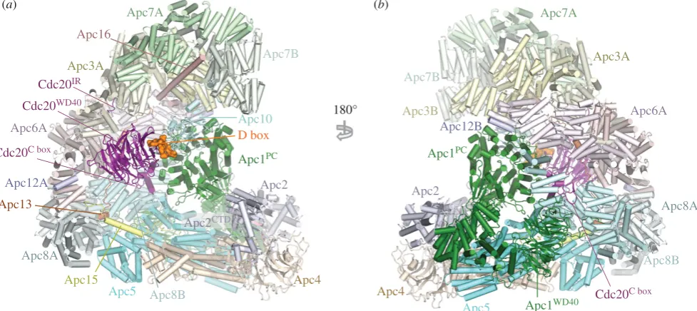
To understand the molecular basis for how phos-phorylation activates APC/CCdc20, a cryo-EM structure of
phosphorylated APC/CCdc20was determined [60]. The
struc-ture of phosphorylated APC/CCdc20is very similar to that of
unphosphorylated APC/CCdh1 (figures 1 and 8). Cdc20 (a)
Apc7A
Apc7B
Apc1PC
Apc4 Apc2
Apc5 Apc15
Apc3A
Apc12A Apc6A
Apc13
Apc16
Cdc20WD40
Apc10
Apc8A
Cdc20IR
Apc8B
(b)
180°
Apc7A
Apc7B
Apc1PC
Apc4 Apc2
Apc3A
Apc12B
Apc8A
Apc5
Apc8B
Apc3B
Apc1WD40
Apc6A
D box Cdc20C box
Apc2CTD
Cdc20C box
Figure 8.
Overall structure of the phosphorylated APC/C
Cdc20.substratecomplex. (a) and (b) Two orthogonal views of the APC/C
Cdc20.substrate. The substrate is the high
affinity budding yeast substrate Hsl1 (residues 667 to 872 containing a D box and KEN box). EM density for Apc11
RINGis weak indicating RING domain flexibility. PDB
5G04, from Zhang
et al.
[60].
rsob.r
oy
alsocietypublishing.org
Open
Biol.
7
:
170204
interacts with the APC/C through three motifs: the C box to Apc8B (augmented by the KILR motif [208]), the IR tail to Apc3A and a region contacting Apc1PC. Relative to Cdh1
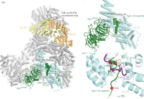
the contacts are fewer. Strikingly, EM density corresponding to phosphorylated regions could not be observed, indicating that phosphorylated regions of the APC/C do not directly or indirectly contribute to increasing the affinity of the APC/C for Cdc20. This implied that APC/C phosphorylation would remove an inhibitory segment from a Cdc20 binding site. To explore this possibility, the structures of phosphory-lated and unphosphoryphosphory-lated apo APC/C were compared. The two structures were very similar, except that in the unphosphorylated apo structure, a segment of EM density occupies the C-box binding site (figure 9). The proximity of this unassigned EM density to the disordered 300s loop of the Apc1 WD40 domain (Apc1WD40) suggested that this
segment corresponded to a region of the Apc1300sloop. In a
structure determined with this loop deleted, the C-box bind-ing site was devoid of EM density [60]. Deletion of the Apc1300sloop constitutively activated APC/CCdc20and
phos-phorylation did not further enhance activity [60], a finding made independently by Kraftet al.[202]. These data convin-cingly showed that a region within the Apc1300s loop (an
auto-inhibitory (AI) segment) represses Cdc20 stimulation of unphosphorylated APC/C activity, further supported by data in Liet al.[110]. Phosphorylation releases this auto-inhi-bition. In support of the idea that direct phosphorylation of the AI segment releases this auto-inhibition, substituting
Glu for Cdk phosphorylation sites within the AI segment constitutively activated APC/CCdc20[60].
The AI segment includes an Arg-Phe dipeptide, analogous to the Arg-Tyr motif of the C box. Modelling of the AI seg-ment into EM density showed that the Arg side chain of the AI segment mimics the Arg of the Arg-Tyr motif of the C box, anchoring the AI segment to the C-box binding site (figure 9c). Mitotic phosphorylation of sites flanking the Arg-Phe motif would destabilize interactions between the AI seg-ment and the C-box binding site through steric hindrance and charge repulsion, leading to the displacement and disordering of the AI segment and relief of auto-inhibition. These findings that an auto-inhibitory segment within the Apc1300s loop
blocks Cdc20 activation and that its mitotic phosphorylation relieves this auto-inhibition are in agreement with biochemical data [202,203] (figure 10). Fujimitsu and colleagues [203] showed that Apc1300s bound to the APC/C in an anaphase
extract, whereas the phosphomimetic mutants abolished this interaction, highlighting how the interaction of Apc1300swith
the APC/C is dependent on its phosphorylation status. The data of Zhanget al.[60] indicated that the critical deter-minant of activation of APC/CCdc20 by mitotic
phosphorylation was displacement of the AI segment to relieve auto-inhibition. However, Apc3 is also highly phosphorylated in mitosis [201,202,205– 207] and Cks stimulates both Cdk-dependent activation of APC/CCdc20[197,209] and Apc1 and
Apc3 phosphorylation [60,209], and interacts with Apc3 [25,203, 209,210]. Deletion of the hyperphosphorylated Apc3
(a)
(c)
Apc1WD40
Apc8B
300s loop Apc3B
Apc3A Cdk-cyclin-Cks recruitment loop
Apc8B
Apc1 AI segment
S372 S373
S364 R368
Cdc20C box
R78
Y79
N370 F369
C-box binding site
(b)
AI segment
C-box binding site
Apc1WD40
Apc8B
300s loop
Apc1 AI segment

![Figure 5. Degron consensus sequences. (a) Sequence motif of D box derived from 68 APC/C substrates [71]](https://thumb-us.123doks.com/thumbv2/123dok_us/8651132.1726530/9.595.89.489.38.366/figure-degron-consensus-sequences-sequence-motif-derived-substrates.webp)