The Capsule of
Cryptococcus neoformans
Modulates
Phagosomal pH through Its Acid-Base Properties
Carlos M. De Leon-Rodriguez,
aMan Shun Fu,
aM. Osman Çorbali,
aRadames J. B. Cordero,
aArturo Casadevall
aaDepartment of Molecular Microbiology and Immunology, Johns Hopkins Bloomberg School of Public Health,
Baltimore, Maryland, USA
ABSTRACT
Phagosomal acidification is a critical cellular mechanism for the
inhibi-tion and killing of ingested microbes by phagocytic cells. The acidic environment
ac-tivates microbicidal proteins and creates an unfavorable environment for the growth
of many microbes. Consequently, numerous pathogenic microbes have developed
strategies for countering phagosomal acidification through various mechanisms that
include interference with phagosome maturation. The human-pathogenic fungus
Cryptococcus neoformans
resides in acidic phagosomes after macrophage ingestion
that actually provides a favorable environment for replication, since the fungus
repli-cates faster at acidic pH. We hypothesized that the glucuronic acid residues in the
capsular polysaccharide had the capacity to affect phagosomal acidity through their
acid-base properties. A ratiometric fluorescence comparison of imaged phagosomes
containing
C. neoformans
to phagosomes containing beads showed that the latter
were significantly more acidic. Similarly, phagosomes containing nonencapsulated
C.
neoformans
cells were more acidic than those containing encapsulated cells.
Acid-base titrations of isolated
C. neoformans
polysaccharide revealed that it behaves as a
weak acid with maximal buffering capacity around pH 4 to 5. We interpret these
re-sults as indicating that the glucuronic acid residues in the
C. neoformans
capsular
polysaccharide can buffer phagosomal acidification. Interference with phagosomal
acidification represents a new function for the cryptococcal capsule in virulence and
suggests the importance of considering the acid-base properties of microbial
cap-sules in the host-microbe interaction for other microbes with charged residues in
their capsules.
IMPORTANCE
Cryptococcus neoformans
is the causative agent of cryptococcosis, a
devastating fungal disease that affects thousands of individuals worldwide. This
fun-gus has the capacity to survive inside phagocytic cells, which contributes to
persis-tence of infection and dissemination. One of the major antimicrobial mechanisms of
host phagocytes is to acidify the phagosomal compartment after ingestion of
mi-crobes. This study shows that the capsule of
C. neoformans
can interfere with full
phagosomal acidification by serving as a buffer.
KEYWORDS
Cryptococcus, capsule, pH, phagosome
I
ngestion of microbes by phagocytic cells results in the formation of a new organelle
called the phagolysosome, a membrane-bound compartment where microorganisms
are subjected to a variety of antimicrobial compounds such as oxidative radicals and
microbicidal proteins. The process of phagolysosome formation results from a complex
cellular choreography that includes phagosome acidification, resulting in microbial
inhibition by creating an unfavorable environment and the activation of various
microbicidal compounds. Consequently, diverse pathogenic microbes have developed
mechanisms to interfere with phagosomal acidification. For example, the bacterium
Mycobacterium tuberculosis
(1), the fungus
Histoplasma capsulatum
(2), and the parasite
Received10 August 2018Accepted18 September 2018 Published24 October 2018 CitationDe Leon-Rodriguez CM, Fu MS, Çorbali MO, Cordero RJB, Casadevall A. 2018. The capsule ofCryptococcus neoformans modulates phagosomal pH through its acid-base properties. mSphere 3:e00437-18.https:// doi.org/10.1128/mSphere.00437-18.
EditorJ. Andrew Alspaugh, Duke University Medical Center
Copyright© 2018 De Leon-Rodriguez et al. This is an open-access article distributed under the terms of theCreative Commons Attribution 4.0 International license.
Address correspondence to Arturo Casadevall, [email protected].
C.M.D.L.-R. and M.S.F. share first authorship.
R.J.B.C. and A.C. share senior authorship.
Host-Microbe Biology
crossm
on September 8, 2020 by guest
http://msphere.asm.org/
phagosome, there is evidence that
C. neoformans
modulates some aspects of
phago-somal maturation, including full phagophago-somal acidification, although the mechanisms
for this effect have not been fully elucidated (7, 8). In fact, for
C. neoformans,
acidifi-cation has been viewed as favoring intracellular growth, since this fungus replicates
faster in acidic environments (9). Its survival inside the phagosome is believed to result
from its ability to withstand oxidative bursts (10), damage the phagosomal membranes
(11), and damage critical host cell homeostasis (12) rather than interference with
phagosomal maturation, although the relative contributions to the overall outcome of
intracellular survival remain to be determined.
Recently, we reported a new role for
C. neoformans
urease in modulating
phago-somal pH (13). Urease-positive
C. neoformans
strains hydrolyzed urea to ammonia,
resulting in pleiotropic changes to the cryptococcal macrophage interaction that
included higher phagosomal pH, delayed intracellular growth, and enhanced nonlytic
exocytosis (13).
C. neoformans
is unusual among intracellular pathogens in that it grows
faster at lower pH, resulting in faster replication inside phagolysosomes than in the
extracellular medium (9). Loss of phagosomal integrity is associated with reduced
acidity in that compartment and the triggering of macrophage death (14). Hence, the
extent of phagosomal acidification is an important variable, which can favor the
microbe or the host cell depending on the state of the interaction (13, 14).
One of the most striking characteristics of
C. neoformans
as a pathogenic microbe is
that it is surrounded by a large polysaccharide capsule that is a critical determinant of
virulence (15). The capsule functions in virulence by interfering with phagocytosis and
immune responses (15, 16). The capsule is also thought to play a major role in
intracellular survival by quenching free radical fluxes in the phagosome (10). The major
capsular polysaccharide is glucuronoxylomannan (GXM), which is composed of a
mannose backbone with xylose and glucuronic acid substitutions (17). The presence of
glucuronic acid residues in cryptococcal polysaccharide imparts a negative charge to
the capsule (18) that is believed to contribute to protection against phagocytosis. In
addition, those glucuronic acid residues can be anticipated to impart considerable
acid-base properties to the cryptococcal GXM. In our recent study on the role of
phagosomal membrane integrity, we observed that even though apoptotic cells had
higher phagolysosomal pH, loss of membrane integrity was not associated with
com-plete loss of acidity, which we hypothesized was due to the acid-based properties of
the capsule (14). In contrast, for
Candida albicans,
which lacks a polysaccharide capsule
and hence has no comparable buffering capacity, phagosome permeabilization
re-sulted in luminal alkalinization (19). In this study, we formally tested that hypothesis
and present evidence that the capsule of
C. neoformans
interferes with full phagosome
acidification. These findings establish a new mechanism for microbial modulation of
phagosomal pH and imply a new role for the capsule in cryptococcal virulence.
RESULTS
pH of phagosomes containing beads and
C. neoformans
.
Ingested
C. neoformans
resides in a mature acidic phagosome (6). However, the extent to which
C. neoformans
modulates the pH of the cryptococcal phagosome is unknown. A comparison of the pH
of phagosomes containing inert beads with phagosomes containing
C. neoformans
cells showed that the latter were significantly less acidic (Fig. 1). On average, the pH of
phagosomes containing inert beads was 4.22
⫾
0.45 (n
⫽
40) at 3 h, which
on September 8, 2020 by guest
http://msphere.asm.org/
sponded to a 0.65 pH unit difference (P
⬍
0.0001 by one-way ANOVA and Tukey’s
multiple-comparison test). To ascertain whether this higher pH was the result of active
pH modulation by
C. neoformans, we compared the pH of phagosomes containing live
and dead
C. neoformans
cells. Comparison of the average pH in phagosomes containing
live and dead
C. neoformans
cells revealed average values of 4.87
⫾
0.58 (n
⫽
62) and
4.78
⫾
0.14 (n
⫽
43) at 3 h, respectively (P
⫽
0.638 by one-way ANOVA and Tukey’s
multiple-comparison test). Phagosomes containing live and dead cells had comparable
pHs to those having live cells, suggesting that the pH modulation in the phagosome is
not the result of secretion of basic compounds by
C. neoformans
(Fig. 1).
The phagosomal pH of encapsulated cells is higher than that of
nonencapsu-lated cells.
To test the hypothesis that pH modulation was the result of the acid-base
properties of
C. neoformans, we sought to compare the phagosomal pH for
encapsu-lated and nonencapsuencapsu-lated cells. However, this presented the practical problem that
nonencapsulated cells could not be opsonized through the FcR, since they lacked a
capsule that would bind GXM-binding antibody. Opsonizing encapsulated cells with
antibody and nonencapsulated cells with complement was not considered acceptable,
since the two opsonins are very different. We tried to label cryptococcal cells with
EZ-Link NHS-biotin and Oregon green 488-conjugated NeutrAvidin, and phagocytosis
was performed using guinea pig complement as an opsonin. However, the signal of
Oregon green was not stable in the phagosome, and after 24 h, it was lost completely,
which we attribute to dye degradation from a combination of the low phagosomal pH
and the oxidative burst (20). We have also tried to measure the phagolysosomal pH
with heat-killed cryptococcal cells labeled with NHS-biotin, but the labeling did not
work with heat-killed cells. Hence, we resorted to coating nonencapsulated cells with
encapsulated
C. neoformans
conditioned media, which results in the attachment of
soluble polysaccharide to the surfaces of nonencapsulated cells to create a
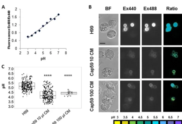
proto-capsule that would allow antibody-mediated opsonization (Fig. 2). The phagosomal pH
of naturally encapsulated
C. neoformans
cells was significantly higher than that of
nonencapsulated cells containing an artificial capsule (Fig. 3).
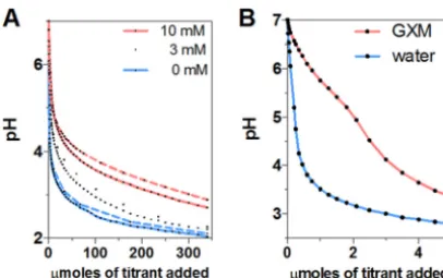
Acid-base properties of glucuronic acid and GXM.
Glucuronic acid is an organic
weak acid with a relatively high pKa. We titrated a sodium salt of glucuronic acid
(sodium D-glucuronate) with HCl and calculated a pKa in the range of 2.5 to 3.11 at the
beginning of our titrations (corresponding to 0.23 to 20
mol of titrant) (Fig. 4A). These
values are comparable to the reported pKa values of 2.9 (21), 2.8 to 2.9 from
13C-nuclear
magnetic spectroscopy (22), and 3.28 measured from standard acid-based titrations
(23). Consequently, the presence of glucuronic acid in solution confers considerable
buffering capacity such that for a 10 mM solution to change from pH 7 to pH 4, it
requires almost 10 times the acid required to achieve the same pH reduction as in a
pure water solution. Similarly, the presence of GXM in water provided considerable
buffering capacity at around pH 5, which is close to the final pH in cryptococcal
FIG 1 pH of phagosomes containing beads or heat-killed or liveC. neoformans. Phagolysosomal pH was measured by using Oregon green dual-excitation ratio fluorescence imaging at the indicated time points. Each dot represents the pH of an individual phagolysosome. Data are from one representative experi-ment. Comparable results were obtained from two additional independent experiments.Pvalues were calculated by one-way ANOVA with Tukey’s multiple-comparison test.
on September 8, 2020 by guest
http://msphere.asm.org/
phagosomes (Fig. 4B). Considering the known polyelectrolyte nature and polydispersity
of GXM preparations in terms of molecular mass (24, 25), together with the mild
inflection point, it is problematic to determine a pKa value for GXM.
DISCUSSION
Many pathogenic microbes express polysaccharide capsules that are essential for
virulence. Microbial capsules are directly antiphagocytic, and efficient ingestion of
FIG 2 Binding of capsular polysaccharide to the nonencapsulatedC. neoformans cap59strain. Capsule material (CM) release into the media byC. neoformanswas incubated with different concentrations of the cap59strain to form a proto-capsule around these cells. Subsequently, the cells were incubated with the monoclonal antibody 18B7 previously conjugated with Oregon green (18B7-OG). Bright-field (top) and immunofluorescence (bottom) images are shown ofC. neoformansH99 and the acapsularcap59strain incubated with 100, 10, or 1l of CM andcap59strain alone. The magnification for this figure is 40⫻.
FIG 3 pH measurement of phagolysosome-containing encapsulated and proto-encapsulatedC. neofor-mans. Macrophages were infected with H99 andcap59strains previously incubated with 100 and 10l of conditioned media (CM) to form a proto-capsule. Opsonization was antibody mediated by using monoclonal antibody 18B7 conjugated to Oregon green. Phagolysosomal pH was measured by using dual-excitation ratio fluorescence imaging. (A) Representative standard curve of mean fluorescence excitation ratio (488-nm excitation/440-nm excitation: 520-nm emission) of Oregon green cryptococcus-loaded macrophages with phagolysosomes equalized in calibration buffers at pH 3 to pH 7. (B) Representative images showing bright-field (BF) microscopy of macrophages containing cryptococcal cells, fluorescence images taken at 440-nm excitation (Ex440) and 488-nm excitation (Ex488), and pseudocolor images of the 488ex/440ex ratio with pH color scale displayed at the bottom of the panel. Scale bar, 10m. (C) Box plot with dots representing pH of individual phagolysosomes. The pH value was 5.19 in H99- containing phagolysosomes, which was less acidic than phagosomes containingcap59strain incubated with 100 and 10l of CM, showing pH values of 4.40 and 4.25, respectively. Fewer data points are plotted for thecap59100-l CM conditions relative to the other conditions because their tendency to aggregate in that experiment but the numbers were sufficient for high statistical significance. One-way ANOVA with Dunnett’s multiple-comparison test was conducted.****,P ⬍0.0001 (Tukey’s multiple comparison).
on September 8, 2020 by guest
http://msphere.asm.org/
encapsulated microbes by phagocytic cells usually requires antibody- or
complement-derived opsonins. However, many microbial capsules are composed of polysaccharides
that are poorly immunogenic that often fail to induce strong responses. Although
microbial capsules are generally thought to contribute to virulence by resisting
inges-tion and killing by host phagocytic cells, there is evidence that capsules mediate other
functions that contribute to pathogenesis and that some of these effects are mediated
by capsule ionic charge. For instance, negatively charged capsular polysaccharides of
Gram-negative bacteria can bind cationic microbicidal peptides and protect bacterial
cells (26). On the other hand, the positively charged modifications in
Streptococcus
pneumoniae
capsular polysaccharide can predispose bacterial cells to enhanced killing
by alpha-defensins (27). Among
Cryptococcus
spp., a comparison of glucuronic acid
residue content among a nonpathogenic species and pathogenic species revealed that
the latter had higher content of this charged residue (28). Hence, while there is
considerable evidence that microbial surface charge can play an important role in
pathogenesis through a variety of mechanisms, their role in phagosomal pH
modula-tion has not been investigated.
It is axiomatic that microbial capsules containing weak acid and basic residues will
exhibit acid-base properties that are reflective of the capacity of these residues to
function as proton donors and acceptors. In this regard, the cryptococcal capsule is
composed of a repeating mannose triad, each of which includes one glucuronic acid
residue, that in turn confers upon the polysaccharide and the resulting capsule a
negative charge (18). Our results confirm the theoretical deduction that the
C.
neofor-mans
capsular GXM buffers the phagolysosomal pH. While titration of the sodium salt
of glucuronic acid (sodium D-glucuronate) shows a rapid initial decrease in pH during
titration, GXM provides a buffer effect around pH 5. This is not surprising, since the pKa
of glucuronic acid is 3.28 and a solution of sodium D-glucuronate will exert its best
buffering effect near the pKa. However, with readily available acidic and basic residues
of glucuronic acid (A
⫺and HA) covalently bound to the GXM polysaccharide backbone,
these residues could experience different electronic milieu that could modify their ionic
properties. Furthermore, GXM molecules are large and structurally complex, and
con-sequently, not all glucuronic acids may be equally exposed to the solvent and in a
position to donate or accept hydronium ions equally. Polyelectrolytes can manifest
complex acid-base properties and the pKa values of the monomer and polymer can
differ (29, 30). Despite these differences, GXM retains considerable weak acid properties
that would confer a maximum buffering capacity at the pH range of approximately 4
to 5, which corresponds to the optimal pH for
C. neoformans
growth (13).
Ingested dead encapsulated
C. neoformans
cells resided in phagosomes that had a
FIG 4 Titration of sodium D-glucuronate and GXM against HCl. (A) Changes in pH of sodium D-glucuronate solutions at 3 and 10 mM as a function of the number of micromoles of titrant added. Initially, sodium D-glucuronate solutions show a rapid pH change by acidic titration. Near the pKa level (pH⬃3.28, it shows the best buffering capacity compared to pure water. Theoretical curves (dashed lines) for glucuronic acid also reveal a similar tendency of rapid pH change and buffering capacity. (B) Change in pH of GXM solution as a function of the number of micromoles of titrant added. GXM provides a substantial amount of buffering capacity around pH 5 compared to ultrapure water.
on September 8, 2020 by guest
http://msphere.asm.org/
outweighed by the fact that we could not be certain that other methods of
opsoniza-tion would result in comparable phagosomes and the fact that natural capsules are
much larger than artificial capsules meant that there would still be much more
polysaccharide in encapsulated cryptococcal phagosomes. Despite this handicap, we
were able to measure a difference in phagosomal pH between encapsulated and
acapsular strains consistent with a strong acid buffering capacity by the polysaccharide
capsule.
In summary, the presence of glucuronic acid residues in the
C. neoformans
capsule
makes the polysaccharide a weak acid capable of modulating pH in the phagosome.
Our experimental observations are consistent with the expected acid-base properties of
the capsule based on its sugar residue composition. The fact that the polysaccharide
capsule of
C. neoformans
is large brings considerable GXM mass into the phagosome
with the potential to mediate considerable buffering capacity. Given that
C. neoformans
has optimal growth rate at acidic pHs (13), the acid-base properties of the capsule can
be expected to promote fungal cell survival in the phagosome by its buffering capacity
during conditions of both phagosomal acidification and phagosomal membrane
leak-age. This mechanism for phagosomal pH modulation based on acid-base properties is
different from other intracellular pathogens that modulate pH by interfering with
phagosome maturation. Our observations suggest that other microbes with charged
microbial capsules could also modulate phagosomal acidification through their
acid-based electrolyte properties.
MATERIALS AND METHODS
Yeast culture.C. neoformansserotype A strain H99 and the acapsular mutantcap59were used for all experiments. Cells were grown from frozen stocks in Sabouraud dextrose liquid medium at 30ºC under agitation (180 rpm) for 2 days.
Coating of acapsular mutant with GXM. For the formation of the proto-capsule, we follow previously published methods (31). Briefly, the supernatant of an overnight culture ofC. neoformansH99 was cleared by centrifugation and filtered using a 0.8-m syringe filter. An overnight culture of 1⫻107
cells/mlcap59acapsular strain was then incubated with 100, 10, or 1l of H99 cleared supernatant (conditioned media) in a total volume of 1 ml medium with rotation for 1 h at room temperature. Images were acquired using an Olympus AX70 microscope (Olympus, Center Valley, PA) with 40⫻objective to visualize the formation of the proto-capsule, which was labeled by Oregon green 488 conjugated 18B7 monoclonal antibody.
Measurement of phagosomal pH.Phagolysosomal pH was measured using ratiometric fluores-cence imaging involving the use of pH-sensitive probe Oregon green 488. Oregon green 488 was first conjugated to monoclonal antibody 18B7 using Oregon green 488 protein labeling kit (Molecular Probes, Eugene, OR) as described previously (13). The Oregon green 488 dye has a succinimidyl ester moiety that reacts with primary amines of proteins to form stable dye-protein conjugates. The labeling procedure was performed according to the manufacturer’s instruction. BMDM were plated (4⫻105cells/well) on 24-well
plate with 12-mm circular coverslip coated with 100g/ml poly-D-lysine. Cells were cultured with completed BMEM medium containing 0.5g/ml LPS and 100 U/ml IFN-␥and then incubated at 37°C with 9.5% CO2
overnight. For infection, H99 andcap59strains (8⫻106cells/ml) were incubated with 10g/ml Oregon
green conjugated mAb18B7 for 15 min. Macrophages were then infected with Oregon green-conjugated 18B7-opsonized yeast in 4⫻105cells per well. Cells were centrifuged immediately at 270⫻gfor 1 min,
and the cells were incubated at 37°C for 10 min to allow phagocytosis. Extracellular cryptococcal cells or beads were removed by washing three times with fresh medium. Samples on coverslip were collected at 24 h after phagocytosis by washing twice with prewarmed HBSS. Annexin V Alexa Fluor 555 staining was performed per the manufacturer’s instructions (Invitrogen, Carlsbad, CA). The coverslip was then placed upside down on a MatTek petri dish (35 mm; 10-mm diameter microwell; MatTek, Ashland, MA) with the Annexin V binding buffer in the microwell. Images were taken by using an Olympus AX70 microscope (Olympus, Center Valley, PA) with 40⫻objective with two excitation wavelengths of 440 nm and 488 nm for Oregon green and wavelength of 550 mm for Annexin V and bright-field microscopy.
on September 8, 2020 by guest
http://msphere.asm.org/
Images were acquired and analyzed using MetaFluor Fluorescence Ratio Imaging Software (Molecular Devices, Downingtown, PA). Relative phagolysosomal pH was determined based on the ratio of 488 nm/ 440 nm. The relative pH was converted to absolute pH by obtaining the standard curve in which the images are taken as described above, but the intracellular pH of macrophage was equilibrated by adding 10M nigericin in pH buffer (140 mM KCl, 1 mM MgCl2,1 mM CaCl2,5 mM glucose, and appropriate
bufferⱕpH 5.0: acetate-acetic acid; pH 5.5 to 6.5: MES;ⱖpH 7.0: HEPES. Desired pH values were adjusted using either 1 M KOH or 1 M HCl). Buffers were used at pH 3 to 7.5 using 0.5-pH unit increments.
Biotinylation of cells.Approximately 1⫻106cryptococcal cells were biotinylated using EZ
Link-sulfo-NHS-biotin (catalog no. 21217; Thermo Fisher Scientific, Rockford, IL). Overnight cultures were washed three times with PBS (pH 8.0) and diluted in PBS (pH 8.0) to 1⫻106cells/ml. EZ
Link-sulfo-NHS-biotin (2 mM) was added to the cell samples and incubated for 30 min at room temperature. Cells were then washed three times with PBS with 100 mM glycine to remove excess biotin reagent and by-product. After biotinylation, cells were labeled with 5g/ml Oregon green conjugate of NeutrAvidin biotin-binding protein (catalog no. A6374; Thermo Fisher Scientific) with rotation for 1 h at room temperature.
GXM isolation. Soluble GXM was obtained from culture supernatants of encapsulated cells by ultrafiltration (32, 33). Briefly, culture supernatants were collected by centrifugation (6,000⫻g, 15 min, 4°C) and filtered using 0.22-m vacuum-driven disposable bottle-top filter (MilliPore) to ensure clearing of cells and other large debris. The cleared supernatant was ultrafiltered sequentially in an Amicon ultrafiltration cell (Millipore, Danvers, MA) using membranes of 100- and 10-kDa nominal molecular weight limits. After filtrating using a 100-kDa membrane, the flowthrough was again filtered through a 10-kDa membrane. On each filtration step, GXM can be recovered from the surfaces of membranes in the form of a viscous gel. This process yields GXM fractions of⬎100 kDa and 100 to 10 kDa that were then dialyzed against ultrapure water, lyophilized, and store until use.
Acid-based titrations.Forty milliliter solutions of 3 and 10 mM sodium D-glucuronate (catalog no. G8645; Sigma) were titrated against 0.1 M HCl in 40-ml glass beakers. Since the pH of ultrapure water was acidic at⬃pH 5, it was adjusted to pH 7 using NaOH before preparation of the glucoronate solutions. GXM solutions were prepared by dissolving the lyophilized GXM fraction 10 to 100 kDa in ultrapure water at 1 mg/ml. Since GXM molecules exhibit wide size distributions, from 1 to⬎100 kDa, we used the 10-to 100-kDa fraction 10-to narrow the range of molecular mass. The 10-total monosaccharide concentrations in GXM solutions were determined by the Dubois method (also known as the phenol-sulfuric acid assay) (34). After the phenol sulfuric assay, GXM solutions were diluted to 2.4 mM total monosaccharide concentration, and 3-ml volumes were titrated against 0.01 M HCl in 10-ml glass beakers. Titrations were conducted in beakers placed inside a water bath equilibrated at a temperature of 20°C with constant stirring. Changes in pH were recorded using an Accumet pH meter.
Calculation of theoretical acid-base titration curve.To calculate the theoretical acid-base titration curve for sodium D-glucuronate (NaA), we assumed that positive charges and negative charges are equal in an ideal solution: [Na⫹]⫹[H⫹]⫽[OH⫺]⫹[A⫺]⫹[CI⫺], such that NaA↔Na⫹⫹A⫺and NaA⫹
HCl↔HA⫹NaCl. Such that [Na⫹]⫽[A⫺]⫹[HA]. Also, C
HA⫽[HA]⫹[A⫺] and CHCI⫽[CI⫺], sinceHCl
totally dissolves. Hence, the equation becomes
CHA⫹
关
H⫹兴
⫽关
OH⫺兴
⫹关
A⫺兴
⫹CHClAt this point, we can turn this equation into a third-degree polynomial, with [H⫹] being the unknown,
x3⫹
冉
nVinitial⫹VHCl⫹
ka⫺CHCl
冊
x2⫺共
kaCHCl⫹kwater兲
x⫺kwaterka⫽0while pKa⫽3.28 (glucuronic acid),kwater⫽0.681⫻10⫺14at 20°C, andnis the initial amount of moles
of sodium D-glucuronate we use for our solution. The positive root of this third-degree polynomial provided us the theoretical [H⫹] value after a certain number of moles of acid was added.
Statistical analysis.All statistical analyses were performed by using one-way ANOVA, followed by Tukey’s or Dunnett’s multiple-comparison test.
ACKNOWLEDGMENTS
A.C. was supported in part by 5R01HL059842, 5R01AI033774, 5R37AI033142, and
5R01AI052733
REFERENCES
1. Wong D, Bach H, Sun J, Hmama Z, Av-Gay Y. 2011. Mycobacterium tuberculosis protein tyrosine phosphatase (PtpA) excludes host vacuolar-H⫹-ATPase to inhibit phagosome acidification. Proc Natl Acad Sci U S A 108:19371–19376.https://doi.org/10.1073/pnas.1109201108. 2. Strasser JE, Newman SL, Ciraolo GM, Morris RE, Howell ML, Dean GE. 1999.
Regulation of the macrophage vacuolar ATPase and phagosome-lysosome fusion by Histoplasma capsulatum. J Immunol 162:6148 – 6154.
3. Vinet AF, Fukuda M, Turco SJ, Descoteaux A. 2009. The Leishmania donovani lipophosphoglycan excludes the vesicular proton-ATPase from phagosomes by impairing the recruitment of synaptotagmin V. PLoS Pathog 5:e1000628.https://doi.org/10.1371/journal.ppat.1000628. 4. Feldmesser M, Kress Y, Novikoff P, Casadevall A. 2000.Cryptococcus
neoformansis a facultative intracellular pathogen in murine pulmonary infection. Infect Immun 68:4225– 4237.https://doi.org/10.1128/IAI.68.7 .4225-4237.2000.
5. Casadevall A, Perfect JR. 1998. Cryptococcus neoformans. American Society for Microbiology, Washington, DC.
6. Levitz SM, Nong SH, Seetoo KF, Harrison TS, Speizer RA, Simons ER. 1999. Cryptococcus neoformansresides in an acidic phagolysosome of human macrophages. Infect Immun 67:885– 890.
7. Smith LM, Dixon EF, May RC. 2015. The fungal pathogen Cryptococcus neoformans manipulates macrophage phagosome maturation. Cell Mi-crobiol 17:702–713.https://doi.org/10.1111/cmi.12394.
8. Qin QM, Luo J, Lin X, Pei J, Li L, Ficht TA, de Figueiredo P. 2011.
on September 8, 2020 by guest
http://msphere.asm.org/
macrophages is accompanied by phagosomal permeabilization and accumulation of vesicles containing polysaccharide in the cytoplasm. Proc Natl Acad Sci U S A 99:3165–3170.https://doi.org/10.1073/pnas .052702799.
12. Coelho C, Souza ACO, da Silveira Derengowski L, de Leon-Rodriguez C, Wang B, Leon-Rivera R, Bocca AL, Gonçalves T, Casadevall A. 2015. Macrophage mitochondrial and stress response to ingestion of Crypto-coccus neoformans. J Immunol 194:2345–2357.https://doi.org/10.4049/ jimmunol.1402350.
13. Fu MS, Coelho C, De Leon-Rodriguez CM, Rossi DCP, Camacho E, Jung EH, Kulkarni M, Casadevall A. 2018. Cryptococcus neoformans urease affects the outcome of intracellular pathogenesis by modulating phagolysosomal pH. PLoS Pathog 14:e1007144.https://doi.org/10.1371/ journal.ppat.1007144.
14. De Leon-Rodriguez CM, Rossi DCP, Fu MS, Dragotakes Q, Coelho C, Guerrero Ros I, Caballero B, Nolan SJ, Casadevall A. 2018. The outcome of the Cryptococcus neoformans-macrophage interaction depends on phagolysosomal membrane integrity. J Immunol 201:583– 603.https:// doi.org/10.4049/jimmunol.1700958.
15. O’Meara TR, Alspaugh JA. 2012. The Cryptococcus neoformans capsule: a sword and a shield. Clin Microbiol Rev 25:387– 408.https://doi.org/10 .1128/CMR.00001-12.
16. Del Poeta M. 2004. Role of phagocytosis in the virulence of Cryptococcus neoformans. Eukaryot Cell 3:1067–1075.https://doi.org/10.1128/EC.3.5 .1067-1075.2004.
17. Cherniak R, Sundstrom JB. 1994. Polysaccharide antigens of the capsule ofCryptococcus neoformans. Infect Immun 62:1507–1512.
18. Nosanchuk JD, Casadevall A. 1997. Cellular charge of Cryptococcus neoformans: contributions from the capsular polysaccharide, melanin, and monoclonal antibody binding. Infect Immun 65:1836 –1841. 19. Westman J, Moran G, Mogavero S, Hube B, Grinstein S. 2018. Candida
albicans hyphal expansion causes phagosomal membrane damage and luminal alkalinization. mBio 9:e01226-18.https://doi.org/10.1128/mBio .01226-18.
20. Bruneau E, Sutter D, Hume RI, Akaaboune M. 2005. Identification of nicotinic acetylcholine receptor recycling and its role in maintaining receptor density at the neuromuscular junction in vivo. J Neurosci 25:9949 –9959.https://doi.org/10.1523/JNEUROSCI.3169-05.2005. 21. Robinson D, Smith JN, Williams RT. 1953. Studies in detoxication. 52. The
apparent dissociation constants of some glucuronides, mercapturic
ac-25. Cordero RJ, Frases S, Guimaraes AJ, Rivera J, Casadevall A. 2011. Evi-dence for branching in cryptococcal capsular polysaccharides and con-sequences on its biological activity. Mol Microbiol 79:1101–1117.https:// doi.org/10.1111/j.1365-2958.2010.07511.x.
26. Llobet E, Tomas JM, Bengoechea JA. 2008. Capsule polysaccharide is a bacterial decoy for antimicrobial peptides. Microbiology 154:3877–3886.
https://doi.org/10.1099/mic.0.2008/022301-0.
27. Beiter K, Wartha F, Hurwitz R, Normark S, Zychlinsky A, Henriques-Normark B. 2008. The capsule sensitizes Streptococcus pneumoniae to alpha-defensins human neutrophil proteins 1 to 3. Infect Immun 76: 3710 –3716.https://doi.org/10.1128/IAI.01748-07.
28. Araujo Gde S, Fonseca FL, Pontes B, Torres A, Cordero RJB, Zancopé-Oliveira RM, Casadevall A, Viana NB, Nimrichter L, Rodrigues ML, Garcia ES, Souza W, Frases S. 2012. Capsules from pathogenic and non-pathogenic Cryptococcus spp. manifest significant differences in struc-ture and ability to protect against phagocytic cells. PLoS One 7:e29561.
https://doi.org/10.1371/journal.pone.0029561.
29. Niu Y, Sun L, Crooks RM. 2003. Determination of intrinsic proton binding constants for poly(amidoamine) dendrimers via potentio-metric pH titration. Macromolecules 36:5725–5731.https://doi.org/ 10.1021/ma034276d.
30. Mafe SG-M, Garcı´´a-Morales V, Ramirez P. 2004. Estimation of pKa shifts in weak polyacids using a simple molecular model: effects of strong polybases, hydrogen bonding and divalent counterion bind-ing. Chemical Physics 296:29 –35.https://doi.org/10.1016/j.chemphys .2003.09.033.
31. Reese AJ, Doering TL. 2003. Cell wall alpha-1,3-glucan is required to anchor the Cryptococcus neoformans capsule. Mol Microbiol 50: 1401–1409.https://doi.org/10.1046/j.1365-2958.2003.03780.x. 32. Nimrichter L, Frases S, Cinelli LP, Viana NB, Nakouzi A, Travassos LR,
Casa-devall A, Rodrigues ML. 2007. Self-aggregation ofCryptococcus neoformans capsular glucuronoxylomannan is dependent on divalent cations. Eukaryot Cell 6:1400 –1410.https://doi.org/10.1128/EC.00122-07.
33. Albuquerque PC, Fonseca FL, Dutra FF, Bozza MT, Frases S, Casadevall A, Rodrigues ML. 2014. Cryptococcus neoformans glucuronoxylomannan fractions of different molecular masses are functionally distinct. Future Microbiol 9:147–161.https://doi.org/10.2217/fmb.13.163.
34. Dubois M, Gilles KA, Hamilton JK, Rebers PA, Smith F. 1956. Colorimetric method for determination of sugars and related substances. Anal Chem 28:350 –357.https://doi.org/10.1021/ac60111a017.