2172
Review On Technologies For Antimicrobial
Testing Methods For Veterinary Origin
B.Keerthi Priya, D.Akhila Reddy, T.Yasaswi, D.V.Rama Koti ReddyAbstract: Antimicrobial Susceptibility Testing (AST) is gaining importance in medical practice as the patients are to be treated with correct Antibiotic with appropriate dosage. For humans one to one communication between the doctor, patient and instrumentation will helps the doctor for the diagnosis but in the case of animals only traditional methods are used for diagnosis and the antibiotics are used randomly, which causes the death of the animals and unnecessary deposition of Antibiotic in the Animal Body. In the case of the humans Isolation and identification of the organisms are important but in the case of animals identification of the right antibiotic in right dosage in less time is important. For the penetration of the systems to the remote location to the farmer’s automation of the systems is the best solution to reduce the experimentation and costs. As there is necessity for resistance surveillance, the zone readers and automated broth dilution system are widely used due to its quantative results and electronic data management. The selection for an AST technique would be based on several factors such as minimal effort of procedure, flexibility, ablity to adapt for automatic system s, expense, robustness and precision.Although slow, the standard methods used for the AST are precise.This review paper summarizes standard techni ques of AST, optical strategies, mechanical methods, semiautomated methods, and fully automated techniques.Techniques currently were using, focusin g on a few hopeful evolving but also coming years technologies in the area of fast profiling of antibiotic susceptibility.
Index Terms: Antimicrobial susceptibility Test, Traditional AST Methods, Sensors Based AST Methods, optical based AST methods, Lab-on-chip AST Methods, Micro fluidics AST Methods, AST Methods currently in use,
————————————————————
1
INTRODUCTION
With advancements in Nano Technology and Biotechnology sophisticated instruments are available for Microbiology labs for the quantitative diagnosis of the patient samples. The Main motivation of this is to get the accurate results in the minimum period. The use of the bio Nano materials, sensors and other techniques like image processing facilitated the above. Veterinary pathogens test Techniques were developed by the Clinical Laboratory & Standards Institute (CLSI) and its Subcommittee is Veterinary Antimicrobial Susceptibility Testing (VAST) for Animal welfare. Research on AST methods for animals and humans are parallel as AST Methods used for human pathogens can be applied to animal diseases. In India, CLSI developed antibacterial MIC breakpoints of susc eptible, intermediate and resistant interpretative parameters, br eakpoints as well as diameter of zone of inhibition the Positive Isolates and Negative Isolates[1].Some tests of AST are either numerical or subjective. Minimum inhibitory concentration (MIC ) of the antibiotic may be calculated in the quantitative tests. MI C is the minimum drug concentration that inhibits bacteria's visi ble growth[2A variety of antimicrobial susceptibility testing (AS T) methods exist to assess antimicrobial susceptibility of bacteria. Method choice depends on numerous considerations like practicability, versatility, effici ency, expense, reproducibility, reliability and personal preferen ce. Standardization, harmonization of AST. Different AST Methods are discussed in below sections. Section 2 describes Traditional Methods, Mechanical methods are discussed in Section 3, and section 4 describes Optical Methods.
AST Methods using Microfluidics are discussed in section 5, AST Methods currently used are discussed in 6, near Future alternatives are discussed in Section 7 and Automated Methods are discussed in section 8
2.
TRADITIONAL
AST
METHODS
2.1 Agar dilution method
Agar plate uses solid medium for organism growth. Different types of agar plate are Blood agar, Nutrient agar and MacConkey agar. Microorganisms are spread on the plate. Agar plates are incubated for 18hours for microorganism growth [3]. After growth on which antibiotics are placed as shown in figure. Anti-biotic C inhibits bacterial growth efficiently. Antibiotic A failed to inhibit bacterial growth. Antibiotic B is inhibiting microorganism growth intermediately. Agar plates before and after growth using agar dilution are shown in figure1. Advantages of this method are up to 30 microorganisms can be tested at a time. This method is cost effective. Automation of this method can be done using image processing. This method can be used for veterinary origin but more time to result
Fig1 Antibiotic disks before and after growth using agar dilution
__________________________________
• B. Keerthi Priya, Assistant Professor, Department of ECE, Gayatri Vidhya Parishad College of Engineering(A) (E-mail: [email protected]) • D. Akhila Reddy, Student, Department of ECE, Andhra University College
of Engineering(A),(E-mail: [email protected])
• T.Yasaswi, JRF in UGC- SAP , Department of Instrument Technology, Andhra University College of Engineering(A) (E-mail: [email protected])
• Prof. D.V. Rama Koti Reddy, Professor, Department of Instrument Technology, Andhra University College of Engineering(A) (E-mail:
2173 2.2 Chromogenic agar medium
The Working guideline of this technique is metabolic transformation of chromogenic mixes in agar medium. This technique empowers quicker CFU identification because of an early color change inside 18h – 24 h for powerless strains. Chromogenic media is an integral asset to fast ID of bacterial agent in water and food. Various shades of microscopic organisms utilizing chromogenic agar dilution are shown in figure2.
Fig 2. Different colors of bacteria using chromogenic agar
2.3 Broth Macro dilution Method
Growth inhibition in fluid medium with anti-biotics utilizes 96 tubes containing two-fold dilution. A prominent measure of suspended microorganism is added Followed by twenty-four hours of incubation [3], the bacterial development is estimated with the turbidity in the wells, using which MIC can be measured. Essential favorable position of the Broth dilution strategy is the capacity to calculate minimum bacterial concentration (MBC) apart from MIC, which is the most minimal measure of medication at which all microbes is killed. This technique can be utilized to test multiple bacteria at different concentration at a time. The benefit of this method is a quantitative outcome (i.e., the MIC). The fundamental inconvenience of this strategy is it requires more time for preparation of anti-biotic solutions. some more drawbacks are it requires more reagents for each test and consume space.
Fig 3. Broth dilution Standard trays contain 96 wells
2.4 Disk diffusion method
In this method on agar plate bacterial inoculum is applied on which anti-biotic strips are kept. Agar plates are placed in incubator and incubated for 16 hours. Around every one of the anti-biotics the zone prohibiting the growth of antibiotic is calculated. The Advantages of this technique are the procedure of this AST Strategy is easy and it doesn't require
extraordinary hardware. The likelihood to alter the sort of anti-biotic by just altering a solitary plate gives a specific level of included adaptability. As of now, this AST strategy has the most reduced expenses. The drawbacks of this plate test are the non-existence of automation of the procedure and another drawback is its unsatisfactory quality for moderate and anaerobically developing microorganisms. and this test results are qualitative but not quantitative
Fig 4. Zone of Inhibition Disk diffusion
2.5 ETest
It utilizes plastic test strips with anti-microbials. This Method is performed comparatively to the disk diffusion strategy. The Etest has a few favorable circumstances over the disk diffusion test. To begin with, on the grounds that the inclination is steady for up to 20 hours, this method is appropriate for an assortment of bacterial pathogens, going from quick developing vigorous microorganisms to very slow-developing exacting microscopic organisms. A different strip is required for every anti-toxin, and in this manner the expense of this technique can be high. Strips containing an anti-microbial angle in E-Test is appeared in figure5. This strategy is easy procedure but is generally costly and doesn't dependably distinguish colistin-resistant isolates. Etest is an advantageous AST technique that gives precise quantitative MICs at a greater expense.
2174 2.6 Polymerase chain reaction technique
It is a research facility technique utilized for making countless duplicates of short areas of DNA from an extremely little example of genetic material. This procedure is classified "enhancing" the DNA and it empowers explicit qualities important to be identified or estimated. DNA is comprised of rehashing successions of four bases – adenine, thymine, guanine, and cytosine as appeared in fig 6. The initial step or cycle of PCR is to isolate the strands of DNA into two single strands by expanding the temperature of the example that contains the DNA of intrigue. This is classified "denaturing" the DNA. When the strands discrete, the sample is cooled
somewhat and forward and turn around preliminaries are added and permitted to tie to the single DNA strands. primers are short successions of bases made explicitly to perceive and tie to the area of DNA to be intensified, which are the quite certain grouping of bases that are a piece of the quality or qualities of intrigue. Primers are classified "forward" and "turn around" in reference to the bearing that the bases inside the area of DNA are duplicated. After the two primers join to each strand of the DNA. Inside 30 to 40 cycles, upwards of a billion duplicates of the first DNA area can be delivered and utilized in various atomic demonstrative test.
Table 1: Summary of Traditional Testing Methods
S.N o
Traditional Testing Technolog
y
Working Principle
Deter mine s Resis tance
Test time (hours)
Easy/ comp
lex
cost
Manual or Automat
ed method
1
Agar dilution method
It uses solid medium for the measurement of
zone of inhibition
No 24-36
hours easy Low
Manual / Automat
ed
2
chromoge nic agar method
Metabolic transformation of chromogenic mixes in
agar medium
yes 18-24 hours easy
Mode
rate Manual
3
Broth dilution method
It uses liquid medium for the measurement of
zone of inhibition
no 24-36
hours easy Low
Manual / Automat
ed
4
Disc diffusion
method
It uses disk for the measurement of zone
of inhibition
yes 24-36 hours easy Low
Manual / Automat
ed
5 Etest
It uses strip for the measurement of zone
of inhibition
no 24
hours easy Low
Manual / Automat
ed
6 Microcalor imetry
it uses the thermal events of bacterial
development
yes
4-5hours easy Mode
rate Manual
7
Polymera se chain reaction technique
it empowers explicit qualities important to be
identified or estimated by enhancing DNA.
yes 2hours Inter medi ate
Mode rate
Manual / Automat
ed
3.
SENSOR
BASED
AST
METHODS
3.1 Asynchronous magnetic bead rotation sensor
Through analyzing the viscosity of a microbial sample, this sensor is often used to assess growth of bacteria. Bacteria is blended with magnetic particles and incubated for 10min. The fluid is poured into well plates after wash stage and put over a magnet array about five minutes to cause magnetic assembly of the samples into sensors. There will be around 106 magnetic bead in each sensor. Their rotational intervals were determined by measuring the varying light using photodiodes via underside from the plate. The AMBR strategy has the favorable position that the underlying development of little centralizations of microscopic organisms can be surveyed inside roughly 1.5 h.
Fig 7: Measurement setup of the OD of a culture.
3.2 Single-cell AMBR biosensor
2175
Schematic portrayal of Single cell AMBR sensor on Microscope is appeared in figure7.
3.3 Surface acoustic wave Sensor (SAW sensor)
It is an AST strategy [6] in which electrodes are held in culture solution to assess the microorganism level, which calculates the shift in frequency caused by increase in resistance of bacterial digestion. The intringue arrangement of the electrodes is set and the interval is estimated before variations in recurrence are recognized. Detecting bacterial growth can be done in 7 hours. No pre-cultures are required as an extra preferred position. This technique doesn't give information about resistance
3.4 Cantilevers
This strategy includes cantilevers by utilizing the buoyant theory of mass. In a reverberating cantilever, microbial suspension move through the path. The bacteria cells location on cantilever affects the overall cantilever mass. The density of the cellular buoyant typically varies by species of the cell. An advantage is it is possible to predict the growth time. It is a fast process, which reports in 20 minutes to couple of hours [7].
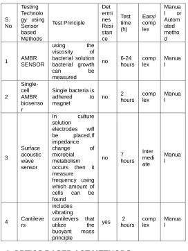
Table 2: Summary of Sensor Based AST Methods
S. No
Testing Technolo gy using Sensor based Methods
Test Principle
Det ermi nes Resi stan ce
Test time (h)
Easy/ comp lex
Manua l or Autom ated metho d
1 AMBR SENSOR
using the viscosity of bacterial solution bacterial growth
can be
measured
no 6-24 hours complex Manual
2
Single-cell AMBR biosenso r
Single bacteria is adhered to magnet
no 2 hours
comp lex
Manua l
3
Surface acoustic wave sensor
In culture solution
electrodes will be placed,If impedance change of microbial metabolism occurs then it measure frequency using which amount of cells can be found
no 7 hours
Inter medi ate
Manua l
4 Cantileve rs
includes vibrating cantilevers that utilize the buoyant mass principle
yes 2 hours
comp lex
Manua l
4.
OPTICS
BASED
AST
METHODS
4.1 Optical density measurement method
This method uses spectro-photometer by finding optical density to know the growth of microbes. The Optical Density
of a microbial suspension has resolved in customary time indicating antibiotic. The primary bit of leeway of this strategy is the quick sign for development. It is economical. The Disadvantages of this technique are more research is expected to approve the strategy for a more prominent assortment of microscopic organisms. Using this method colony forming units can be measured but Minimum Inhibitory concentration cannot be measured This technique isn't appropriate for exceptionally low centralizations of microscopic organisms; Measurement of the OD of a culture as appeared in Figure7
4.2 Biomimetic polymer sensor method
It is a quick computerized measure which uses traditional Agar dilution Method. Nano particles and polymers like Polydiacetylene which gives fluorescence are added. To attract bacteria in agar medium. A color change of blue to red can be observed with instrument. Change in shade can be seen under 6 hours much faster compared to traditional agar dilution Method. Drawback is it cannot differentiate type of microorganism.
4.3 Fluorescence activated cell Sorting (FACS)
By utilizing this strategy, type of cell can be identified and it assess the viability of bacteria and tally with dispersion of light. Consolidating microscopic organisms, anti-toxins and applicable luminous stains, Pathogenic practicality & anti-microbial vulnerability can be find. Sample is kept in incubator for couple of hours, subsequently investigation will be done. Because some reflective stains also tie to nucleotides inside the microbes, permeable cells provide more grounded fluorescent emanation compared to non-permeable cells[8], totally lysed cells gives more prominent measure of fluorescence.
4.4 Image analysis software
Traditional Methods like agar dilution which uses solid growth medium and broth dilution, which uses liquid medium can be automated by image analysis method. Since Watching number of samples by eye is tedius job. Some softwares like cell profiler, Yeast Xtract are already developed. MIC can be calculated quicker and number of colony forming units also will be measured. This technique is moderately modest, with simple handling. Despite their high affectability, image processing programming, advanced plate perusing frameworks can identify early development of pathogens. MICs can be calculated quicker.
4.5 Optical tweezers
In this method using 1064nm laser shaft, single bacteria cell will be captured to track the effects of synthetic specialists. Contingent upon the centralization of the compound operator and microscopic organisms type[10], the single-cell reaction can be resolved inside several minutes, which is a significant bit of leeway. Minimum Inhibitory concentration can be calculated without developing colonies. Framework is valid to moving microorganisms.
4.6 Raman spectroscopy
2176
be identified by vibration groups. This method is favorable to all types of organisms and it is po ssible to break down antimicrobial compounds as long as nucl eic acids and proteins are available to an adequate degree an d consideration is given to size and relocation attributes.One burden is the significant expenses of spectroscope, despite relatively low operational costs.
4.7 Matrix-assisted laser desorption/ionisation time-of-flight mass spectrometry (MALDI-TOF MS)
This system utilizes delicate ionization laser pulses to examine bio-molecules. Bacterial strains blend inside around 40 min. Tests must be examined inside 48 h of assortment[12]. The Working chief is Detection of anti-toxin debasement items. This innovation permits precise and delicate recognition and recognizable proof of microscopic organisms. It has various points of interest, including straightforwardness of example test readiness, a base handling time and extremely low consumable expenses. this strategy requires cheap consumables and is financially focused with different methodologies. Figure8 shows Schematic of MALDI-TOF MS.
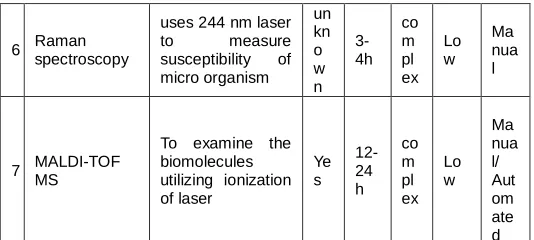
Table 3: Summary of optics based AST methods
S . N o Testing Technology using Optics based Method Test Principle D et er mi ne s R es ist an ce Tes t tim e Te st pr oc ed ur e cos t Ma nua l or Aut om ate d met hod 1 Optical density (OD) measurement
In a liquid medium growth curves of microorganisms can be calculated
ye s 2-3h Ea sy mo der ate Ma nua l/ Aut om ate d 2 Biomimetic polymer sensor
It uses nano particles on agar plate to determine CFU count ye s 6-18 h Ea sy mo der ate Ma nua l
3 FACS
Susceptibility be
known by
consolidating microorganisms, anti-biotics and applicable fluorescent stains
ye s 2h
Int er m ed iat e mo der ate Ma nua l/ Aut om ate d 4 Image analysis Tool
development of micro organism
could be
investigated through
photographing of various points of time to evaluate it with medical imaging un kn o w n De pe nd s on pro ce dur e Int er m ed iat e Hig h Ma nua l/ Aut om ate d
5 Optical tweezers
uses a 1064nm laser beam so that single bacterium could be caught, N o 10 0-20 0s co m pl ex Hig h Ma nua l
6 Raman spectroscopy
uses 244 nm laser to measure susceptibility of micro organism un kn o w n 3-4h co m pl ex Lo w Ma nua l
7 MALDI-TOF MS
To examine the biomolecules utilizing ionization of laser Ye s 12-24 h co m pl ex Lo w Ma nua l/ Aut om ate d
5.
AST
METHOD
THAT
CAN
BE
USED
FOR
LAB
ON
CHIP
5.1 Microfluidic agarose channels tracking single- cell Growth
This Method is Rapid AST technique to find Minimum Inhibitory Concentration with time lapse pictures for single cells [13]. Microbes are immobilized in culture and presented to various anti-infection culture, allowing the analysis of single cell growth (Figure.8). Using CCD camera pictures are taken and the relative pace of the growth will be resolved to decide Minimum Inhibitory concentration. MIC qualities can be gotten inside 3–4h, which is profoundly profitable. One burden is that clinical examples need a few arrangement ventures before the investigation can be performed.
5.2 Microfluidic pH sensor
Chitosan is coordinated in a microfluidic channel. Limited quantities of microscopic organisms are obliged in little channels[14]. As glucose-containing development medium turns out to be progressively acidic with an expanding measure of microorganisms, The pH of the medium may be correlated to the survival and development of the bacteria. growing and contracting of chitosan might be surveyed continuously by Fourier transform reflective interferometric spectroscopy (FTRIFS).
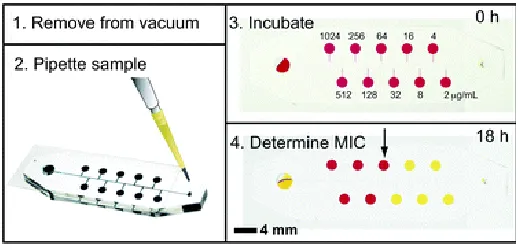
5.3 Self-loading microfluidics device
It is a convenient microfluidics purpose of care gadget(Figure. 8) that decides the Minimum Inhibitory Concentration. The unit is kept in incubator for eighteen hours as microorganism develop, it will lowers the glucose which in turn reduces the solution’s pH in turn. The minimum concentration that changed shading from red to yellow color is the MIC. The chip allows the monitoring of a number of bacteria from Gram-positive to Gram-negative. The chip empowers testing of an assortment of microbes running from Gram-positive to Gram negative ones. a significant disadvantage is only few anti-toxins at specific fixations is offered by this AST Method, in this way leaving no exploratory adaptability.
5.4 Microdroplets
2177
plan is that the level of blending is controlled by the length of the channel. Electric control offers favorable circumstances, for example, protection of room and adaptability from a restricted channel plan. One of the key focal points of bead microfluidics is the capacity to produce novel drops that can be moved and broke down independently.
Fig8. Self-loading microfluidics device
Table 4: Summary of Microfluidics method based AST
S. No
Testing Technology that can be used for Lab on chip
Test Principle
Deter mine s Resis tance
Test time (h)
Easy/ comp lex
Manual or Automated method
1
MAC tracking single cell
Growth
Using time lapse imaging susceptibility can be measured
Yes
3 to 4hour s
Inter medi ate
Manual
2 Microfluidic pH sensor
development medium turns out to be progressively acidic with an expanding measure of microscopic organisms
unkn own
2hour s easy
Manual/ Automated
3 Self-loading microfluidics device
a compact microfluidics purpose of care gadget is developed that decides the MIC of anti-toxins
Yes 18ho
urs easy Manual
4 Microdroplets
microorganisms are implanted in reverse emulsion beads can be utilized to evaluate susceptibility
Yes
Less than 4hour s
comp lex
Manual/ Automated
6.
AST
METHODS
CURRENTLY
IN
USE
6.1 Fluorescent live/dead staining
The Working rule is Microscopy of (non)permeable cells within the sight of fluorescent stains. The Live/Dead Cell Double Staining Kit is used for synchronous fluorescence recoloring of feasible and dead cells. Using Cell-staining system cells can be visualized in a better way with magnifying instrument [16]. Cell staining methods [17] and arrangement rely upon the kind of stain and investigation used. Once cell staining is done and slides are prepared, they might be put away in obscurity and potentially refrigerated to protect the slide, and afterward saw with a magnifying instrument. Favorable circumstances of utilizing such a unit are a fast strategy, quantitative investigations, just as the likelihood to gauge utilizing different instruments, for example, flow cytometer or microscope
6.2 PCR gene detection (Polymerase chain reaction)
This technique takes a shot at the head of DNA amplification. Quantitative-PCR or Q-PCR (frequently alluded to as real time PCR) is currently generally utilized in microbial biology to
decide quality and additionally transcript numbers present
inside ecological examples. A comparing increment in the fluorescent sign related with item development during each cycle in the PCR. Important screening apparatus of PCR is as yet viewed as an assistant test for certain analytic tests that still depend on smear and culture, for example, tuberculosis. PCR testing alone might be constrained as a symptomatic instrument, still need culture for testing for sedate growth.
6.3 Real-time microscopy
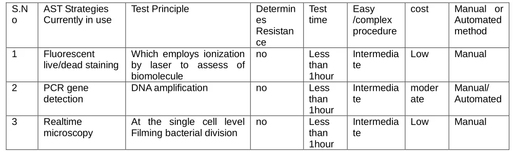
2178 Table 5: Outlines of AST Strategies that are Currently in use
S.N o
AST Strategies Currently in use
Test Principle Determin es Resistan ce
Test time
Easy /complex procedure
cost Manual or Automated method
1 Fluorescent live/dead staining
Which employs ionization by laser to assess of biomolecule
no Less than 1hour
Intermedia te
Low Manual
2 PCR gene detection
DNA amplification no Less than 1hour
Intermedia te
moder ate
Manual/ Automated
3 Realtime microscopy
At the single cell level Filming bacterial division
no Less than 1hour
Intermedia te
Low Manual
7.
FUTURE
ALTERNATIVES
FOR
AST
7.1 Calorimetrics
The Working principle is Detection of heat created by microscopic organisms. The benefits of this procedure incorporate the accompanying: (a) testing is directed in fixed ampules, (b) check during test is latent and does not require manual control of the test bottles (c) finished investigation gives data about the most extreme development pace of bacteria, static versus cidal action of the bacteria, & deferrals to logarithmic-phase development brought about by the agent
7.2 FACS(fluorescence-activated cell sorting)
It Works on the head of Sizing and estimating differential fluorescence among living and dead cells. This method licenses morphological changes[19], and survival of bacteria after exposure to anti-microbials. It is a technique for considering cells and chromosomes. A portion of the apparent obstacles for the strategy are, among others, the capacity to
separate cell harm brought about by cidal versus static anti-infection agents, auto fluorescence of some bacteria, and the enormous efforts are essential to confirm/approve observational data collected on patients to meet the criteria.
7.3 Magnetic bead spin
In the Magnetic field according to the number of joined microbes spin of the will be altered. This technique quickly distinguishes bacteria development, decides sedate affectability by estimating variation in the suspension thickness. This sensor that recognizes bacterial development dependent on the turn of a bunch of magnetic miniaturized scale particles framed through self-get together[20]. Utilizing these self-collected AMBR biosensors, the MIC esteem was estimated. At the point when microscopic organisms develop, they adjust the drag of the pivoting magnetic bead. This is a key element of the AMBR sensor, as changes in the drag can be because of changes in consistency, volume or potentially shape.
Table 6: Summary of future alternatives of AST methods
future Alternatives
of AST Test Principle
Determines Resistance
Test Time
Easy/Comple
x Cost
Manual / Automated Calorimetrics Detection of heat created by
microscopic organisms. No
<5
Hours Intermediate Moderate Automated FACS
(fluorescence-activated cell sorting)
Sizing and measuring differential fluorescence between living and dead cells
No < 5
Hours Intermediate Moderate Automated
Magnetic bead spin Finds out sensitivity of the
antibiotic by measuring viscosity No
< 5
Hours Intermediate Moderate Automated
8.
AUTOMATED
AST
A few automated systems for AST are economically accessible the models are Vitek System, Phoenix-Automated Microbiology System, Micro Scan Walkaway System(Siemens Healthcare Diagnostics), SensititreAris 2X (Trek Diagnostic System). General points of interest of Automated AST are Quantitative outcomes (MIC values), Reproducibility, Cost-powerful for research facilities with high throughput, Reduction in labor, simplicity of execution, Faster detailing of defenselessness results and Convenient interface with the lab data framework. But the main disadvantage are equipment cost as well as test cost is more. And studies reveal that results also not accurate as manual methods.
9.
CONCLUSION
2179
10
REFERENCES
[1]. Jorgensen JH, Ferraro MJ (2009) Antimicrobial susceptibility testing:a review of general principles and contemporary practices.Clin Infect Dis 49(11):1749– 1755.
[2]. European Committee for Antimicrobial Susceptibility Testing of the European Society of Clinical Microbiology and Infectious Disease (September 2000). "Determination of minimum inhibitory concentrations (MICs) of antibacterial agents by agar dilution". Clinical Microbiology and Infection. 6 (9): 509–515.
[3]. Ericsson JM, Sherris JC. Antibiotic sensitivity testing: report of aninternational collaborative study. ActaPatholMicrobiolScand1971;217 (Suppl):1–90. [4]. Kinnunen P, Sinn I, McNaughton BH, Newton DW,
Burns MA,Kopelman R (2011) Monitoring the growth and drug susceptibilityof individual bacteria using asynchronous magnetic bead rotation sensors. BiosensBioelectron 26(5):2751–2755.
[5]. Kinnunen P, CareyME, Craig E, Brahmasandra SN, McNaughton BH (2014) Rapid bacterial growth and antimicrobial response using self-assembled magnetic bead sensors. Sensors Actuators B Chem 190:265– 269.
[6]. Chang K-S, Chang C-K, Chen C-Y (2007) A surface acoustic wave sensor modified from a wireless transmitter for the monitoringof the growth of bacteria. Sensors Actuators B Chem 125(1): 207–213.
[7]. Godin M, Delgado FF, Son S, Grover WH, Bryan AK, Tzur A, Jorgensen P, Payer K, Grossman AD, KirschnerMW,Manalis SR (2010) Using buoyant mass to measure the growth of single cells. sNat Methods 7(5):387–390.
[8]. Hoefel D, Grooby WL, Monis PT, Andrews S, Saint CP. Enumeration of water-borne bacteria using viability assays and flow cytometry: a comparison to culture-based techniques. J Microbiol Methods. 2003;55(3):585–97.
[9]. Shah NA, Laws RJ, Wardman B, Zhao LP, Hartman JLT 4th (2007) Accurate, precise modeling of cell proliferation kinetics from time-lapse imaging and automated image analysis of agar yeast culture arrays. BMC Syst Biol 1:3.
[10]. Samadi A, Zhang C, Chen J, Reihani SN, Chen Z (2014) Evaluating the toxic effect of an antimicrobial agent on single
[11]. bacterial cells with optical tweezers. Biomed Opt Express 6(1): 112–117.
[12]. Neugebauer U, Schmid U, Baumann K, Holzgrabe U, ZiebuhrW, Kozitskaya S, Kiefer W, Schmitt M, Popp J (2006) Characterization of bacterial growth and the influence of antibioticsby means of UV resonance Raman spectroscopy.Biopolymers 82(4):306–311. [13]. Clark AE, Kaleta EJ, Arora A, Wolk DM (2013)
Matrix-assisted laser desorption ionization-time of flight mass spectrometry: a fundamental shift in the routine practice of clinical microbiology. Clin Microbiol Rev 26(3):547– 603.
[14]. Choi J, Jung YG, Kim J, Kim S, Jung Y, Na H, Kwon S (2013) Rapid antibiotic susceptibility testing by tracking single cell growth in a microfluidic agarose channel system. Lab Chip 13(2):280–287.
[15].Tang Y, Zhen L, Liu J,Wu J (2013) Rapid antibiotic susceptibility testing in a microfluidic pH sensor. Anal Chem 85(5):2787–2794.
[16].Jiang L, Boitard L, Broyer P, Chareire AC, Bourne-Branchu P,Mahé P, Tournoud M, Franceschi C, Zambardi G, Baudry J, Bibette J (2016) Digital antimicrobial susceptibility testing using the MilliDrop technology. Eur J Clin Microbiol Infect Dis 35(3): 415– 422.
[17].Hoerr V, Ziebuhr W, Kozitskaya S, Katzowitsch E, Holzgrabe U. Laser-induced fluorescence - capillary electrophoresis and fluorescence microplate reader measurement: Two methods to quantify the effect of antibiotics. Anal Chem. 2007;79(19):7510–8.
[18].Berney M, Hammes F, Bosshard F, Weilenmann HU, Egli T. Assessment and interpretation of bacterial viability by using the LIVE/DEAD BacLight Kit in combination with flow cytometry. Appl Environ Microbiol. 2007;73(10):3283–90.
[19].Auty MA, Gardiner GE, McBrearty SJ, O’Sullivan EO, Mulvihill DM, Collins JK, et al. Direct in situ viability assessment of bacteria in probiotic dairy products using viability staining in conjunction with confocal scanning laser microscopy. Appl Environ Microbiol. 2001;67(1):420–5.
[20].Shrestha NK, Scalera NM,Wilson DA, Procop GW(2011) Rapid differentiation of methicillin-resistant and methicillin-susceptible Staphylococcus aureus by flow cytometry after brief antibiotic exposure. J Clin Microbiol 49(6):2116–2120.