27
Dysplastic Koilin Causing Proventricular Obstruction
in an Eclectus Parrot (Eclectus roratus)
Ryan De Voe, DVM, Laurel Degernes, DVM, Dipl ABVP (Avian), and
Kristie Karli, DVM
Abstract: A 7-year-old male eclectus parrot (Eclectus roratus) was presented for evaluation of
acute onset of dyspnea, tachypnea, and lethargy. Proventricular dilatation was observed on survey radiographs. Contrast fluoroscopic examination revealed decreased gastrointestinal motility and re-tention of contrast material in the proventriculus. Despite supportive care, the bird died. At necropsy, a tubular diverticulum of the ventriculus extended orad into the proventriculus. This diverticulum caused almost complete obstruction of proventricular outflow. No inflammation, infectious organ-isms, or neoplasia were associated with the lesion. The cause of the ventricular diverticulum in this parrot is unknown.
Key words: dysplastic koilin, gastrointestinal obstruction, contrast fluoroscopy, eclectus parrot, Eclectus roratus
Introduction
The ventriculus, or muscular stomach of birds, is lined on its mucosal surface by a cuticle layer, the koilin. The structure of both the ventriculus and the koilin varies according to diet between species of birds. The koilin serves as a grinding surface, en-abling the ventriculus to mechanically digest food as it contracts.1Insectivorous, herbivorous, and gra-nivorous birds possess a well-developed, muscular ventriculus with a thick, abrasive koilin layer. In contrast, birds that eat foods not requiring signifi-cant mechanical digestion (ie, carnivores and pis-civores) have an almost rudimentary ventriculus and soft koilin.1–4
Koilin consists of a carbohydrate-protein com-plex that is secreted from glands in the mucosa of the ventriculus.1–4 After this complex is secreted, it hardens in response to exposure to hydrochloric acid produced by the proventriculus. This process results in a tough, wear resistant material.1,2,4 Dis-ruption of normal koilin architecture or production resulting in structural defects has been reported in many species of birds.5–17Various etiologic factors
From the Departments of Clinical Sciences (De Voe, Deger-nes) and Population Health and Pathobiology (Karli), College of Veterinary Medicine, North Carolina State University, 4700 Hillsborough Street, Raleigh, NC 27606, USA.
have been implicated in causing defects in the koilin layer, including a variety of infectious agents.
The structure of the koilin layer of the ventriculus is complicated. The basic structure consists of ver-tical rodlets of hard koilin that are produced by clus-tered tubular glands found in mucosal crypts. These rodlets collectively form the rods that are the ver-tical scaffolding of the koilin layer. The rods are surrounded by a horizontal matrix of desquamated cells and softer material produced by the surface epithelium.1 The koilin layer is continually worn down by grinding action and replaced by glandular secretion.1,2 In most birds with a well-developed ventriculus, the koilin is asymmetrically formed, be-ing thickest opposite the craniodorsal and caudov-entral thick muscles.1,2,4
In this report, we describe a ventricular divertic-ulum and koilin dysplasia of unknown cause in an eclectus parrot (Eclectus roratus) that resulted in gastrointestinal obstruction and death.
Case Report
A 7-year-old male eclectus parrot was examined because of dyspnea, tachypnea, and lethargy. The parrot was hand-raised and had been with the cur-rent owner for 1 year. It was housed in a room with two conures (Aratinga auricapilla and Nandayus
been obtained at once from the same source. No abnormalities were reported with the other birds. The diet consisted of commercial pellets supple-mented with seed, fresh fruits, and vegetables. All other husbandry practices were considered ade-quate. There was no known history of toxin expo-sure. The dyspnea and tachypnea had developed the previous day, and the owner had presented the bird to an emergency clinic late that evening. At the emergency clinic, it was placed in an oxygen cage and given intramuscular injections of enrofloxacin (Baytril, Bayer Corp, Shawnee Mission, KS, USA) and vitamin A, D, and E. The next morning, the bird was transferred to our hospital for evaluation.
On examination, the bird was in good body con-dition and weighed 352 g. The rhinotheca was mod-erately overgrown, and the choanal papillae were severely blunted. Feather loss from feather plucking was present over most of the body. The bird was tachypnic (approximately 60 breaths per minute) and moderately dyspneic at rest, with a tail-bob and an audible click on expiration. It was very lethargic and rested with its beak on the ground to help sup-port its weight. The coelom was mildly distended. Dehydration was estimated at 8–10%.
Jugular venipuncture was performed to obtain a blood sample for a complete blood count (CBC) and a plasma biochemical analysis. Results of the CBC revealed a mildly increased packed cell volume (56%; reference range 45–55%).18 Results of the biochemical analysis revealed high concentrations of aspartate aminotransferase (555 IU/L; reference range, 135–230 IU/L), creatine kinase (4277 IU/L; reference range, 200–625 IU/L), and lactate dehy-drogenase (944 IU/L; reference range, 100–280 IU/ L).18All other CBC and plasma biochemical values were within reference ranges. Results of a fecal Gram stain were unremarkable. A cloacal swab specimen was submitted for aerobic and fungal cul-ture. No pathogens were isolated.
The bird became severely dyspneic after exami-nation and sample collection, necessitating hospi-talizing the bird in a warm oxygen cage. After the bird’s condition stabilized, fluid, antibiotic, and an-tifungal therapy was begun. The treatment regimen consisted of enrofloxacin (25 mg/kg SC q24h; Bay-tril, Bayer), itraconazole (10 mg/kg PO q12h; Spor-anox, Janssen Pharmaceutica Products, Titusville, NJ, USA), and lactated Ringer’s solution (50 ml/kg SC q12h). The bird was also fed 20 ml of a nutri-tional supplement by gavage (Emeraid II, Lafeber Company, Cornell, IL, USA).
The next morning, the parrot’s clinical condition appeared slightly improved. It had passed scant green feces and yellow urates, and it had eaten a
small amount of seed overnight. To prevent strug-gling and dyspnea during handling, the bird was anesthetized with isoflurane to take whole-body sur-vey radiographs. Radiographic abnormalities in-cluded a significantly dilated proventriculus (;2–3 times the normal diameter) and loss of serosal detail in the caudal coelomic cavity (Fig 1).
Contrast fluoroscopy was then done to evaluate the coelomic structures and gastrointestinal motility. Barium sulfate was administered (12 ml, 1:1 barium and water) by gavage into the crop. The bird was placed in a closed, narrow cardboard box during imaging to prevent excessive movement. Standing lateral and dorsal-ventral images were taken at 20, 50, 110, and 200 minutes. Motility of the crop, pro-ventriculus, and ventriculus motility was dimin-ished. When the final images were taken at 200 minutes, only a small amount of contrast material had reached the ventriculus. A filling defect was apparent in the proventriculus.
With the exception of gavage feeding, previously prescribed treatments were continued after the ini-tial survey radiographs and fluoroscopy. A small amount of regurgitated barium was observed in the cage that evening. The bird was found dead late that night.
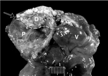
At gross necropsy, abnormalities were restricted to the proventriculus and ventriculus. Grossly, a thin-walled conical diverticulum, approximately 2.5 cm long and 1.5 cm wide, protruded from the ven-triculus into the provenven-triculus. Proventricular out-flow was almost completely obstructed by this verticulum. A small opening at the apex of the di-verticulum allowed communication between the proventriculus and ventriculus. The ventriculus and diverticulum contained a moderate amount of grit, seed, and barium sulfate (Fig 2). Barium sulfate and a scant amount of seed were present in the proven-triculus.
Figure 1. Lateral radiograph of an eclectus parrot with dysplastic koilin that resulted in obstruction of the proventric-ulus. The proventriculus is moderately enlarged (outlined by arrows).
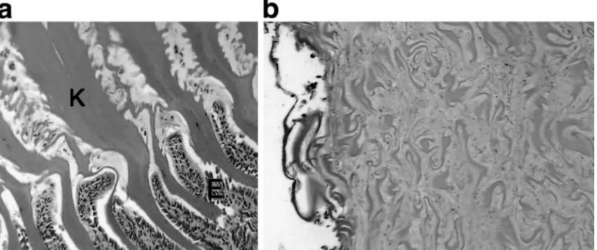
Figure 3. Photomicrograph of the normal ventricular koilin (a) and the dysplastic koilin of the ventricular diverticulum (b) from the eclectus parrot described in Figure 1. Note the regular, columnated structure of the normal koilin layer and its association with the glandular epithelium of the ventriculus. The koilin matrix of the diverticulum shows no distinct organization. K indicates koilin layer; E, glandular epithelium (hematoxylin and eosin3400).
Discussion
In this eclectus parrot, a diverticulum of the ven-triculus caused almost complete obstruction of pro-ventricular outflow, resulting in death. On histologic examination, the koilin layer of the diverticulum was thin, and the normal columnated structure was absent. No evidence of inflammation, infectious or-ganisms, or neoplasia was found, and the cause of the ventricular diverticulum was not identified.
Gastrointestinal diseases are relatively common in avian species and can be challenging to diagnose. Infectious gastrointestinal diseases frequently occur and can be bacterial, fungal, viral, or parasitic in nature. Other common gastrointestinal disorders in avian patients include toxicosis, foreign body in-gestion, papillomatosis, and neoplasia.19Many clin-ical signs observed in birds with gastrointestinal disease are nonspecific, such as lethargy, anorexia, and a fluffed appearance. Illness can be acute or chronic. For these reasons, developing a specific list of differential diagnoses at presentation can be dif-ficult.
In this parrot, the presenting complaints were dyspnea, tachypnea, and lethargy. The owner re-ported no regurgitation or vomiting, diarrhea, an-orexia, weight loss, or other signs typically associ-ated with gastrointestinal disease.20 This bird showed no overt signs of gastrointestinal disease initially, and gastrointestinal involvement was not suspected until the radiographic and fluoroscopic findings of proventricular dilatation, decreased ventricular and ventricular motility, and slow pro-ventricular emptying of contrast material were
iden-tified. Our initial differential diagnoses included pri-mary respiratory disorders such as a tracheal or sy-ringeal mass or foreign body, aspergillosis, chlamydiosis, sarcocystis, or an inhalant toxicosis (eg, polytetrafluoroethylene gas).21 Initial therapy was directed toward possible respiratory disease. Oxygen therapy, as well as administration of anti-microbials effective against most of the common respiratory pathogens, was instituted. In retrospect, the dyspnea and tachypnea seen in this bird were likely secondary to proventricular enlargement or associated discomfort. Respiratory difficulty is of-ten observed in birds with organomegaly, as air sac volume is diminished.21
ac-cumulated secondary to functional ileus. Withhold-ing solid food and feedWithhold-ing this bird a liquid diet for 24 hours or more before fluoroscopic evaluation may have been helpful. An acute obstruction was suggested by the fact that the bird was in good body condition and passing feces until less than 24 hours before its death.
Heavy metal analysis, especially for lead and zinc, would have been appropriate in this case but was not performed. Lead and zinc toxicosis have been associated with ileus and proventricular im-paction.3Zinc toxicosis associated with ventriculitis and erosions of the koilin has been reported in var-ious avian species.10–12Ingesting excessive amounts of other metals, such as copper, also has been as-sociated with ventricular and koilin abnormalities.3 The koilin layer can be disrupted from a variety of causes. Mycotic proventriculitis or ventriculitis, such as that produced by avian gastric yeast or
Can-dida species, can disturb koilin production.5,6,14,17 Adenovirus infections in poultry have resulted in erosion of koilin layers.7,8Other potential causes of koilin disruption are inappropriate diet (eg, hypo-vitaminosis A, excessive polyunsaturated fatty ac-ids),13parasitism,15neoplasia,24–26and toxicosis (eg, zinc, copper, ferrous sulfate).3,10–12,16The koilin layer can also be eroded by ingestion of excessively abra-sive objects or substances.13
The cause of the dysplastic koilin in this bird is unknown. Idiopathic intussusception of the proven-triculus into the venproven-triculus has been described in chickens.33 However, the anatomic deformity ob-served in this eclectus parrot has not been previ-ously described in other avian species. In this par-rot, the ventricular diverticulum was intussuscepted into the proventriculus. In the absence of any in-flammatory or malignant changes associated with the defect, the underlying cause was unlikely to be infectious or neoplastic. Possibly, a previous insult disrupted the koilin and mucosa of the ventriculus, initiating development of the diverticulum.
Normally, ingesta passes back and forth between the proventriculus and ventriculus, allowing com-bined chemical and mechanical digestion.34 In this bird, the strong contractions of the ventriculus might have progressively forced more and more in-gesta into the diverticulum during normal reflux. The accumulated solid ingesta in the diverticulum eventually could have prevented the relatively weak contractions of the proventriculus from expelling the contents of the diverticulum into the ventriculus. By the time the diverticulum became clinically sig-nificant, the inciting cause might have resolved. An-other possibility is that this ventricular diverticulum was a congenital anatomic defect that did not
inter-fere with gastrointestinal function until it became distended with ingesta over time, ultimately causing a mechanical obstruction between the proventricu-lus and ventricuproventricu-lus.
This case illustrates the potential challenges of diagnosing gastrointestinal obstruction in avian pa-tients. Also, structural abnormality involving the koilin should be considered as a differential diag-nosis for gastrointestinal obstruction in psittacine birds.
References
1. King AS, McLelland J. Birds: Their Structure and
Function. London, England: Bailliere Tindall; 1984:
84–109.
2. Akester AR. Structure of the glandular layer and ko-ilin membrane in the gizzard of domestic fowl
(Gal-lus gal(Gal-lus domesticus). J Anat. 1986;147:1–25.
3. Lumeij JT. Gastroenterology. In: Ritchie BW, Harri-son GJ, HarriHarri-son LR, eds. Avian Medicine:
Princi-ples and Application. Lake Worth, FL: Wingers;
1994:483–521.
4. Denbow, MD. Alimentary canal: anatomy, regulation of feeding and motility. In: Whittow, GC, ed.
Stur-kie’s Avian Physiology. 5th ed. New York, NY:
Ac-ademic Press; 2000:299–321.
5. Jeffrey JS, Chin RP, Shivaprasad HL, et al. Proven-triculits and ventriculitis associated with zygomycos-is in ostrich chicks. Avian Dzygomycos-is. 1994;38:630–634. 6. Anderson, NL. Candida/megabacteria proventriculitis
in a lesser sulphur-crested cockatoo (Cacatua
sulphu-rea sulphusulphu-rea). J Assoc Avian Vet. 1993;7:197–201.
7. Abe T, Nakamura K, Tojo T, Yuasa N. Gizzard ero-sion in broiler chicks by group 1 avian adenovirus.
Avian Dis. 2001;45:234–9.
8. Goodwin MA. Adenovirus inclusion body ventricu-litis in chickens and captive bobwhite quail (Colinus
virginianus). Avian Dis. 1993;37:568–571.
9. Fosum O, Sandstedt K, Engstrom BE. Gizzard ero-sions as a cause of mortality in white leghorn chick-ens. Avian Pathol. 1988;17:519–525.
10. Zdiarski JM, Mattix M, Bush RM, Montali RJ. Zinc toxicosis in diving ducks. J Zoo Wildl Med. 1994;25: 438–445.
11. Romagnano A, Grindem CB, Degernes L, Mautino M. Treatment of a hyacinth macaw with zinc toxicity.
J Avian Med Surg. 1995;9:185–189.
12. Droual R, Meteyer CU, Galey FD. Zinc toxicosis due to ingestion of a penny in a gray-headed chachalaca (Ortalis cinereiceps). Avian Dis. 1991;35:1007– 1001.
13. Keymer IF. Disorders of the digestive system. In: Sa-mour J, ed. Avian Medicine. Philadelphia, PA: Mos-by; 2000:193–210.
14. Hubbard GB, Schmidt RE, Eisenbrandt DL, et al. Fungal infections of ventriculi in captive birds. J
Wildl Dis. 1985;21:25–28.
ven-triculitis in mallards (Anas platyrhynchos). J Wild
Dis. 1987;23:680–682.
16. Wallner-Pendleton DP, Froman P, Hedstrom O. Iden-tification of ferrous sulfate toxicity in a commercial broiler flock. Avian Dis. 1986;30:430–432.
17. Prattis SM, Cioffee CJ, Reinhard G, Zaoutis TE. A retrospective study of disease and mortality in zebra finches. Lab Anim Sci. 1990;40:402–405.
18. Clubb SL, Schubot RM, Joyner K, et al. Hematologic and serum biochemical reference intervals in juvenile eclectus parrots (Eclectus roratus). J Assoc Avian
Vet. 1990;4:218–225.
19. Morrisey JK. Gastrointestinal diseases of psittacine birds. Semin Avian Exotic Pet Med. 1999;8:66–74. 20. Rupley AE. Diagnostic techniques for
gastrointesti-nal diseases of psittacines. Semin Avian Exotic Pet
Med. 1999;8:51–65.
21. Rupley AE. Manual of Avian Practice. Philadelphia, PA: WB Saunders; 1997:55–90.
22. Speer BL. Chronic partial proventricular obstruction caused by multiple gastrointestinal foreign bodies in a juvenile umbrella cockatoo (Cacatua alba). J Avian
Med Surg. 1998;12:271–275.
23. Adamcak A, Hess LR, Quesenberry KE. Intestinal string foreign body in an adult umbrella cockatoo (Cacatua alba). J Avian Med Surg. 2000;14:257– 263.
24. Schmidt RE, Dustin LR, Slevin RW. Proventricular adenocarcinoma in a budgerigar (Melopsittacus
un-dulatus) and a green-cheeked parakeet (Brotogeris pyrrhopterus). AAV Today. 1988;2:140–142.
25. Leach MW, Paul-Murphy J, Lowenstine LJ. Three cases of gastric neoplasia in psittacines. Avian Dis. 1989;33:204–210.
26. Campbell TW, Turner O. Carcinoma of the ventric-ulus with metastasis to the lungs in a sulphur-crested cockatoo (Cacatua galerita). J Avian Med Surg. 1999;13:265–268.
27. Wilson GH, Greenacre CB, Howerth EW, et al. As-caridiosis in a group of psittacine birds. J Avian Med
Surg. 1999;13:32–39.
28. Greenwood AG, Storm J. Intestinal intussusception in two red-tailed hawks (Buteo jamaicensis). Vet Rec. 1994;134:578–579.
29. Frasca S, Khan MI. Multiple intussusceptions in a juvenile rhea (Rhea americana) with proventricular impaction. Avian Dis. 1997;41:475–480.
30. McMillan MC. Avian gastrointestinal radiography.
Comp Contin Educ Pract Vet. 1983;5:273–278.
31. McMillan MC. Imaging techniques. In: Ritchie BW, Harrison GJ, Harrison LR, eds. Avian Medicine:
Principles and Application. Lake Worth, FL:
Wing-ers; 1994:246–326.
32. Van Sant F. Zinc toxicosis in a hyacinth macaw. Proc
Annu Conf Assoc Avian Vet. 1991:255–259.
33. Sharma UK. Intussusception of the proventriculus of chickens. Avian Dis. 1972;16:453–457.