Effect of
in ovo injection of L-Arginine in different
chicken embryonic development stages on
post-hatchability,
immune
response
and
muscle
development proteins
Sivakumar Allur Subramaniyan 1, Da Rae Kang 1, Jin Ryong Park 1, Sharif Hasan Siddiqui 1, Palanisamy Ravichandiran 2, Chong Sam Na1 and Kwan Seob Shim 1,*
1 Department of Animal Biotechnology, College of Agriculture and Life Sciences, Chonbuk National University, Jeonju, 561-756, Republic of Korea.
2 Graduate School, Department of Energy Storage/Conversion Engineering, Hydrogen and Fuel Cell Research Center, Chonbuk National University, Jeonju 561-756, Republic of Korea
* Correspondence: [email protected]; Tel: +82-63-270-2609; Fax +82-63-270-2614
Simple Summary: In the current study, we hypothesized that the in ovo injection of L-arginine (L-Arg) at different stages of embryonic development which would have positive effects on survival rate, hatching rate, immunoglobulin (IgM) levels and heat shock proteins such as HSP-47, 60 and 70 as well muscle development markers mainly myoD and myogenin. As indicated, the in ovo injection of L-Arg resulted in increased hatch rate and weight, survival rate, higher levels of IgM, and myogenin and MyoD expression in the muscles. At the same time, a decrease in the level of HSP-47,60 and 70 expressions in the tissues at 14th day of injection compared to 8th and 18th day of injection
peroid. In addition, the in ovo injection of L-Arg decreased the SGOT and SGPT concentration in serum as well micronuclei and nuclear abnormality in the blood of 14th day incubation period.
Hence, 14th day would be suitable day for injection of L-Arg to promote the hatching rate and muscle
growth of broiler chickens (i.e. animals).
Abstract: The objective of this study was to evaluate the effect of in ovo injection of L-arginine (L-Arg) into Ross broiler eggs at different embryonic developmental stages on their survival, hatchability, and body weight (BW). Additionally, we have analyzed the levels of serum glutamic-oxaloacetic transaminase (SGOT) and serum glutamic-pyruvic transaminase (SGPT), protein expression of heat shock proteins (HSPs), also we have the determined micronuclei (MN) and nuclear abnormality (NA). Results showed that survival and hatching rates as well as body weight were increased on the 14th day incubation compared to 8th and 18th day incubation at lower concentration of L-Arg. Moreover, the levels of SGOT and SGPT were also significantly (P < 0.05) increased at 14th day incubation at the same concentration (100μg/μl/egg) of injection. In addition,
IgM levels were increased on the 14th day incubation compared to other days. The protein expressions of HSP-47, HSP-60, and HSP-70 in the liver were significantly down-regulated whereas the expression of myogenin and MyoD were significantly up-regulated on the 14th day after
incubation in treated with all different doses such as 100μg, 1000μg and 2500μg/μl/egg namely 3T1,
3T2 and 3T3 respectively. However, the treatment with low dose of L-Arg down-regulated expression levels of those proteins on the 14th day incubation. Histopathology of liver by hematoxylin and eosin (H&E) straining showed that the majority of liver damage, specifically intracytoplasmic vacuoles, were observed in 3T1, 3T2, and 3T3. The minimum dose of 100
μg/ml/egg on the 14th day of incubation significantly prevented intracytoplasmic vacuole damages.
These results demonstrate that in ovo administration of L-Arg at (100μg/μl/egg) may be an effective
method to increase chick BW, hatch rate, increasing muscle growth related proteins and promote the immune response through increasing IgM on the 14th day of incubation period.
Keywords: L-arginine; embryonic development; intracytoplasmic vacuoles; immunoglobulin; heat shock proteins
1. Introduction
Selection of chickens (Gallus gallus) for meat production has led to the generation of inbred strains that show accelerated growth performance, particularly enhanced muscle growth that mostly occurs during embryogenesis [1,2]. During embryogenesis, nutrients and energy are mainly getting from yolk mainly containing lipids and low level of carbohydrates [3]. Subsequently, the embryo and post-hatch chicken depends up on gluconeogenesis from essential amino acids [4,5].In recent years, found that administration of amino acids into fertilized broiler eggs called in ovo feeding may provide poultry companies with an alternative method to increase hatchability and muscle growth weight of newly hatched chick [6,7]. Supplementation of nutrients into fertilized broiler egg influences the growth and embryo development during incubation and post-hatch growth performance of chicks [7]. The nourishment and supplementation with bioactive substances such as bioactive amino acids, polyphenols, and prebiotics can enhance the immune system, decrease osteoporosis, and decrease the risk of heart health [8]. Similarly, previous report indicates that the amino acids, carbohydrates, and vitamins that are applied to egg through in ovo feeding can improve hatching rate, body weight, survival rate, growth performance, and marketing size [9]. Moreover, earlier study demonstrated that in ovo feeding site and time can affect hatchability [10].
During embryonic development, developed the chorioallantoic membrane which can vascularize on 12th day of incubation period. Moreover, the embryo is surrounded by the amniotic
fluid that remains in contact with the embryonic gastrointestinal tract which allows transport of substances from the air chamber into the intestine [11]. Several genes are associated with cellular interactions and differentiation during organogenesis of the eye, ear, brain, skin, and tissues such as bones and cartilages those genes expression is either transient or initiated during later stages of embryogenesis [12].Some authors have indicated that injection of amino acids into the egg on the first day is sufficient to fully support embryonic development [13,14] leading to increased hatching and breast weight [15].It has been demonstrated that injection of sucrose and dextrin into chicken embryos can result in greater percentage of pectoral muscle weight than the control [5,16]. Recently, it has been reported that chicken embryos injected with L-glutamine on the first day of incubation can increase fiber area, pectoral muscle mass, and endothelial cell proliferation while stimulating vasculogenesis and angiogenesis [17]. Not only that in ovo administration of amino acids/peptides may increase expression levels of myoblast determination protein 1 (MyoD1) and paired box protein 7 (Pax7) which are necessary for myogenesis, muscle maturation (muscle growth), and breast muscle yield during embryogenesis [18].
Standardization of injection site, needle length, and embryonic age using amino acid (Lys+Met+Cys, Thr+Gly+Ser or Ile+leu+Val) with 11 mm and 24 mm needles at 7th and 14th day of incubation has resulted in poor hatchability and poor muscle growth markers [19].There was some previous study about in ovo injection of conjugation of glutamine with Ag NPs on first day of incubation, results increased the muscle mass [20]. Similarly, in ovo injection of (L-Arg) into 1d old quail embryos enhancing the hatchability rate and productive performance of quail [21]. To our best knowledge there were no studies about different embryonic stages with different concentration of (L-Arg) on chicken embryos. Therefore, we are trying first time to check the effects of different concentrations of L-Arginine (L-Arg) through in ovo injection at different embryonic stages (8th, 14th,
and 18th day) to which embryonic stages suitable to improve the survival rate, hatchability,
biochemical profile in blood, muscle accretion and muscle growth related proteins such as myogenin and MyoD also heat shock protein and Immunoglobulin M (IgM) levels.
2. Materials and Methods
2.1. Ethics Statement
sampling procedures complied with the “Guidelines on Ethical Treatment of Experimental Animals”
(2015) No. CBNU 2015-048 set by the Ministry of Science and Technology, Republic of Korea.
2.2. Chemicals
L-arginine was purchased from Sigma-Aldrich (Sigma-Aldrich, St. Louis, MO, USA). iScriptTM cDNA synthesis kit (170–8891) and SsoFast Eva-Green Supermix (172–5202) were purchased from BIO-RAD (BIO-RAD Laboratories, Inc., Hercules, CA, USA). Chemiluminescent for band detection purchased from Thermo Scientific (Rockford, IL, USA). Antibodies were purchased from ENZO Life Science (Farmingdale, NY, USA). Hematoxylin and eosin (H&E)1 were purchased from Sigma-Aldrich (St. Louis, MO, USA). All laboratory glassware was acquired from Falcon Lab ware (Becton, Dickinson and Company, Franklin Lakes, NJ, USA).
2.3. Experimental Design and Incubation
Ross 1040 broiler chicken eggs were obtained from Samhwa-Won Jong, South Korea. On the 1st
day of incubation, eggs were weighed (60 ± 1.36 g) and separated to different groups. Eggs were randomly divided into thirteen groups (4 × 20 × 3 = replication × eggs × injection) detail described in in followings. L-Arg was injected at three concentrations (100 µg, 1,000µg, and 2,500µg/100µl/egg) at 8th, 14th, and 18th day of incubation period, respectively.A 0.100 mL of L-Arg solution (dissolved in
1% PBS) was injected into the air sac of egg. Immediately after the injection, the hole was sealed with liquid paraffin. Eggs were then placed in an incubator for 20 days under standard conditions (temperature, 37.8°C; humidity, 60%). On the 18th day, eggs were transferred to hatching boxes and
promptly placed in a hatcher incubator with humidity maintained at 60% and temperature set at 37°C to hatch chicks. At the end of the rearing period, the one day old chicks were sacrificed by cutting the jugular and carotid veins, after which further slaughter processing was conducted. During the slaughter process, 2 mL of blood as well as tissue from the breast muscle were collected and washed in ice-cold saline. Small amount of collected blood were immediately smeared on clean grease free microscope slides and air-dried for Micronuclei (MN) and Nuclear Abnormality.
2.4. In ovo injection and treatment groups
Group Dosage n = 20×4 Total No. of eggs
1C Control 20 eggs × 4 replicates 80
1C1 (8th day) PBS/100 µl/egg 20 eggs × 4 replicates 80 1T1 (8th day) 100 µg/100 µl/egg 20 eggs × 4 replicates 80 1T2 (8th day) 1000 µg/100 µl/egg 20 eggs × 4 replicates 80 1T3 (8th day) 2500 µg/100 µl/egg 20 eggs × 4 replicates 80 2C1 (14th day) PBS/100 µl/egg 20 eggs × 4 replicates 80 2T1 (14th day) 100 µg/100 µl/egg 20 eggs × 4 replicates 80 2T2 (14th day) 1000 µg/100 µl/egg 20 eggs × 4 replicates 80 2T3 (14th day) 2500 µg/100 µl/egg 20 eggs × 4 replicates 80 3C1 (18th day) PBS/100 µl/egg 20 eggs × 4 replicates 80 3T1 (18th day) 100 µg/100 µl/egg 20 eggs × 4 replicates 80 3T2 (18th day) 1000 µg/100 µl/egg 20 eggs × 4 replicates 80 3T2 (18th day) 2500 µg/100 µl/egg 20 eggs × 4 replicates 80 2.5. Survival rate measurement
Embryos’ survival rates during the incubation period were measured on the 8th day. Treated
eggs were checked to determine the number of live and dead eggs as well as fertilized and non-fertilized ones among the total number of eggs. At 18th day of incubation, after injection the eggs live
2.6. Hatching rate, body weight, and liver weight measurements
On the 21st day, hatched chicks were moved from the hatcher incubator to hatching boxes to
determine hatching rates. These hatched chicks were kept without feed and water at 32 °C. They were then weighed to record their live body weights and liver weights. The hatching rate was calculated with the following formula:
2.7. Biochemical indices
On the 21st day of the post hatching period, chicks were sacrificed under anesthesia (diethyl
ether). Blood was collected from the jugular vein into tubes containing ethylenediaminetetraacetic acid (EDTA) for serum separation. Liver samples were excised, washed with ice-cold saline, weighed, and then homogenized with cold 0.1 M phosphate buffer, pH 7.4. Concentrations of serum glutamic-oxaloacetic transaminase (SGPT) and serum glutamic-pyruvic transaminase (SGOT) in sera were measured using commercial kits (Asan Pharamaceuticals Co., Ltd., Seoul, South Korea).
2.8. Micronuclei (MN) and nuclear abnormality (NA) tests using periodic acid Schiff’s (PAS) staining
Blood samples collected from the first day of hatching period were immediately smeared on clean grease-free microscope slides and air-dried. Afterwards, slides were fixed with methanol for 5 min at room temperature, gently rinsed with running tap water for 1 min, and immersed in a periodic acid solution for 5 min at room temperature. These slides were then rinsed using D.H2O, immersed
in PAS Schiff’s reagent for 15 min at room temperature, and gently washed with running tap water
for 5 min. Finally, counter-staining was performed with a hematoxylin solution for 90s. Slides were then rinsed in running tap water for 30 s, air dried, and examined with a light microscope (100X) using mineral oil.
2.9. Measurement of IgM concentration in plasma
Plasma samples were collected from individual experimental animals to determine plasma immunoglobulin (Ig) M levels using chicken IgM ELISA kit (Abcam) following the manufacture’s
specification. IgM levels were analyzed based on absorbance values measured at 450 nm.
2.10. Analysis of heat-shock proteins (HSPs) by western blot
dilutions) for 60 min at room temperature and then washed thrice with TBST (10 min each wash). Protein bands were visualized using a Chemiluminescent assay kit from Thermo Scientific for 1 to 5
min. Bands were imaged with an iBright™ CL1000 Imaging System (Invitrogen) and quantified using Image J Software. Relative density of band was normalized to that of β-actin as an internal control.
2.11. In silico molecular docking studies
To understand the mechanism of interaction of L-Arg with heat shock protein, crystal structures of GroEL mutant A109C (PDB ID: 5OPW) [22] and human HSP70 substrate binding domain L542Y mutant (PDB ID: 5XIR) [21] were downloaded from protein data bank. Molecular docking studies were performed using the GLIDE program [23] (version 8.5, Schrodinger LLC, New York, NY, USA). To analyze docking results and execute the protocol, the Maestro user interface (version 8.5, Schrodinger LLC, New York, NY) was employed. Validation of the protocol was performed by redocking. The structure of L-Arg was sketched using ACD/chemsketch (Freeware version). GLIDE grid generation wizard was used to define the docking space. Docking was performed using XP (Extra Precision mode) docking protocol.
2.12. Histopathological Study of the Liver
Livers were collected after chickens were sacrificed, immediately fixed with 10% neutral buffered formalin (NBF), and processed in an auto processor (Excelsior ES, Thermo Scientific, Waltham, MA, USA). After embedding in paraffin, 5 μm sections were made and subjected to H&E
staining. Digital images were obtained using a Leica DM2500 microscope (Leica Microsystems, Wetzlar, Germany) at fixed 100 × (200×) magnification.
3. Statistical analysis
Data were analyzed using Tukey's HSD test post-hoc following ANOVAs with SAS® software,
version 9.4. Student’s t-tests were used to determine statistical differences between groups. Values are presented as mean ± SD from twelve determinations.
4. Results and discussion
4.1. Survival rate and hatchability
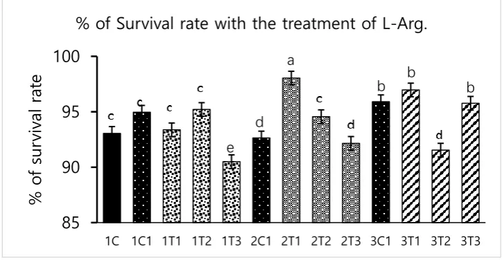
Survival rate was significantly (p < .05) increased in 2T1 and 3T1 groups than that in other groups. The lowest survival rate was observed in 1T3, 3T2, and 3T3 groups. There was no difference among the IC, IC1, 2C1 and 3C1. These results showed that the survival rates were differed depending upon the injection period and the concentration of L-Arg. Embryos may utilize in ovo administered amino acids to improve energy status and save muscle protein to improve enteric development, hatching, and survival rate [24]. In our study, the same mechanism might have occurred administration of L-Arg could improve the survival rate at the minimal concentration (3T1) of 14th day injection period (Figure 1). However, during incubation, excess of amino acids such as
glycine and proline failed to the embryo development [25]. The same attributes could have been in our current study that maximum concentration of L-Arg affect the embryonic growth.
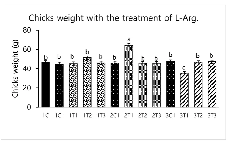
Different concentration of L-Arg injected in embryos which can influence biological molecules and toxicity during embryogenesis. However, several studies have reported that higher doses of L-Arg become toxic which can cause significantly increased mortality rates and impaired weight gain. Lower concentration of L-Arg (1.0%) injected chicks showed better growth performance than higher concentration (1.5%) of L-Arg injected chicks [25]. The parallel effect revealed in our current study, that lower concentration of L-Arg (2T1) at 14th day injection can increase hatching rate (96.29%), body
might have stimulated utilization of amino acids with concomitant decrease in degradation of amino acids by the embryo [26]. In-ovo feeding of L-Arg resulted in higher embryo weight. This might be due to increase in muscle mass known to be affected by administration of in-ovo feeding. It has been reported that L-Arg could stimulate muscle development [27].Thus, in-ovo injected L-Arg could be utilized by the embryo, resulting in increased muscle mass and heavy embryo that can increase the hatching rate [28]. L-Arg may attenuate adverse effects of rearing chickens under cold ambient temperatures or at high altitudes. Furthermore, feeding broiler chickens with diet that is deficient of L-Arg under cold stress at high altitudes can depress nitric oxide synthesis, decrease feed intake, reduce body weight gain, increase right ventricle to total ventricle weight ratio, mortality rate, and ascites mortality [29].Previous study reported that lower percentage (1.36%) of L-Arg supplemented to broiler easily digestible than higher percentage of arginine, and it could obtain the highest egg weight [30]. Albeit, the low dose of L-Arg stimulates the secretion of the growth hormone could be increased the body weight [21]. The same mechanism might be occurred in our present study, L-Arg appeared to improve body weight of chicks in group 2T1 (Figure 3). However, body weights did not significantly vary among other groups. Hence, low dose (2T1) of L-Arg injected on the 14th day
incubation period could improve body weight compared to 8th and 18th day of incubation period.
Figure 1. Effects of in ovo injections at different concentrations of L-Arg with different developmental embryonic stages on survival rate. Small characters indicate significant differences among experimental groups at p < .05. Values are presented as mean ± SD from twelve determinations.
Figure 2. Effects of in ovo injections at different concentrations of L-Arg with different developmental embryonic stages on hatching rate. Small characters indicate significant differences among experimental groups at p < .05. Values are presented as mean ± SD from twelve determinations.
85
90
95
100
1C 1C1 1T1 1T2 1T3 2C1 2T1 2T2 2T3 3C1 3T1 3T2 3T3
%
o
f s
ur
vi
val
r
at
e
% of Survival rate with the treatment of L-Arg.
a
b
b
b
d
e
0
50
100
150
1C 1C1 1T1 1T2 1T3 2C1 2T1 2T2 2T3 3C1 3T1 3T2 3T3
%
o
f
Ha
tch
ing
r
at
e
% of Hatching rate with the treatment of L-Arg.
a
Figure 3. Effects of in ovo injections at different concentrations of L-Arg with different developmental embryonic stages on Chicks weight. Small characters indicate significant differences among experimental groups at p < .05. Values are presented as mean ± SD from twelve determinations.
4.2. Biochemical indices (SGOT and SGPT)
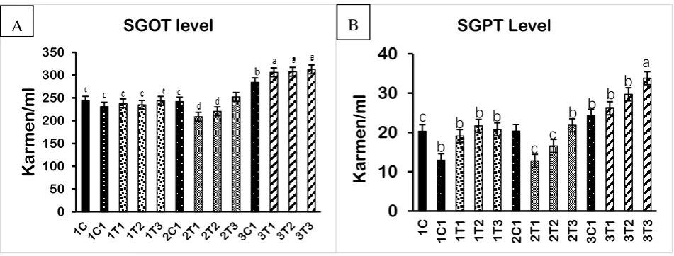
Elevated SGOT and SGPT levels which indicates improper liver functions due to damages of cell integrity and cell membrane in liver. Our results revealed that injection of L-Arg at all doses except lower dose which has affected SGOT and SGPT levels at 8 and 18th day embryonic stages (Figure 6a).
SGOT and SGPT levels were significantly decreased in 2T1 and 2T2 groups of embryos compared to those in other groups. There was no difference among the IC, IC1, 2C1 and 3C1. Increased levels of SGOT and SGPT in the blood are conducive to liver function damage [31]. In fact, free radicals can attack hepatocytes and release stored ALT to re-enter the blood serum [32]. Lower concentration of L-Arg supplementation caused greater percentage reduction in SGOT and SGPT levels in sickle cell anaemia subjects [31]. Supplementation of L-Arg to mice with higher concentration showed increased the SGOT and SGPT levels had been linked to damage to hepatic cells and hemolysis [33]. The cause of liver damage is unclear. Hence, confirming that higher concentration of L-Arg might have damaged the hepatic cell through elevation of SGOT and SGPT in 1T1, 1T2, 1T3, 3T1, 3T2 and 3T3 group. Other hand 2T1 group of injected chicken embryos could re-back the SGOT and SGPT levels compared to the other groups. Stimulating action of nitric oxide (NO) production by L-Arg results showed that improved the degree of the heapto-cellular structure by blocking of Bcl-2 and Tumor necrosis factor-α (TNF-α) [34]. In addition, L-Arg at 1g/day decreased the liver enzymes such as SGOT and SGPT through increased the sitric oxide (NO) synthesis. NO synthase plays an important role in liver injury through Inducible Nitric Oxide Synthase (iNOS) pathways [35]. The same mechanism could be involved in our curent study too.This same mechanism would be occurred in our study that the production of NO reduces necrosis and apoptosis by attenuation of inflammatory pathway to prevent the hepatotoxicity, improved the hepatobiliary function and the ultrastructure of liver results reduced the SGOT and SGPT levels in L-Arg treatment in the lower dose (2T1) at 14th
day injection of embryos (Figure 4).
0
20
40
60
80
1C 1C1 1T1 1T2 1T3 2C1 2T1 2T2 2T3 3C1 3T1 3T2 3T3
C
hi
cks
w
ei
ght
(
g)
Chicks weight with the treatment of L-Arg.
b
Figure 4. and 4B. Effect of in ovo feeding on broiler eggs with different concentrations of L-Arg at different developmental embryonic stages and effects on SGOT and SGPT concentrations in blood plasma. Small characters indicate significant differences among the experimental groups at p < .05. Values are presented as mean ± SD from twelve determinations.
4.3. Micronuclei (MN) and nuclear abnormality (NA) tests using periodic acid Schiff’s (PAS) staining
The wide use of different doses of L-Arg at three different incubation period requires to examine the genotoxic activity in peripheral blood [36]. MN and NA tests were conducted to examine peripheral blood cells in all groups of experimental chicks (Figure 5). MN test can measure subcellular processes of chromosomal breaks (clastogenesis) or cell spindle malfunctions (aneugenesis) as well as the formation of mitochondrial disruption and nuclear DNA than can lead to mitochondria-dependent apoptosis in chicken embryos as an indicator of chromosomal damage [37]. Similar results were obtained in our current experiment that the MN and NA in peripheral blood erythrocytes were observed which has clearly demonstrating higher genotoxicity of high dose of L-Arg on the 8th, 14th
and 18th day incubation period. Moreover, 2T2 group showed no harmful effects at low doses of
L-Arg (2T1 & 2T2) at 14th day incubation time found normal architecture of nuclei in peripheral blood
cells, similar to control group.
0 50 100 150 200 250 300 350
K
a
rm
e
n
/m
l
SGOT level
bA
0
10
20
30
40
1C 1C1 1T1 1T2 1T3
2 C 1 2 T 1 2 T 2 2 T 3 3 C 1 3 T 1 3 T 2 3 T 3
K
a
rm
e
n
/m
l
SGPT Level
b
b
b
b
b
c
c
c
b
b
b
a
4.4. Protein analysis by western blot
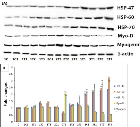
Western blot was performed in muscles to determine whether the different doses of L-Arg supplement at various days of incubation period may alter the protein levels of HSPs family such as HSP-47, HSP-60 and HSP-70. As shown in Figure (6), the protein expressions of HSP-47, HSP-60 and HSP-70 were significantly down-regulated in the 2T1 group compared to other groups. Moreover, their levels in 3T2 and 3T3 groups were significantly up-regulated compared to 2T1 and 2T2 group, although protein expressions of HSP-60 and HSP-70 showed no significant difference among 1T1, 1T2, 1T3, and 2T1 groups. Moreover, HSP-46, HSP-60 and HSP-70 were down-regulated in 2T1 group compared to those in other groups whereas there was no significant difference in their levels between 1C and 2T1. HSP-70 is a reliable index of stress in chickens while “3-hydroxyl-3-methyl-glutaryl
coenzyme A reductase” which has been used as an indicator of stress [38]. Pretreatment with L-Arg markedly reduced the dramatic down regulation of HSP-60 and HSP-70 in hypoxic rat model. The increased expression of HSP-60 and HSP-70 might be related to their leakages of tissue in response to tissue injury [39,40].Hence, present result may suggest that the increased level of HSP-47, HSP-60 and HSP70 in high doses of L-Arg may have the major role in tissue injury.The results of the study also show that the increase of HSP-60 and 70 may be involved in tissue injury in 3T1, 3T2, and 3T3 group. 2T1-group can prevent tissue injury via down-regulation of HSP-46, HSP-60 and HSP-70. Moreover, the protein expressions of myogenin and MyoD were significantly up-regulated in 2T1 group, whereas it was down regulated in 3T1, 3T2 and 3T3 groups compared to other experimental groups. Oxidative stress, can cause muscle atrophy by reducing myogenic differentiation markers such as Myogenin and MyoD in skeletal muscles [41]. Some growth factors, namely cytokines and oncogenes, suppress the activity of myogenin and MyoD, thus resulting in decreased in the mass of muscle which defined as muscle atrophy [42]. Previous study reported that myogenic regulatory factor mainly MyoD and MRF4 is only expressed later in different embryonic muscle groups [43]. Pro-oxidant compounds especially L-Arg are increased in muscle cell under oxidative stress and reduced-to-oxidized glutathione ratio, which suggests the impairment of myogenic regulatory factors such as Myogenin and MyoD. Not only that, results from the previous experiment demonstrated that L-Arg supplementation could promoted the HSP70 expression [44]. However,
high concentration of branched chain amino acids, especially L-Arg can enhance expression of HSPs through whey protein hydrolysate [45]. Hence, our present study has proved that increasing the concentration of L-Arg at 8th and 18th day injection period could up-regulate the expression of
HSP-60 and HSP-70 might be through whey protein hydrolysate that means improper of functional food ingredient. Moreover, the L-Arg at 14thday with (100μg/100μl/egg) promote the muscle mass through
Figure 6. Effects of expression levels of L-Arg, HSP-47, HSP-60, and HSP-70 as well Myogenin and MyoD protein expressions in different stages of chicken embryos at different doses. Small characters indicate significant differences among experimental groups at p < .05.(B) Bar graph represents the quantitative comparison among the groups. Data are expressed as a relative intensity ratio compared to β-actin. Values are presented as mean ± SD from twelve determinations.
4.5. Measurement of IgM concentration in plasma
Concentrations of immune response markers such as IgM in all experimental group were analyzed. The duration and amount of L-Arg supplementation may influence immune status. Short-term supplementary L-Arg can influence the immunity power because L-Arg has antioxidant and anti-inflammatory effects [46,47]. It can attenuate inflammatory reactions by suppressing the generation of inflammatory mediators such as inflammatory cytokines and C-reactive protein which play a major role in the progression of tissue damage and organ dysfunction [48]. The treatment of L-Arg shows improved renal function through improve immune function [49]. Levels of IgM could provide an overall picture of immune function.It has been recently demonstrated that L-Arg can increase specific immune response against infectious bursal disease (IBD) in chickens [50]. L-Arg provided by treatment has been reported to be the sole precursor of nitric oxide with lots of immune
0 0.5 1 1.5 2 2.5 3 3.5 4
C 1C1 1T1 1T2 1T3 2C1 2T1 2T2 2T3 3C1 3T1 3T2 3T3
Fo
ld
chan
g
es
HSP-47HSP-60
HSP-70
Myo-D
functions and growth performance [51]. These same biological attributes might be present after low dose L-Arg injection on the 14th day of incubation period. It may improve immunity via generation
of IgM and suppression of inflammatory cytokines and C-reactive protein (Figure 7).
Figure 7. L-Arg induces IgM levels in different stages of chicken embryos at different doses. Small characters indicate significant differences among experimental groups at p < .05. Values are presented as mean ± SD from twelve determinations.
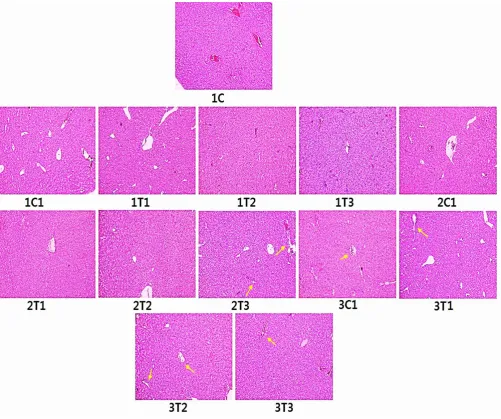
4.6. Histopathology (H&E) staining
Figure 8 shows histology of liver of all experimental groups. Sections from the control group exhibited complete structure with regular shape of liver cells. Section from 1T1 and 2T1 shows normal hepatocyte gap compared to 1C and 1C1 group. Section from 1T3, 2T3, 3T1, 3T2, and 3T3 appears with intracytoplasmic vacuoles in hepatocytes around centrilobular regions. Moreover, hepatocyte tubes were surrounded by inflammatory cells and showed necrosis with nuclear fragmentation in 3T2 and 3T3 groups. The hepatocyte gap was increased in 2T3, 3T1, 3T2, and 3T3 groups. The hepatocyte gap appears in normal architecture in 1C, 1C1, 2C1, 3C1, 1T1, and 2T2. Degeneration of livers were observed for birds when treated with 167 and 334 mg/L of L-Arg had adverse effects on organs [52]. The liver after treatment with L-Arg (334mg/L) had congested vascular spaces and periportal mononuclear inflammatory infiltration [53]. Addition of L-Arg to poultry diets is required to avoid harmful influences of excessive free radicals produced during normal metabolism [54]. Dietary L-Arg supplementation plays a key role in enhancing meat quality. Increased L-Arg and betaine supplementation alleviates total body fat deposition and fatty liver [55,56]. Additionally, supplementation with high doses (50 and 100%) of L-Arg has negative effects on the structure of the liver of Sasso birds proved by H&E staining. However, our current results showed that in ovo injection with low doses (2T1) of L-Arg on the 14th day of egg embryo did not have any negative effects
compared to higher doses of L-Arg on the 8th or 18th day of incubation period.
0 50 100 150 200 250
1C 1C1 1T1 1T2 1T3 2C1 2T1 2T2 2T3 3C1 3T1 3T2 3T3
Ig
M
(n
g
/mL)
a
c
c
c
c
b
b
b
b
b
c
a
Figure 8. Histopathology of liver using hematoxylin and eosin straining. Sections from control chicks’ hepatic
lobule have complete structure. Liver cell has regular shape that is within normal limits. Intracytoplasmic vacuoles are shown in hepatocytes around the centrilobular region in 1T3, 2T3, 3T1, 3T2, and 3T3. Hepatocyte tubes arranged haphazardly with inflammatory cell infiltration, liver cells appear necrosis with nuclear fragmentation in 3T2, 3T3. The hepatocyte gap is also increased in 2T3 3T1, 3T2, 3T3 groups. There was no significant difference between control and 2T1.
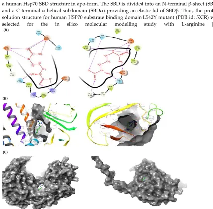
4.7. In silico molecular docking studies.
a human Hsp70 SBD structure in apo-form. The SBD is divided into an N-terminal β-sheet (SBDβ)
and a C-terminal α-helical subdomain (SBDα) providing an elastic lid of SBDβ. Thus, the protein,
solution structure for human HSP70 substrate binding domain L542Y mutant (PDB id: 5XIR) were selected for the in silico molecular modelling study with L-arginine [57].
Figure 10. Possible mechanism of high dose and low dose of L-Arg on toxicity and muscle growth in chicken embryo.
5. Conclusion
In this study, we described a suitable embryonic developmental stage for accessibility of in ovo injection using L-Arg at different concentrations for the first time. The 14th day injection of L-Arg at
100 μg/μl/egg enhanced both hatching and survival rates. It also increased body weight and immune
response (IgM). Histology from control and 2T1 groups sowed normal architecture while injection on the 18th day of incubation of 1st day chicks showed liver tissue damage. Overall results demonstrate
that the optimum dose is 100μg/μl/egg and optimum injection stage is 14th day to improve the
immunity, hatching, and survival rate which can be used for the poultry industry. In ovo injection in early and late embryonic stages could not offer good benefit for survival, hatching rate, or muscle development. If we choose middle stage of embryonic development for in ovo injection, L-Arg might be able to promote muscle growth and improve the immune power without inducing adverse effect on the liver.
Acknowledgments: This work was supported by the Basic Science Research Program, National Research Foundation of Korea (NRF), funded by Ministry of Education (Project No. NRF-2017R1D1A1B03032217 and 2017R1D1A3B03028490) and the “Research Funds of Chonbuk National University in 2018”. We thank to Mrs. Choi, Eun-Jin, Center for University Research Facility (CURF) at Chonbuk National University.
Author Contributions: Conceived and designed the experiments: S.A.S., K.S.S. Performed the experiments and measurements of serum biochemical parameters: S.A.S, D.R.K., J.R.P., and S.H.S. Protein analysis by western blot: S.A.S. Histopathology (H&E) staining: S.A.S. In silico molecular docking studies: P.R and C.S.N.
Competing interests: The authors declare no competing interests.
References
1.
Berri, C.; Wacrenier, N.; Millet, N.; Le Bihan-Duval, E. Effect of selection for improved body composition on muscle and meat characteristics of broilers from experimental and commercial lines. Poult. Sci. 2001, 80, 833-838.2. Sobolewska, A.; Elminowska-Wenda, G.; Bogucka, J.; Szpinda, M.; Walasik, K.; Bednarczyk, M.; Paruszewska-Achtel, M. Myogenesis–possibilities of its stimulation in chickens. Folia biologica. 2011,59, 85-90.
3. Dos Santos, T. T.; Corzo, A.; Kidd, M. T.; McDaniel, C. D.; Torres Filho, R. A.; L. F. Araujo, L.F. Influence of in ovo inoculation with various nutrients and egg size on broiler performance. Journal of Applied Poultry Research 2010, 19, 1-12.
4. Dransfield, E.; Sosnicki, A.A. Relationship between muscle growth and poultry meat quality. Poultry science. 1999, 78, 743-746
5. Uni, Z.; Ferket, P. R.; Tako, E.; Kedar, O. In ovo feeding improves energy status of late-term chicken embryos. PoulT. Sci. 2005.84, 764-770.
6. Ohta, Y.; Kidd, M. Ishibashi, T. Ishibashi. Embryo growth and amino acid concentration profiles of broiler breeder eggs, embryos, and chicks after in ovo administration of amino acids. Poult. Sci. 2001, 80, 1430-1436.
7. Shafey, T. M.; Sami, A. S.; Abouheif, M. A. Effects of in ovo feeding of L-glutamine on hatchability performance and hatching time of meat-type breeder eggs. Journal of Animal and Veterinary Advances.
2013, 12, 135-139.
8. Chalamaiah, M.; Yu, W.; Wu, J. Immunomodulatory and anticancer protein hydrolysates (peptides) from food proteins: A review. Food chemistry. 2018, 245, 205-222.
9. Kadam, M. M.; Barekatain, M. R.; K Bhanja, S.; Iji, P.A. Prospects of in ovo feeding and nutrient supplementation for poultry: the science and commercial applications—a review. Journal of the Science of Food and Agriculture. 2013, 93, 3654-3661.
10. Ohta, Y.; Kidd, M.T. Optimum site for in ovo amino acid injection in broiler breeder eggs. Poult. Sci. 2001,80, 1425-1429.
11. Sobolewska, A.; Elminowska-Wenda, G.; Bogucka, J.; Dankowiakowska, A.; Kułakowska, A.; Szczerba, A.; Bednarczyk, M. The influence of in ovo injection with the prebiotic DiNovo® on the development of histomorphological parameters of the duodenum, body mass and productivity in large-scale poultry production conditions. Journal of animal science and biotechnology. 2017, 8, 45.
12. Spurlin III, J.; Lwigale P.A. A Technique to Increase Accessibility to Late‐Stage Chick Embryos for In Ovo Manipulations. Developmental Dynamics. 2013,242, 148-154.
13. Ohta, Y.; Tsushima, N.; Koide, K.; Kidd, M. T.; Ishibashi, T. Effect of amino acid injection in broiler breeder eggs on embryonic growth and hatchability of chicks. Poult. Sci. 1999,78, 1493-1498.
14. Zielinska, M.; Sawosz, E.; Grodzik, M.; Wierzbicki, M.; Gromadka, M.; Hotowy, A.; Chwalibog, A. Effect of heparan sulfate and gold nanoparticles on muscle development during embryogenesis. International journal of nanomedicine. 2011,6, 3163.
15. Noy, Y.; Uni, Z. Early nutritional strategies. World's Poultry Science Journal. 2010, 66: 639-646.
16. Foye, O. T.; Uni, Z.; Ferket, P.R. Effect of in ovo feeding egg white protein, β-hydroxy-β-methylbutyrate, and carbohydrates on glycogen status and neonatal growth of turkeys. Poult. Sci. 2006, 85, 1185-1192. 17. Miller, A. L. Therapeutic considerations of L-glutamine: a review of the literature. Alternative medicine
review: a journal of clinical therapeutic. 1999, 4, 239-248.
18. Uni, Z.; Ferket. P.R. U.S. Patent No. 6,592,878. Washington, DC: U.S. Patent and Trademark Office. 2003. 19. Bhanja, S. K.; Mandal, A.B. Effect of in ovo injection of critical amino acids on pre and post hatch growth,
20. Sawosz, F., Pineda, L., Hotowy, A., Jaworski, S., Prasek, M., Sawosz, E., & Chwalibog, A. (2013). Nano-nutrition of chicken embryos. The effect of silver nanoparticles and ATP on expression of chosen genes involved in myogenesis. Archives of animal nutrition, 67(5), 347-355.
21. Al-Daraji, H. J.; Al-Mashadani, A. A.; Al-Mashadani, W. K.; Al-Hassani, A. S.; Mirza, H.A. Effect of in ovo injection with L-arginine on productive and physiological traits of Japanese quail. South African Journal of Animal Science. 2012,42, 139-145.
22. Yan, X.; Shi, Q.; Bracher, A.; Miličić, G.; Singh, A. K.; Hartl, F. U.; Hayer-Hartl, M. GroEL ring separation and exchange in the chaperonin reaction. Cell. 2018, 172, 605-617.
23. Umehara, K.; Hoshikawa, M.; Tochio, N.; Tate, S.I. Substrate binding switches the conformation at the lynchpin site in the substrate-binding domain of human hsp70 to enable allosteric interdomain communication. Molecules. 2018,23, 528.
24. Friesner, R. A.; Murphy, R. B.; Repasky, M. P.; Frye, L. L.; Greenwood, J. R.; Halgren, T. A.; Sanschagrin, P.C.; Mainz, D.T. Extra precision glide: Docking and scoring incorporating a model of hydrophobic enclosure for protein− ligand complexes. Journal of medicinal chemistry. 2006, 49, 6177-6196.
25. Shafey, T.M.; Sami, A.S; Abouheif, M.A. Effects of in ovo feeding of L-glutamine on hatchability performance and hatching time of meat-type breeder eggs. Journal of Animal and Veterinary Advances.
2013,12, 135-139.
26. Shafey, T. M.; Mahmoud, A. H.; Alsobayel, A. A.; Abouheif, M.A. Effects of in ovo administration of amino acids on hatchability and performance of meat chickens. South African Journal of Animal Science. 2014,44, 123-130.
27. Azhar, M.; Rahardja, D. P.; Pakiding, W. Embryo Development and Post-Hatch Performances of Kampung Chicken by in Ovo Feeding of L-Arginine. Media Peternakan. 2016, 39, 168-172.
28. Bhanja, S. K.; Baran Mandal, A.; Agarwal, S. K.; Majumdar, S. Modulation of post hatch-growth and immunocompetence through in ovo injection of limiting amino acids in broiler chickens. Indian Journal of Animal Sciences. 2012, 82, 993.
29. Izadinia, M.; Nobakht, M.; Khajali, F.; Faraji, M.; Zamani, F.; Qujeq, D.; Karimi, I. Pulmonary hypertension and ascites as affected by dietary protein source in broiler chickens reared in cool temperature at high altitudes. Animal Feed Science and Technology. 2010,155, 194-200.
30. Duan, X., Li, F., Mou, S., Feng, J., Liu, P., & Xu, L. (2015). Effects of dietary L-arginine on laying performance and anti-oxidant capacity of broiler breeder hens, eggs, and offspring during the late laying period. Poultry science, 94(12), 2938-2943.
31. Jaja, S. I.; Ogungbemi, S. O.; Kehinde, M. O.; Anigbogu, C.N. Anigbogu. Supplementation with l-arginine stabilizes plasma arginine and nitric oxide metabolites, suppresses elevated liver enzymes and peroxidation in sickle cell anaemia. Pathophysiology. 2016, 23, 81-85.
32. Nsiah, K.; Dzogbefia, V. P.; Ansong, D.; Akoto, A. O.; Boateng, H.; Ocloo, D. Pattern of AST and ALT changes in relation to hemolysis in sickle cell disease. Clinical medicine. Blood disorders. 2011,4, CMBD-S3969.
33. Pandey, S.; Sharma, A.; Dahia, S.; Shah, V.; Sharma, V.; Mishra, R. M.; Pandey S.W.; Saxena, R. Biochemical indicator of sickle cell disease: preliminary report from India. Indian Journal of Clinical Biochemistry. 2012, 27, 191-195.
34. Chattopadhyay, P.; Shukla, G.; Wahi, A.K. Protective effect of L-arginine against necrosis and apoptosis induced by experimental ischemic and reperfusion in rat liver. Saudi journal of gastroenterology: official journal of the Saudi Gastroenterology Association. 2009,15, 156.
35. Ozsoy, Y., Ozsoy, M., Coskun, T., Namlı, K., Var, A., & Özyurt, B. (2011). The effects of L-arginine on liver
damage in experimental acute cholestasis an immunohistochemical study. HPB Surgery, 2011.
36. Subramaniyan, S. A.; Belal, S. A.; Choe, H. S.; Shim, K.S. A Comparative Study of Biologically and Chemically Fabricated Synthesized AgNPs’ Supplementation with Respect to Heat-Shock Proteins, Survival, and Hatching Rates of Chicken Embryos: An In Ovo Study. Journal of Cluster Science. 2018, 29, 129-139.
38. Hasheimi, S. R.; Zulkifli, I.; Somchit, M. N.; Zunita, Z.; Loh, T. C.; Soleimani, A. F.; Tang, S.C. Dietary supplementation of Zingiber officinale and Zingiber zerumbet to heat‐stressed broiler chickens and its effect on heat shock protein 70 expression, blood parameters and body temperature. Journal of animal physiology and animal nutrition. 2013, 97, 632-638.
39. Sohn, S. H.; Subramani, V. K.; Moon, Y. S.; Jang I.S. Telomeric DNA quantity, DNA damage, and heat shock protein gene expression as physiological stress markers in chickens. Poult. Sci. 2012,91, 829-836.
40. Al-Rasheed, N. M.; Fadda, L.; Mohamed, A. M.; Attia, H. A.; Al-Rasheed, N.M. Regulating Effect Of Carnosine And/Or L-Arginine On The Expression Of Inflammatory Molecules Induced Nephropathy In The Hypoxic Rat Model. Brazilian Archives of Biology and Technology. 2016, 59
41. Murray, J.; Auwerx, J.; Huss, J.M. Impaired myogenesis in estrogen-related receptor γ (ERRγ)-deficient skeletal myocytes due to oxidative stress. The FASEB Journal. 2013, 27, 135-150.
42. Wust, R.C.; Degens, H. Factors contributing to muscle wasting and dysfunction in COPD patients. Int. J. Chron. Obstruct. Pulmon. Dis. 2007, 2, 289–300.
43. Mok, G.F.; Mohammed, R.H.; Sweetman, D. Expression of myogenic regulatory factors in chicken embryos during somite and limb development. Journal of anatomy. 2015, 227, 352-360.
44. Wu, X.; Ruan, Z.; Gao, Y.; Yin, Y.; Zhou, X.; Wang, L.; Wu.G. Dietary supplementation with L-arginine or N-carbamylglutamate enhances intestinal growth and heat shock protein-70 expression in weanling pigs fed a corn-and soybean meal-based diet. Amino acids. 2010,39, 831-839.
45. Moura, C. S.; Lollo, P. C. B.; Morato, P. N.; Risso, E. M.; Amaya-Farfan, J. Modulatory effects of arginine, glutamine and branched-chain amino acids on heat shock proteins, immunity and antioxidant response in exercised rats. Food & function. 2017,8, 3228-3238.
46. Deng, K.; Wong, C. W.; Nolan, J.V. Long-term effects of early life L-arginine supplementation on growth performance, lymphoid organs and immune responses in Leghorn-type chickens. British poultry science.
2005, 46, 318-324.
47. Ivanov, V.; Cha, J.; Ivanova, S.; Kalinovsky, T.; Roomi, M. W.; Rath, M.; Niedzwiecki, A. Essential nutrients suppress inflammation by modulating key inflammatory gene expression. International journal of molecular medicine. 2008, 22, 731-741.
48. Wu, G. H.; Zhang, Y. W.; Wu, Z.H. Modulation of postoperative immune and inflammatory response by immune-enhancing enteral diet in gastrointestinal cancer patients. World journal of gastroenterology.
2001,7, 357.
49. Klahr, S. Can L-arginine manipulation reduce renal disease?. In Seminars in nephrology 1999,19, 304-309. 50. Kline, J. A.; Watts, J.; Courtney, D.; Lee, Y. Y.; Hwang, S. Severe pulmonary embolism decreases plasma
L-arginine. European Respiratory Journal. 2014,43, 906-909.
51. Kang, K.; Shu, X. L.; Zhong, J. X.; Yu, T. T.; Lei. T. Effect of L-arginine on immune function: a meta-analysis. Asia Pacific journal of clinical nutrition. 2014, 23, 351-359.
52. Alabi, O. O.; Shoyombo, A. J.; Animashahun, R. A.; Olawoye, S. O.; Abdulazeez, J. O.; Faduhunsi, O. O.; Oladehinbo, D.O. Effects of L-Arginine supplementation of drinking water on the kidney and liver of Sasso chickens. International Journal of Livestock Production. 2018,9, 160-164.
53. Pacher, P.; Beckman, J. S.; Liaudet, L. Nitric oxide and peroxynitrite in health and disease. Physiological reviews. 2007, 87, 315-424.
54. Atakisi, O.; Atakisi, E.; Kart, A. Effects of dietary zinc and l-arginine supplementation on total antioxidants capacity, lipid peroxidation, nitric oxide, egg weight, and blood biochemical values in Japanase quails. Biological trace element research. 2009,132, 136-143.
55. Oh, H. S.; Oh, S. K.; Lee, J. S.; Wu, C.; Lee, S.J. Effects of l-arginine on growth hormone and insulin-like growth factor 1. Food Science and Biotechnology. 2017, 26: 1749-1754.