1
High mobility group protein 1 and dickkopf-related protein 1 in schizophrenia and
treatment-resistant schizophrenia: associations with interleukin-6, symptom domains, and neurocognitive
impairments.
Arafat Hussein Al-Dujaili a, Rana Fadhil Mousa b, Hussein Kadhem Al-Hakeim c, Michael
Maes d,e,f.
a Senior Clinical Psychiatrist at the Faculty of Medicine, University of Kufa, Iraq. E-mail:
b A biochemist at the Faculty of Veterinary Medicine, University of Kerbala, Iraq. E-mail:
c Department of Chemistry, College of Science, University of Kufa, Iraq. E-mail:
d Corresponding Author: Department of Psychiatry, Faculty of Medicine, Chulalongkorn
University, Bangkok, Thailand;
e Department of Psychiatry, Medical University of Plovdiv, Plovdiv, Bulgaria;
f School of Medicine, IMPACT Strategic Research Centre, Deakin University, PO Box 281,
Geelong, VIC, 3220, Australia. E-mail: [email protected].
Corresponding author:
Prof. Dr. Michael Maes, M.D., Ph.D.
IMPACT Strategic Research Center
Barwon Health, Deakin University
3
Abstract
Background: Schizophrenia and treatment-resistant schizophrenia (TRS) are associated with
aberrations in immune-inflammatory pathways. Increased High Mobility Group Protein 1
(HMGB1), an inflammatory mediator, and Dickkopf-Related Protein (DKK1), a Wnt/β-catenin
signaling antagonist, affect the blood-brain-barrier and induce neurotoxic effects and
neurocognitive deficits.
Aim of the study: The present study aims to examine HMGB1 and DDK1 in non-responders to
treatments with antipsychotics (NRTT, n=60), partial RTT (PRTT, n=55) and healthy controls
(n=43) in relation to established markers of schizophrenia including IL-6, IL-10 and CLL11
(eotaxin); and to delineate whether these proteins are associated with the schizophrenia symptom
subdomains and neurocognitive impairments.
Results: HMGB1, DKK1, IL-6 and CCL11 were significantly higher in schizophrenia patients
than in controls. DKK1 and IL-6 were significantly higher in NRTT than in PRTT and controls
while IL-10 was higher in NRTT than in controls. Binary logistic regression analysis showed that
schizophrenia was best predicted by increased DDK1 and HMGB1 while NRTT (versus PRTT)
was best predicted by increased IL-6 and CCL11 levels. A large part of the variance in psychosis,
hostility, excitation, mannerism and negative (PHEMN) symptoms, and formal thought disorders
was explained by HMGB1, IL-6, and CCL11 while most neurocognitive functions were predicted
by HMGB1, DDK1 and CCL11.
Conclusion: The neurotoxic effects of HMGB1, DKK1, IL-6 and CCL11 including effects on the
blood-brain-barrier and the Wnt/β-catenin signaling pathway may cause impairments in executive
functions, and working, episodic and semantic memory and explain, in part, PHEMN symptoms
4
Keywords: schizophrenia, treatment resistance, neuro-immune, inflammation, cytokines,
5
Introduction
The World Health Organization reported that schizophrenia (SCZ) patients die at a younger
age as expected due to preventable physical diseases, such as cardiovascular disease, metabolic
disease, and infections.1 These diseases have an immune-inflammatory etiology and, therefore, those comorbidities may be explained by the neuro-immune theory of SCZ. The first
comprehensive neuro-immune theory of SCZ was introduced by Smith and Maes in 1995 2 as the “macrophage-T-lymphocyte theory” considering that activated macrophages and T lymphocytes
are key phenomena in the pathophysiology of SCZ.2 Two years later, Maes and colleagues reported that SCZ is accompanied by an ongoing inflammatory response as indicated by increased
plasma acute-phase proteins, such as fibrinogen, haptoglobin, hemopexin, antitrypsin, and
α1-acid glycoprotein, as well as complement component 3 and complement 4 and immunoglobulins
G and M. 3 In addition, these authors reported that increased plasma levels of interleukin (IL)-6,
sIL-1 receptor antagonist (sIL-1RA), IL-8 and IL-10 are associated with treatment-resistant
schizophrenia (TRS). 4-6
Those early findings on the immune-inflammatory response system (IRS) in SCZ are now
well-replicated and synthesized in meta-analytic studies 7,8 while new studies also showed activated M1 macrophage (increased production of tumor necrosis factor-alpha (TNF-α) and
IL-6); T helper (Th)-1 (increased levels of interferon (IFN)-γ and IL-2); Th-2 (IL-4 and IL-5), Th-17
(increased IL-17), and T regulatory (Treg) cell activation (increased IL-10), coupled with
increased IgA levels to Gram-negative bacteria and neurotoxic tryptophan catabolites (TRYCATs)
in SCZ.9-14 Furthermore, new data show that TRS is characterized by activated M1 and Th-1 phenotypes, increased IL-6 trans-signaling (increased IL-6 and sIL-6R) and elevated chemokine
6
Recently, it was reviewed that SCZ and its phenotypes including first-episode psychosis,
the acute episodes, TRS and chronic and deficit schizophrenia are not only accompanied by an
activated IRS, but also by activation of the compensatory immune-regulatory system (CIRS).14 Indicants that immune-regulatory processes are activated in SCZ include activated Th-2 and Treg
responses (see above), increased levels of some acute-phase proteins, which show
immune-regulatory effects (including haptoglobin), and increased levels of sIL-2R, sIL-1RA, and
sTNF-R2.14 These immune-regulatory CIRS mechanisms are secondary to IRS activation and downregulate the primary IRS . Moreover, TRS is not only characterized by an activated IRS, but
also by activated CIRS pathways as indicated by increased levels of sIL-1RA, sTNFR1, and
sTNFR2. 14
Importantly, products of M1, Th-1 and Th-2 have multiple neurotoxic effects and as such
may cause deficits in executive functions, and episodic and semantic memory and as a
consequence also SCZ symptom domains including negative and PHEM (psychosis, hostility,
excitation, and mannerism) symptoms.15-17 Neurotoxic compounds that are upregulated in SCZ or SCZ phenotypes comprise IL-1β, TNF-α, IL-6, IFN-γ, CCL11, IL-4, IL-13, TRYCATs, and
LPS.11,15-19
Severe IRS responses, as in sepsis, are mediated by high mobility group protein (HMGB)1,
a damage-associated molecular pattern (DAMP) released by injured or necrotic cells, which acts
as a pro-inflammatory cytokine promoting the release of IL-6, TNF-α and IFN-γ.20,21 In neurological disorders, HMGB1 is a biomarker of neuroinflammation and neurodegeneration,
which may cause blood-brain barrier (BBB) dysfunctions.22 Likewise, HMGB1 may impair memory and behaviors in mice mediated via the Toll-like receptor (TLR)4 complex and/or the
7
HMGB1 is increased in patients with SCZ or TRS and whether this protein is associated with
increased IL-6 and IL-10 and impaired cognitive functions.
Inflammation is also accompanied by an upregulation of Dickkopf-related protein 1
(DKK1), a pro-inflammatory glycoprotein secreted by platelets and endothelial cells. 24 DKK1 antagonizes the canonical Wnt signaling transduction pathway and therefore may interfere with
tissue regeneration and repair and, additionally, may induce a rapid disassembly of synapses in
mature neurons. 24, 25 This is further underscored by recent findings that circulating DKK1 is associated with cognitive decline in older adults.26 Moreover, in a Japanese population, DKK1 genetic variants are associated with SCZ.27 However, there are no data whether increased DKK1 levels are associated with SCZ, TRS, and neurocognitive impairments and symptom severity.
Hence, the aims of the study are to 1) examine whether HMGB1 and DDK1 are increased
in SCZ and TRS, and 2) delineate the association between both proteins and established markers
of SCZ (IL-6, IL-10, and CCL11), SCZ symptom subdomains and neurocognitive impairments.
Participants and Methods
Participants
Sixty TRS patients and fifty-five non-TRS patients, as well as 43 healthy controls (both sexes,
ages between 18 and 65 years), were recruited to participate in the current study. All patients were
recruited at the Psychiatry Unit at Al-Imam Al-Hussain Medical City in Karbala Governorate-Iraq
in 2019. All patients complied with the diagnostic criteria of SCZ according to the DSM-IV-TR .
TRS is defined as two periods of treatment nonresponse to two different antipsychotic treatments
8
Clinical Global Impression (CGI) Improvement (CGI-I).28 Forty-three healthy controls participated in the study, namely family members or friends of the staff. Patients and controls were
recruited from the same catchment area, namely Karbala city, Iraq. We excluded SCZ patients who
showed axis-1 DSM-IV-TR diagnoses other than SCZ including psycho-organic disorders,
schizoaffective disorder, autism, major depression, and bipolar disorder. Healthy controls were
excluded when they showed a lifetime or current diagnosis of axis I diagnosis or a positive family
history of SCZ. Moreover, patients and controls were excluded when they (a) presented with
neuro-immune, neuroinflammatory or neurodegenerative disorders including Parkinson’s disease,
stroke, multiple sclerosis, and Alzheimer’s disease; (b) suffered from medical illnesses including
diabetes type 1, psoriasis, COPD, rheumatoid arthritis, and inflammatory bowel disease.
Furthermore, we excluded patients and controls who had ever been using medications that interfere
with immune functions, such as glucocorticoids and immunosuppressive, or therapeutic doses of
antioxidant supplements three months prior to the study. All subjects showed serum C-reactive
protein (CRP) concentrations lower than 6 pg/mL excluding subjects with overt inflammation.
All controls and patients, as well as the guardians of patients (parents or the closest family
members), gave written informed consent prior to participation in our study. The study was
conducted according to International and Iraq ethics and privacy laws. Approval for the study was
obtained from the Institutional Review Board of the University of Karbala (418/2019) and Karbala
Health Department (1331/2019), which is in compliance with the International Guidelines for
Human Research protection as required by the Declaration of Helsinki, The Belmont Report,
Council for International Organizations of Medical Sciences (CIOMS) Guideline and International
9
Measurements
Clinical assessments
The diagnosis of SCZ was made by a senior psychiatrist specialized in SCZ according to
DSM-IV-TR diagnostic criteria using the Mini-International Neuropsychiatric Interview
(M.I.N.I.), in a validated Arabic translation (Iraqi dialect). The same day as the M.I.N.I., the same
psychiatrist employed a semi-structured interview to assess demographics as well as clinical data
in all participants and he also measured the CGI-I and Severity (CGI-S) scale.28 The CGI-I was used to define patients who were non responders to treatment (NRTT), namely those who did not
show any change in the CGI-I or showed worse scores after treatment (minimally worse, much
worse, very much worse) and those who were partial RTT (PRTT), namely those with improved
scores (minimally, much or very much). Not one of the patients showed complete remission after
treatment. He also assessed the Scale for the Assessments of Negative Symptoms (SANS) to assess
the severity of negative symptoms.29 We computed scores reflecting PHEM (psychosis, hostility, excitation, and mannerism) symptoms, FTD (formal thought disorders) and PMR (psycho-motor
retardation) as explained previously.16,19 Towards this end we also assessed the Brief Psychiatric
Rating Scale (BPRS),30 the Hamilton Depression Rating Scale31 and the positive and negative syndrome scale (PANSS) for schizophrenia. 32
On the same day, a well-trained research psychologist, blinded to the clinical diagnosis,
completed the Brief Assessment of Cognition in SCZ (BACS) 33 to assess episodic memory using the List Learning test; working memory with the Digit Sequencing Task; verbal fluency and
semantic memory employing Category Instances and Controlled Word Association tests; attention
using the Symbol Coding test, and executive functions using the Tower of London. DSM-IV-TR
10
was measured on the same day as the clinical interview and was scored as body weight (kg) /
length (m2).
Assays
After an overnight fast, five milliliters of venous blood were sampled, utilizing disposable
needle and plastic syringes, between 8.00 and 9.00 a.m. The samples were transferred into a clean
plain tube; blood was left at room temperature for 15 min for clotting, centrifuged 3000 rpm for
10 min, and then serum was separated and transported into two Eppendorf tubes to be stored at
-80 °C until thawed and analyzed. Commercial ELISA sandwich kits were used to measure serum
CCL11, DKK1, HMGB1, and IL-10 (Elabscience®, Inc. CA, USA) and IL-6 (Melsin Medical Co,
Jilin, China). All measured concentrations of CCL11 (sensitivity=9.38 pg/mL), DKK1
(sensitivity=18.75 pg/mL), HMGB1 (sensitivity=18.75 pg/mL), and IL-6 (sensitivity=0.1 pg/mL)
were greater than the sensitivity of the assays. There was only one IL-10 concentration (4.05
pg/mL in a normal volunteer) that was lower than the sensitivity of the assay (sensitivity=4.69
pg/mL). We did not apply left-censoring and used the actual measurements in the statistical
analyses.12 The procedures were followed exactly without modifications according to the manufacturer’s instructions. The intra-assay coefficient of variation (CV) (precision within an
assay) were < 10.0%. Serum CRP was measured using a kit supplied by Spinreact®, Spain. The test is based on the principle of latex agglutination.
Statistical analysis
Analysis of variance (ANOVA) was used to check differences in scale variables between
11
variables. In order to assess associations among the biomarkers, clinical and cognitive scores we
examined correlation matrices based on Pearson’s product-moment and Spearman’s rank-order
correlation coefficients. We used multivariate general linear model (GLM) analysis to delineate
the associations between diagnosis and the biomarkers while controlling for confounding variables
including nicotine dependence, sex, age, BMI and education. Consequently, we carried out tests
for between-subject effects to delineate the associations between diagnosis and each of the
biomarkers. The effect size was estimated using partial eta-squared values. We also computed
model generated (GLM analysis) estimated marginal mean (SE) values and protected pairwise
comparisons among treatment means. We employed binary logistic regression analysis to delineate
the best predictors of NRTT (PRTT as reference group) and SCZ (controls as reference group)
using the biomarkers as explanatory variables. Odd’s ratios with 95% confidence intervals were
computed as well as Nagelkerke values as pseudo-R2 values. We used multiple regression analysis
to assess the significant biomarkers, which predict the symptom domains and neurocognitive tests
while allowing for possible effects of age, sex, and education. We used an automatic stepwise
method with the inclusion of variables with a p-to-entry of 0.05 and p-to-remove of 0.06 while
checking the R2 change. All regression analyses were checked for collinearity using tolerance and VIF values. Variables were also z transformed and the mean z scores were displayed in bar plots.
Tests were 2-tailed and a p-value of 0.05 was used for statistical significance. All statistical
analyses were performed using IBM SPSS windows version 25, 2017.
Results.
12
Table 1 shows the sociodemographic data of the NRTT, PRTT and healthy controls. In
this study, we recruited 142 SCZ patients who were treated with antipsychotic drugs during two
trials with antipsychotic drugs. During the first trial, patients were treated for 8 weeks and after
this trial divided into those without a clinical response (n=84) and a partial response (n=51) (we
lost 7 patients during this first trial). The partial responders continued to take the same medication
for another 2 months while we lost again 7 patients in the follow up yielding a final PRTT study
group of n=55. The non-responders to a first antipsychotic agent were switched to another
antipsychotic agent for another 8 weeks and during this follow-up period we lost again 13 patients.
Two months later, 11 patients showed a partial response to treatment and were classified as PRTT,
whereas 60 did not show any improvement on the CGI-I and, therefore, were classified as NRTT.
Consequently, 55 PRTT and 60 NRTT were recruited to participate in the study.
There were no significant differences in age, sex ratio, BMI, and TUD between PRTT and
NRTT and normal controls. There were somewhat more NRTT who were single than normal
controls. Significantly more SCZ patients were unemployed as compared with controls while years
of education were somewhat lower in NRTT. There were no differences in age at onset between
both SCZ subgroups.
All 6 cognitive test scores were significantly different between the three study groups and
the scores decreased from controls to PRTT to NRTT. The total SANS score was significantly
different between the three study groups. Figure 1 displays a plot of all symptom domains
examined in this study (shown as z scores). Psychosis (F=772.55, df=2/152, p<0.001), hostility
(F=498.12, df=2/152, p<0.001), excitement (F=320.71, df=2/152, p<0.001), mannerism
(F=204.41, df=2/152, p<0.001), FTD (F=414.15, df=2/152, p<0.001) and PMR (F=297.46,
13
controls to PRTT to NRTT. Table 1 shows also the measurement of the clinical global impression
(CGI) score in the SCZ patients. Both the CGI-I and CGI-S scores were significantly higher in
NRTT than in PRTT. All CGI-I scores in PRTT equaled 2 (much improved) or 3 (minimally
improved) and in NRTT 4 (no change) or 5 (minimally worse). The table also shows the current
medication patients were taking. Thus, NRTT were more often treated with clozapine, quetiapine,
and risperidone than PRTT who were more often treated with olanzapine and haloperidol.
Biomarkers between the study groups
In the total study group, there were significant correlations between IL-6 and DKK1
(r=0.641, p<0.001, n=158) and HMGB1 (r=0.249, p=0.002, n=158) and a significant association
between IL-10 and HMGB1 (r=0.465, p<0.001, n=158). The correlation between IL-6 and
HMGB1 was established in controls (r=0.649, p<0.001, n=43) and SCZ patients (r=0.561,
p<0.001, n=115). A significant correlation between IL-10 and DKK1 was detected in controls
(r=0.395, p=0.009, n=43) and SCZ patients (r=0.430, p<0.001, n=115).
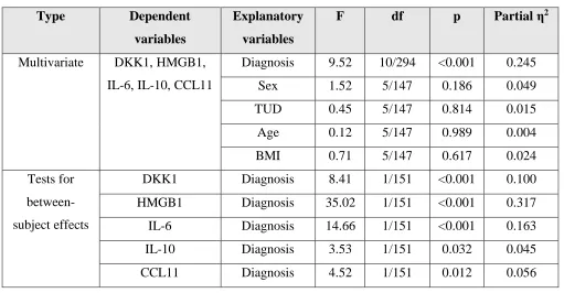
Table 2 shows the results of multivariate GLM analyses comparing the differences in the
biomarkers between the three study groups while adjusting for sex, age, BMI and TUD. There
were highly significant differences in the biomarkers between the groups with an effect size of
0.245, while the 4 covariates had no significant effects. Tests for between-subject effects and
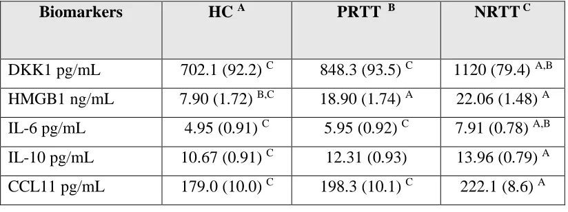
Table 3, which shows the estimated marginal means, indicate that DKK1 and IL-6 were
significantly higher is NRTT than in PRTT and controls and that HMGB1 was significantly
different between SCZ and controls. IL-10 was higher in NRTT than in controls while PRTT
14
Table 4 shows the results of two binary logistic regression analyses examining the best
predictors of SCZ (versus controls) and NRTT (versus PRTT) using an automatic stepwise method
with biomarkers as explanatory variables while allowing for the effects of age, sex and education.
The first regression analysis showed that SCZ was best predicted by increased levels of DDK1 and
HMGB1 (χ2=60.58, df=2, p<0.001, Nagelkerke=0.462) while the accuracy was 74.7% with a
sensitivity of 72.2% and a specificity of 81.4%. The second regression shows that IL-6 combined
with CCL11 were the best predictors of NRTT versus PRTT (χ2=25.84, df=2, p<0.001,
Nagelkerke=0.268).
Effects of background variables.
As described above there were no significant effects of age, sex, BMI and TUD on the
biomarkers included. In patients with SCZ, we also examined the effects of the use of
antipsychotics on the biomarkers. Multivariate GLM analysis followed by tests for
between-subjects effects showed that there were no significant effects of use of haloperidol (F=0.54,
df=5/104, p=0.743, partial eta squared=0.025), quetiapine (F=0.58, df=2/104, p=0.713, partial eta
squared=0.027), clozapine (F=0.59, df=5/104, p=0.708, partial eta squared=0.028), and
risperidone (F=1.17, df=5/104, p=0.331, partial eta squared=0.053) on the biomarkers. There was
a significant effect of olanzapine (F=2.45, df=5/104, p=0.039, partial eta squared=0.105), although
after p-correction for multiple testing this effect was no longer significant (p=0.195). Tests for
between-subjects effects showed a significant effect on CCL11 only (F=7.67, df=1/108, p=0.007,
partial eta squared=0.066). This effect remained significant after p-correction for false discovery
15
220.1 ±9.1 pg/ml). However, the differences between the diagnostic groups in CCL11 were not
affected after covarying for use of olanzapine.
Prediction of symptom domains by biomarkers
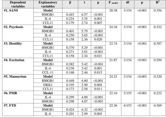
Table 5 shows different stepwise multiple regression analyses with the symptom domains
as dependent variables and the 5 biomarkers as explanatory variables while allowing for the effects
of age, sex, and education. Regression #1 shows that 35.6% of the variance in the total SANS score
was explained by the regression on HMGB1, IL-6, and CCL11. Regressions #2, #3 and #4 show
that the same variables explained a considerable part of the variance in psychosis (32.2%), hostility
(30.7%), and excitation (29.9%). Regression #5 shows that 32.0% of the variance in mannerism
was explained by HMGB1, DKK1, and CCL11. Figure 2 shows the partial regression plot of the
mannerism scores on HMGB1 levels. IL-6 and HMGB1 together explained 22.2% of the variance
in PMR (regression #6). Regression #7 shows that 36.9% of the variance in FTD was explained
by the combined effects of HMGB1, IL-6, CCL11, and education. In all regressions, HMGB1 was
the variable with the highest impact (except for PMR).
Prediction of cognitive impairments by biomarkers
Table 6 shows the outcome of 6 multiple regression analyses with the cognitive test results
as dependent variables and biomarkers as explanatory variables while allowing for the effects of
age, sex and education. We found that (regression #1) 20.6% of the variance in List Learning
scores was explained by the regression on HMGB1, DKK1 and CCL11 (all inversely associated)
and education (positively associated). Up to 33.5% of the variance in Digit Sequencing Task scores
16
education (positively). Part of the variance (20.7%) in Category Instances scores was explained by
HMGB1, DKK1 (negatively) and education (positively). We found that 33.4% of the variance in
the COWA test (regression #4) scores was explained by the cumulative effects of HMGB1 and
IL-6 (both negatively) while 39.5% of the variance in Symbol Coding scores (#5) was associated with
HMGB1 and DKK1. Figure 3 shows the partial regression plot association between Symbol
Coding scores and HMGB1 levels. Regression #6 shows that part of the variance in the Tower of
London scores was explained by HMGB1 and DKK1 (inversely) and education (positively).
Discussion
The first major finding of this study is that SCZ is characterized by increased levels of
HMGB1, DKK1, IL-6, and CCL11 as compared with healthy controls. Our results that IL-6 and
CCL11 are increased in SCZ are in accordance with findings in previous studies. 5,12,13, 17-19, 34,35
This is the first study that HMGB1 is significantly increased in SCZ versus normal
volunteers. HMGB1 is a transcriptional modifier that acts as a pro-inflammatory cytokine
promoting the release of other cytokines, including IL-6.21 HMGB1 is normally localized in the
nucleus but following immune-inflammatory signals, including LPS and TNF-α, HMGB1 is
translocated to the cytoplasm and may aggregate and accumulate in secretory lysosomes to be
secreted from activated monocytes, macrophages and natural killer cells.36-38 Moreover, extracellular HMGB1 engages membrane receptors leading to immune activation and
neuroinflammation.39 HMGB1 is a DAMP that may activate the TLR2 and TLR4 complex thereby
contributing to danger signaling leading to immune-inflammatory signals.20 Moreover, hemoglobin released from lysed red blood cells may synergize with HMGB1 to stimulate the
17
a complex with HMGB1 thereby stimulating an immune-regulatory response with increased levels
of IL-10.40 In this respect, we found that HMGB1 levels are significantly and positively associated with IL-6 and IL-10, indicating that increased HMGB1 is part in the immune-inflammatory
pathophysiology of SCZ.
This is also the first report that serum DKK1 concentrations are significantly increased in
SCZ. DKK1 is secreted by endothelial cells and platelets while the latter are essential to raising
plasma DKK1 levels.24,41 In humans, a dramatic increase in systemic DKK1 is observed in acute infections42 while platelet-derived DKK-1 contributes to elevated systemic levels in infectious models.41 In mice, LPS may increase the expression of DKK1 and IL-6 43 while in the present study, there were significant and positive correlations between IL-6 and DKK1. Nevertheless,
there is a study reporting lowered DKK1 levels in SCZ patients while these authors found
upregulated mRNA expression of Wnt signaling pathway genes. 44 It is important to note that both
DKK1 and IL-6 and other cytokines, which are increased in SCZ (including TNF-α), may inhibit
activation of the Wnt-β-catenin signaling pathway.44
The second major finding of our study is that NRTT was associated with increased DKK1,
IL-6, and CCL11 concentrations as compared with NTRR and that IL-10 is, additionally,
significantly increased in NRTT as compared with controls. These findings indicate that a partial
treatment response is characterized by attenuated immune-inflammatory pathways (IL-6 and
CCL11) and maybe by inhibition of the Wnt pathway (as a consequence of increased DKK1).
While our DKK1 results are novel, increased IL-6 and IL-10 levels were previously reported in
patients with TRS. 4,6 Our results further extend the findings that TRS is accompanied by IRS activation as indicated by increased levels of sIL-6R, IL-8, CCL2 and CCL3, and by CIRS
18
reported that drug naïve first-episode psychosis is characterized by significant IRS
(M1 + Th-1 + Th-17) and CIRS (Th-2 and Treg) responses and that treatment with risperidone attenuates
both IRS and CIRS responses while increased baseline levels of some CIRS biomarkers may
predict clinical improvement. All in all, the above results and the current study indicate that
antipsychotic agents may attenuate the immune response in some SCZ patients, namely in PRTT,
thereby improving symptoms and neurocognitive deficits.
The third major finding of this study is that the different symptom domains of SCZ
(including PHEM and negative symptoms and FTD) are highly significantly predicted by
increased HMGB1, IL-6 and CCL11, while impairments in executive functions, working memory
and episodic and semantic memory, and attention are predicted by increased levels of especially
HMGB1 and DKK1 but also CCL11. Previously, we reported that CCL11 and IL-6 and other
neurotoxic immune compounds (see Introduction) significantly predict PHEM and negative
symptoms, FTD and PMR suggesting that immune-inflammatory pathways are involved in the
pathophysiology of SCZ.12,18,19
There is now evidence that HMGB1 plays an important role not only in the propagation of
immune-inflammatory responses (see above), but also in neuroinflammatory and
neurodegenerative processes and the associated memory impairments in Parkinson’s and
Alzheimer's disease and multiple sclerosis.46 HMGB1 released from necrotic neurons or inflamed microglia may act on microglia macrophage antigen complex 1 thereby stimulating the production
of multiple neurotoxic factors.47 HMGB1 may cause neurite degeneration via the TLR4 complex
and phosphorylation of MARCKS via MAP kinases.48 On the other hand, HMGB1-specific antibodies protect against lethal endotoxaemia20 and preserve BBB integrity thereby attenuating
19
damage to hippocampal neurons, neuronal degeneration, and brain damage.49, 50 Furthermore, blocking of HMGB1 signaling improves neuroprotection in neurodegenerative disorders.51 Clinical studies show that sepsis survivors have permanent cognitive deficits, which are probably
mediated via elevated HMGB1 levels.52
As described above, DKK1 is a natural antagonist of the Wnt/β-catenin signaling pathway,
53 which is a key regulator of BBB function and contributes to its formation, maturation, and
function.54-56 Elevating β-catenin signaling leads to lowered permeability of the endothelial cells of the BBB,56 whereas DKK1-induced aberrations in Wnt/β-catenin signaling may induce BBB breakdown.57, 58 Moreover, administration of DKK1 to a mixture of human neurons and astrocytes in culture results in downregulation of neuronal processes explaining that lowering DKK1 protects
against neurotoxicity.59 DKK1 mediates amyloid-ß-associated synaptic loss, causes a rapid disassembly of synapses in mature neurons,25 induces BAX and decreases Bcl-2 thereby causing
cell death.60 Moreover, loss of DKK1 may counteract the downregulation of hippocampal neurogenesis and accompanying cognitive impairments that are associated with increasing DKK1
levels with age. 61 This may explain that DKK1 deficient mice show improved working memory,
memory consolidation and affective behaviors. 61 It is important to note that the effects of HMGB1 and DKK1 affecting BBB functions may aggravate the effects of CCL11, neurotoxic TRYCATs
and LPS which all lead to breakdown of the BBB in SCZ.11
Limitation of the study
The results of our study should be discussed with respect to its limitations. First, we
performed a case-control study and, therefore, no firm causal inferences may be established.
20
relation to HMGB1 as well as a broader panel of cytokines. Finally, we found that treatment with
olanzapine significantly increased CCL11 levels from 176.3 ±14.4 to 220.1 ±9.1 pg/ml, which if
replicated in ex vivo and in vivo studies, would suggest that olanzapine may augment the
detrimental effects of CCL11. Nevertheless, our intergroup analyses were adjusted for the drug
state, which did not affect the differences in CCL11 between the study groups.
Conclusions
HMGB1, DKK1, IL-6, and CCL11 were significantly higher in SCZ patients, whereas a
non-response to treatment with antipsychotics was associated with increased DKK1, IL-6 and
CCL11. Both HMGB1 and DKK1 were significantly correlated with IL-6 levels. HMGB1 and
DKK1 participate in the immune pathophysiology of SCZ and may explain, in part, the phenome
of SCZ (neurocognitive impairments and various symptom clusters) via their detrimental effects
on the BBB and multiple neurotoxic effects as well.
Acknowledgment
We acknowledge the staff of the Psychiatry Unit at Al-Imam Al-Hussain Medical City in Karbala
city for their help in the collection of samples and the high-skilled staff members of Asia Clinical
Laboratory, Najaf city, for their help in the ELISA measurements .
Funding
There was no specific funding for this specific study .
21
The authors have no conflict of interest with any commercial or other association in connection
with the submitted article .
Author’s contributions
All the contributing authors have participated in the preparation of the manuscript .
References
1. World Health Organization (WHO) | Schizophrenia. In: World Health Organization; World
Health Organization; 2018.
2. Smith RS, Maes M. The macrophage-T-lymphocyte theory of schizophrenia: Additional
evidence. Med Hypotheses 1995;45:135-141. doi:10.1016/0306-9877(95)90062-4.
3. Maes M, Delange J, Ranjan R, et al. Acute phase proteins in schizophrenia, mania and major
depression: modulation by psychotropic drugs. Psychiatry Res. 1997;66:1-11.
4. Lin A, Kenis G, Bignotti S, et al. The inflammatory response system in treatment-resistant
schizophrenia: increased serum interleukin-6. Schizophr Res 1998;32:9-15.
5. Maes M, Bocchio Chiavetto L, Bignotti S, et al. Effects of atypical antipsychotics on the
inflammatory response system in schizophrenic patients resistant to treatment with typical
neuroleptics. Eur Neuropsychopharmacol 2000;10:119-124.
6. Maes M, Bocchio Chiavetto L, Bignotti S, et al. Increased serum 8 and
interleukin-10 in schizophrenic patients resistant to treatment with neuroleptics and the stimulatory effects of
22
7. Miller BJ, Buckley P, Seabolt W, Mellor A, Kirkpatrick B. Meta-analysis of cytokine
alterations in schizophrenia: clinical status and antipsychotic effects. Biol
Psychiatry 2011;70(7):663-671. doi: 10.1016/j.biopsych.2011.04.013.
8. Goldsmith DR, Rapaport MH, Miller BJ. A meta-analysis of
blood cytokine network alterations in psychiatric patients: comparisons between schizophrenia,
bipolar disorder and depression. Mol Psychiatry 2016;21(12):1696-1709. doi: 10.1038/mp.2016.3.
9. Noto C, Ota VK, Gouvea ES, et al. Effects of risperidone on cytokine profile in drug-naïve
first-episode psychosis. Int J Neuropsychopharmacol 2014;18:4.
10. Li H, Zhang Q, Li N, et al. Plasma levels of Th17-related cytokines and complement C3
correlated with aggressive behavior in patients with schizophrenia. Psychiatry Res
2016;246:700-706. Doi: 10.1016/j.psychres.2016.10.061.
11. Maes M, Sirivichayakul S, Kanchanatawan B, Vodjani A. Breakdown of the paracellular tight
and adherens junctions in the gut and blood brain barrier and damage to the vascular barrier in
patients with deficit schizophrenia. Neurotox Res 2019, in press.
12. Al-Hakeim, H.K, Al-Mulla, A.F, Maes, M. The immune fingerprint of major
neuro-cognitive psychosis or deficit schizophrenia: a supervised machine learning study. Preprints, 2019,
doi:10.20944/preprints201905.0285.v1. Neurotox. Res., in press.
13. Noto C, Maes M, Ota VK, et al. High predictive value of immune-inflammatory biomarkers
for schizophrenia diagnosis and association with treatment resistance. World J Biol Psychiatry
2015;16:422-429. doi: 10.3109/15622975.2015.1062552.
14. Roomruangwong C, Noto C, Kanchanatawan B, Anderson G, Kubera M, Carvalho AF, Maes
23
compensatory immune-regulatory reflex system (CIRS) in different phenotypes of schizophrenia:
the IRS-CIRS theory of schizophrenia. Mol Neurobiol. 2019. doi: 10.1007/s12035-019-01737-z.
15. Sirivichayakul, S, Kanchanatawan, B, Thika, S, Carvalho, A.F, Maes, M. A new schizophrenia
model: immune activation is associated with induction of different neurotoxic products which
together determine memory impairments and schizophrenia symptom dimensions. CNS. Neurol
Disord Drug Targets 2019;18:124-140.
16. Maes, M, Kanchanatawan, B, Sirivichayakul, S, Carvalho, A.F. In schizophrenia, increased
plasma IgM/IgA responses to gut commensal bacteria are associated with negative symptoms,
Neurocognitive Impairments, and the Deficit Phenotype. Neurotox Res 2019;35:684-698.
17. Maes, M, Sirivichayakul, S, Kanchanatawan, B, Carvalho, A.F. In schizophrenia, psychomotor
retardation, with executive and memory impairments, negative and psychotic symptoms,
neurotoxic immune products and lower natural IgM to malondialdehyde. Preprints, 2019, doi:
10.20944/preprints201901.0108.v1. World J Biol Psychiatry In press.
18. Al-Hakeim, H.K, Almulla, A.F, Al-Dujaili, A.H, Maes, M. Construction of a
Neuro-Immune-Cognitive Pathway-Phenotype Underpinning the Phenome of Deficit Schizophrenia. Preprints,
2019, 2019100239. doi: 10.20944/preprints201910.0239.v1. In press.
19. Sirivichayakul S, Kanchanatawan B, Thika S, Carvalho AF, Maes M. Eotaxin, an endogenous
cognitive deteriorating chemokine (ECDC), is a major contributor to cognitive decline in normal
people and to executive, memory, and sustained attention deficits, formal thought disorders, and
psychopathology in schizophrenia patients. Neurotox Res 2019;35:122-138. doi:
10.1007/s12640-018-9937-8.
20. Yang H, Wang H, Levine YA, et al. Identification of CD163 as an anti-inflammatory receptor
24
21. Lotze MT, Tracey KJ. High-mobility group box 1 protein (HMGB1): Nuclear weapon in the
immune arsenal. Nat Rev Immunol 2005;5:331-342.
22. Festoff BW, Sajja RK, van Dreden P, Cucullo L. HMGB1 and thrombin mediate the
blood-brain barrier dysfunction acting as biomarkers of neuroinflammation and progression to
neurodegeneration in Alzheimer’s disease. J Neuroinflammation 2016;13:194.
23. Mazarati A, Maroso M, Iori V, Vezzani A, Carli M. High-mobility group box-1 impairs
memory in mice through both toll-like receptor 4 and Receptor for Advanced Glycation End
Products. Exp Neurol 2011; 232(2): 143-148.
24. Chae WJ, Bothwell ALM. Dickkopf1: An immunomodulatory ligand and Wnt antagonist in
pathological inflammation. Differentiation. 2019;108:33-39. doi: 10.1016/j.diff.2019.05.003.
25. Dickins EM, Salinas PC. Wnts in action: from synapse formation to synaptic maintenance.
Front Cell Neurosci. 2013;5:162. doi: 10.3389/fncel.2013.00162.
26. Ross RD, Shah RC, Leurgans S, Bottiglieri T, Wilson RS, Sumner DR. Circulating Dkk1 and
TRAIL are associated with cognitive decline in community-dwelling, older adults with cognitive
concerns. J Gerontol A Biol Sci Med Sci. 2018; 73:1688-1694.
27. Aleksic B, Kushima I, Ito Y, et al. Genetic association study of KREMEN1 and DKK1 and
schizophrenia in a Japanese population. Schizophr Res. 2010;118:113-117. doi:
10.1016/j.schres.2010.01.014.
28. Guy W. "Clinical Global Impressions". ECDEU Assessment Manual for
Psychopharmacology—Revised. Rockville, MD: U.S. Department of Health, Education, and
Welfare; National Institute of Mental Health; Psychopharmacology Research Branch; Division of
25
29. Andreasen NC. The scale for the assessment of negative symptoms (SANS): conceptual and
theoretical foundations. Brit. J. Psychiatry Suppl. 1989;7:49-58.
30. Overall JE, Gorham DR. The brief psychiatric rating scale. Psychol Rep 1962;10:799-812.31.
31. Hamilton M. A rating scale for depression. J. Neurol. Neurosurg. Psychiatry, 1960;23:56-62.
32. Kay SR, Fiszbein A, Opler LA. The positive and negative syndrome scale (PANSS) for
schizophrenia. Schizophr Bull 1987;13:261-276.
33. Keefe RSE, Goldberg TE, Harvey PD, Gold JM, Poe MP, Coughenour L. The brief assessment
of cognition in schizophrenia: reliability, sensitivity, and comparison with a standard
neurocognitive battery. Schizophr Res 2004;68:283-297.
34. Maes M, Meltzer HY, Bosmans E. Immune-inflammatory markers in schizophrenia:
comparison to normal controls and effects of clozapine. Acta Psychiatr Scand 1994;89:346-351.
35. Teixeira AL, Reis HJ, Nicolato R, et al. Increased serum levels of CCL11/eotaxin in
schizophrenia.Prog Neuropsychopharmacol Biol Psychiatry. 2008;32:710-714.
36. Gardella S, Andrei C, Ferrera D, et al. The nuclear protein HMGB1 is secreted by monocytes
via a non-classical, vesicle-mediated secretory pathway. EMBO Rep 2002;3:995-1001.
37. Semino C, Ceccarelli J, Lotti LV, Torrisi MR, Angelini G, Rubartelli A. The maturation
potential of NK cell clones toward autologous dendritic cells correlates with HMGB1 secretion. J
Leukoc Biol 2007; 81:92-99.
38. Zhao G, Zhang J, Nie D, et al. HMGB1 mediates the development of tendinopathy due to
mechanical overloading. PLoS ONE 2019;14:e0222369. doi:10.1371/journal.pone.0222369.
39. Fossati S, Chiarugi A. Relevance of high-mobility group protein box 1 to neurodegeneration.
26
40. Lin T, Sammy F, Yang H, Thundivalappil S, Hellman J, Tracey KJ, Warren HS. Identification
of hemopexin as an anti-inflammatory factor that inhibits synergy of hemoglobin with HMGB1 in
sterile and infectious inflammation. J Immunol 2012;189:2017-2022.
41. Guo, Y, Mishra, A, Howland, E, et al. Platelet derived Wnt antagonist Dickkopf-1 is implicated
in ICAM-1/VCAM-1-mediated neutrophilic acute lung inflammation. Blood 2015;126,
2220-2229.
42. Mazon, M, Larouche, V, St-Louis, M, Schindler, D, Carreau, M. Elevated blood levels of
Dickkopf-1 are associated with acute infections. Immun Inflamm Dis 2018;6:428-434.
43. Zhang R, Real CI, Liu C, et al. Hepatic expression of oncogenes Bmi1 and Dkk1 is
up-regulated in hepatitis B virus surface antigen-transgenic mice and can be induced by treatment
with HBV particles or lipopolysaccharides in vitro. Int J Cancer. 2017;141:354-363. doi:
10.1002/ijc.30742.
44. Malysheva K, de Rooij K, Löwik CW, et al. Interleukin 6/Wnt interactions in rheumatoid
arthritis: interleukin 6 inhibits Wnt signaling in synovial fibroblasts and osteoblasts. Croat Med J
2016;57: 89-98.
45. Noto MN, Maes M, Nunes SOV, et al. Activation of the immune-inflammatory response
system and the compensatory immune-regulatory system in antipsychotic naive first episode
psychosis. Eur Neuropsychopharmacol. 2019;29:416-431. doi: 10.1016/j.euroneuro.2018.12.008.
46. Fang P, Schachner M, Yan-Qin S. HMGB1 in development and diseases of the central nervous
system. Mol Neurobiol 2012;45:499-506.
47. Gao HM, Zhou H, Zhang F, Wilson BC, Kam W, Hong JS. HMGB1 Acts on Microglia Mac1
to Mediate Chronic Neuroinflammation That Drives Progressive Neurodegeneration. J Neurosci.
27
48. Fujita, K, Motoki, K, Tagawa, K. et al. HMGB1, a pathogenic molecule that induces neurite
degeneration via TLR4-MARCKS, is a potential therapeutic target for Alzheimer’s disease. Sci
Rep 2016;6:31895 doi:10.1038/srep31895.
49. Hei Y, Chen R, Yi X, Long Q, Gao D, Liu W. HMGB1 Neutralization attenuates hippocampal
neuronal death and cognitive impairment in rats with chronic cerebral hypoperfusion via
suppressing inflammatory responses and oxidative stress. Neuroscience 2018;383:150-159. doi:
10.1016/j.neuroscience.2018.05.010.
50. Yang L, Wang F, Yang L, et al. HMGB1 a-Box Reverses Brain Edema and Deterioration of
Neurological Function in a Traumatic Brain Injury Mouse Model. Cell Physiol
Biochem. 2018;46:2532-2542. doi: 10.1159/000489659.
51. Andersson U, Tracey KJ. HMGB1 is a therapeutic target for sterile inflammation and infection.
Annu Rev Immunol. 2011; 29():139-62.
52. Iwashyna TJ, Ely EW, Smith DM, Langa KM. Long-term cognitive impairment and functional
disability among survivors of severe sepsis. JAMA. 2010;304:1787-1794.
53. Logan CY, Nusse R. The Wnt signaling pathway in development and disease. Annul Rev Cell
Dev Biol. 2004; 20():781-810.
54. Liebner S, Corada M, Bangsow T, et al. Wnt/beta-catenin signaling controls development of
the blood-brain barrier. J Cell Biol. 2008;183:409-417.
55. Artus C, Glacial F, Ganeshamoorthy K, et al. The Wnt/planar cell polarity signaling pathway
contributes to the integrity of tight junctions in brain endothelial cells. J Cereb Blood Flow Metab.
28
56. Wang Y, Sabbagh MF, Gu X, Rattner A, Williams J, Nathans J. Beta-catenin signaling
regulates barrier-specific gene expression in circumventricular organ and ocular vasculatures.
eLife 2019;8:e43257 DOI: 10.7554/eLife.43257.
57. Liu L, Wan W, Xia S, Kalionis B, Li Y. Dysfunctional Wnt/β-catenin signaling contributes to
blood-brain barrier breakdown in Alzheimer's disease. Neurochem Int. 2014;75:19-25. doi:
10.1016/j.neuint.2014.05.004.
58. Na KS. Jung HY, Kim YK. The role of pro-inflammatory cytokines in the neuroinflammation
and neurogenesis of schizophrenia. Prog Neuropsychopharmacol Biol Psychiatry.
2014;48:277-86. doi: 10.1016/j.pnpbp.2012.10.022.
59. Orellana JA, Sáez JC, Bennett MV, Berman JW, Morgello S, Eugenin EA. HIV increases the
release of dickkopf-1 protein from human astrocytes by a Cx43 hemichannel-dependent
mechanism. J Neurochem. 2014;128:752-763. doi: 10.1111/jnc.12492.
60. Scali C1, Caraci F, Gianfriddo M, et al. Inhibition of Wnt signaling, modulation of Tau
phosphorylation and induction of neuronal cell death by DKK1. Neurobiol Dis. 2006;24:254-65.
61. Seib DR, Corsini NS, Ellwanger K, et al. Loss of Dickkopf-1 restores neurogenesis in old age
and counteracts cognitive decline. Cell Stem Cell 2013;12:204-14. doi:
29
Figure 1. Bar plot displaying the scores on the SANS (scale for the assessment of negative
symptoms) psychosis, hostility, excitement, mannerism, FTD (formal thought disorders) and PMR
(psychomotor retardation) were significantly different between the three study groups and
increased from controls to partial responders to treatment (PRTT) to non-responders to treatment
30
Figure 2. Partial regression plot of the mannerism scores on High Mobility Group Box (HMGB)1
31
Figure 3. Partial regression plot of the Symbol Coding test scores on High Mobility Group Box
32
Table 1: Demographic and clinical data of healthy controls (HC) and partial (PRTT) and non (NRTT) responders to
treatment.
Variables HC A
(n=43)
PRTT B (n=55)
NRTT C (n=60)
F/ψ/χ2 df p
Age (years) 33.2 (11.1) 36.5 (9.5) 36.2 (12.3) 1.29 2/155 0.280
Sex (Female/Male) 19/24 15/40 22/38 3.08 2 0.214
Single/married 12/31 C 35/30 32/28 A 6.69 2 0.035
BMI (kg/m2) 27.9 (4.1) 29.6 (4.3) 28.4 (4.9) 1.90 2/155 0.153
TUD (No/Yes) 30/13 44/11 40/20 2.71 2 0.258
Employment (No/Yes) 17/26 B,C 36/19 A 43/17 A 11.63 2 0.003
Education (years) 11.1 (3.6) C 10.8 (4.5) C 8.9 (4.7) A,B 4.21 2/155 0.017
Age at onset (years) - 27.5 (7.5) 29.3 (10.2) 1.14 1/113 0.287
List learning * 54.9 (1.7) 48.2 (1.5) 21.4 (1.4) 142.21 1/151 <0.001
Digit sequencing task * 18.1 (0.5) 6.8 (0.4) 2.7 (0.4) 301.03 1/151 <0.001
Category instances * 50.5 (1.6) 41.4 (1.4) 29.7 (1.3) 52.09 1/151 <0.001
COWA * 49.1 (1.1) 20.3 (0.9) 6.5 (0.9) 447.92 1/151 <0.001
Symbol coding * 76.4 (1.1) 8.1 (0.9) 3.3 (0.9) 1564.46 1/151 <0.001
Tower of London * 16.4 (0.5) 8.6 (0.5) 2.5 (0.5) 198.70 1/151 <0.001
SANS total score * 4.4 (0.3) 52.5 (12.2) 91.95 (16.9) 591.70 2/155 <0.001
CGI-I - 2.73 (0.45) 4.20 (0.40) 342.92 1/113 <0.001
CGI-S - 4.38 (0.49) 5.95 (0.70) 190.63 1/113 <0.001
Clozapine (No/Yes) - 55/0 46/14 Ψ=0.356 - <0.001
Quietiapin (No/Yes) - 55/0 54/6 Ψ=0.225 - 0.016
Haloperidol (No/Yes) - 43/12 60/0 Ψ=0.357 - <0.001
Olanzapine (No/Yes) - 2/53 25/35 Ψ=0.448 - <0.001
33
Results are shown as mean (SD), except the neuropsychological test scores which are shown as estimated marginal mean (SE) values after considering the effects of age, sex and education
*The test scores are significant different between the three study groups.
34
Table 2: Results of multivariate GLM analysis showing the associations between biomarkers and diagnosis while adjusting for background variables
Type Dependent
variables
Explanatory
variables
F df p Partial η2
Multivariate DKK1, HMGB1,
IL-6, IL-10, CCL11
Diagnosis 9.52 10/294 <0.001 0.245
Sex 1.52 5/147 0.186 0.049
TUD 0.45 5/147 0.814 0.015
Age 0.12 5/147 0.989 0.004
BMI 0.71 5/147 0.617 0.024
Tests for
between-subject effects
DKK1 Diagnosis 8.41 1/151 <0.001 0.100
HMGB1 Diagnosis 35.02 1/151 <0.001 0.317
IL-6 Diagnosis 14.66 1/151 <0.001 0.163
IL-10 Diagnosis 3.53 1/151 0.032 0.045
CCL11 Diagnosis 4.52 1/151 0.012 0.056
Diagnosis: partial responders to treatment versus non responders to treatment versus healthy controls
BMI: body mass index; CCL11: CC-motif chemokine 11 or eotaxin; DKK1: Dickkopf protein 1; HMGB1: high mobility group box 1 protein; IL: interleukin; TUD: Tobacco use disorder.
35
Table 3. Model-generated estimated marginal means values (SE) of the biomarkers in partial responders to treatment (PRTT),
non responders to treatment (NRTT) and healthy controls (HC)
Biomarkers HC A PRTT B NRTT C
DKK1 pg/mL 702.1 (92.2) C 848.3 (93.5) C 1120 (79.4) A,B
HMGB1 ng/mL 7.90 (1.72) B,C 18.90 (1.74) A 22.06 (1.48) A
IL-6 pg/mL 4.95 (0.91) C 5.95 (0.92) C 7.91 (0.78) A,B
IL-10 pg/mL 10.67 (0.91) C 12.31 (0.93) 13.96 (0.79) A
CCL11 pg/mL 179.0 (10.0) C 198.3 (10.1) C 222.1 (8.6) A
A,B,C: pairwise comparisons between group means
36
Table 4: Results of two different binary logistic regression analyses with schizophrenia (versus healthy controls) and non-responders to treatment (NRTT) versus partial non-responders to treatment (PRTT) as dependent variables and the biomarkers as explanatory variables.
Dichotomies Explanatory variables B SE Wald df p OR 95% CI
Schizophrenia/ controls DKK1 HMGB1 0.459 1.602 0.240 0.282 5.60 32.26 1 1 0.018 <0.001 1.77 4.96 1.10-2.83 2.86-8.63
NRTT / PRTT IL-6
CCL11 1.039 0.512 0.221 0.211 16.44 5.37 1 1 <0.001 0.020 2.83 1.67 1.71-4.67 1.08-2.57
OR: Odds ratio, 95% CI: 95% confidence intervals
37
Table 5: Results of multiple regression analysis with schizophrenia symptom domains as dependent variables.
Dependent variables
Explanatory variables
β t p F model df p R2
#1. SANS Model 28.38 3/154 <0.001 0.356
HMGB1 0.443 6.57 <0.001
IL-6 0.224 3.35 0.001
CCL11 0.179 2.74 0.007
#2. Psychosis Model 24.34 3/154 <0.001 0.322
HMGB1 0.401 5.79 <0.001
IL-6 0.250 3.65 <0.001
CCL11 0.158 2.36 0.020
#3. Hostility Model 22.74 3/154 <0.001 0.307
HMGB1 0.370 5.29 <0.001
IL-6 0.271 3.91 <0.001
CCL11 0.158 2.34 0.021
#4. Excitation Model 21.87 3/154 <0.001 0.299
HMGB1 0.382 5.42 <0.001
IL-6 0.238 3.42 0.001
CCL-11 0.168 2.46 0.015
#5. Mannerism Model 24.21 3/154 <0.001 0.320
HMGB1 0.448 6.60 <0.001
DKK1 0.195 2.90 0.004
CCL11 0.173 2.58 0.011
#6. PMR Model 22.16 2/155 <0.001 0.222
IL-6 0.299 4.09 <0.001
HMGB1 0.298 4.07 <0.001
#7. FTD Model 22.36 4/153 <0.001 0.369
HMGB1 0.424 6.32 <0.001
38
CCL11 0.194 2.98 0.003
Education -0.138 -2.10 0.037
39
Table 6: Results of multiple regression analysis with neurocognitive test scores as dependent variables.
Dependent variables
Explanatory variables
β t p F model df p R2
#1. List learning Model 9.95 4/153 <0.001 0.206
HMGB1 -0.228 -3.08 0.002
Education 0.244 3.35 0.001
DKK1 -0.179 -2.45 0.015
CCL11 -0.152 -2.08 0.039
#2. Digit
sequencing task
Model 19.30 4/153 <0.001 0.335
HMGB1 -0.402 -5.84 <0.001
IL-6 -0.192 -2.78 0.006
Education 0.163 2.42 0.017
CCL11 -0.160 -2.40 0.018
#3. Category instances
Model 13.37 3/154 <0.001 0.207
HMGB1 -0.292 -4.01 <0.001
DKK1 -0.242 -3.33 0.001
Education 0.165 2.28 0.024
#4. COWA Model 38.83 2/155 <0.001 0.334
HMGB1 -0.490 -7.23 <0.001
IL-6 -0.208 -3.07 0.003
#5. Symbol coding Model 50.34 2/154 <0.001 0.395
HMGB1 -0.583 -9.20 <0.001
DKK1 -0.165 -2.61 0.010
#6. Tower of London
Model 26.36 3/154 <0.001 0.339
HMGB1 -0.436 -6.56 <0.001
40
DKK1 -0.153 -2.30 0.023