Sterilisation
Submitted for examination by The University of London for Degree
of Doctor of Medicine.
Î.V I
Roger Hart
All rights reserved
INFORMATION TO ALL USERS
The quality of this reproduction is dependent upon the quality of the copy submitted.
In the unlikely event that the author did not send a complete manuscript and there are missing pages, these will be noted. Also, if material had to be removed,
a note will indicate the deletion.
uest.
ProQuest U643073
Published by ProQuest LLC(2016). Copyright of the Dissertation is held by the Author.
All rights reserved.
This work is protected against unauthorized copying under Title 17, United States Code. Microform Edition © ProQuest LLC.
ProQuest LLC
789 East Eisenhower Parkway P.O. Box 1346
For over 100 years gynaecologists have attempted to effect sterilisation via a
transcervical approach, however most had limited success. Our aim was to
develop a safe and simple method of hysteroscopic sterilisation, which could
be performed as an out-patient.
A tubal device based on a self tapping screw was developed as it was felt that
such a design would be less likely to become dislodged with time. In-vivo and in-vitro application studies were performed to ensure correct positioning of the screw at the tubal ostia and to test the strength of application. The equipment
used to apply the tubal screw initially consisted of a modified hysteroscope,
superseded by a cystoscope; the original application technique used a tubular
cannula and ‘push-rod’ for screw disimpaction, this was replaced by mounting
the screw with a titanium bayonet loosely in a metal coil applicator. The
means of providing effective uterine distension evolved from the use of fixed
and variable flow hysteromat pumps to the use of a large pressure bag to
provide for excellent cornual visualisation.
In women undergoing concurrent laparoscopic sterilisation tubal patency was
tested pre and post screw application and to monitor screw retention interval
pelvic ultrasonography was performed. If the tubal screws were still present
after 1 year they were removed hysteroscopically under local anaesthesia.
In-vitro and in-vivo screw application demonstrated that the screws were held firmly at the uterine cornu. However, despite demonstrating effective tubal
occlusion with the tubal screws, in-vivo screw retention even in the later stages of the development process was poor.
Further refinements of the equipment are essential prior to testing as a
method of sterilisation.
Acknowledgements 7-8
Declaration 8
Tables 9-10
Figures 11-19
Chapter 1. The need for a simple, outpatient method of 20-45
female sterilisation.
Chapter 2. Review of intrauterine methods of female 46-101
sterilisation.
Chapter 3. Review of the anatomy of the uterotubal junction, 102-172
studies of uterine morphology and review of the safety
of insertion of a PTFE tubal screw.
3A. Anatomy and physiology of the fallopian tube 102-121
3B. Studies performed to derive the deflection
required to cannulate the tubal ostia 122-151
screw and insertion system.
Chapter 5. In-vitro studies of a new tubal screw sterilisation 238-254
device.
Chapter 6. In-vivo hysterectomy studies of the tubal screw 255-281
sterilisation device.
Chapter 7. The retention of tubal screws in patients 282-312
undergoing simultaneous laparoscopic sterilisation
Chapter 8. The future of the hysteroscopic tubal screw to 313-319
effect sterilisation.
Publications 371
Presentations 372
Protocol for in-vivo hysterectomy studies 373
Patient information for in-vivo hysterectomy studies 374
Consent form for in-vivo insertion at hysterectomy 375
Protocol for sterilisation study 376
Patient information for sterilisation study 377
Consent form for sterilisation study 378
I would like to thank Rimmer Brothers, London UK, for the manufacture of the
original modified hysteroscope and application system. I am grateful to Mr
Snook, senior technician in the medical physics department of The Royal
Free Hospital, for his help in the development of the application system and
alteration of the tubal screw. Without his invaluable help the development of
our equipment would have been profoundly limited.
I would also like to express my gratitude to Sister Roseanne Spry, sister in
charge of the out-patient hysteroscopy suite, and to her staff for their help
with the follow-up of the patients in the concurrent laparoscopic sterilisation
study and their eventual hysteroscopic retrieval of their screws.
I would also like to thank the operating theatre staff for their patience in the
operating theatre and their help sterilising our equipment.
I would also like to thank the staff of the medical illustration department at
The Royal Free Hospital for their help in recording of the development of our
equipment.
Finally I would like to express my gratitude to my supervisor, Mr Adam Magos
BSc MD FRCOG, for his continual enthusiasm and support with the
development of our method of sterilisation through times of occasional
This work was carried out solely by myself with the assistance and
supervision of my supervisor, Mr Adam Magos BSc MD FRCOG at The Royal
Chapter 3
Table 3.1 The angle of deflection required for perpendicular approach to
the uterine cornu derived from reviewing old
hysterosalpingograms.
Table 3.2 Statistics for the angle deflection required for access to the
uterine cornu.
Table 3.3 Table of uterine specimens used for extirpated
hysterosalpingogram assessment.
Table 3.4 The angle of deflection required in extirpated
hysterosalpingogram assessment.
Table 3.5 Statistics for the angle deflection required for access to the
uterine cornu performed on extirpated uteri.
Table 3.6 Morphological measurements of uteri as made by ultrasound
assessment, in parous women with dysfunctional uterine
bleeding.
Chapter 5
Table 5.1 Description of uterine specimens used in the in-vitro application
study.
Chapter 6
Table 6.1 The histological assessment study to assess correct placement
of in-vivo applied screws.
Table 6.2 The weight study to assess strength of application of in-vivo
applied plugs.
Chapter 7
Table 7.1 Method of hysteroscopic tubal screw application, follow-up
ultrasound reports and interval screw removal.
Table 7.2 Duration of screw retention or until removal at the time of
hysteroscopy (cases 1-20).
Table 7.3 Duration of screw retention or until removal at the time of
Figures
Chapter 1
Figure 1.1 The intratubal screw (from original plans).
Figure 1.2 The intratubal screw mounted on applicator and applied to
uterine cornu (from original plans).
Figure 1.3 The intratubal screw mounted on applicator (from original
plans).
Figure 1.4 The tubal screw used in the in-vitro-studies.
Chapter 2
Figure 2.1 Intratubal devices and their method of fixation
Figure 2.2 The Essure ™ pbc device, Conceptus, San Carlos, USA .
Figure 2.3. The Adiana catheter is inserted into the intramural portion of the
fallopian tube, and a mild lesion is created.
Figure 2.4 After the lesion is created, the sheath of the catheter retracts,
depositing a matrix into the scarred area, and the catheter is
then withdrawn.
Figure 2.5. Surrounding tissue grows into the matrix over the next few
Chapter 3
Figure 3.1 Diagrammatic representation of the uterus demonstrating the
angle of approach required to cannulate the tubal ostia.
Figure 3.2 Position of uterus in the female pelvis. Gray's Anatomy (37th
Edition, 1989).
Figure 3.3 Blood supply to uterus and fallopian tube. Gray's Anatomy (37th
Edition, 1989).
Figure 3.4 Venous drainage of the fallopian tube via uterine veins to the
internal iliac vein. Gray's Anatomy (37th Edition, 1989).
Figure 3.5 Lymphatic drainage of the pelvis. Gray's Anatomy (37th Edition,
1989).
Figure 3.6 Schematic representation of measurements performed on
hysterosalpingogram to gauge required deflection for uterine
cornual access.
Figure 3.7 Uterus deviated to right.
Figure 3.8 Uterus deviated to right with incorrect uterine axis drawn.
Figure 3.9 Hysterosalpingogram on extirpated uterus. (Poor projection as
background is radiolucent and uterus has many fibroids)
Figure 3.10 The uterine cavity ultrasound measurements.
Figure 3.11 Plot representing the angle of deflection required to approach
uterine cornu.
Figure 3.12 Scatter of cornual angles as measured by reviewing old
Figure 3.13 Graph showing the distribution of the angle of deflections
required to approach the uterine cornu as demonstrated by
hysterosalpingography on uterine specimens immediately after
hysterectomy.
Figure 3.14 The difference in angle of deflection required comparing in-vivo hysterosalpingograms of subfertile women and
hysterosalpingograms performed on extirpated uteri.
Figure 3.15 Plot of uterine morphological measurements.
Figure 3.16 The effect of rotation of the uterus reducing the apparent
deflection required to cannulate the tubal ostia by artefact.
Figure 3.17 Demonstration of the projection of the hysterosalpingogram.
The cornual angles subtended from the mid-line are dependant
on the degree of uterine anteversion.
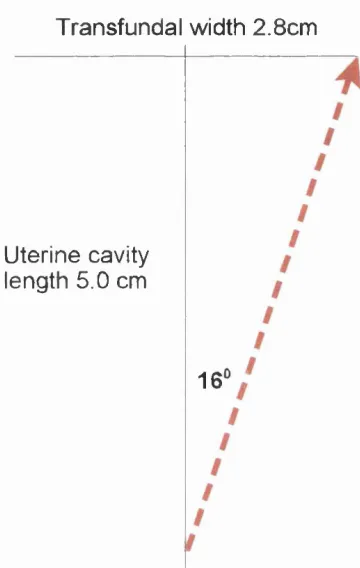
Figure 3.18 Using the ultrasound study the angle of deflection that would be
required to approach the uterine cornu is 16°.
Figure 3.19 Creation of polymer polytetrafluoroethylene (PTFE) from
tetrafluroethylene.
Figure 3.20 Demonstration of the non-stick and non-wettability properties of
PTFE which is used in medicine as an inert implant.
Figure 3.21 The medical uses of PTFE.
Chapter 4
Figure 4.1 The original design of the tubal screw developed by Adam
Magos (British Patent Application no.980634.7)
Figure 4.2 The hysteroscopic tubal screw.
Figure 4.3 A reverse screw thread scored into base of the tubal screw,
which is mounted on the applicator with a screw thread.
Figure 4.4 Hexagonal PTFE mounting on applicator and corresponding
recessed area in base of tubal screw.
Figure 4.5 Tubal screw with titanium bayonet mounted on coil applicator.
Figure 4.6 Diagrammatic representation of device in applicator showing
bayonet pin locking mechanism
Figure 4.7 Demonstration of the field of view with the 15° and 70° lens
optical hysteroscopes, however with the natural tendency to
‘point’ towards the area being treated the 30° hysteroscope was
favoured.
Figure 4.8 The original Rimmer Brothers modified single channel
hysteroscope.
Figure 4.9 The fixed deflector at the distal end of the Rimmer
hysteroscope.
Figure 4.10 Distal end of Rimmer hysteroscope with PTFE cannula and
tubal screw demonstrating fixed deflector.
Figure 4.11 Tubal screw mounted on distal end of PTFE cannula with push-
rod for disimpaction.
Figure 4.12 Original green PTFE cannula demonstrating water tight seal of
push-rod at proximal end to enable disimpaction
Portex Limited) after placement of the screw at the tubal ostia.
Figure 4.14 Demonstrating that a proximal small rotational torque when
applied to a curved cannula is amplified distally to describe a
large arc.
Figure 4.15 Scoring in outer sheath of hysteroscope.
Figure 4.16 The coil with rubber casing to prevent fluid leakage.
Figure 4.17 Demonstration of the lateral frictional force generated by the
natural elasticity of the application cannula.
Figure 4.18 Demonstrating the effect of using a coil mounting to hold the
tubal screw.
Figure 4.19 Standard Hamou Hysteromat.
Figure 4.20 Leakage of distending fluid around the oval diameter of the
hysteroscope as compared to the conventional hysteroscope.
Figure 4.21 Leakage of distending fluid around rubber seal to the operating
channel of the hysteroscope.
Figure 4.22 Distension fluid pours into the application cannula after the tubal
device is disimpacted and out of the operating channel of the
hysteroscope.
Figure 4.23 Litechnica Statflo Liquid infusion system.
Figure 4.24 The 3 litre pressure bag - the preferred method of uterine
distension. VBM Medizintechnik GMBH, Germany.
Figure 4.25 The use of two separate systems to effect deflection towards the
tubal ostia. Operating hysteroscope not shown for clarity.
Figure 4.27 The technique of altering the angle of deflection with the
‘trombone outer sheath’.
Figure 4.28. The ‘trombone’ outer sheath.
Figure 4.29 The ‘trombone’ outer sheath overlying the hysteroscope.
Figure 4.30 Tubal screw mounted on cannula, which adopts a
predetermined angle of deflection once extruded from the
hysteroscope.
Figure 4.31 An active method to alter deflection with a proximal ‘joy-stick’.
Figure 4.32 A passive method to alter deflection with a proximal ‘joy-stick’.
Figure 4.33 The extra deflecting bridge attachment on the cystoscope with
tubal screw mounted on coiled applicator with rubber outer
coating.
Figure 4.34 Application of the tubal screw to the tubal ostia.
Figure 4.35 The bayonet mounted tubal screw prior to disimpaction after
being screwed into place at the tubal ostia.
Figure 4.36 Bilaterally placed tubal screws demonstrating the bayonet
mounting of the tubal screws.
Figure 4.37 The tubal screw with bayonet mounting enabling easy insertion
and disimpaction.
Figure 4.38 Developmental changes in the equipment in chronological order.
Figure 4.39 The development of the hysteroscopic plug sterilisation
Chapter 5
Figure 5.1 The uterus fixed on board and suture attached to tubal screw.
Figure 5.2 The applicator to screw tubal screw in place.
Figure 5.3 The applicator with tubal screw mounted prior to performing
in-vitro study.
Figure 5.4 The dissecting board and jig system used in the in-vitro weights
study
Figure 5.5 Weights attached to the plug suspended by the jig.
Figure 5.6 The weight required to dislodge the tubal screw from the uterine
cornu after in-vitro application.
Figure 5.7 The force required to dislodge the tubal screw from the uterine
cornu after in-vitro application. (The gravitational force acting
upon the weights applied to the screw is assumed to be 10 m/s^
downward force).
Chapter 6
Figure 6.1 A prototype pointed tubal screw approximated to the tubal ostia
prior to insertion.
Figure 6.2 The push-rod to disimpact the tubal screw.
Figure 6.3 The push-rod to disimpact the tubal screw close-up.
Figure 6.4 Tubal screw in-situ at the uterotubal junction. Note presence of
Patient 1 in the histological assessment study.
Figure 6.5 Tubal screw at uterine cornu after serial sections have been
performed. Patient 1 in the histological assessment study.
Figure 6.6 Uterine cornu incised and tubal screw transected to confirm
correct placement by the methylene blue dye and subsequently
by histology (with 1cm scale).
Figure 6.7 Patient 2 in the histological assessment study with uterine cornu
bisected after screw placement.
Figure 6.8 Patient 2 with close-up of uterine cornu.
Figure 6.9 Patient 3 in histological assessment study after bisection of the
uterus showing tubal screw at uterine cornu.
Figure 6.10 Preparing to perform the weights study on an in-vivo applied
tubal screw in the operating theatre.
Figure 6.11 The purpose built weights and their support for the in-vitro and
in-vivo weights studies.
Figure 6.12 Graph demonstrating the force required to dislodge the in-vivo
hysteroscopically applied tubal screws.
Chapter 7
Figure 7.1 Flow chart showing the changes in the equipment and
technique of hysteroscopic screw sterilisation.
screw viewed on ultrasound scan 3 months post insertion.
Figure 7.3 Ultrasound appearance of a hysteroscopically placed tubal
screw viewed on ultrasound scan 3 months post insertion.
Figure 7.4 Further ultrasound scan demonstrating a single tubal screw
Figure 7.5 Further ultrasound scan demonstrating bilaterally placed tubal
screws
Figure 7.6 3-D ultrasonography demonstrating the presence of a tubal
screw at the uterine cornu. (Patient number 1)
Figure 7.7 3-D ultrasound scan demonstrating bilateral placement of tubal
screws.
Figure 7.8 Life table plot of right tubal screw retention (cases 1-20).
Figure 7.9 Life table plot of left tubal screw retention, (cases 1-20)
Figure 7.10 Life table plot of retention of all tubal screws (cases 1-20).
Figure 7.11 Life table plot of right tubal screw retention (cases 21-35).
Figure 7.12 Life table plot of left tubal screw retention (cases 21-35).
Figure 7.13 Life table plot of retention of all tubal screws (cases 21-35)
Figure 7.14 Log rank test to compare the life table plots between the initial
Chapter 1
1.1. Introduction
For well over one hundred years physicians have struggled to effect a surgical
technique whereby they render a woman sterile (Blundell, 1823, cited by
Bordahl 1985). The driving force behind the technique employed was that it
was to be simple and rapid, it was only many years later that concerns of
safety in the choice of technique were really addressed (Phillips et al., 1977, Chamberlain and Brown, 1978). Now at the beginning of the century the
commonest method of sterilisation is a laparoscopic technique performed
under a general anaesthetic.
1.2. History of Tubal Sterilisation
The current technique of occluding the fallopian tubes has its origins in the
early 19**^ century. Ligation of the fallopian tubes was first discussed in 1827,
and then subsequently in 1829, by Blundell. Although he did not mention
whether he actually performed the procedure it would seem likely (Blundell,
1828 and 1829). He described his technique thus;
"draw up the fallopian tube, which is easily done, first on one side, then the
other, cutting out a portion of it, so as to render it impervious, by which the
woman would for ever afterwards become sterile".
From then on various doctors advocated sutures around the fallopian tube,
and simple technique of tubal sterilisation. Reasons for sterilisation were the
risk of repeated caesarean section, severe chronic illness and occasionally
mental sub-normality (Wilson 1995). The procedure was often performed at
the time of caesarean section (Irving 1924).
Although sterilisation was still practised infrequently, the drive for a rapid
irreversible method of contraception continued for sinister purposes. From
1907 onwards various US states adopted legislation to practice eugenic
sterilisation for people with hereditary diseases (Skajaa, 1932), as indeed was
practised by the Nazis from 1933-45. Evidence of this is shown in this extract
from the proceedings of the Annual Meeting of the Eugenics Association in
New York in 1929:
‘the sterilization of subnormal women to protect racial integrity is regarded
much more important by eugenicists than sterilization of the male’
(DeVilbiss, 1935).
Throughout the early part of the 20*'^ century various eponymous techniques
to effect surgical ligation for sterilisation were developed. The most widely
known being ligation-and-crushing, Madlener technique (Madlener, 1919), the
Irving technique (Irving, 1924) where the tubes are divided and proximal
stump buried in myometrium, and the Pomeroy technique (Bishop and Nelms,
1930), not reported until after his death.
fimbrial ends of the tubes in the broad ligament (Aldridge, 1934), cornual
resection (Overstreet, 1964), fimbriectomy (Kroener, 1969) and the Uchida
procedure, postpartum fimbriectomy (Uchida, 1975).
Baird introduced sterilisation as a technique of family planning in Aberdeen in
the 1930s. However they were criticised for their liberal use and hence a
woman had to be near 40 years and have more than eight children (Baird,
1965). The first reported case of puerperal sterilisation was in 1939, the Oslo
method’ (Adair and Brown 1939), which led to an increase in the practice of
sterilisation elsewhere.
The use of electrosurgery to effect laparoscopic tubal sterilisation was first
described by Boesch (Boesch et al., 1936) and in 1941 by Power and Barnes using an early laparoscope under local anaesthetic. With the advent of the
newer laparoscopes electrocautery became more frequently used initially
using high frequency monopolar electrodiathermy but this was superseded by
the safer use of bipolar electrodiathermy due to the occurrence of accidental
visceral thermal injury with monopolar surgery (Peterson at a!., 1983a). After the early laparoscopes of Power and Barnes, Raul Palmer was a pioneer in
the development of more advanced laparoscopic techniques and
instrumentation (Palmer, 1946; Palmer, 1947). These newer laparoscopes
used improved endoscopic lenses and advances in the technology of cold
light transmission. Palmer also introduced the Trendelenberg position along
with safer methods of peritoneal insufflation and monitoring, to provide a view
performed monopolar electrocoagulation and transection of the fallopian
tubes aided by his biopsy drill forceps bearing his name. Steptoe was another
pioneer in laparoscopic surgery publishing the first English language textbook
in the field containing a report of his experience of electrocoagulation of the
fallopian tubes. (Steptoe, 1967).
A modification of the Madlener technique to convert a laparotomy approach to
a less invasive laparoscopic procedure was developed by Fragenheim in
Germany. This technique used a prolene suture to ligate the fallopian tubes,
but this procedure required a high degree of endoscopic expertise and skill to
manipulate the instruments (Fragenheim and Kleindienst, 1974; Wortman,
1976). Fragenheim, who had learnt his laparoscopic techniques from Raul
Palmer, went on to develop many laparoscopic instruments and made one of
the first protoype carbon dioxide insufflators. A milestone in the development
of gynaecological laparoscopy was the introduction at the end of the 1950s of
electrocoagulation for tubal sterilisation by Palmer in Paris and Frangenheim
in Konstanz each working independently. The technique of laparoscopic
fulguration of the fallopian tubes involved either inserting the monopolar
instrument either down a special channel in the laparoscope or via a
secondary entry site after insufflation with 2-4 litres of gas. The fallopian tube
was then picked up and either coagulated or transected using a cutting
current. A coagulation current causes cellular dehydration and charring
without division whilst the intense heat of cutting current causes the tube to
divide. Both forms of current are potentially dangerous as they may produce
of using monopolar current Frangenheim developed bipolar
electrocoagulation for sterilisation and for controlling bleeding in 1972. The
benefit of bipolar diathermy is that the current passes only between the
blades of the instrument and does not pass through the patient to a return
plate electrode thus minimising sparking. This technique was demonstrated to
be as effective as transection of the fallopian tube using monopolar current.
Indeed Yuzpe encountered no pregnancies in 335 women followed-up for up
to 10 months (Yuzpe, 1974).
A safer method of laparoscopic sterilisation by coagulation and transection of
the fallopian tube using monopolar electrodiathermy was developed by Semm
and Steptoe (Steptoe, 1976 cited by Wortman, 1976) by using a fulguration
instrument, which is heated from the inside using a low voltage and low
temperature technique. With this technique there was less risk of thermal
injury to surrounding structures, and indeed the remainder of the fallopian
tube by thermal spread, leaving the possibility of reversal an option by
surgical reanastomosis of the remaining ends.
The Pomeroy method of interval tubal sterilisation by laparotomy, mini
laparotomy, culdoscopy or colpotomy was the favoured technique to effect
sterilisation at this time (Wortman, 1976). However Clark (Clark, 1972),
Loeffler (Loeffler, 1974), Greene (Greene, 1974) and Alexander (Alexander,
1975) described Pomeroy ligation via laparoscopy whereby the fallopian tube
was either tied within the abdomen or brought out through the abdominal
consequently the approach of mini-laparotomy persisted (Wortman, 1976)
Clips or rings have also been used to effect tubal closure via laparoscopy.
The clips that were used initially were made of tantalum, a non-tissue reactive
metal. These clips were previously used to provide haemostasis at open
surgery. Failure to cause tubal occlusion was as high as 27% (Davidson and
Donald, 1972; Wheeless, 1974). The first reported purpose built clip was the
Hulka clip (Hulka et al., 1973), the Falope ring was reported by Yoon at a!., in 1974 and the Filshie clip was introduced in 1975 and is probably the most
widely used clip today (Wilson, 1995).
Until the advent of laparoscopy and culdoscopy the main route to approach
the fallopian tube had been via a mini-laparotomy incision 2-3 cm above the
symphysis pubis (Osathanondh at a!., 1974). Culdoscopy was abandoned as the favoured route for sterilisation due to the high incidence of failures and
due to an unacceptably high complication rate (WHO 1982).
1.3. Complications of Tubal Sterilisation
The advent of laparoscopic surgery to gynaecology, as in other surgical
specialities, has brought some undisputed benefits to patient care. It is
claimed that advantages over laparotomy include reduced postoperative
discomfort, shorter hospitalisation, faster recovery all leading to savings in
treatment cost, and of course a better cosmetic result. This is undoubtedly
indications such as tubal pregnancy in gynaecology and appendicectomy in
general surgery (Vermesh et al., 1989; Kum et al., 1993; Nagele et al., 1996; Golub et al., 1998). This also follows for a technique as simple and widespread as sterilisation. However, laparoscopic management can also be
associated with potential disadvantages. It is well documented that
endoscopic procedures can have their unique complications rarely seen at
laparotomy which can be life threatening. Examples include vascular or bowel
trauma from the insertion of trocars; (Bassil et al., 1993; Nordestgaard et al., 1995; Soderstrom, 1993; Soderstrom, 1997) and electrosurgical and laser
burns to the gastro-intestinal tract; (Tucker, 1995; Harkki-Siren and Kurki,
1997). The complication rate for the procedure of laparoscopic sterilisation is
up to 1.8% (DeStefano et al., 1983).
Although these complication rates are very low it is important to understand
how frequently laparoscopic sterilisation is performed. There are in excess of
50,000 laparoscopic tubal sterilisations performed each year in the UK
(hospital in-patient episode statistics 1997), approximately 1 million annually
in the United States of America (Tulandi, 1997), and enormous numbers are
performed each year in developing countries. Indeed worldwide there were
predicted to have been 159 million sterilisations performed in the 1990s (Ross
1992).
Complications in laparoscopic surgery arise with the insertion of the Verres
needle, insertion of the trocar and complications related to the operation itself.
25,764 laparoscopies performed in 72 hospitals in Holland (Jansen et al., 1997). The commonest complication was laceration of the inferior epigastric
vein (0.15%), gastro-intestinal injury (0.11%) and intra-abdominal vascular
injury (0.11%). Most injuries occurred during the laparoscopic approach,
rather than the procedure proper. When the rates were calculated by type of
procedure the rate for diagnostic laparoscopy was 0.27%, for sterilisation
0.45%, and for operative laparoscopy 1.8%. A recent report detailing the
different complications rates according to method of laparoscopic sterilisation
was produced by the United States Collaborative Review of Sterilization which
demonstrated that the spring clip method had the lowest complication rate,
0.47 per 100 procedures. This rate was not significantly lower than the
complication rates of other procedures although in adjusted analysis, diabetes
mellitus, general anaesthesia, previous abdominal or pelvic surgery and
obesity were all independent predictors of complications (odds ratio 4.5, 3.2,
2.0 and 1.7 respectively) (Jameison et a!., 2000).
There have been many large scale reviews of the mortality rate for
laparoscopic sterilisation, although many relate to the now out moded practice
of monopolar diathermy to the fallopian tubes. The mortality rate of tubal
sterilisation is approximately 1.5 per 100,000 procedures as reported by the
Centre for Disease Control in the USA (Escobedo et a!., 1989). However a more recent survey of 22,966 sterilisations performed in the United States
reported one death, deriving a mortality rate of 4 per 100,000 procedures, but
only 13% of gynaecological surgeons surveyed replied to the questionnaire
sterilisations performed (Hulka etal., 1990).
Reports in the literature detail fatality rates of up to 10.2 per 100,000
sterilisations performed (Chamberlain and Brown, 1978). These initial reports
of the Royal College of Obstetricians and Gynaecologists and the
Collaborative Review of Sterilization in the United States and the International
Project / Association for Voluntary Sterilization attributed the high mortality
rate to the use of monopolar diathermy to transect fallopian tubes, a practice
now rarely used and to the use of general anaesthesia (Chamberlain and
Brown, 1978; Aubert et al., 1980; Peterson et al., 1982; Peterson et al., 1983a; Peterson et al., 1983b). However the death rates can be much greater in developing countries due to their use of local anaesthetic with sedation. In
one series of 170,000 sterilisations performed over a two-year period, the
death rate was 99 per 100,000 procedures (0.1%), 66 times as great as in the
developed world (Rosenberg et al., 1982). In the largest series of camp sterilisation deaths Bhatt (1991) reported a mortality rate of 0.02% for 3
million laparoscopic sterilisations performed over three years. The mortality
rate was 0.05% for those gynaecologists with less than six months experience
and was greatest in higher parity women. However the results were markedly
better with a very experienced single operator, being as low as 4.8 per
100,000 (Mehta et ai, 1989)
1.4. Failure Rates of Tubal Sterilisation
sterilisation it is important to report the rate of failure of sterilisation and the
subsequent ectopic pregnancy rate if the woman did conceive. The
cumulative probability of conceiving 10 years after sterilisation is 1.34% and
one third of these failures are ectopic pregnancies (US Collaborative Review
of Sterilization, CREST) (Peterson et al., 1996). In this report the failure rate was greatest with laparoscopic spring clip application and least after
monopolar tubal coagulation. A subsequent analysis showed that the rate of
ectopic pregnancy was greatest in women sterilised when less than thirty
years of age by bipolar cautery of the fallopian tubes (Peterson at a!., 1997). However the results of laparoscopic sterilisation may be better if performed by
experienced operators, 1.7 per 1000 for tubal ring and Filshie clip application
at one year using life-table analysis (Sokal at a!., 2000). The Royal College Working Party on Sterilisation quote a failure rate for Filshie clip sterilisation
of 1 in 200 procedures, as described later in this chapter (RCOG, 1999).
Other reported failure rates of tubal sterilisation record the failure rate of tubal
sterilisation of approximately one in 100 procedures at between two and eight
years; Mumford and Bhiwandiwala (1981) with the majority of procedures
performed by monopolar diathermy, Aranda at al, (1985) using the Rocket clip and Koetsawang at a/., (1990) who used electrocoagulation, the Hulka clip and the tubal ring to effect sterilisation.
Another important factor in the management of a woman undergoing
sterilisation is the incidence of sterilisation regret. It is known that women
under 30 are most likely to return requesting reversal of sterilisation (Winston
the incidence was found to be between 1.5% and 15% (Schwyart and Kutner,
1973). In this study the highest dissatisfaction occurred in women with less
than four children and in those women sterilised at the time of termination of
pregnancy. The CREST group reported the cumulative incidence of
sterilisation regret in a cohort of women followed-up for five years after their
sterilisation as 7.0%. They also recorded the incidence of regret in a group of
women whose husbands had undergone a vasectomy as 6.1% (Jamieson et al., 2002).
The chance of a successful sterilisation reversal depends upon the length of
the remaining fallopian tube, the location of the anastomosis and the
experience of the surgeon (Siegler et a!., 1985). All methods of tubal sterilisation cause damage to the fallopian tube, either due to excision of a
short part of its length, thermal damage or by crushing and pressure necrosis.
Hence up to about 40% of patients may have their request for sterilisation
reversal denied as their fallopian tubes are too damaged (Antoine et a!., 1983). Therefore a technique that potentially causes minimal damage to the
fallopian tube, which could in theory be reversible would appear to be very
desirable. Several procedures have been described which involved either
carefully dividing the fallopian tube, with a view to a potential later
anastomosis, or buried the ovary or fallopian tube within a peritoneal fold
1.5. Guidelines of Royal College of Obstetricians and Gynaecologists for
female sterilisation
In 1999, the Royal College of Obstetricians and Gynaecologists
(RCOG) published evidence-based guidelines for female sterilisation
developed from a multidisciplinary body and after peer review.
1.5.1. Indication and timing of sterilisation.
There are no absolute contraindications to sterilisation providing the patient is
able to give adequate consent. Caution is advised for sterilisation at the time
of Caesarean section, in association with abortion, in nulliparous women and
women under 25 years of age in view of an increased incidence of
sterilisation failure and post sterilisation regret. Sterilisation can be performed
at any stage of the menstrual cycle providing the woman continues
contraceptive precautions until the next period. A pregnancy test is imperative
if a woman believes she has missed a period. All women should be offered
comprehensive information as to all contraceptive choices available including
the use of freely available, impartial and easily understood information. The
latter is very important, as in a UK study in 65% of patients had not received
any written information regarding their sterilisation (Walsh etal., 1998).
1.5.2. Information
reversible methods of contraception, as some methods are as successful as
sterilisation at preventing conception. For instance, the TCu380A intrauterine
device, which has a 12-year cumulative pregnancy rate of 1.9/100 women
treated (Anonymous, 1997). This is similar to the success of laparoscopic
sterilisation (Peterson et al., 1996). It is imperative that couples are aware that male sterilisation is a local anaesthetic, outpatient procedure, which is
very successful at preventing conception and is safer. The failure rate of
vasectomy is approximately 1 in 1,000 in the first year (RCOG, 1999). The
surgeon should ensure that the medical records include a full gynaecological
and medical history, and the examination findings are recorded, along with
the preoperative counselling for sterilisation:
Out-patient consultation and counselling:
Parity and any complex obstetric history
Gynaecological history and current symptoms
Pelvic examination
Discussion of long-term contraceptive alternatives to tubal occlusion
Expected method of access to tubes and method of occlusion (and reason for
method of occlusion if not mechanical)
Risk of extended procedure, if non-life-threatening problems occur
Extent of consent to alternative methods of tubal occlusion if first intended
method not possible
Failure rate 1 in 200
Irreversibility, potential for reversibility with expected method and availability
of reversal locally on NHS
Information leaflet given
Immediate pre-procedure;
Date of last menstrual period
Pregnancy test result if performed
Confirm outpatient details and other preoperative discussions
Confirm valid consent form with the patient’s name, name of doctor obtaining
consent - to be countersigned by surgeon performing the procedure
(Fitness for day case surgery)
Operation notes:
Name of operating surgeon(s) including surgeon present in theatre taking
overall responsibility
Ease of access to the tubes
Clarity of identification of the tubes
Accurate placement of occlusive method
Post-procedure:
Method actually used
Discharge letter to GP
Patient informed of method used and any intra-operative findings / events
Whether further contraception advised eg. up to next period, or pending result
of tubal patency test.
Royal College of Obstetricians and Gynaecologists
Evidence Based Guidelines on Female Sterilisation, 1999
Prior to undergoing sterilisation of the female partner the surgeon should
establish the date of the last menstrual period to ensure she is not pregnant,
and perform a pregnancy test if indicated. It is imperative that the operating
surgeon ensures that the patient has been appropriately counselled, is aware
of the risks of the surgery and the failure rate of the procedure. The patient
should be warned that the risk of a laparotomy due to visceral injury is 1.9 per
1,000 and the mortality rate is 1 in 12,000 as described previously
(Chamberlain and Brown, 1978; Jansen et al 1997). The woman should be informed about the method of sterilisation performed prior to discharge, any
surgical difficulties encountered and if there was any difficulty in clip
application or if an extra clip was required. If there is any doubt as to the
success of the sterilisation then the woman should be informed
postoperatively, and advised to continue contraceptive precautions until a
patient should be warned prior to discharge to attend the emergency
department or her General Practitioner should she suffer abdominal pain or
begins to feel unwell due to the potential late presentation of bowel injury.
The woman should be informed both preoperatively and again prior to
discharge that the risk of sterilisation failure is 1 in 200 women overall lifetime
risk (RCOG, 1999). This figure is based on the CREST study (Peterson et al., 1996). This prospective study of 10,685 women, described earlier, derived a
ten-year life table probability of failure of 16.6 per 1,000 procedures. This
failure rate is substantially greater than the failure rates previously reported of
3-6 per 1,000 at up to one year (RCOG, 1999). After multivariate analysis the
spring clip (Hulka clip), bipolar coagulation, decreasing age and particularly
women less than 28 years of age were all associated with significantly
increased chances of sterilisation failure. However the Filshie clip was not
available in the USA at the time of the CREST study. The best available
evidence suggests that it has a failure rate of 2-3 per 1,000 at two years
(personal communication. Professor John Guillebaud). The RCOG guidelines
extrapolate this figure using the CREST model to derive a lifetime risk of
sterilisation failure of 1 in 200 for Filshie clip application and the chance of
failure does not diminish with time. With regard to the risk of an ectopic
pregnancy post laparoscopic sterilisation the RCOG Guidelines (1999)
suggest that women should be warned that the risk of an ectopic gestation is
greater if tubal diathermy has been used and less likely if mechanical
occlusion or tubal ligation has been performed. Whilst women who have been
women, if they conceive their chance of the pregnancy being ectopic is up to
nine times greater than a woman who has not been sterilised (RCOG, 1999).
The RCOG review provides reassurance that for women over thirty years of
age, tubal occlusion does not cause a significant change to the menstrual
cycle. All women should be informed that it is possible to reverse the
sterilisation if required, although it should be stressed that sterilisation is
intended to be permanent.
The guidelines state that culdoscopy should not be used as an approach to
sterilisation, and that laparoscopic mechanical occlusion of the fallopian tubes
by either clips or rings as a day case procedure should be is the method of
choice in the UK. The guidelines state that ‘transcervical application of
chemicals, adhesives or synthetic plugs are still under evaluation and have
not been considered for these recommendations on methods of tubal
occlusion’.
With regard to analgesia the RCOG guidelines state that it is probable that
pain after surgery is greatest after ring application, with diathermy being the
least painful and with clip application being intermediate. Of benefit at
alleviating pain is; instillation of a local anaesthetic agent over the fallopian
tubes, into the mesosalpinx or on the Filshie clip. With regard to sterilisation
under local anaesthesia, benefits were noted with regard to postoperative
pain and earlier return to normal activities post procedure. It is cheaper,
anaesthetic sterilisation reported that 91% of patients would recommend the
procedure to a friend (Mackenzie et al., 1987).
1.5.3. Training in sterilisation
Due to the increasing incidence of litigation against gynaecologists the RCOG
recommends that the operator should be properly trained and that the
operation should be carried out technically correctly. The Guidelines
recommend that a trainee should not perform the laparoscopic sterilisation
until they have successfully completed 25 supervised cases and are able to
perform a laparotomy unsupervised. It is also advised that trainees learn an
alternative method of sterilisation in case it is impossible to apply an occlusive
clip at the time of the operation. It is also imperative that sterilisation only
takes place in site with properly maintained equipment where there is
equipment available to perform a laparotomy if necessary. Regarding audit
the working group of the RCOG advocate establishing a national database of
failed sterilisations to provide accurate failure rates for counselling patients,
and it would also identify an area of substandard practice at an early stage.
1.5.4. Areas for more research
The working party recommends performing long-term studies of the failure
rate, risk of ectopic pregnancy and the effect on the menstrual cycle of all
sterilisation techniques, particularly Filshie clip sterilisation. A study, of women
cycle and risk of hysterectomy should be performed. Interestingly, the RCOG
working party made no suggestion that there should be research into the
development of a hysteroscopic method of sterilisation.
1.5.5. Compliance of the study with the RCOG guidelines
These guidelines were published after the completion of our study of a novel
method of hysteroscopic sterilisation. Despite this fact the preoperative
counselling was as described in the Guidelines, however the failure rate of
laparoscopic sterilisation quoted to our patients was that the failure rate of the
procedure over the long-term was unknown but that it could be as high as one
failure in 100 patients. This figure was derived as Vessey et al., reported a failure rate of one per 100 procedures over a seven year period after ring
sterilisation. When the CREST study was reported we were obliged to inform
our patients that the failure rate of laparoscopic sterilisation maybe higher
than the quoted figure of 1% over seven years previously quoted (Peterson et a!., 1997). All other facets of the pre-procedure counselling were as recommended in the Guidelines of the working party, including providing the
patient the current information leaflet from the Family Planning Association to
read prior to their sterilisation.
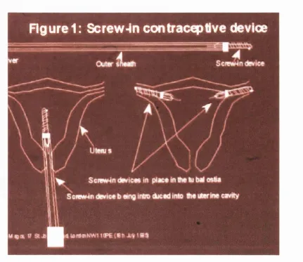
1.6. The Need for a Simple, Outpatient Method of Sterilisation
In view of the complications of laparoscopy it would appear that the ideal
outpatient-setting, avoiding abdominal instrumentation and general
anaesthesia. If this technique were highly effective and potentially reversible it
would become the ’gold-standard’ method of female sterilisation. With these
aims in mind we developed a tubal screw’, which would be applied
hysteroscopically into the uterine ostia and would be immediately effective
A
Handle
Thread Head
Side view
View from "A"
Ad an Laszloiy/bgcB.17 St J (tin s Road, lo n c t n N W 1 1 OPE (1 8 th J ü y 1 9 Ë 5 )
Rgurel: Screw-In contracepllve device
■ B K B ^ ^ H
1
Figure 1.2 The intratuba I screw mounted on applicator and applied to
Figure 4; Outer applicator
Side view
View from "A" * d j n 1 ~ C t J c h n s R o it ! L o n t t n f A M ) l ^)PE > W t n ,1 d y 1 9 5 . '
Figure 1.3 The intratuba! screw mounted on applicator (from original
e
- n '
A - B = 15mm
A - C = 3mm
Chapter 2
2.1. Introduction
Due to the risks associated with general anaesthesia and the potential risks of
visceral trauma on instrumentation of the abdomen, a permanent method of
female contraception avoiding these pitfalls has been sought for many years.
However the initial attempts at sterilisation were via a uterine approach as
opposed to being an abdominal procedure.
2.2. Initial intra uterine techniques
Proximal obstructions of the fallopian tubes were first recanalised by Smith in
1849. This was performed using a whalebone guide passed through a silver
directing tube, which due to the shape of the uterus he thought ‘would always
pass to the ostia’ to treat cornual block.
At around the same time attempts to cause obliteration of the uterine cornu
were made by Frioriep (1849). This technique relied on infusing silver nitrate
into the uterine cavity towards the cornua via a cannula. Electrocautery was
employed by Kocks in 1878, to cause thermal injury and then potentially
destruction of the endometrium to prevent implantation of an embryo.
Electrocautery of the tubal cornua remained unchanged over the following
years apart from improvements in the controlled delivery of the electric
current to cause tissue damage (Risquez and Confine, 1993). Further reports
Dickinson (1916), as an introduction to his new technique, provides an
interesting insight into the background and requirements for a technique of
sterilisation almost 100 years ago.
‘Section of the vas deferens, though simple, is not accepted by the
male...’
‘Ovarian shrinkage by X-ray is neither sure nor its duration determined.
Chemical slough stricture inside the upper uterine angles as advised
by Froriep in 1850 was given up ’
‘..various operations on the fallopian tubes involve opening the
abdominal cavity....which is never justifiable for this purpose....’
‘The only outlook for a simple and sure method, and that without risk or
loss of time and with but little pain, seems through closure of the tube,
where it enters the uterus...’
Dickinson 1916
It would appear that we are still striving to achieve this ideal almost 90 years
later. The technique reported by Dickinson was to apply local anaesthetic into
the uterine cavity and to the cervix. Then cauterisation of the cervix was
performed. Note was taken of the cervix to see how long scarring and slough
took to form. Then with touch only he attempted to find the tubal ostia and
repeated this cautery for the same duration of time, using a semi-flexible loop
of wire, at the cornua of the uterus. Confirmation of tubal occlusion was
technique are reported although he reported that he had performed 40
procedures between 1916 and 1950 without complication (Dickinson and
Gamble, 1950, cited by Hulka and Omran, 1972). At this time Prudnikoff was
also performing electrocoagulation of the uterine cornu (1912).
DeVilbiss reported her results in 1935 using a similar technique to Dickinson
employing a semi-flexible uterine electrode placed by touch at the uterine
cornua. A 3.5 amp current was applied for 15 seconds. Tubal patency was
assessed using the Rubin’s test, whereby if the uterus holds an air pressure
of 150-200 mm Hg for up to 5 seconds the fallopian tubes are presumed
blocked. Of 30 patients treated 17 cases were reported with bilateral blocked
fallopian tubes, however there were 3 conceptions in this group. She
described that the treatment is contraindicated in un-treated syphilis due to
the liability to haemorrhage. Two case histories are described below which
appear to demonstrate that sterilisation was used as a method of eugenics at
this time:
“Case 14.-White paralytic moron, husband when drunk kicked her
about. Reported blocked tubes returned to the clinic pregnant, with
signs of an impending abortion....staff surgeons recommended a
Case 7.-Colored syphilitic moron, aged twenty-four...5 pregnancies, 4
living children, 1 dead...reported four months pregnant.”
DeVilbiss 1935
2.3. The advent of hysteroscopy
The first attempt at visualisation of an abdominal organ was by Bozzini in
1806. This was performed by illumination of the urethra with a candle.
However the future of endoscopy was jeopardised as the Medical Faculty of
Vienna reprimanded Bozzini for his undue curiosity. After this, procedures
were performed by touch rather than with visual guidance until hysteroscopy
was developed in 1869 as a diagnostic tool for intrauterine disease
(Pantaleoni, 1869, cited by Silander 1962). Illumination was by an external
kerosene lamp.
The first report of ‘out-patient’ hysteroscopy was described in 1908 by David
who wrote his thesis on the uses, dangers and contraindications of
hysteroscopy, although he did not use this approach to perform sterilisations
(David, 1908 cited by Rubin 1925). With development of hysteroscopy for the
purpose of cornual electrocoagulation by Rubin in 1925 the possibility of
effecting sterilisation by an intra-uterine technique became increasingly a
realistic objective. He described the visualisation of the uterotubal ostia with
carbon dioxide gas (CO2) and water and suggested the possibility of
at its distal end.
To reduce the problems, noted by previous hysteroscopists, of poor uterine
distension with the uterine wall being too near the objective lens and profuse
bleeding interfering with the view of the uterine cavity, Silander in 1962
devised a technique of uterine distension and tamponade. His hysteroscope
had at its distal end a balloon, which inflated to distend the uterine cavity to
improve visualisation of the endometrium (Silander, 1962).
2.4. Sterilisation by tubal electrocoagulation
Hyams reported his success of cornual electrocoagulation at almost 100%.
The procedure was reportedly performed without anaesthesia and perforation
of the uterus was thought to be impossible (Hyams, 1934). This technique
consisted of blind introduction of an insulated probe to find the uterine
cornua. Then a trocar was advanced enabling an electrode to protrude and a
current of 200mA was activated for 5 seconds. No anaesthetic was used for
this procedure. After one month tubal obliteration was confirmed by
performing a hysterosalpingogram. Later the technique was changed to use
X-ray screening to aid visualisation of the uterine cornua. Iodine dye was
initially injected into the uterus, and then the cauterisation would be
performed under X-ray screening (Hyams, 1934).
blind technique as described above, as 40% of fallopian tubes were patent
after the procedure. Schroeder in 1934 described his technique of sterilisation
of the intramural portion of the fallopian tubes by electrocoagulation under
direct vision in 2 patients. Unfortunately follow-up hysterosalpingograms
demonstrated no success. The warmth of the uterus felt through the
abdomen determined the intensity and duration of the current required for
coagulation. Although it appeared that these techniques were using women in
experiments, there were attempts made to perform in-vitro testing on
extirpated uteri; for instance, Mickulicz-Radecki and Freund in 1927 used an
instrument they called a 'curettoscope'. In their opinion to effect sterilisation
they needed to destroy the endosalpinx and part of the myometrium to stop
the fallopian tube recanalising. However this degree of thermal damage
although, undoubtedly causing severe tubal damage in the in-vivo situation,
would potentially lead to severe local visceral thermal injury. If this occurred to
the bowel, bladder or ureter then fistulae and peritonitis would ensue.
The most reported method to effect sterilisation via an intrauterine approach
at this time was using a thermal modality. Norment and Greensboro in 1956
described their hysteroscope using water as the distension medium and
reported 100 cases of hysteroscopy. They attempted sterilisation by cornual
fulguration but described too few to cases to report their results (Norment and
Greensboro, 1956). Using the Hyams technique, Sheares in 1958 described
the results of tubal electrocautery. In 48 patients where an attempt at
due to peritonitis caused by a bowel injury. He described the new technique
whereby he listened for sizzling and crackling over the abdomen with a
stethoscope for 15 seconds. At this point it was felt that sufficient thermal
injury had occurred to cause sterilisation (Sheares, 1958). The fallopian tubes
were tested for occlusion using a CO2 manometer, the Rubin’s test.
Other authors who reported attempts at tubal electrocoagulation were Bowers
in 1938 and Porter etal. (1957, cited by Quinones et ai, 1973). Yasui in 1952 described 299 patients mostly after termination of pregnancy, using a
transcervical procedure, and reported an 80% bilateral occlusion rate. There
were 13 pregnancies but no complications. Effective sterilisation was thought
to have occurred when a 'vibrational sound' was heard after 20-30 seconds of
treatment.
The technique of cornual coagulation under either hysteroscopic or X-ray
monitoring was popular in Japan at this time, although success rates were
poor (Hayashi, 1972). New electrodes were developed to overcome the curve
required to pass to the tubal ostia (Ishikawa and Hayashi parabola
electrodes), rather than the straight cone electrodes developed initially by
Hyams in 1934. Hayashi reported that using the Hyams’ cones the pregnancy
rate in his treated patients was 83%, after refinement by using a round
electrode the pregnancy rate was 39% and using a curved electrode the
pregnancy rate was 5%. The temperature that was required was 110-120°C
Pasricha in 1968 described her blind detection of the uterine cornua and
cautery for up to 20 seconds in 89 patients. Initially she followed Dr. Hyams’
technique but due to the difficulties of working in a very busy hospital in New
Delhi she felt that she was able to accurately locate the cornua blindly. As
described by Sheares in 1958 she would listen with a stethoscope for sizzling
followed by crackling where upon sterilisation was presumed to have
occurred. A refinement that she described was to place a finger in the rectum
and fix the uterus in a retroverted position to aid the cautery procedure. Of 34
patients attending for a follow-up hysterosalpingogram a success rate of 47%
was initially reported. The procedure was repeated in those women who had
undergone a failed sterilisation and then overall success rate was reported as
76%. Unfortunately several patients suffered serious complications; salpingitis
in two patients, 1 patient suffered with profuse bleeding and underwent an
emergency hysterectomy, three patients suffered with peritonitis, one patient
had a bowel perforation and two patients required a laparotomy (Pasricha,
1968).
In a consensus statement after a hysteroscopic sterilisation workshop of 54
scientists from 9 countries Williams and Sciarra (1973) reported that:
1 The ostia are best visualised in the post menstrual phase
2.A uterine relaxant aids dilatation of the uterine cavity
3.Dextran is advantageous
5. A 45° or 60° hysteroscope is beneficial
B.Tubal cannulation was possible in all cases
y.Tubal blockage occurs only if the endosalpinx is completely
destroyed
S.Thirty watts for 3 or 4 seconds or 40 watts for 2 seconds is sufficient
for treatment
9.Side effects were minimal
10.Long term clinical evaluation of the sterilising effect is premature.
Williams and Sciarra, 1973
In their review of the literature Hulka and Omran, in 1972, reported on 603
patients who had been treated worldwide to date with an overall pregnancy
rate of 8.4%. All patients underwent a test of tubal patency and the rate of
success of successful occlusion at the initial attempt was 40-80%. Most
authors recommended a repeat procedure if the initial attempt did not cause
bilateral tubal occlusion. This series reported a total major complication rate
of 2.5% and a mortality rate of 0.1%.
With the development of fibre optical leads, the light source was held distant
to the patient and did not risk thermal injury. These new optics also provided
a better view of the endometrial cavity leading to a renewed interest in
hysteroscopy. However to effect thermal cautery to the uterine cornua using
electrocoagulation, which relies on the heating effect of a monopolar current,
percent dextran had been used in Sweden (Edstrom and Fernstrom, 1970)
and been found to be an acceptable solution for uterine distension. It is
immiscible with blood and highly viscous. Levine and Neuwirth validated this
method of hysteroscopy in 1972. They described excellent visualisation of the
tubal ostia and the cannulation of the ostia with a Teflon catheter and
injection of indigo carmine dye.
The following year using their technique of hysteroscopy with 30% dextran
and employing a specially developed serrated cautery tip they reported
treating the tubal ostia of 18 patients with a 25% failure rate (Neuwirth et al., 1973). They subsequently tested their technique in Thailand, in order to
evaluate the feasibility of a large-scale ambulatory program, (Richart et al. 1973). Their success rate was 84% bilateral tubal closure, 34 of the 44 also
had a tubal mesh or plug placed concurrently to the cauterised area of the
ostia.
With the development of CO2 hysteroscopy by Lindemann and Mohr in 1970
(Lindemann, 1974 and Lindemann and Mohr, 1976), the modality that Rubin
initially described in 1925 was revisited (Rubin 1925). They described their
adaptor which, by means of a vacuum attached to the cervix maintained a
pneumometra and their ‘Hysteroflator 1000', maintained a constant inflation
pressure. When they developed CO2 hysteroscopy they performed initial
studies on dogs to determine the safe limits of uterine insufflation. They