The Effect of a Squamous Cell Carcinoma Associated
p i Integrin Mutation on Cell Behavior
Richard D. Evans
University College London
And
Cancer Research UK
Cancer Research UK Supervisor: Dr. Fiona M Watt UCL Supervisor: Prof. Alan Hall
A thesis submitted in partial fulfillment of the requirements for the
degree of Doctor of Philosophy, University of London
All rights reserved
INFORMATION TO ALL USERS
The quality of this reproduction is dependent upon the quality of the copy submitted. In the unlikely event that the author did not send a complete manuscript and there are missing pages, these will be noted. Also, if material had to be removed,
a note will indicate the deletion.
uest.
ProQuest 10016079
Published by ProQuest LLC(2016). Copyright of the Dissertation is held by the Author. All rights reserved.
This work is protected against unauthorized copying under Title 17, United States Code. Microform Edition © ProQuest LLC.
ProQuest LLC
789 East Eisenhower Parkway P.O. Box 1346
Abstract
Integrins are the main receptors for extracellular matrix proteins and are responsible for mediating attachment of most cell types to their surrounding matrix. In addition integrins regulate growth and differentiation of many cell types, including epidermal kératinocytes, through a variety o f signalling pathways. Integrin loss or overexpression contributes to the pathogenesis o f benign epidermal disorders and influences the incidence and prognosis of squamous cell carcinomas (SCC) and other tumours. However, no integrin mutations have yet been reported in tumours of any origin. In my thesis I have studied the SCC4 cell line isolated from a human oral SCC. This cell line has normal integrin expression and is poorly differentiated. When these cells are infected with a retrovirus encoding the wild-type chick (31 integrin they regain the ability to differentiate. I found that these cells are heterozygous for a (31 mutation, T188I, mapping to the A domain of the subunit. T188I results in increased ligand binding, irrespective of the partner a subunit, both in solid phase assays with recombinant protein and in living cells. The mutation promotes cell attachment and spreading but not invasion or motility. It also alters integrin signalling as shown by increased levels of MAPK phosphorylation. When introduced into the SCC4 cell line the mutant (31 integrin fails to stimulate differentiation. Activation o f pi integrins in normal kératinocytes also suppresses differentiation.
I would like to thank Fiona, my supervisor, for all her help over the last four years. I would also like to thank Simon and Liz for all the times they have fed cells and helped out far beyond the call of duty. Also the various members of bay three over the last few years, John, Liz, Robin and Kristen for making it such as fun (and often surreal) place to work. The other integrin people in the lab (Ingo, Douglas, Sam and Dave) have been o f great help, as have all the other members of the lab.
The support staff at LIT have been of immense help, especially Cathy, Ayad, Gary and Derrick from the FACS lab, Colin, Debbie and Peter from microscopy and all the crew of the equipment park and cell production.
I would also like to acknowledge the help provided by Viv Perkins, Alistair Henry, Paul Stephens and Martyn Robinson of CellTech PLC, Judith Jones o f HMDS and Claire Taylor of Cancer Research UK Leeds.
Table of Contents
ABSTRACT... 2
ACKNOWLEDGMENTS...3
TABLE OF CONTENTS...4
TABLE OF FIGURES... 10
LIST OF TABLES...13
LIST OF ABBREVIATIONS... 14
1 INTRODUCTION...15
Overview... 15
1.1 Integrins...15
1.1.1 Integrin families...17
1.2 Integrin Structure and Function... 19
1.2.1 a subunit...19
1.2.2 P subunit...23
1.2.3 Ligand binding and activation...28
1.3 Integrin Interacting Proteins, Signalling and Focal Adhesion Assembly 34 1.3.1 Transmembrane and extracellular Integrin interacting proteins...34
1.3.2 Integrin cytoplasmic domain binding proteins...35
1.3.3 Focal Adhesions...38
1.3.4 Focal adhesion assembly/disassembly...41
1.3.5 Integrin Signalling...43
1.4.1 The Dermis...46
1.4.2 The Epidermis...47
1.4.2 The basal layer kératinocytes...48
1.4.3 The upper epidermal layers...49
1.4.4 Squamous Cell Carcinoma...51
1.5 Epidermal Integrins...53
1.5.1 Functions o f Epidermal Integrins...55
1.5.2 Epidermal Stem Cells...57
1.5.3 Integrins and Squamous Cell Carcinoma...60
1.5.4 Regulation o f terminal differentiation...61
1.6 A im s...65
2 MATERIALS AND METHODS... 66
2.1 Cell Biology...66
2.1.1 General solutions...66
2.1.2 J2-3T3 andJ2-3T3 Puro Cell Culture...68
2.1.3 Culture o f human epidermal kératinocytes...69
2.1.4 Squamous cell carcinoma cells... 72
2.1.5 GD25 p i null mouse fibroblasts...72
2.1.6 Retroviral Producer Cells...73
2.1.7 MOLT-4...75
2.2 Assays of proliferation and differentiation...75
2.2.1 Proliferation Assay... 75
2.2.2 SCC4 Differentiation A ssay.... 75
2.2.3 Suspension differentiation assay... 76
Table of Contents
2.3.1 ECM proteins... 77
2.3.2 Cell Attachment Assay...78
2.3.3 Recombinant Integrin Adhesion Assay...79
2.4 Cell motility and spreading...80
2.4.1 Time lapse microscsopy...80
2.4.2 Determination o f rate o f spreading...80
2.4.3 Cell motility towards serum...80
2.5 Invasion A ssay... 81
2.6 Immunological Methods... 81
2.6.1 General Solutions...81
2.6.2 Antibodies...83
2.6.3 Preparation o f cells fo r immunofluorescence staining...84
2.6.4 Staining protocols...84
2.7 Flow cytometry... 85
2.7.1 Staining protocol...85
2.7.2 Sorting on basis o f extracellular epitope expression...85
2.8 Biochemistry...86
2.8.1 Protein Extraction fo r MAPK assay...86
2.8.2 Protein assay...86
2.9 Polyacrylamide Gel Electrophoresis (SDS P age)...86
2.9.1 General Solutions...86
2.9.2 SDS-PAGE...87
2.9.3 Western Blotting...88
2.10 Molecular Biology... 89
2.10.2 Transformation o f bacteria...90
2.11 DNA techniques... 90
2.11.1 General solutions...90
2.11.2 Isolation o f DNA from agarose gels...91
2.11.3 Maxi/mini preps...91
2.11.4 Genomic DNA preparation...91
2.11.5 Restriction enzyme digestion...92
2.12 PCR techniques... 92
2.12.1 PCR...92
2.12.2 Site directed mutagenesis...92
2.12.3 RT-PCR...93
2.12.4 Sequencing...93
2.12.5 Primers...93
2.12.6 SNP analysis...95
2.13 List of Suppliers and Distributors... 97
3 ANALYSIS OF THE SCC4 PHENOTYPE...99
3.1 Introduction... 99
3.2 R esults...100
3.2.1 Infection o f SCC4 cells with chick p i subunits induces differentiation. 100 3.2.2 Expression o f endogenous integrin is normal when compared to SCC12B2...102
3.2.3 SCC4 form focal adhesions when plated on ECM proteins... 104
Table of Contents
3.2.5 Sequencing o f p i cDNA in SCC4...106
3.3 Discussion... 109
4 ANALYSIS OF THE EFFECT OF THE T188I MUTATION ON LIGAND
BINDING BY THE p i INTEGRIN SUBUNIT...115
4.1 Introduction... 115 4.2 R esults...116
4.2.1 Analysis o f the effect o f the T188I mutation on integrin affinity in cell free
assays...116
4.2.2 Construction o f chick p i T 1881 equivalent retroviral construct. 119
4.2.3 Expression o f chick T188I in p i null fibroblasts...120 4.2.4 Adhesion o f GD25 cells expressing W Tand T188I integrin subunits.... 122
4.2.5 Adhesion ofSCC4 cells to ECM proteins...125 4.2.6 Homology modelling o f the p i integrin A domain...128
4.3 Discussion... 132
5 THE EFFECT OF INTEGRIN ACTIVATION ON CELL BEHAVIOUR.. 137
5.1 Introduction... 137 5.2 R esults...138
5.2.1 Analysis o f the effect o f the T 1881 mutation on spreading, migration and
invasion...138 5.2.2 The effect o f integrin activation on cell signalling...148
5.2.3 Effect o f integrin activation on SCC and normal kératinocytes...150
5.2.4 The activating anti- p i antibody TS2/16 can effect normal keratinocyte
differentiation...156
6 SCREENING FOR FURTHER INTEGRIN MUTATIONS... 167
6.1 Introduction... 167
6.2 R esults...168
6.3 Discussion... 172
7 GENERAL DISCUSSION... 174
7.1 The T188I mutation activates the pi subunit... 174
7.2 Integrin activation affects different aspects of p i null fibroblast behaviour. 175 7.3 Integrin activation by expression of the T188I subunit or treatment with TS2/16 influences keratinocyte differentiation...176
7.4 Mutation of the A domain of the p i subunit is not a common event in human SCC... 177
7.5 General Discussion... 178
APPENDIX 1: DETAILS OF p i MUTATION SCREEN...181
Table of Figures
Fi g 1 .1 : St r u c t u r eo ft h e a v s u b u n i t...2 2
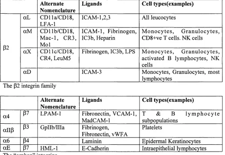
Fi g 1.2: St r u c t u r eo ft h e p 3 s u b u n i te x t r a c e l l u l a r d o m a i n...2 7
Fi g 1 .3 ; Co n f o r m a t i o n a lc h a n g e sint h e I d o m a i no ft h e o 2 s u b u n i t... 3 2 Fi g 1 .4 : KERATINOCYTES EXPRESSING ENDOGENOUS W T HUMAN INTEGRIN (R E D ) AND Y P R F MUTANT
CHICK INTEGRIN (G R EEN ) SPREAD ON COLLAGEN... 38 Fi g 1 .5 : Sc h e m a t i cs t r u c t u r ea n dc o m p o n e n t so f (A ) f o c a lc o n t a c t sa n d ( B ) f i b r i l l a r
ADHESIONS... 3 9 Fi g 1 .6 ( A ) F A K DEPENDENT AND INDEPENDENT ACTIVATION OF E R K BY LIGATION OF INTEGRINS. (B )
In t e g r i n s i g n a l l i n g t o Ak tt h r o u g h F A K /P I 3 K ...4 5 Fi g 1.7: Sc h e m a t i cr e p r e s e n t a t i o no f t h es t r u c t u r eo fh u m a n s k i n...4 7 Fi g 1.8: Sc h e m a t i cr e p r e s e n t a t i o no ft h e s t r u c t u r eo ft h e e p i d e r m i s...4 8 Fi g 1 .9 Hi s t o l o g yo f ( A ) No r m a lh u m a n s k i na n d ( B ,C ) s q u a m o u sc e l lc a r c i n o m a...52 Fi g 1 .1 0 (A ) p i HIGH EXPRESSING CELLS ARE FOUND IN CLUSTERS WITHIN EPIDERM IS(B) THE FATE OF
EPIDERMAL STEM CELLS... 59 Fi g 1 .1 1 Su p p r e s s i o n o fs u s p e n s i o n i n d u c e dd i f f e r e n t i a t i o n b y p i i n t e g r i nl i g a t i o n...6 2
Fi g 1 .1 2 Th e 8 c y t o p l a s m i cd o m a i n a n d D 1 5 4 A e x t r a c e l l u l a rd o m a i n c p l c o n s t r u c t su s e d
TO EXAMINE THE ROLE OF THE P l SUBUNIT IN REGULATION OF DIFFERENTIATION (LEV Y E T A L ,
2 0 0 0 ) ... 6 4 Fi g 2 .1 El u t i o no f Vn f r o mh e p a r i n s e p h a r o s ec o l u m n...7 8 Fi g 3 .1 : Ex p r e s s i o n o ft h e c p l s u b u n i ti nt h e S C C 4 c e l l s... 10 0
Fi g 3 .2 : (A ) S C C 4 CELLS INFECTED WITH C p l W T UPREGULATE THE KERATINOCYTE DIFFERENTIATION MARKER INVOLUCRIN... 101
Fi g 3 .3 : Th e S C C 4 c e l ll i n ee x p r e s s e s e q u i v a l e n tl e v e l so f p l i n t e g r i nt o S C C 1 2 B 2 ...103 Fi g 3 .4 S C C 4 IS CAPABLE OF FORMING FOCAL ADHESIONS ON COLLAGEN COATED SURFACES... 103 Fi g 3 .5 Cl u s t e r i n g o fe n d o g e n o u s p l o c c u r s w h e n S C C c o n t a i n i n ge i t h e r W T o r Y P R F c p i
F i g 3 .7 C l u s t a l W a l i g n m e n t o f t h e A d o m a i n o f a l l e i g h t k n o w n p s u b u n i t s 114
F i g 4.1 : Th e T 1881 MUTANT p i s u b u n i t e x h i b i t s ag r e a t e ra f f i n i t y f o rf i b r o n e c t i n c o a t e d
SURFACES THAN THE WT SUBUNIT...118
Fig 4.2: PBABE-PURO r e t r o v i r a l v e c t o r s h o w i n g p o s i t i o n o f c p l i n t e g r i n C D N A AND THE POSITION OF THE BASES CORRESPONDING TO MUTATION T1881... 119 F i g 4 .3 Th e T1 881 m u t a n ts u b u n i tf o r m sise x p r e s s e do n t h es u r f a c e o ft h ec e l la n d is
CAPABLE OF FORMING FOCAL ADHESIONS...121
Fig 4.4: A d h e s i o n o f GD25 c e l l s e x p r e s s i n g WT (GD25WT), T188I c p l (GD25 T188I) OR EMPTY VECTOR (GD25) TO ECM PROTEINS...124
F i g 4.5: SCC4 SHOWS GREATER ADHESION TO (A) FN AND (B) COL THAN SCC13, A WT/WT
UNDIFFERENTIATED CELL LINE. (C ) THE H U T S 2 1 P l INTEGRIN ACTIVATION REPORTER EPITOPE IS EXPRESSED ON S C C 4 IN THE PRESENCE OF Mn2 + . IT IS ALSO PRESENT ON THE POSITIVE
CONTROL LINE M 0LT4 BUT NOT ON NORMAL KERATINOCYTES...127
Fi g 4.6 T h e C-C l o o p ( r e d ) b e a r i n g T1 881 ( w h i t e ) l i e s a d j a c e n t t o t h e l i g a n d b i n d i n g s i t e (LIGHT BLUE) ON THE SURFACE OF THE Pl A DOMAIN...130 F ig 4.7: (A) T h e o r e t i c a l m o d e l o f a v p i p p r o p e l l e r a n d p s u b u n i t A d o m a i n b o u n d t o a n
RGD PEPTIDE (PRODUCED BY SWISS-MODEL BASED ON 1L5GB)...131
F ig 5.1 : E x p r e s s i o n o f T1 881 c p i c a u s e s a n i n c r e a s e i n t h e r a t e o f s p r e a d i n g o f GD25 c e l l s . . ... 141
F i g 5.2: Th ee f f e c to f W T o r T 1881 c p i o nt h e m o v e m e n to f p i n u l lf i b r o b l a s t s...142
F i g 5.3: T h e e f f e c t o f W T o r T1 881 c p l e x p r e s s i o n o n GD25 m o v e m e n t t h r o u g h t r a n s w e l l s
IN THE PRESENCE OR ABSENCE OF MATRIGEL...147 F i g 5 .4 : ERK PHOSPHORYLATION IN Pl WT AND T1881 EXPRESSING GD25 CELLS...149
F i g 5 .5 (A) E x p r e s s i o n o f t h e T 1 8 8 1 c p i d o e s n o t i n h i b i t g r o w t h m o r e t h a n t h e W T ... 150
F ig 5.6: INVOLUCRIN e x p r e s s i o n i n SCC4 CELLS RETROVIRALLY INFECTED WITH THE WT, T1881 CPl
SUBUNITS OR EMPTY VECTOR...152
F i g 5.7: CORNIFIN EXPRESSION IN S C C 4 CELLS RETROVIRALLY INFECTED WITH THE WT OR T1881 C P l
Table of Figures
Fig 5.8: E x p r e s s i o n o f t h e W T o r T 1 8 8 I c p l c o n s t r u c t s d o e s n o t i n d u c e d i f f e r e n t i a t i o n i n
EITHER UP OR SCC12B2...155 Fig5 .9 : T h e a c t i v a t i n g m A b T S 2 /1 6 c a u s e d a d e c r e a s e in t h e n u m b e r o f i n v o l u c r i n p o s i t i v e
KERATINOCYTES IN ADHERENT CULTURES AFTER INCUBATION FOR 4 8 HR... 158 Fig 5.10 I n c u b a t i o n o f k e r a t i n c o y t e s i n s u s p e n s i o n w i t h TS2/16 r e d u c e s s u s p e n s i o n i n d u c e d
INVOLUCRIN e x p r e s s i o n BY THE SAME DEGREE AS F N ... 159 Fig 6.1: (A) E x a m p l e o f SSCP t r a c e s . U p p e r t r a c e is s a m p l e 51 c a r r y i n g t h e 776 O T A239V.
M u t a t i o n . L o w e r t r a c e is WT c o n t r o l . (B) S e q u e n c i n g r e s u l t s o f t h e s a m e s a m p l e s . ...170 F ig 6.2: S t a i n i n g o f p i s u b u n i t in SCC s a m p l e s w i t h t h e (A) 776 C>T A239V, (B) 471 O T
AND (C) IVS2-36 O A m u t a t i o n s . (D) H U T S -2 1 STAINING OF THE SCC CONTAINING THE 776 C>T A239V MUTATION... 171 Fig 7.1 T h e i n v e r s e r e l a t i o n s h i p b e t w e e n a d h e s i o n a n d d i f f e r e n t i a t i o n o b s e r v e d i n
Ta b l e 1.1: H u m a n i n t e g r i n d i m e r s d i v i d e d b y s u b u n i t c o m p o s i t i o n s h o w i n g t y p i c a l l i g a n d s
AND CELLS ON WHICH THEY ARE EXPRESSED... 18
Ta b l e 1.2 : E p i d e r m a l P h e n o t y p e s o f i n t e g r i n t r a n s g e n i c m ic e ( W a t t , 2 0 0 2 ) ... 56
Ta b l e 2 .1 : A n t i b o d i e s u s e d i n t h i s t h e s i s ... 83
Ta b l e 2 .2 : SDS-P AGE g e l r e c i p e...88
Ta b l e 2 .3 P r i m e r s u s e d i n t h i s t h e s i s ...95
Ta b l e 3.1 : S e q u e n c e c h a n g e s f o u n d i n t h e SCC4 p i cDNA f r a g m e n t s ...107
Ta b l e 5.1 A n t i b o d i e s s c r e e n e d f o r a n a b i l i t y t o i n f l u e n c e d i f f e r e n t i a t i o n i n c u l t u r e d KERATINOCYTES...156
List of Abbreviations
Abbreviation Definition
AM12 Gag, pol +env AM 12 packaging cells
BSA Bovine serum albumin
cPl Chick p i integrin
CD Cluster of differentiation antigen
Col Collagen
DCS Donor calf serum
DMEM D u lb e c c o ’s m o d ificatio n of Eagle’s medium
DMSO Dimethyl sulphoxide ECM Extracellular Matrix EOF Epidermal growth factor
FACS Fluorescence activated cell sorter
FAD FI 2 +adenine +DMEM
FCS Foetal calf serum
Fn Fibronectin
hpl Human p l integrin
fflCE H y d ro x y c o rtis o n e , i nsul i n, cholera enterotoxin and EOF HRP Horseradish peroxidase IVS Intron Variable Sequence
kDa Kilo dalton
Ln Laminin
mAb Monoclonal antibody
MAPK Mitogen activated protein kinase
MT Mutant
pAb Polyclonal antibody PBS Phosphate buffered saline S.D. Standard deviation
TBS Tris buffered saline
Vn Vitronectin
VWF Von Willebrand Factor
Introduction
Overview
The importance o f the p i family o f integrin extracellular matrix receptors in regulating the terminal differentiation of human kératinocytes has been known for many years. In my thesis I describe the effects o f an integrin mutation on the behaviour of a cell line derived from a human squamous cell carcinoma. This introduction will begin with an overview of integrins and then proceed to discuss their role in the skin and how they regulate keratinocyte behaviour.
1.1 Integrins
Integrins are a family of cation dependent extra cellular matrix (ECM) receptors. An integrin consists of a heterodimer of a single a and p subunit (Hynes, 1992). Each subunit is a large (>100kDa) integral membrane glycoprotein (Tamkun et a l, 1986). Integrins have been implicated in a diverse range o f processes including platelet aggregation, phagocyte adhesiveness, leucocyte extravasation and angiogenesis (Hynes, 1987; Hynes, 1992). In all of these cases the essential property of the integrin is its ability to bind extracellular proteins and mediate the adhesion of the cell. The term integrin was coined denote its role as an integral membrane complex involved in the transmembrane association between the extracellular matrix
Chapter 1
Introduction
Integrins are amongst the most widespread family of proteins known with every nucleated human cell type expressing at least one family member. Integrin like proteins are found in many organisms. Drosophila has 4 a (aP S l, aPS2, aPS3 and av) and 2 P (PPS and Pv) subunits (Gotwals et a l, 1994). These show changing patterns of expression during development. Their functional importance is shown by the effects of mutations of pPS {lethal myospheroid), a P S l {multiple edematous
wings) and aPS2 {inflated) which result in embryonic lethality due to defects in
muscle junctions (Gotwals et al, 1994). Two integrins homologous to a5 and a v have been found in C. elegans (Sulston et al., 1992). Even primitive organisms such as the sponge have genes that have high degrees o f similarity to mammalian integrins (W immer et a l, 1999). The importance of integrins, particularly the p l family is further illustrated by the pi knockout phenotype which is lethal prior to implantation of the embryo (Fassler and Meyer, 1995; Stephens et a l, 1995).
Integrins function by binding to defined motifs in their ligands (such as the RGD or REDV motifs in fibronectin, (Pierschbacher and Ruoslahti, 1984; Humphries et a l,
and by clustering (Yauch et a l, 1997; van Kooyk and Figdor, 2000; Hogg and Leitinger, 2001).
1.1.1 Integrin families
Integrins can be loosely divided into families depending on their subunit composition (Hynes, 1992) (Tablet. 1). While this is a convenient division it does not show the complexity o f the tissue distribution and ligand specificity of the different dimers, nor does it necessarily equate with their function.
Alternate Nomenclature
Ligands Cell types(examples)
p l
a l CD49a/CD29 VLA-1
Collagen Fibroblasts, A ctivated T&B lymphocytes
a2 CD49b/CD29 VLA-2
Collagen F i b r o b l a s t s , P l a t e l e t s , Endothelial and Epithelial cells. Monocytes
a3 CD49c/CD29 VLA-3
Laminin Fibroblasts, Epithelial cells. a4 CD49d/CD29
VLA-4
Fibronectin, VCAM-1 M elanocytes, Lym phocytes, monocytes
a5 CD49e/CD29 VLA-5
Fibronectin Fibroblasts, Endothelial and epithelial cells, monocytes
a6 CD49f/CD29 VLA-6
Laminin M onocytes, T lym phocytes, platelets
a l Laminin Muscle
a8 Fibronectin, Tenascin Neurons, Epithelial cells a9 Tenascin Muscle, hepatocytes av CD51/CD29 Fibronectin,
vitronectin
Fibroblasts
Chapter 1
Introduction
Alternate Nomenclature
Ligands Cell types(examples)
p i CD51/CD29 Fibronectin, Vitronectin Fibroblasts
av
P3 CD51/CD61 F ibrinogen, vW FA, Thrombospondin, Vitronectin,
F i b r o b l a s t s , P l a t e l e t s , Endothelial and Epithelial cells. Monocytes
P5 Vitronectin Epithelial cells. Fibroblasts
P6 Fibronectin, Epithelial cells
P8 Fibronectin, Laminin Sensory neurones The a v integrin family, ligands and typical cells on which they are expressed.
Alternate Nomenclature
Ligands Cell types(examples)
aL CDlla/CD18, LFA-1
ICAM-1,2,3 All leucocytes
P2
aM GDI lb/CD 18, M ac-1, CR3, Mol
ICAM-1, Fibrinogen, IC3b, Heparin
Mo n o c y t e s , Gr anul ocyt es, CD8+ve T cells. NK cells
aX CD11C/CD18, CR4, LeuM5
Fibrinogen, IC3b, EPS Mo n o c y t e s , Gr anul ocyt es, activated B lymphocytes, NK cells
aD ICAM-3 Monocytes, Granulocytes, most lymphocytes
The P2 integrin family
Alternate Nomenclature
Ligands Cell types(examples)
a4 P7 LPAM-1 Fibronectin, VCAM-1, M ade AM-1
T & B l y m p h o c y t e subpopulations
a lip P3 GpIIb/IIIa Fibrinogen,
Fibronectin, vWFA
Platelets
a6 P4 Laminin Epidermal Kératinocytes
aE P7 HML-1 E-Cadherin Intraepithélial lymphocytes The “orphan” integrins
1.2 Integrin Structure and Function
At its simplest an integrin dimer resembles a globular head and pair of stalks leading to the plasma membrane. This structure was first seen by electron microscopy (Nermut et a l, 1988). The recent solution of the avp3 extracellular domain crystal structure has given a clearer picture of the structure of an integrin (Xiong et a l,
2001). The receptor is a large multi-domain structure capable o f undergoing radical conformational changes which allows it to react both to signals from the cell and from the surrounding matrix (Xiong et a l, 2002).
1.2.1 a subunit
The a integrin subunit consists of multiple domains (Fig 1.1). At the extracellular terminus there is a large P propeller structure made up o f seven blades o f four P sheets (Springer, 1997). Two of these blades (2 and 3) are involved in forming part of the ligand binding site in the majority of integrins (Mould et a l, 2000). Four of these blades (4-7) also contain calcium-binding motifs. These were once thought to be involved directly in ligand binding however it has now been shown that these lie on the underside of the propeller, well away from the regions interacting with either the P chain or the ligand (Xiong et al, 2002).
Chapter 1
Introduction
the atomic level (Lee et a l, 1995b). The domain has a MIDAS site (Metal Ion Dependent Adhesion Site) which mediates ligand binding.
The stalk region of the a subunit comprises a series of 3 P sandwich domains known as the thigh and the calf 1 and 2 domains (Xiong et a l, 2001) (Fig 1.1). A single transmembrane region connects the extracellular domains to the cytoplasmic tail of the subunit. In a subset of a subunits the extracellular region is cleaved at a point in the calf 2 domain just before the transmembrane region and the subunit is then held intact by a disulphide bond ( a 3,5,6,7,8). a 4 also exists in a cleaved form («4 80/70) however this cleavage is further out at the base of the propeller domain (Rubio et a l, 1992; Teixido et a l, 1992). A truncated form o f a6, a6p, has also been reported which is synthesised from a normal, full length, a 6 mRNA (Davis et a l, 2001). This smaller form of a 6 lacks the first 13 exons including the ligand binding domains. The authors suggest that it is formed either by post-transcriptional modification of the full length a 6 or that it results from translation o f the mRNA starting from a second internal translation start site. The role of this form is not well understood but it appears to be upregulated in differentiating kératinocytes and it has been suggested to be a dominant negative form of the subunit involved in down regulating cell adhesion (Davis et al, 2001).
Chapter 1
Introduction
p
propeller
thigh
- r # ' h
hinge
calf1
calf2
Fig 1.1: Structure of the av subunit, a-helixes in red, p-sheets in yellow, random coil in grey. Based on 1JV2 (Xiong et al, 2001).
1.2.2 p subunit
All p subunits have a highly conserved complex structure (Fig 1.2). The subunit consists o f several domains, a ligand binding domain, an Ig fold, several cysteine rich domains that comprise the “stalk”, a short transmembrane domain and a cytoplasmic domain (Xiong et al, 2001).
Chapter 1
Introduction
is then thought to stabilise the ligand bound state (X iong et al, 2002). Another
important area o f the ligand binding domain is the specificity loop, a small cysteine
bridged loop not found in other vWFA domains which has been shown to control the
specificity o f both the ligand binding and signalling performed by the receptor. This
loop also cross-links to integrin peptide ligands indicating it forms part o f the ligand
binding surface (Bitan et al, 2000; Yahalom et a i, 2002). Substitution o f this loop in
pi for the corresponding sequence from p3 has been shown to alter ligand specificity
(Takagi et al, 1997), signalling (M iao et a l, 2002) and to influence folding and
dimérisation (Takagi et al, 2002). Another region, the 3io helix, is also not found in
a subunit 1 domains. An W k bicm e sidechain from this loop fits into the cavity at the
centre o f the P-propeller o f the a subunit and is crucial for subunit dim érisation
(Xiong et al, 2001).
The region that lies below the pA domain is a hybrid Ig fold consisting partly o f the
N term inus o f the protein and partly from the region that lies C term inal to the PA
domain (Xiong et al, 2001). This domain makes extensive contacts across the lower
surface o f the PA domain. It is thought that there is little m ovem ent between these
two structures. The extrem e N term inus o f the protein form s a PSI ( P le x u \
Semaphorin and Integrin) domain and this connects the N term inus back onto itself
to the EGF-1 domain in the stalk below the Hybrid domain. To date the atomic level
structure o f this domain has not been determined.
The stalk o f the p subunit com prises four EGF like units w hich together are often
referred to as the cysteine rich region as each unit contains m ultiple internal
disulphide bonds (_Shih,«zTd |qs3 ^Calvete et a l, 1991). The four units appear to
divide into 2 tandem repeats (1 and 2, 3 and 4). The junction between the two units inside the tandem repeat appears inflexible and includes a disulphide bridge as well as H-bonding whereas the interface between units 2 and 3 appears more flexible (Xiong et al, 2001). In addition to the EGF-like repeats there is a final extracellular domain known as PTD (Xiong et al, 2001). This comprises a four stranded P sheet and a N terminal helix and has little homology to any other known structure. The stalk region contains the epitopes for many o f the affinity reporter antibodies (Bazzoni et a l, 1995; Luque et a l, 1996). These antibodies function by binding to an epitope that only becomes exposed once the integrin undergoes structural rearrangement following ligand binding.
Chapter 1
Introduction
Hybrid Ig domain
A domain ( R
■V' S ',
f %
Cysteine
rich stalk
Specificty Loop
3io helix
Fig 1.2: Structure of the P3 subunit extracellular domain, a-helixes in red, P-sheets in yellow, random coil in grey. Based on 1JV2 (Xiong et al, 2001). The PSI domain is not shown as the atomic structure has not been resolved. Inset is an alternative view of the A domain in the ligand bound form (based on 1L5G (Xiong et al, 2002)) showing the specificity loop, the 3io helix and the LIMBS, MIDAS and ADMIDAS cation sites (blue spheres, left to right)
Chapter 1
Introduction
1.2.3 Ligand binding and activation
The heterodimeric structure of integrins is more complex than many other types of cell adhesion molecule and this increased complexity allows modulation of integrin function in a more subtle manner than simple up or down regulation at the protein level. One of the requirements of integrin function in many situations is the necessity to perform their adhesive functions in a very rapid and specific manner, often in limited time frames and locations. Integrins achieve this regulation of their activity through two main methods, receptor clustering and changes in receptor conformation (Hynes, 1992; van Kooyk and Figdor, 2000; Hogg and Leitinger, 2001).
Integrin clustering is often associated with integrin ligation and this clustering of receptors leads to an increased avidity of the cell for its ligand. As an example showing the importance of clustering, deletion o f the a 4 cytoplasmic domain prevents the clustering o f a 4 p l into focal contacts and hence prevents cell attachment despite not preventing the binding of the truncated integrin to VC AM-1 (Yauch et al, 1997). In this case the clustering of the receptor appears to be the important step in regulating the interaction between the cell and the ECM. In other cases structural rearrangements in the receptor itself lead to changes in affinity for ligand. The platelet integrin a llb p 3 exists in a low affinity form on unactivated platelets. In this state it is capable o f binding to immobilised fibrinogen but not to any of its soluble ligands. This means the platelet can only bind to pre-existing sites of clotting. Exposure of the platelet to activating stimuli result in the aIIbP3 receptor switching into a high affinity state capable of binding to soluble ligands (Kieffer and Phillips, 1990; Phillips et a l, 1991). Similar activation events have been observed in
the p2 integrins (Arnaout, 1990; Larson and Springer, 1990). The multiple conformational states can be measured by the use of reporter antibodies that specifically recognise one o f the affinity states (Luque et al, 1996) and other antibodies have been produced that are capable o f inducing or stabilising a high affinity conformation (K ovach et a l, 1992; Petruzzelli et a l , 1995). Different activating antibodies have also been reported to promote different active states recognising different ligands (Ortlepp et a l, 1995)
There is no current consensus of whether the conformational shift or receptor clustering is the initial event in integrin activation. It is possible that ligand binding to activated, high affinity integrins causes recruitment o f further receptors resulting in clustering. It is also conceivable that the initial interactions are between low affinity integrins and ligand and that clustering o f these receptors results in changes in conformation leading to strengthening of the interaction due to increases in both avidity and affinity. The two mechanisms of integrin adhesion regulation are likely to have a synergistic effect with conformational changes increasing individual integrin-ligand interactions and integrin clustering increasing the avidity of the interaction and supporting cell attachment. It is also likely that whether integrin clustering or changes in conformation is the dominant factor in activation varies depending on the integrin dimer in question.
The crystal structures o f individual integrin domains have helped to reveal the changes in conformation that occur on activation and ligand binding. Comparisons of the crystals o f a subunit I domains in the presence o f Mg2+ and Mn2+ (Lee et a l ,
Chapter 1
Introduction
mutants that lock the domain in either the low or high affinity conformation (Shimaoka et a l, 2000; Shimaoka et a l, 2001) have provided a mechanism for regulation of I domains. The crystals of the extracellular portion o f the avP3 in the presence of an RGD peptide have shed light on how changes are transmitted throughout the structure (Xiong et al, 2002).
Information on the mechanisms of regulation of integrin activation has also been obtained through studies using antibody epitopes that either cross the boundary between the a and P chains (Zang et al, 2000), block function (Greve and Gottlieb, 1982; Mould et a l, 1997; Shih et a l, 1997; Mould et al, 2000), report affinity (Bazzoni et al, 1995; Lu et a l, 2001a; Mould et a l, 2002) or activate the integrin (Faull et a l, 1996). Other experiments involved the use of ligand mimetic peptides to map the critical residues in the ligand binding domain (Humphries et a l , 2000; Mould et al, 2000). These results led to a number of models o f different regions of the integrin structure being proposed (Huang and Springer, 1997; Tuckwell and Humphries, 1997; Huang et a l, 2000a; You et a l, 2002).
The quaternary structure of the dimer has recently been resolved in both the ligand bound and an unbound form for the avp3 dimer (Xiong et al, 2001; Xiong et al,
2002). The interaction of the p subunit A domain and the p propeller domain closely resembles the structure of the G-protein a /p subunit interface with the 3io helix of the pA domain located in the gap in the axis of the P-propeller domain (Xiong et al,
domains in conducting signals both into the cell and out to the ligand binding domains is crucial in understanding integrin function (Takagi et a l, 2001).
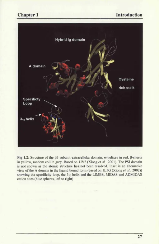
In integrins where an a subunit I domain is the principal site of ligand binding the I domain undergoes some very specific changes upon activation (Fig 1.3). In the alI domain a single turn of helix is transferred from helix C to helix a 6 which allows the opening o f the ligand binding domain (Emsley et al, 2000). The MIDAS cation moves, its co-ordination sphere changes and it interacts directly with the ligand. Most notably small changes in other regions o f the structure result in a large movement o f the C terminal al helix which drops downwards by 10 Â in the liganded structure (Emsley et al, 2000). Mutated domains where this helix has been restrained in either the up or down position show that this movement is capable of activating or inactivating the entire I-domain (Lu et a l, 2001b; Shimaoka et al,
Chapter 1
Introduction
Fig 1.3: Conformational changes in the I domain of the a2 subunit (after (Emsley et al, 2000)). On ligand binding the cation moves towards helix a5, the aC helix unwinds and transfers a loop to a6 and the a l helix is displaced downwards and away from a l . a-helix in red, P sheets in yellow and random coil in grey. Cations shown as blue spheres. Prepared using SWISSPdB viewer ft-om structures lAOX (left) and IDZI (right).
In subunits lacking an a subunit I domain the principal site of ligand binding is the p subunit A domain. This domain is similar in structure to the a I domain, however activation appears to occur in a different manner. In the p A domain the major events are rearrangement o f the cation binding sites with the MIDAS site becoming occupied and co-ordinating with the ligand (Xiong et al, 2002). The LIMBS cation binding site forms and becomes occupied to stabilise the conformation and the region containing the specificity loop moves towards the MIDAS site (Xiong et al, 2002). It has also been suggested that the a l helix may become displaced in a similar fashion to the a l movement seen in a subunit I domains (Mould et al, 2002). Sequences in the second and the third repeats of the a subunit P-propeller domain play a role in defining ligand specificity by interacting with “synergy sequences” in integrin ligands that surround the RGD motif (Mould et al, 1997; Humphries et al,
2000; Mould et al, 2000)..
Chapter 1
Introduction
1.3 Integrin Interacting Proteins, Signalling and Focal
Adhesion Assembly
Integrins are capable of assembling large complexes of proteins through interactions mediated by their cytoplasmic, transmembrane and extracellular domains. These complexes function not only as structural junctions between the cell and ECM but also control complex signalling systems allowing the cell to respond to changes in its environment.
1.3.1 Transmembrane and extracellular Integrin interacting proteins
Integrins mediate several processes through cis-interactions with other membrane proteins. Caveolin has been shown to interact with a subset of a subunits (al,a5,ocv) via the integrin transmembrane domains. Its ability to bind Src family kinases, such as Fyn, allows a signalling pathway to ERKl/2 to be activated (Wary et a l, 1998). This pathway is associated with caveolin’s ability to bind to cholesterol and glycosphingolipids and organise membrane rafts that are rich in the Src family members which are both myristolated and palmitoylated such as Fyn, Yes and Lck (Harder and Simons, 1997). Integrins which are in the active conformation have been shown to interact with these lipid rafts in certain cell types (Leitinger and Hogg,
2002).
lAP (CD47) is a large 5 transmembrane Ig family protein which is widely expressed. It has been shown to act as a receptor for thrombospondin. It interacts with a 2 p l (Wang and Frazier, 1998) and avP3 (Lindberg et a l, 1996) and appears to act to regulate cell migration by signalling through pathways involving FAK and heterotrimeric G proteins.
Many a integrin subunits form highly stable interactions with members o f the tetraspanin or transmembrane-4 superfamily (TM4SF). These interactions are mediated through the one o f the extracellular loops o f the tetraspanin and the extracellular domain of the a subunit although the exact point of interaction seems to differ in different tetraspanin/integrin complexes (Hemler et a l, 1996; Yauch et a l,
2000). The cytoplasmic domains of the tetraspanins are associated with signalling pathways involving PI-4-K and PKC and have been implicated in processes including cell spreading, migration, MMP induction and differentiation in different cell types (Hemler, 1998; Sugiura and Berditchevski, 1999; Stipp and Hemler, 2000; Berditchevski, 2001),
1.3.2 Integrin cytoplasmic domain binding proteins.
Chapter 1
Introduction
adhesions which interact with the p subunit tail (reviewed in (Petit and Thiery,
2000)).
Other proteins that interact directly with the p cytoplasmic domain have been identified by yeast two hybrid screens; ICAP, RACK-1, caspase-8 and ILK. ICAP has been shown to interact with the region between the cyto-2 and 3 motifs of p i (Chang et a l, 1997; Zhang and Hemler, 1999). Overexpression of ICAP inhibits cell spreading but this is reversed by overexpression of consitutively active Cdc42. ICAP interacts with both cdc42 and Rac as an inhibitor of GDP dissociation (Degani
et a l, 2002).
RACK-1 interacts with an area just before the cyto-1 motif and has been shown to co-immuno-precipitate with p subunits under certain conditions (Liliental and Chang, 1998). RACK-1 is an adapter protein for the active form o f PKC and recruits activated PK C aand e (Liliental and Chang, 1998; Besson et a l , 2002). PKC isoforms are involved in processes governing transport o f p subunits to the membrane (Ng et a l, 1999). PK Ca may interact directly with some p subunits through the NPXY motifs in the pi tail (Parsons et a l, 2002).
Caspase-8 is reported to bind directly to the membrane proximal region of unligated pi and p5 subunits (Stupack et a l, 2001).
et a l , 2001) and tumor^genicity (Marotta el a l , 2001). O ver expression o f ILK
induces an epithelial/mesenchym al transition in mammary epithelial cells (Somasiri
et a l, 2001). There is also evidence that ILK can promote P-catenin signalling (Tan
et a l, 2001). The kinase activity o f ILK does not appear to be essential for its
function in integrin signalling (Zervas and Brown, 2002) and it is now thought to act
primarily as an adapter molecule (Tu et al, 1999; Guo and Wu, 2002).
The kinase FAK (Focal Adhesion Kinase) has also been reported to bind to the p i
tail (Lew is and Schwartz, 1995; H annigan et al, 1996) and has a role in focal
adhesion assembly/disassembly and in integrin mediated signalling .
Chapter 1
Introduction
1.3.3 Focal Adhesions
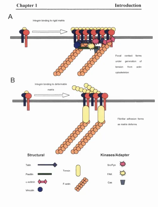
Focal adhesions were first identified in the 1970s as regions of the cell that were in close contact with the substratum (Abercrombie et a l, 1971; Couchman and Rees, 1979). These are large protein structures that assemble around integrin tails to mediate adhesion and motility. Focal adhesions can be divided into the two types: focal contacts and fibrillar adhesions (Zamir el al, 1999). Focal contacts are the “classical” focal adhesion, enriched in scaffolding proteins such as talin and paxillin and are highly tyrosine phosphorylated. Fibrillar adhesions are enriched for tensin, another scaffold protein, and are less highly phosphorylated.
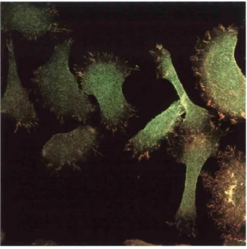
Fig 1.4: Kératinocytes expressing endogenous WT human integrin (Red) and YPRF mutant chick integrin (Green) spread on collagen. WT integrin forms large focal adhesions. The YPRF disrupts the cyto-2 motif in the pi tail and this prevents recruitment of the chick subunit to these focal adhesions.
A
Integrin binding to rigid matrix
Paxillin
a-actinin
Vinculin
Focal contact forms
under generation of
tension from actin
cytoskeleton
Integrin binding to deformable
matrix
Structural Kinases/Adapter
O
Src/FynTensin
o
F-actin
Fibrillar adhesion forms
as matrix deforms
Fig 1.5 : Schematic structure and components of (A) focal contacts and (B) fibrillar adhesions.
Chapter 1
Introduction
Focal contacts
The structure of the focal contact (Fig 1.5A) is provided by a series o f scaffolding proteins such as talin, vinculin, filamin and a-actinin upon which is built a series of signalling systems. Talin is present as a dimer and may associate directly with the P subunit tail. Cells expressing integrins that lack the binding sites for a-actinin and talin are deficient in migration and adhesion (Bodeau et ai, 2001). In culture focal contacts appear to form at the periphery o f the cell and can be promoted by plating the cells onto rigid matrixes such as glass bound vitronectin. These contacts apply significant amounts o f force onto the matrix generated by myosin II driven cytoskeleton contraction (Zamir et al, 1999).
Fibrillar adhesions
Fibrillar adhesions are more elongated structures (Hynes and Destree, 1978) containing tensin, a protein with an actin filament capping function (Lo et a l, 1994; Chuang et a l, 1995) (Fig 1.5B). These adhesions also appear to be simpler, and not to involve as many other proteins as the talin containing focal contacts (Katz et a l ,
1.3.4 Focal adhesion assembly/disassembly
As previously described focal adhesions are large complexes of proteins, the primary role of which is to anchor the cell to the basement membrane. However this strong adhesion has the effect o f severely limiting the migration of the cell. In order for effective cell movement the stability of focal adhesions has to be tightly regulated (Rodriguez Fernandez et al, 1992; Rodriguez Fernandez et al, 1993), allowing the cell to attach to the matrix, apply sufficient force across the adhesion to move the cell body over it then detach from the matrix at the rear o f the cell and recycle components to the leading edge.
The initial step of focal adhesion formation is clustering o f integrin subunits. This process is highly dependent on the p cytoplasmic tail and is mediated by adapters such as talin binding to the cyto2/3 motifs (Horwitz et a l, 1986). Subunits in which these motifs are disrupted are incapable of being recruited to integrin clusters and are present diffusely across the whole membrane surface (Solowska et a l, 1989; Hayashi
et al, 1990). FAK, talin, tensin, a-actinin and vinculin are recruited to early
Chapter 1
Introduction
tension is also essential for the proper formation of the focal adhesion. Experiments have shown that the type of adhesion that forms, either fibrillar adhesion or focal contact, is directly dependent on the amount of contractile force that the matrix can support. If the matrix is non-rigid little tension is exerted on the focal adhesion as the cell deformations the matrix. If the matrix is rigid the force across the focal adhesion increases and this leads to recruitment of proteins such as talin and vinculin which stabilise the structure (Katz et al, 2000; Zamir et al, 2000; Riveline et a l, 2001). The degree of phosphorylation seen in the structure also appears to be dependent on the degree of tension (Jockusch et a l, 1995; Zamir et al, 1999).
1.3.5 Integrin Signalling
Unlike many other cell surface receptors integrins have no intrinsic kinase activity. For many years it was thought that, because of this, they had no signalling function beyond their role in cell adhesion and focal adhesion formation. It is now understood that, by recruiting a range of adapter proteins and kinases through interactions with both extracellular and cytoplasmic domains, integrins are involved in many different signalling pathways (reviewed in Giancotti and Ruoslahti, 1999; Schwartz, 2001; Schwartz and Ginsberg, 2002).
One o f the major kinases involved in integrin mediated signalling is the Focal Adhesion Kinase (FAK). As its name suggests FAK plays an important role both in focal adhesion formation and disassembly but it is also involved in several signalling cascades leading from the integrin. One of the best characterised roles for FAK is in FAK mediated MAPK activation (Fig 1.6A). FAK is initially recruited to focal adhesions through its interactions with talin and vinculin (Chen et a l, 1995) and possibly the p subunit tail (Lewis and Schwartz, 1995). Auto phosphorylation of Y397 activates FAK as a tyrosine kinase. This allows binding o f the membrane bound kinase Src via an SH2 domain interaction with Y397 (Schaller et a l, 1994; Schlaepfer et a l, 1994). Further phosphorylation events lead to the recruitment of the adapter proteins such as CAS and CRK leading to activation of the JNK pathway. Recruitment of the Grb2/S0S complex leads to activation o f the ERK l/2 MAP Kinase via Ras and Raf (reviewed in Giancotti and Ruoslahti, 1999).
Chapter 1
Introduction
leading to phosphorylation of She and recruitment of the Grb2/S0S complex. This allows activation of Ras and triggers the MAPK cascade .The two pathways leading to Ras are thought to act in tandem, She initially triggering the high levels of MAPK activation seen shortly after adhesion and FAK causing the sustained activation following it (reviewed in Giancotti and Ruoslahti, 1999).
Integrin ligation can influence signalling to MAPK stimulated by ligation of the EGF receptor. Studies have shown that in the absence o f integrin ligation EGF induced signalling is largely inhibited (Miyamoto et al., 1996), in contrast integrin derived signalling to MAPK can occur in the absence of EGF, albeit at lower levels than when growth factors are present. Other work has suggested an association between the EGF receptor and various integrins (Miyamoto et al, 1996). It has been shown that co-clustering of the EGF receptor and the a 2 p l integrin at sites o f cell-cell contact induce tyrosine phosphorylation and activation o f the EGF receptor in the absence of its ligand (Moro et a l, 1998; Yu et a l, 2000). EGF receptor signalling uses many of the same components found in integrin mediated signalling pathways, such as She, Grb and SOS so a synergistic role for these two pathways is likely. In addition the PDGF (Schneller et al, 1997; Woodard et a l, 1998) and VEGF (Soldi et a l, 1999) receptors have also been reported to form complexes with integrins.
A
S R C phosphorylates CAS Caveolin
G rb/S O S
> Rac
Fyn phosphorylates She
B
RAS
FAK Auto-P Y 3 9 7 and binds SR C and
G rb/S O S
JNK
ERK
PTEN FAK
PI3K
Fig 1.6 (A) FAK dependent and independent activation of ERK by ligation of integrins. (after (Giancotti and Ruoslahti, 1999)) (B) Integrin signalling to Akt through FAK/PI3K. PTEN is thought to act as a repressor on this pathway.
Chapter 1
Introduction
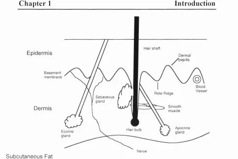
1.4 Structure and Function of the Skin
Human skin consists of several layers (Fig 1.7). The outermost layer is the epidermis. Below the epidermis lies a basement membrane that separates the epidermis from the underlying dermis. The epidermis and the dermis interdigitate, forming the rete ridge and dermal papillae (Fig 1.7).
1.4.1 The Dermis
Hair shaft
Epidermis Dermal
'papilla
Basement membrane
Blood Vessel Rete Ridge
Sebaceous gland
Smooth muscle Dermis
Apocrine gland Hair bulb
Eccrine gland
Nerve Subcutaneous Fat
Fig 1.7: Schematic representation o f the structure o f hum an skin.
1.4.2 The Epidermis
The epidermis is the main barrier between the body and the surrounding environment
(Fig 1.8). It is predominantly made up o f kératinocytes although sm aller numbers o f
m elanocytes (pigment cells)(Jim bow c/ ai, 1991), M erkel cells (sensory cells) and
Langerhans cells (a type o f antigen presenting cell) (H auser et a l, 1991) are also
present. The outer layers o f the epidermis consist o f tough squames that provide the
barrier function o f the skin. Below these are layers o f living cells that arise from the
population o f dividing kératinocytes in the basal layer. W ithin the epiderm is and
dermis are also found hair follicles, apocrine and eccrine sweat glands and sebaceous
glands (Odland, 1991). These are all derived from the epidermis.
Chapter 1
Introduction
— ^ —PT-r—-1
czzzzz
Cr--- — IT
--- ---3L__
"---- , ——!r
1
o o
o
3
o
V J
f \
#
k VV # J
r N
#
k---Jk --- V#
Cornified Granular Cytoplasmic keratohyaiin granules r ~s # #
k J
#
k J
f %
#
k
_______ / # \ --- / Spinous Basal Shedding Terminal differentiation Proliferation Basement membraneFig 1.8: Schematic representation o f the structure o f the epidermis.
1.4.2 The basal layer kératinocytes
The cells o f the basal layer (Fig 1.8) are in contact with an underlying membrane
comprised o f a range o f ECM proteins. The most abundant are collagens, especially
type IV collagen (Timpl, 1989; Odland, 1991). In addition there are large amounts of
laminins and other proteoglycans (Burgeson and Christiano, 1997). Fibronectin is not
norm ally present except in wounded epiderm is (M osher, 1989; Fine, 1994). The
basement membrane com ponents are synthesised both by kératinocytes and dermal
fibroblasts (Marinkovieh et al., 1993). The basal kératinocytes adhere to the basal
membrane through (34 mediated hem idesmosomes and (31 mediated focal adhesions
(Carter et ai, 1990a; Carter et ai, 1990b; Stepp et ai, 1990).
1.4.3 The upper epidermal layers
The various layers of the skin can be distinguished by the range o f proteins they express.
The spinous layer comprises the first 4-8 cell layers above the basal layer and is characterised by an increase in cell size and an increase in desmosomal junctions between the kératinocytes (Holbrook, 1994). Most suprabasal layers express involucrin (Rice and Green, 1979), a protein synthesised early in the differentiation pathway, which is part of the cornified envelope of a mature squame.
The granular layer is identified by the synthesis o f electron dense keratohyaiin granules of loricrin and profilaggrin (Holbrook, 1994). Loricrin is a component of the cornified layer (Mehrel et a l, 1990) and profilaggrin is the precursor to filaggrin, a protein involved in the aggregation of keratin filaments (Rothnagel et a l, 1987; Rothnagel and Steinert, 1990).
Chapter 1
Introduction
The cells of all layers adhere to each other in a calcium dependent manner through adherens junctions and desmosomes (Cowin, 1994). Adherens junctions, also known as zonula adherens are found as a belt around the cell and appear as electron dense regions where the membranes of adjacent cells run parallel about 20 nm apart (Geiger and Ginsberg, 1991). This type of cell-cell junction is found in many tissues including the epithelium (Geiger et a l, 1987).
The intracellular section o f adherens junctions consists of dimers of cadherin family proteins (Bussem akers et a l , 1993; Kemler, 1993). These are transmembrane proteins that form calcium dependent head-to-tail dimers through their extracellular domain with corresponding proteins from adjacent cells. Kératinocytes of all epidermal layers express E-cadherin while P-cadherin is expressed only in the basal layer (Hirai et a l, 1989). Adherens junctions link to the actin cytoskeleton through vinculin, a-actinin and catenin family proteins (Kemler, 1993).
Desmosomes link adjacent cells through desmoglein and desmocollin, members of the cadherin superfamily (Schwarz et a l, 1990; Kowalczyk et a l, 1999). In vivo
Gap junctions are also present in the kératinocytes. These are small transmembrane channels that permit the transfer of small molecules between adjacent cells (Evans,
1988). These are found in all layers of the epidermis
1.4.4 Squamous Cell Carcinoma
Squamous cell carcinomas (SCC) are common tumours of the skin and epithelia (Fig 1.9). The incidence of SCC is so high and the mortality rate so low that, along with basal cell carcinomas of the skin, they are excluded from the national statistical analysis o f cancer cases. The combined incidence o f these two types of non melanoma skin cancer was over 40,000 in England and Wales in 1997 and around 20% of these were recorded as SCC. Less than 500 mortalities per year are recorded as being directly due non-melanoma skin cancer (Source: National Office of Statistics).
Chapter 1
Introduction
mmi%
Fig 1.9 Histology of (A) Normal human skin and (B) squamous cell carcinoma. (C) SCC showing keratin pearls (arrow heads).
Treatment is normally by excision followed by chemo- or radiotherapy and survival rates are normally 90% over 5 years if treated early. If the condition is not treated promptly large scale tissue destruction, can occur, especially in oral tumours (Nasemann et al, 1983). SCC show disruption of the normal skin architecture with islands of kératinocytes present within the dermis and loss or disruption o f the basement membrane (Wheater, 1991). The keratinocyte differentiation program is also perturbed. Well-differentiated SCC produce large amounts of keratin and frequently large masses of protein known as keratin pearls can be seen (Fig 1.9C). Poorly differentiated tumours show reduced keratin expression (Wheater, 1991). Spindle like cells may be present in more severe undifferentiated tumours (McGee et a l, 1992)
1.5 Epidermal Integrins
Kératinocytes express a range of integrin subunits. In vivo a2, a3, a6, a9 and a v are expressed along with the p i, P4 and pS subunits (Watt and Hertle, 1994; Watt, 2002). These make up the a 2 p i collagen receptor, the a 3 p i laminin receptor, a6P4 laminin receptor, « 9 p i tenascin receptor and the av P S vitronectin receptor. Upregulation of a S p i and avp6 is seen in wounded or hyperproliferative epidermis as well as in culture (Adams and Watt, 1991; Watt and Hertle, 1994). a v p s is also reported to be present (Stepp, 1999). Despite expressing both the a 6 ,a v and p i subunits neither a 6 p i or a v p i is expressed in kératinocytes (Adams and Watt, 1991).
Chapter 1
Introduction
exception to this is avpS which is expressed only in the suprabasal layers of the epidermis (Stepp, 1999). a 6 p 4 is concentrated on the basal membrane of kératinocytes where it forms hemidesmosomes, anchoring the cell to the basement membrane (Watt and Hertle, 1994). Wholemount labelling o f basal kératinocytes shows some focal adhesion like clusters of p i integrins interspersed with the hemidesmosomes on the basal surface of the cell but most p i integrins are present in a ring around the cell periphery (Jensen et a l, 1999). Although pi integrins appear to be concentrated at cell-cell borders this is likely to be a result o f the heavily interdigitated membranes at these sites rather than due to any role in cell-cell adhesion (Braga et a l, 1998). In wounded or hyperproliferative skin integrin expression may be seen in the suprabasal layers of the epidermis (Watt and Hertle,
1994).
Alternative splicing events occur in the integrin subunits expressed by kératinocytes. The most common p i expressed is P lA although kératinocytes are also one of the few cell types to express piB . This subunit has a unique 12 amino acid sequence that replaces the last 21 amino acids of piA . p iB is expressed on the cell surface but does not mediate cell adhesion and acts in a dominant negative manner against pi A function in transfection experiments (Balzac et a l, 1994). The importance of piB is questionable as it is expressed at very low levels in human kératinocytes (Kee et a l ,
2000) and is not conserved in the mouse genome (de Melker and Sonnenberg, 1999).
expressed in kératinocytes although trace amounts of (34B (Hogervorst et al, 1993) and P4E are present. (34E has a shorter (232 amino acid) tail and lacks the structures required for hemidesmosome formation (van Leusden et a l, 1997; de Melker and Sonnenberg, 1999).
1.5.1 Functions of Epidermal Integrins
The primary function of integrins in the epidermis is to secure the cells of the basal layer to the basement membrane through their interactions with the actin and keratin cytoskeletons. Integrins also play a vital role in mediating keratinocyte migration over ECM (Watt and Hertle, 1994; Watt, 2002). a 3 p l is required at the leading edge of the cell for keratinocyte migration over laminin (DiPersio et a l, 1997; Goldfinger
et a l, 1999). In migrating cells a 6 p 4 is present at the leading of edge of the cell where it can be associated with membrane protrusions and show a reduced association with hemidesmosomes on the basal surface (Nguyen et a l, 2000; Mercurio et a l, 2001).
Chapter 1
Introduction
Subunit Phenotype Reference
a3 D iso rg a n ise d basem en t m em brane, occasional blistering.
(DiPersio e t a l , 1997) (36 Juvenile hair loss (Huang e t a l , 1996) a9 No defects (Huang et a l , 2000c) (35 Reduced migration of kératinocytes (Huang et a l , 2000b) (31 Floxed p i X K5Cre
Abnormal hair follicles, hair loss, blistering, reduced p ro liferatio n and abnorm al differentiation, disruption of basement membrane, impaired wound healing
(Grose et a l, 2002)
Floxed p i X K14Cre
Blistering, basement membrane disruption, reduced hemidesmosomes, thin epidermis, reduced hair follicles, reduced a6P4
(Brakebusch et a l , 2000)
a6 Severe blistering (Georges-Labouesse et a l ,
1996)
P4 Severe blistering (Dowling et a l , 1996; van der Neut et a l , 1996) a3 + a6 Blistering (DiPersio et a l , 2000)
a 2 No defects (Holtkotter e t a l , 2002)
Table 1.2 : Epidermal Phenotypes of integrin transgenic mice (Watt, 2002)
Loss of a3 by targeted deletion in mice also causes a disruption o f the basement membrane and occasional epidermal blistering on the feet and legs (DiPersio et a l ,
1997). The double knockout of « 3 p i and a6P 4 does not result in a more severe phenotype than either alone (DiPersio e t a l , 2000). (x2, a 9 and pS knockouts do not result in skin phenotypes (Huang et a l , 2000b; Huang et a l , 2000c; Holtkotter e t a l ,
2002) although loss of p5 does result in severely impaired migration o f kératinocytes (Huang et a l , 2000b).
The p i knockout is early embryonic lethal (Fassler and Meyer, 1995; Stephens e t a l ,
expressing Cre under the control of either the keratin 5 or 14 promoter. This results in Cre activity in the basal layer of the epidermis (Brakebusch et al, 2000; Raghavan
et a l, 2000). These mice suffer from epidermal blistering but not to the degree
reported in the a 6 p 4 null mice. Wound healing studies performed on these mice confirm that |3l is essential for keratinocyte migration in vivo (Grose et a l, 2002). Mouse kératinocytes lacking p i due to K5-Cre expression show an increase in the proportion of cells expressing differentiation markers, the number of cells expressing involucrin increased from 1% to 20-40% (Grose et al, 2002). Other than the increase in the number of differentiated cells the differentiation program itself appears to proceed normally. The mice also show a general reduction in proliferative cells in their hair follicles and by 7 weeks lack both hair follicles and sebaceous glands (Brakebusch et al, 2000). No change in the differentiation program was seen in K14- Cre mice, however the proportion o f differentiated cells was not measured (Raghavan et al, 2000).
1.5.2 Epidermal Stem Cells.