1
Long-term urological follow-up of multicystic dysplastic kidneys: Is it still necessary in 2007?
Karen J. Psooy, MD
University of Manitoba, Winnipeg, MB
Background:To determine if the current indications for the long-term uro-logical follow-up of children with multicystic dysplastic kidneys (MCDK) are supported by the literature. Observation of MCDK began in the 1980s. In 1993, the Multicystic Kidney Registry published that observation of MCDK was safe, and that prophylactic nephrectomy was not indicated. However, it has not been clarified if observation is necessary either. The author believes that after 20 years of observation, the answer to this question can now be answered.
Methods:The membership of the Pediatric Urologists of Canada was sur-veyed to determine if long-term urological follow-up was being performed, and if so, for what indications. A literature search using PubMed, EMBASE and a Conference Papers Index was performed to determine if the indi-cations listed were supported by the current literature.
Results:A response rate of 72% was achieved for the survey, with 82% of responders (23/28) following children with MCDK long-term. The main indications listed for long-term follow-up were the increased risk of Wilms’ tumor (54%), the increased risk of hypertension (32%), observation of the contralateral kidney (43%) and to observe involution of the MCDK (36%). The literature search did not support the need for long-term urological follow-up for any of these indications, provided unilateral MCDK was an isolated genito-urinary abnormality. There are were only 5 document-ed cases of Wilms’ tumor in the Unitdocument-ed States between 1983–1998, and none since. No case series of MCDKs have observed a Wilms’ tumor, sug-gesting the risk of developing Wilms’ tumor with MCDK is nil. Hypertension, although rare, could be monitored for by a primary care physician. When unilateral MCDK is an isolated genitourinary abnormal-ity and compensatory hypertrophy is confirmed, the risk of developing urinary tract infection, chronic renal failure or end stage renal disease is very low. With current radiological techniques, observation of invo-lution is not required for confirmation of diagnosis. Additionally, not all MCDKs involute.
Conclusion:Twenty years of observation has shown that long-term uro-logical follow-up of children with “simple” MCDK is no longer indicat-ed once the diagnosis has been confirmindicat-ed with a follow-up renal ultra-sound at 12–24 months. Blood pressure monitoring by a primary care physician is recommended.
2
Single and multiple layer small intestine submucosa in the repair of severe chordee and complicated hypospadias
Matthew H. Hayn, MD; Mark F. Bellinger, MD; Francis X. Schneck, MD University of Pittsburgh, Pittsburgh, PA
Background:Small intestine submucosa (SIS) has been described for corporal body grafting in cases of severe chordee. We describe our expe-rience with single and multiple layer SIS in the repair of proximal hypospa-dias and severe chordee.
Methods:Between 2000 and 2004, 15 boys with proximal hypospadias and/or severe ventral chordee (> 40°) underwent SIS grafting to correct the curvature. Both single layer (Surgisis, Cook Biotech) and a 4-layer (Stratasis, Cook Biotech) were used for corporal grafting. In each case the ventral defect in the corporal bodies was measured, and the SIS graft was measured approximately 20% larger than the defect. Reassessment was performed by artificial erection test at the time of the second-stage reconstruction or at follow-up visit in the clinic.
Results:First-stage corporal body grafting was performed on 15 patients between June 2000 and March 2004. Surgisis was used in 6 cases and Stratasis was used in 9 cases. Of the 15 boys, 12 underwent a planned second-stage repair with subsequent urethroplasty 6 to 25 months after the initial first-stage repair. Median age at first-stage repair of the 12 boys was 12 months. Native meatus location was penoscrotal in 5 boys, mid-scrotal in 6 boys, and perineal in 1 boy. A 15-month-old boy under-went single stage combined chordee correction with SIS for penoscro-tal hypospadias repair. Two boys without hypospadias, ages 1 and 4.5 years, underwent chordee correction with SIS. There were no medical or surgical complications related to the use of SIS for chordee repair. Median age of the 12 boys at second-stage repair was 24 months. At the time of the second-stage hypospadias repair, the graft site did not show any significant scarring and no patients had recurrence of chordee. All chordee correction has remained durable with follow-up ranging from 2 to 75 months. Postoperative complications associated with second-stage repairs occurred in 6 patients, including urethrocutaneous fistula in 4 patients requiring repair, separation of glans closure in 1 patient ing distal revision, and stricture of buccal urethroplasty in 1 patient requir-ing redo-hypospadias repair.
Conclusions:Although this study includes a small population of patients, our outcomes suggest that SIS is safe and effective for corporal body graft-ing in the correction of severe chordee, especially in the settgraft-ing of a multistage hypospadias and chordee repair.
3
Is prestenting necessary for upper tract ureteroscopic access in prepubertal children?
Anthony T. Corcoran, MD; Dev Mally, BS; Marc C. Smaldone, MD; Mark F. Bellinger, MD; Francis X. Schneck, MD; Steven G. Docimo, MD; Hsi-Yang Wu, MD
University of Pittsburgh Medical Center, Pittsburgh, PA
Background:The ability to access the upper tract is limited in some pre-pubertal children by the small caliber of the ureter and concerns regard-ing development of stricture with aggressive dilitation. We investigated if age, height, weight or BMI were associated with failure to access the upper tract and need for ureteral stenting prior to definitive ureteroscopy. Methods:We retrospectively reviewed all ureteroscopic procedures for upper tract calculi at 1 institution from 2001–2005. A 6.9 Fr flexible ureteroscope was used in all cases. Demographic, intraoperative and post-operative data were analyzed using SAS 9.1 statistical software. Results:Twenty-nine patients (48% male), mean age 9.3 years (range 3–14) who underwent ureteroscopy for proximal ureteral or renal cal-culi were identified. Eighteen patients (62%) were prestented due to acute renal colic (78%) or infection (22%). Of the 11 patients (38%) that were not prestented, successful ureteroscopic access to the upper tract was achieved in 7 patients (64%). 8/10 Fr coaxial ureteral dilators were uti-lized in 91%. The youngest child with successful access was 3 years old, 94 cm tall, weighed 14 kg, with a BMI of 15.8. There was no sig-nificant difference in mean age (10 ± 4.8 v. 9.5 ± 2.6 yr, p = 0.8), height (128 ± 31 v. 131 ± 21 cm, p = 0.75) weight (32 ± 21 v. 42 ± 26 kg, p = 0.34) or BMI (17.6 ± 4.2 v. 22.3 ± 6.1, p = 0.12) between the patients with successful upper tract access versus access failures. These physical findings were similar in the group that was prestented (age 8.8 yr, height 130.4 cm, weight 33.9 kg, BMI 18.5). Of the unstented group, 91% were stone free (86% after a single ureteroscopic procedure) with no long-term complications at a mean follow-up of 13.3 months.
Conclusions:In our series, age, height, weight and BMI were not asso-ciated with difficulty accessing a naïve upper tract. Ureteroscopy is a
rea-PODIUM SESSION 1: PEDIATRIC UROLOGY
Friday, September 7, 8:15–9:00 am
sonable first line therapy for upper tract calculi in prepubertal children with placement of a ureteral stent for passive dilatation only in cases of primary access failure. This approach reduces the number of necessary procedures while maintaining a low complication rate.
4
Comparative analysis of ventral penile lengthening versus dor-sal plication for severe ventral curvature: single center experi-ence with 100 cases
Luis H. Braga, MD; Darius J. Bagli, MD; Joao L. Pippi Salle, MD; Armando J. Lorenzo, MD; Walid A. Farhat, MD; Antoine E. Khoury, MD The Hospital for Sick Children, Toronto, ON
Background:Corporal disproportion (with a shorter ventral surface) is often the intrinsic cause of severe ventral curvature (sVC) (> 45°) once all other intraoperative manoeuvres have been exhausted. The 2 main approaches to correct the persisting sVC are dorsal plication (DP) of the corpora or ventral corporal lengthening by tunica albuginea patching. Controversy persists as neither technique has been proven to be superi-or to the other with respect to initial superi-or long-term outcome. However, a direct comparison of outcome of these 2 procedures has not been pre-viously reported.
Methods:A retrospective review of 100 consecutive patients who under-went repair of penoscrotal or more proximal defects from 1996 to 2004 was performed. Children were divided into 2 groups: 32 had a ventral penile lengthening (VPL) procedure and 68 dorsal plication (DP-Nesbitt). Meatal location, penoscrotal transposition, preoperative testosterone stim-ulation, sVC (> 45°) at the beginning of operation and after degloving, and recurrent VC were compared between the 2 groups. Recurrent VC was assessed by direct history (children’s or parents’ reports) and obser-vation in follow-up examination as documented in patients charts. Results:Mean age was 17 months (9–56) for VPL patients and 17.8 months (10–58) for DP boys. Mean follow-up was 65 months (29–120) and 62 months (30–116), respectively.
Conclusions: Although VPL-patients had higher proportion of scrotal/per-ineal defects and preoperative sVC, and despite showing less intraoper-ative improvement following degloving (v. the DP-patients), they devel-oped significantly less postoperative recurrent VC. We therefore recommend VPL over DP for patients with sVC.
5
Evaluation of flap perfusion during complex hypospadias repair using near infrared fluorescence of intravenous indocyanine green
Dragan J. Golijanin, MD; Eric A. Singer, MD, MA; Ralph R. Madeb, MD; Ronald W. Wood, PhD; Vikram S. Dogra, MD; William C. Hulbert
Jr., MD; Robert A. Mevorach, MD; Ronald Rabinowitz, MD University of Rochester Medical Center, Rochester, NY
Background:Assessment of the viability of local tissue flaps in complex hypospadias repairs is difficult and is dependent on a surgeon’s experi-ence. Intraoperative detection of insufficient skin flap perfusion is high-ly desirable as it can improve hypospadias repair success and decrease complications. This video represents the intraoperative use of intravenous (IV) indocyanine green (ICG) in complex hypospadias repair.
Methods:The intraoperative use of laser-induced near infrared fluores-cence (NIRF) of IV ICG allows for objective quantification of skin perfu-sion and viability with precise details of vascularity. We used Cardio-Green (Acorn, Buffalo Grove, IL) for intravenous administration of ICG and the SPY 2000 imaging system (Novadaq Technologies Inc., Mississauga, Ontario, Canada).
Results:Three boys (ages 2, 4 and 9 yr) underwent repair of complex hypospadias, 2 after previous attempts at repair. Preoperative assess-ment and intraoperative planning outlined the shape of the flaps to be used. ICG fluorescence was performed at the beginning of the flap draw-ings to visualize the feeding vessels, at the end of the flap formation, and at the conclusion of the operation by intravenously injecting 200 mL of ICG (0.25 mg/kg). The images were taken to outline perfusion of the flaps. In 2 out of 3 cases, near infrared imaging helped tailor the flaps and prevent the use of nonperfused tissue for reconstruction. There were no areas of nonperfusion detected at the end of each operation. Patients tolerated the procedures well and there were no complications or side effects from ICG use. Median follow up is 3 months (2–4 mo). Cosmetic and functional results of 2 complete and 1 first stage hypospadias repair are excellent.
Conclusions:IV use of ICG allows a detailed near infrared analysis of flap perfusion. Our preliminary results are encouraging and may support the use of ICG in complex hypospadias repairs to improve surgical outcomes.
6
Fetal MRI demonstrates similar volume evolution but different shape in right and left kidneys during evolution
Guy Bogaert, MD; Joke Meersschae, MD; Mieke Cannie, MD; Frederik De Keyzer, MD; Valerie Neirynck, MD; Steven Dymarkowski, MD Univ. Hospital Gasthuisberg, Leuven, Belgium
Background:To assess the changes at MRI in shape and volume of the fetal kidneys during gestation and to establish a normative curve for kidney growth during gestation.
Methods:A total of 142 human singleton fetuses without urogenital abnor-malities between 20 and 36 weeks of gestational age (GA) underwent a prenatal MRI. T2-weighted images (Single Shot Turbo Spin Echo) were used to measure the following kidney variables: bipolar diameter, antero-posterior (AP) diameter and volume. All variables were correlated to GA and a comparison was performed between the values in left and right kidneys.
Results:Linear regression analysis showed a correlation of both bipolar and AP diameter, area and volume with GA in weeks (p < 0.0001). The bipolar diameter increases twice as fast as the AP diameter with increas-ing GA. The bipolar diameter also expressed the least variability around the regression line. No substantial volume differences between left and right kidney could be identified, however, a significantly larger area and greater bipolar diameter (p < 0.01) of the right kidney compared to the left kidney was found.
Conclusions:This study proves that fetal kidney volumes increase lin-early with increasing GA. The proffered normative curve can be used to detect volumetric abnormalities in pathologic cases and assess growth normalization after therapy. At the same time, the similar volumes, but significantly different areas and bipolar diameters of left and right kidneys, clearly demonstrate the different morphology of left and right kidneys in fetuses.
Podium Session 1: Pediatric Urology
Table 1. Abstract 4
Variables VPL n = 32 (%) DP n = 68 (%) p
Initial meatal position Scrotal/perineal Penoscrotal
14 (43.8) 18 (56.2)
13 (19.1) 55 (80.9)
0.009
Penoscrotal transposition
29 (90.6) 41 (60.3) 0.002
Testosterone stimulation
13 (40.6) 27 (39.7) 0.93
Preop sVC (> 45°) 31 (96.8) 44 (64.7) < 0.001
Preop sVC (> 45°) 29 (94.0) 19 (27.9) < 0.001
7
Transvaginal biofeedback and electrical stimulation: effective treatment for refractory urinary urgency and frequency asso-ciated with pelvic floor muscle spasm
James M. Belarmino, MD1; Emma Ester F. Bendana, MS1; Jenny H. Dinh,
R-PA1; Cynthia L. Cook, ANP2; Brian P. Murray, MD2; Paul Feustel, PhD1;
Elise J.B. De, MD1
1Albany Medical Center, Albany, NY; 2Bellevue Woman’s Hospital,
Albany, NY
Background:Urinary urgency and frequency (U&F) can be debilitating and affect quality of life. A subset of patients have identifiable pelvic floor mus-cle spasm. Previous work has suggested a relationship between pelvic floor resting tone and irritative bladder symptoms; therapy aimed at relaxing the pelvic floor should decrease these symptoms. We report our results with transvaginal biofeedback and electrical stimulation (TVBEstim) in women. Methods:Eighty-six women referred with diagnoses of refractory void-ing dysfunction had pelvic floor muscle spasm on exam. Sixty-seven patients were eligible (e.g., no neurologic disease) and TVBEstim was rec-ommended. Fifty-two patients (average age 44.9 ±17) underwent ther-apy. Overlapping referring diagnoses included U&F (43), interstitial cys-titis (8) and UTI symptoms with sterile cultures (20). TVBEstim consisted of 6 sessions: education, exercises monitored by graphic representation of vaginal probe activity, and passive electrical stimulation. Data was
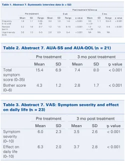
col-lected prospectively pre treatment and 3 months post treatment, includ-ing the American Urological Association Symptom Score (AUA-SS) and Quality of Life Score (AUA-QOL), 10-point Visual Analogue Scales (VAS) of symptom severity and effect on daily life, and systematic interview data. Results: At completion of the TVBEstim sessions, 52 patients reported a mean symptom improvement of 64.5% (SD 27, range 0–100); while 27 patients at 6 weeks post-treatment showed a durable improvement of 75.1% (SD 24, range 0–100).
Conclusions:Early results show TVBEstim is a promising treatment for refractory urgency and frequency in women identified to have pelvic floor muscle spasm.
8
Magnetic resonance microscopy of the murine urinary tract Ronald W. Wood, PhD1; Raymond B. Baggs, DVM, PhD2; Robert D.
Mayer, MD1; Scott Kennedy, PhD1
1University of Rochester School of Medicine, Rochester, NY; 2Oregon
State University, Corvallis, OR
Background:Several transgenic mouse strains display slow onset outlet obstruction associated with enlargement of tissues arising from the uro-genital sinus. Several of these strains display profound increase in prostate mass. Identifying the site of partial outlet obstruction in the tissue obtained at necropsy is extremely labor intensive using conventional histologic methods. Since magnetic resonance imaging can achieve levels of spa-tial resolution that approach or are comparable to light microscopy, we thought this modality might be an efficient way to identify areas of out-let obstruction within the tumour mass.
Methods:The N20 TRAMP FVB mouse is a rapid onset transgenic ade-nocarcinoma model that has a life expectancy of approximately 5 months. After documenting reduced uroflow in a mouse whose lower urinary tract was subsequently fixed by immersion in 10% NBF, MR imaging was performed at 9.4 tesla using a Bruker Omega PSG instrument. Samples in tubes were placed in either a 4-cm diameter bird cage coil or a 2.5-cm diameter Alderman-Grant resonator. The imaging gradients have an inter-nal diameter of 56 mm and maximum magnetic field gradients of 500 mTesla/metre. Three-dimensional gradient-echo images were acquired with the following initial parameters: first sample – TR/TE = 50/5 msec, flip angle approximately 25°, field of view was 4.0 ×2.8 ×2.8 cm with 512 ×256 ×256 resolution (voxel dimensions = 78 ×109 ×109 μm, 2 signal averages; second sample – TR/TE = 275/5 msec, flip angle approx-imately 45°, field of view was 3.2 ×1.4 ×1.4 cm with 512 ×256 ×256 resolution (voxel dimensions = 62 ×55 ×55 μm), 2 signal averages. Imaging parameters are adjusted from initial values for optimal contrast. Results:Extraordinary detail was obtained with this imaging modality. Segmentation of the bladder and urethral lumen provided evidence of patency that extended to an area near the opening of the seminal vesicles to the urethra.
Conclusions:Prostate and suburethral gland enlargement is observed in probasin-driven transgenic mouse model systems, and the present obser-vations are consistent with an obstruction resulting from suburethral gland enlargement. MR microscopy permits examination of fine detail of the murine lower urinary tract that can assist in blocking tissues for subse-quent higher-resolution imaging and conventional histology.
9
Outcomes with the Prolieve Thermodilation System for BPH William P. Conners, III, MD; Ronald Kaufman, MD
Albany Medical College, Albany, NY
Background:The Prolieve Thermodilation System is a minimally-invasive
PODIUM SESSION 2: BPH / NEUOROUROLOGY
Friday, September 7, 3:30–4:00 pm
Podium Session 2: BPH / Neuorourology
Table 1. Abstract 7. Systematic interview data (n = 52)
Post-treatment follow-up
Pre-treatment 6 wk 3 mo
Mean SD Range Mean SD Range p value Mean SD Range p value Frequency
(h to void)
1.8 1.1 0.25– 4.0
3.0 1.0 1–6 < 0.001 3.0 n = 31
1. 1 0.5–4 < 0.001
Nocturia (per h)
2.2 1.6 0–7 1.2 1.5 0–10 < 0.001 1.2 n = 29
1.2 0–5 < 0.001
Urge Intensity (0–5)
3.6 1.3 0–5 2.6 0.9 0–4 < 0.001 NA NA NA
Table 2. Abstract 7. AUA-SS and AUA-QOL (n = 21)
Pre treatment 3 mo post treatment
Mean SD Mean SD p value
Total symptom score (0–35)
15.4 6.9 7.4 8.0 < 0.001
Bother score (0–6)
4.3 1.2 2.8 1.7 < 0.001
Table 3. Abstract 7. VAS: Symptom severity and effect on daily life(n = 23)
Pre treatment 3 mo post treatment
Mean SD Mean SD p value
Symptom severity (0–10)
6.0 2.3 3.5 2.6 < 0.001
Effect on daily life (0–10)
therapy for moderate BPH that combines microwave ablation with ure-thral balloon dilation. Industry data reported a 30% improvement in AUA score in > 50% of patients by 2 weeks, durable to 12 months, with 70% of patients able to discontinue medical therapy following treatment and only 20% of patients requiring a catheter post procedure. Our previous short-term results were largely consistent with these findings. Here, we reexamine our long-term outcomes.
Methods:A retrospective chart review was conducted for those who under-went this procedure at our institution since its use began in August 2005. Pre-procedure and follow-up history, uroflow testing, AUA symptom score, and PVR were obtained. Uroflow data was not analyzed for volumes less than 100 mL. We limited data analysis to those with at least 6 months follow-up.
Results:Forty-one patients underwent the procedure, the majority of whom were being successfully medically managed for their BPH. The average follow-up period was 10.9 months (range 6–16 mo). Eighty point nine per-cent of patients were taken off of medical therapy, with 76.4% able to suc-cessfully stay off of medication. The average improvement in the AUA score was 2.3 points, with only 31% demonstrating at least 30% improvement.
There was essentially no change in the average Qmax(range –11 to 10.3 mL/s). The average PVR actually increased by 10 mL. The sub-group which fared the best were those who underwent Prolieve treatment in order to avoid starting on medical therapy for BPH with an average 4.5 mL/sec-ond increase in Qmax, 32 mL decrease in PVR and 4.3 point increase in AUA score, all improving at least 30% by AUA. While prostate volume did not significantly impact outcomes, those with urethral lengths of 3 cm or greater tended to do more poorly. Twelve percent of patients required a catheter post procedure. Seventeen percent of patients in this study sub-sequently went on to TURP for either retention or failure to improve following therapy. Twelve percent of patients were started on anticholin-ergic medications for new irritative voiding symptoms.
Conclusions:While our early outcomes with the Prolieve system were favourable, it has not proven to be effective long term. Of concern, a num-ber of patients also developed new irritative voiding symptoms follow-ing treatment. The patients who did best were not previously on med-ical therapy and had urethral lengths less than 3 cm. During the study period the Prolieve catheter had been modified, implicating possible equip-ment failure in the outcomes seen here.
10
Efficacy of sildenafil at 8 and 12 hours postdose in men with mild to moderate erectile dysfunction
Andrew R. McCullough III, MD, FACS1; Christopher P. Steidle, MD2; Brian
Klee, MD3; Li-Jung Tseng, PhD3
1New York University School of Medicine, New York, NY; 2Northeast
Indiana Urology, PC, Fort Wayne, IN; 3Pfizer Inc., New York, NY
Background: Empiric observation suggests that the period of effectiveness of sildenafil may be longer than its half-life of 4 hours. We sought to better define the period of responsiveness to sildenafil in 2 double-blind, placebo-controlled (DBPC) trials.
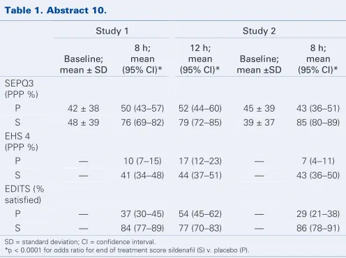
Methods: Study 1 had a crossover design with 2 4-week DBPC phases, while study 2 included a single 4-week DBPC phase. In both studies, men with mild to moderate ED (6-question IIEF Erectile Function domain score of 11−25) were given sildenafil (100 mg) or placebo and asked to have sexual intercourse 8 hours postdosing (7−9h) during an initial 4-week phase. In the first study, men crossed over to the other medication and were asked to have sexual intercourse 12 hours postdosing (11−13h) in a second 4-week phase. The primary end point was the mean per-patient proportion of occasions (PPP) of “yes” responses to question 3 of the Sexual Encounter Profile (SEP Q3), “Did your erection last long enough for you to have successful intercourse?” Other end points were the mean PPP of occasions of erection hardness score (EHS) of 1 to 4 and percentage of men satisfied with treatment (Erectile Dysfunction Inventory of Treatment Satisfaction [EDITS] Index > 50).
Results:A total of 351 men were randomized to placebo (n = 177, mean age ± SD = 52.8 ± 9.2 yr) or sildenafil (n = 174, mean age ± SD = 52.5 ± 10.5 yr) in the first study, and 250 men were randomized to placebo (n = 124; mean age ± SD = 52.9 ± 9.0 yr) or sildenafil (n = 126; mean age ± SD = 52.3 ± 8.8 yr) in the second study. Sildenafil demonstrated significant efficacy over placebo at both 8 and 12 hours postdose.
Conclusions:These findings illustrate that the period of responsiveness after a single dose of sildenafil in men with mild to moderate ED may be much longer than the 4 hours currently thought by many clinicians and patients to be the effective time period of sildenafil.
11
Return of nocturnal erections and erectile function after bilat-eral nerve-sparing radical prostatectomy in men treated night-ly with Viagra (sildenafil citrate): subananight-lysis of a randomized, double-blind, placebo-controlled trial
Andrew R. McCullough, MD, FACS1; Laurence A. Levine, MD2; Harin
Padma-Nathan, MD3
1New York University School of Medicine, New York, NY; 2Rush University
Medical Center, Chicago, IL; 3Keck School of Medicine at the University
of Southern California, Beverly Hills, CA
Background:After bilateral nerve-sparing radical retropubic prostatec-tomy (BNSRRP), nocturnal and sexually mediated erections may help to preserve normal erectile function (EF). To investigate nocturnal penile tumescence and rigidity (NPTR) in a subset (n = 54 men) from a ran-domized double-blind trial (n = 76) of nightly Viagra (sildenafil citrate) after BNSRRP.
Methods:Inclusion required preoperative “normal” EF (defined as a com-bined score of ≥8 for International Index of Erectile Function questions 3 [penetration] and 4 [maintained erection after penetration]) and NPTR testing (≥ 10 continuous minutes of > 55% rigidity [R > 55%] at the base of the penis). Postoperative assessments were at weeks 4 (pretreat-ment), 16, 28, 40 (during 36 weeks of nightly prophylaxis: Viagra 50 mg [n = 17], 100 mg [n = 18] or placebo [n = 19]), and 48 (after 8 weeks of no erectile dysfunction therapy). “Responders” were delineat-ed at week 48 by the defindelineat-ed normal EF and a “yes” response to “Over the past 4 weeks, have your erections been good enough for satisfacto-ry sexual activity?” Base and tip rigidity and tumescence of the penis were measured using RigiScan monitoring. Main outcome measures were dura-tion of R > 55% and areas under the curve for rigidity and tumescence. Results:Postoperatively, rapid profound reduction in nocturnal EF was noted in all groups. There was a gradual dose-dependent improvement in base and tip rigidity in the Viagra groups but little improvement in the placebo group. Eight weeks after treatment termination (48 wk post-operatively), 24% (4/17) of 50-mg Viagra recipients, 33% (6/18) of 100-mg Viagra recipients, and 5% (1/19) of placebo recipients were respon-ders. Tip R > 55% was the most discriminating NPTR measure between nonresponders and responders to Viagra, in whom it regained preoper-ative levels whereas base R > 55% did not. Tip R > 55% was most pro-longed in responders to Viagra 100 mg.
Conclusions:Although further study is needed, the results of this trial showed that nightly Viagra for 9 months post-BNSRRP objectively improved nocturnal erections and pharmaceutically unassisted EF in a significant
Podium Session 3: Impotence / Infertility
PODIUM SESSION 3: IMPOTENCE / INFERTILITY
Friday, September 7, 4:00–4:30 pm
Table 1. Abstract 10.
Study 1 Study 2
Baseline; mean ± SD
8 h; mean (95% CI)*
12 h; mean (95% CI)*
Baseline; mean ±SD
8 h; mean (95% CI)* SEPQ3
(PPP %)
P 42 ± 38 50 (43−57) 52 (44−60) 45 ± 39 43 (36−51) S 48 ± 39 76 (69−82) 79 (72−85) 39 ± 37 85 (80−89) EHS 4
(PPP %)
P — 10 (7−15) 17 (12−23) — 7 (4−11)
S — 41 (34−48) 44 (37−51) — 43 (36−50) EDITS (%
satisfied)
P — 37 (30−45) 54 (45−62) — 29 (21−38)
S — 84 (77−89) 77 (70−83) — 86 (78−91)
SD = standard deviation; CI = confidence interval.
*p < 0.0001 for odds ratio for end of treatment score sildenafil (S) v. placebo (P).
Table 1. Abstract 11. Mean duration of Tip R > 55%, as a percentage of preoperative levels, in responders (R) and nonresponders (NR)
4 wk 16 wk 28 wk 40 wk 48 wk
Viagra 100 mg or 50 mg (R), n = 10
16% 36% 62% 39% 145%
Viagra 100 mg or 50 mg (NR), n = 25
4% 16% 10% 28% 21%
proportion of men after BNSRRP, compared with the slow and incom-plete recovery in placebo recipients.
12
Developing a prognostic tool for Peyronie’s disease: validation of a percutaneous aspiration technique
Trustin Domes, MD1; Ling De Young, MD2; Gerald Brock, MD1 1Division of Urology, Department of Surgery, University of Western
Ontario, London, ON; 2St. Joseph’s Health Care, Lawson Health Research
Institute, London, ON
Background:One of the greatest challenges in the treatment of Peyronie’s disease (PD) is the lack of prognostic tools to help guide patient man-agement. Previous data from our laboratory demonstrated differential pro-tein expression between cell cultures of normal tunica albuginea and PD plaque tissue using surface enhanced laser desorption/ionization time-of-flight mass spectrometry (SELDI). The aim of this project is to vali-date a percutaneous penile plaque aspiration technique by correlating SELDI spectral data between aspiration and surgical biopsy specimens and to determine if a less invasive office-based needle aspiration tech-nique will provide an adequate amount of protein for analysis. Methods:Aspiration specimens were obtained from patients undergo-ing reparative surgery for PD. Aspiration was accomplished by movundergo-ing
a 25 gauge needle with a negative pressure syringe in and out of the palpable plaque within the tunica albuginea layer. During the surgery, a biopsy specimen of PD plaque was obtained for comparison purpos-es. Protein extracts were prepared using tissue protein extract buffer with protease inhibitor and homogenization. Total protein was quantified by BCA protein assay. Approximately 2 μg of protein from each sample was incubated on CM10 (weak cation exchange) array and read by the SELDI-PCS 4000 system. Spectra were analyzed using Ciphergen Express 3.0 software.
Results:Between 2–10 μg of protein was obtained from each sample by using the percutaneous aspiration technique. Similar spectral pat-terns were demonstrated between the surgical tissue and aspiration sam-ples. The spectral peaks at molecular weight of 2.4 kDa, 6.5 kDa and 6.7 kDa appeared in all samples. Peaks at 15.8 kDa and 66.6 kDa appeared in most samples.
Conclusions:Our aspiration technique is a valid means of procuring an adequate amount of protein for SELDI analysis. It is efficient, less invasive and provides similar proteomic information as the surgical biopsy spec-imen. This may serve as a means of detecting protein alterations in men with Peyronie’s disease. Further work to identify the proteins represent-ing these spectral peaks and to correlate the expression pattern of these proteins with disease severity is ongoing in our laboratory.
13
Cold scissor dissection simplified repair technique for partial nephrectomy
Adam Perlmutter; Satish Sharma, MD; Khurshid A. Guru, MD; Hyung L. Kim, MD
Roswell Park Cancer Institute, Buffalo, NY
Background:Goals of laparoscopic partial nephrectomy (LPN) include complete resection of the renal mass, and minimizing warm ischemia time and blood loss. We advocate use of cold scissor dissection to minimize the risk of positive margins and present a technique for suture closure, which achieves hemostasis without need for bolsters.
Methods:Thirty-one consecutive patients underwent laparoscopic par-tial nephrectomy with hilar clamping. Cold scissor dissection was per-formed followed by suture repair using 0 Vicryl suture on a partially straight-ened CPX needle. The needles were placed under the surface of the tumour bed in an overlapping horizontal mattress fashion and secured with LapraTy suture clips. The hilar clamp was released and additional sutures were placed across the tumour bed when necessary. Gelatin matrix sealant and Surgicel gauze were placed over the resection bed only after comfirm-ing complete hemostasis. The collectcomfirm-ing system was not repaired sepa-rately even when entry into the collecting was clearly noted.
Results:The mean patient age was 60 years (30–92 yr). There were 18 left-sided and 13 right-sided tumours. Mean tumour size was 2.3 cm (0.6–5.4). Thirteen tumours were located on the posterior surface of the kidney. Mean total operative time was 219 minutes (142–333 min). Mean warm ischemia time was 23 minutes (0–40 min). Mean blood loss was 316 mL (30–1000). Renal carcinoma was identified in 22 (71%) patients. All margins were negative. Mean length of stay was 1.6 days (1–6 d). No patient in this series required intraoperative transfusion; however 1 patient experienced delayed bleed requiring 4 units of blood. No patient developed a urine leak.
Conclusion:Hilar clamping and cold scissor dissection provide a clear view of the dissection plane through normal renal parenchyma, avoid-ing positive margins. Our suturavoid-ing technique simplifies the reconstruc-tion, achieving hemostasis and preventing urine leaks without use of bolsters.
14
Construct validity of the biometric smoothness in the ProMIS system: impact evaluation in a urology training program Andrew Feifer, MD; Jossée Delisle, BSc, RN; Maurice Anidjar, MD, FRCPC McGill University, Montreal, QC
Background:The use of simulation for the advancement of laparoscop-ic skill among urology residency programs continues to advance. One purported benefit of simulation is that it allows for objectification of tech-nical skill, enabling the documentation of performance improvements as experience increases. The aim of this study was to demonstrate the con-struct validity of the instrument smoothness parameter in the ProMIS (Haptica Ltd., Dublin, Ireland) augmented reality simulator using 5 stan-dardized laparoscopic suturing tasks.
Methods:Fourteen urology residents ranging from R5–R1 were assessed using the ProMIS system on 3 occasions using 5 standardized laparoscop-ic tasks, including peg transfer, intra and extra corporeal suturing, ves-sel cannulation and laparoscopic cutting. Smoothness of movement was measured by detecting the changes of instrument velocity over time (unit-less) for each task. The values were recorded and subjected to statisti-cal analysis using the Student’s t test. Senior residents with standard-ized laparoscopic experience greater than 50 hours were compared to
junior residents with less than 50 hours of cumulative experience. Results:The senior resident cohort demonstrated superior laparoscopic smoothness of movement in all 5 standardized laparoscopic tasks, demon-strating strong statistical significance (p < 0.05). This was further reflect-ed in an improvement in overall task completion among the senior resi-dent cohort as compared to the junior resiresi-dent cohort. The senior resiresi-dent group also demonstrated greater consistency of movement in this param-eter, as evidenced by the standard deviations across tasks. This resulted in a 38% reduction in unnecessary laparoscopic instrument manipulation. Conclusions:These preliminary results of construct validity for the smooth-ness biometric parameter of the ProMIS simulator demonstrate its abili-ty to distinguish between more experienced and novice urologic laparo-scopists in an urology teaching program. This is a compelling feature of ProMIS that should facilitate its further incorporation into urology train-ing programs worldwide. It further demonstrates that ProMIS can be used to assess, train and follow a variety of laparoscopic technical skills, and will enhance efficiency of laparoscopic movement, and possibly decreased operative time for patients.
15
Laparoscopic partial nephrectomy: functional and oncologic out-comes with up to 6 years follow-up
Jean-Baptiste Lattouf, MD1; Avi Beri, MD2; Oswald F.J. D’Ambros2;
Manfred Gschwendtner, MD2; Josef Ziegerhofer2; Karl Leeb2; Günter
Janetschek, MD2
1Centre Hospitalier de l’Université de Montréal, Montréal, QC; 2Krankenhaus der Elisabethinen, Linz, Austria
Background:We present functional and oncologic outcomes of laparo-scopic partial nephrectomy from one institution with a follow-up of up to 6.5 years.
Methods: Ninety-four patients underwent laparoscopic partial nephrec-tomy between August 2000 and September 2006. Of these, 53 patients with at least 1 year follow-up were included in this study. Mean patient age was 62.5 years. In 4 (7.5%) of the cases indication for partial nephrec-tomy was imperative. Mean tumour size was 2.4 cm (range 0.6–4.3). In 46 (86.8%) of the cases, postoperative histopathological examination was positive for renal cell carcinoma. A 2-tailed paired t test or Wilcoxon signed rank test were carried out for pre- and postoperative continuous parameters’ comparisons. A p value inferior to 0.05 was considered sta-tistically significant.
Results:Median follow-up was 36 months (mean 35.9; range 12–79 mo). Calculated creatinine clearance (CCT) decreased from a mean of 87.0 mL/minute preoperatively to 74.5 mL/minute in the immediate postop-erative period (p < 0.001). Three months following the surgery, CCT improved significantly to 80.8 mL/minute (p < 0.003). Postoperative nuclear scans showed functional kidney moiety in all but 1 case. A mean cal-culated postoperative split MAG-3 clearance was significantly lower on the operated side than on the contralateral side (74.0 mL/min v. 110.7 mL/min respectively; p < 0.001). However a mean postoperative peak concentration time was similar on the both sides (6.42 min v. 6.05 min; p = 0.652). The rate of positive surgical margins was 2.2% (1 patient). No cases of disease progression, local or port-site recurrence were observed. Two patients (3.8%) died 11 and 20 months after the surgery of unrelated causes. Overall survival was 96.2% and disease-free survival 100% at a median of 36 months of follow-up.
Conclusions:At a median follow-up of 3 years, laparoscopic partial nephrectomy demonstrates oncologic and functional results similar to that of open surgery.
Podium Session 4: Endourology / Laparoscopy
16
Percutaneous surgery for treatment-resistant biliary calculi A. Andrew Ray, BSc, MSc, MD1; Mordechai Duvdevani, MD2; Hassan
Razvi, MD1; John D. Denstedt, MD1
1University of Western Ontario, London, ON; 2The Hebrew University,
Jerusalem, Israel
Background:Patients that have failed open or endoscopic (ERCP) treat-ment of biliary stones have few remaining treattreat-ment options. Due to exten-sive experience with percutaneous treatment of renal calculi, these patients may also be referred to urologists following percutaneous biliary tract drainage. Here we report the results of all biliary calculi treated by endouro-logic methods at a single institution over the past 10 years.
Methods:We conducted a retrospective study of all patients that under-went percutaneous, endoscopic treatment of biliary calculi since January 1, 1997. Both hospital and clinic charts were systematically reviewed. We investigated both the endoscopic technique used as well as type and loca-tion of tract as predictors of success. Primary outcomes of interest were symptom and stone free rates, length of hospital stay and complications. Results:Over the past 10 years, 17 patients underwent 19 percutaneous treatments of their biliary calculi. The primary indication for treatment was: cholangitis (5), retained stone (10), and biliary colic (2). Of these, 15 patients (88.2%) had failed prior endoscopic or open attempts at treat-ment of their stones, while the remaining 2 patients (11.8%) were unable to tolerate a general anesthetic. Patients had experienced a mean of 1.79 prior failed attempts at stone removal. Several treatment modali-ties were utilized including Ho:YAG laser (68.4%), electrohydrolic lithotripter (15.8%), ultrasound (10.5%), basket extraction (10.5%) and balloon dilatation of the ampulla (21.1%). Overall, treatment led to symp-tom relief in 84.2% of patients and 73.7% were stone-free. Biliary tract imaging was conducted an average of 20.9 days after treatment. Average length of hospital stay was 1.97 days. One patient experienced a peri-operative AMI and another prolonged biliary drainage. Both had success-ful endoscopic treatment of their calculi. There were no cases of treatment-related sepsis and no other complications were observed. Conclusions:Biliary calculi may be successfully treated using standard endourologic methods with high stone-free rates. This technique is gen-erally well-tolerated even among high-risk patients.
17
Intraoperative imaging of renal cortical tumors using near infrared fluorescence of intravenous indocyanine green
Dragan J. Golijanin, MD; Ralph R. Madeb, MD; Eric A. Singer, MD, MA; Jonah S. Marshall, MD; Ronald W. Wood, PhD; Jay E. Reeder, PhD; Vikram S. Dogra, MD; Jorge L. Yao, MD; Erdal Erturk, MD; Guan Wu, MD, PhD; Jean V. Joseph, MD, MBA; Edward M. Messing, MD University of Rochester Medical Center, Rochester, NY
Background:Near infrared fluorescence (NIRF) of indocyanine green (ICG) has proven useful in the detection and treatment of various malignan-cies including breast and brain tumours. The objective of this study was to determine if renal cortical tumours can be detected by laser-induced NIRF of intravenous (IV) ICG.
Methods:Ten patients undergoing radical (2) or partial (8) nephrecto-my had NIRF intraoperative imaging of their renal tumours following intra-venous injection of ICG. After kidney mobilization, approximately 10 mL (2.5mg/mL) of ICG was injected intravenously. Shortly after injection, NIRF was recorded using a LUNA NIRF system (Novadaq Technologies Inc., Mississauga, Canada).
Results:All tumour and non-tumorous lesions were clearly delineated by NIRF in the partial nephrectomy patients. Mean tumour size was 3.6 cm (range 1.5–6.5 cm). A total of 14 lesions were found in 10 patients (9 solid and 5 cystic). All tumours (clear cell [8], papillary renal cell [2], chro-mophobe renal cell carcinoma [1]) were seen as hypo- or non-fluores-cent areas which were clearly demarcated from the surrounding nor-mal parenchyma. Benign cysts were characterized by increased fluorescence compared to the normal parenchyma. Simple, thick walled cysts had similar characteristics to normal parenchyma. Tumors exam-ined during radical nephrectomy were not well visualized because of the
thick layer of perinephric fat preventing NIRF detection. NIRF microscopy confirmed ICG presence in normal parenchyma, and less fluorescence inside tumorous tissue. H&E histopathology confirmed differential ICG fluorescence in tumour versus normal renal tissue. All surgical margins were negative and averaged 4 mm (range 2–10 mm). Intravenous ICG administration resulted in no hypersensitivity reactions or changes in post-operative hepatic or renal function.
Conclusions:NIRF after IV ICG permits accurate intraoperative detection of renal cortical tumours during partial nephrectomy. This modality may permit urologists to diagnose additional lesions, decrease positive sur-gical margin rates, and minimize resection of normal parenchyma thus preserving renal function.
18
Robot-assisted radical cystectomy and lymph node dissection: Is there a learning curve?
Kevan M. Sternberg, MD; Scott Wilbur; Lawrence Jenkins; Hyung L. Kim, MD; Wei Tan; Gregory Wilding, PhD; James L. Mohler, MD; Khurshid A. Guru, MD
Roswell Park Cancer Institute, Buffalo, NY
Background:A learning curve was sought for robot-assisted radical cys-tectomy for surgical treatment of invasive bladder cancer.
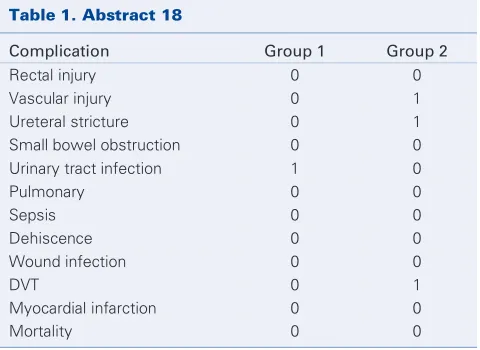
Methods:Between October 2005 and March 2007, 45 patients under-went robot-assisted radical cystectomy (RARC) and pelvic lymph node dissection. Diversion was performed open. Preoperative parameters (age, sex and BMI) were similar in both groups. Intra-operative parameters, immediate postoperative results and oncological efficacy were used to compare the first 10 (group 1) and the last 12 patients (group 2). Results:Total overall operative time was lower in the group 2 (441 min v. 324 min).The incision to RARC and diversion times were lower, while the robot-assisted PLND times were not different in the 2 groups (45 min v. 43 min). Overall median estimated blood loss increased in group 2 (475 mL v. 597 mL). Median hospital stay was similar in both groups (7 d). One patient was converted in the first group due to inabil-ity to tolerate steep Trendelenberg position. Two patients (group 1) with pathological disease greater than T- had positive margins while no pos-itive margins were reported in group 2.
Conclusion:RARC was safe and feasible during the initial part of a learn-ing curve, but increaslearn-ing experience was associated with shorter opera-tive times.
19
Evaluation of an elevated creatinine phosphokinase following da Vinci robotic assisted radical prostatectomy. What does it mean, and when should we look for it?
Podium Session 4: Endourology / Laparoscopy
Table 1. Abstract 18
Complication Group 1 Group 2
Rectal injury 0 0
Vascular injury 0 1
Ureteral stricture 0 1
Small bowel obstruction 0 0 Urinary tract infection 1 0
Pulmonary 0 0
Sepsis 0 0
Dehiscence 0 0
Wound infection 0 0
DVT 0 1
Myocardial infarction 0 0
Andrew T. Zabinski, MD
Albany Medical Center, Albany, NY
Background and Introduction:Rhabdomyolysis is muscle necrosis and the release of intracellular contents into circulation which can ultimate-ly lead to acute renal failure. This is a rare but serious complication that is most often encountered after operations in patients who have under-gone prolonged procedures in exaggerated positions. We encountered several patients with rhabdomyolysis after robotically assisted prostate-ctomy and therefore reviewed our experience with this. The purpose of this study was to determine if a change in patient positioning would lead to lower postoperative creatinine phosphokinase levels.
Materials and Methods:The charts and records of all patients who have undergone a robotically assisted radical prostatectomy between June 27, 2005 and December 30, 2006 were reviewed retrospectively with regards to rhabdomyolysis. There were a total of 20 patients who had their serum creatinine phosphokinase (CPK) measured (elevated > 3000 IU/L). Hospital records were reviewed for, age, body mass index (BMI), OR time, EBL, patient positioning, serum creatinine and CPK.
Results:A total of 20 patients were included in the study ranging from 43–66 years of age (mean 56.7 yr). OR time ranged from 5–9 hours (mean 6.6 h). EBL ranged from 50–1300 (mean 295). The mean preoperative serum creatinine was 1.06, mean immediate postoperative creatinine was 1.32 and mean creatinine upon discharge was 1.14. The initial 10 patients included in the review were positioned in stirrups, placed on a bean bag and placed in an exaggerated lithotomy position. The mean OR time was 6.725 hours (SD 1.2 h), mean BMI 30.1 (SD 4.75) and the mean post operative CPK was 2071 (SD 2779). At the end of the review, the final 10 patients were positioned with split legs, without a beanbag and then put into the exaggerated Trendelenberg position. The mean OR time for these patients was 6.3 hours (SD 0.8), mean BMI 26.9 (SD 2.99) and the mean post operative CPK was 800 (SD 637).
Discussion:Rhabdomyolysis is a rare but serious complication that can be encountered after urological procedures. We observed elevated CPK levels, peaking 16 hours after surgery in patients placed in exaggerated lithotomy positions undergoing robotically assisted radical prostatectomies. Although 9 patients had an elevated serum creatinine postoperatively, none required dialysis and no patient experienced permanent renal dam-age. This data suggests that re-positioning patients leads to lower post-operative creatinine phosphokinase levels.
20
Trends in the treatment of localized prostate cancer in Rochester, New York
Joy K. Knopf, MD1; Dragan J. Golijanin, MD1; Ganesh S. Palapattu, MD1;
Guan Wu, MD, PhD1; Hani H. Rashid, MD1; Gregory Oleyo, MD2; Louis
Eichel, MD2; John R. Valvo, MD2; Ralph Madeb, MD2
1Division of Urology, Rochester General Hospital, Rochester, NY; 2University of Rochester Medical Center, Rochester, NY
Background:Surgical telerobotic systems allow surgeons to visualize the operative field with great accuracy and manipulate instruments in a more intuitive and ergonomic fashion compared to pure laparoscopy. Urologic robotic surgery was adopted in Rochester, NY in 2001. Since then, Rochester urologists have been increasingly embracing robotic tech-nology and have emphasized robotic surgery in the residency training curriculum. We retrospectively analyzed changes in treatment of local-ized prostate cancer as the urologic community has transitioned from an open surgical environment to a robotic one.
Methods:We retrospectively reviewed OR case logs for all surgeons performing open and robotic prostatectomies in all Rochester, NY hos-pitals from 2003–2006. We assessed other modalities of treatment for localized prostate cancer and analyzed the influence robotic
prostatec-tomy had on the other modalities. The only oncologic parameter used to assess proficiency was positive surgical margin status — and was done only for those surgeons that had greater than 50 open and robotic cases. Other parameters evaluated were surgical logs of the graduating chief res-idents with respect to their open and robotic case numbers.
Results:Twenty surgeons in Rochester, NY regularly (> 10 cases per year) perform radical prostatectomy in Rochester’s 4 hospitals. Two of the 4 hospitals have robotic systems. In 2003–2004 there were approximate-ly 30 open radical prosatatectomies performed each month and less than 10 performed via the robotic approach. In 2006, the situation was reversed with approximately 50 robotic cases performed each month and less than 10 open cases performed. The rate of brachytherapy fluctuat-ed over time and increasfluctuat-ed in centres without a surgical robot. The number of open prostatectomies performed in those centres without a sur-gical robot dropped significantly with less than 10 cases performed per year. Those surgeons that had sufficient experience with both open and robotic prostatectomies were able to at least halve their positive margin rate with this new technology (p < 0.05). Also notable is the significant decrease in the number of open prostatecomies performed by our grad-uating chief residents between 2003 and 2006.
Conclusions:Since the adoption of robotic prostatectomy in Rochester, significant changes have been seen in surgical outcomes, individual and group practice patterns, and resident training. Training of residents has become more analytical and progressive rather than intuitive. Robotic systems will continue to be an important asset in the urologic arma-mentarium and will likely continue to influence practice patterns and training of urologists in our community.
21
Robot-assisted radical cystectomy and pelvic lymph node dis-section: comparison with open radical cystectomy
Scott Wilbur, BA; Jannah Thompson, MD; Lawrence Jenkins; Wei Tan; Gregory Wilding, PhD; Pamela Piacente, PA; Hyung L. Kim, MD; Robert P. Huben, MD; James L. Mohler, MD; Khurshid A. Guru, MD
Roswell Park Cancer Institute, Buffalo, NY
Background:Few series of robot-assisted radical cystectomy (RARC) have been reported. Robot-assisted radical cystectomy was compared to open radical cystectomy at Roswell Park Cancer Institute as part of a robot-assisted surgical quality assurance program.
Materials & Methods:Robot-assisted radical cystectomy and pelvic lymph node dissection was offered to all patients who were candidates for an open radical cystectomy since October 2005. Forty-five consecutive patients from March 2002 to September of 2005 who underwent open radical cystectomy were compared to 45 consecutive patients who under-went robot-assisted radical cystectomy and pelvic lymph node dissection. Demographics, operative times, hospital course, complications and patho-logic outcomes were compared in the 2 groups.
Results:Age, body mass index and ASA scores were similar between groups. The overall operative times were similar (Open: 384 min; Robot: 364 min). The 2 groups differed (p < 0.0001) in estimated blood loss (Open: 1731 mL; Robot: 542 mL), intra-operative transfusions (Open: 1.8; Robot: 0.3), time of removal of naso-gastric tube (Open: 5 d; Robot: 2 d), and time to diet (Open: 6 d; Robot: 4 d). The 2 groups had similar complication rates (Open: 45%; Robot: 35%). Pathologic stages were sim-ilar in the 2 groups; however, only 14 open patients had a lymph node dissection (LN yield: Open: 7; Robot: 17). Margin positive rates were 12% open and 9% robotic.
Conclusion:The early experience with robot-assisted radical cystecto-my compared to open radical cystectocystecto-my showed lower blood loss and similar pathological efficacy. Careful follow-up is required in order to establish long-term oncological efficacy.
22
Long-term durability of an in-office, nonsurgical transurethral radiofrequency treatment for female stress urinary incontinence: a retrospective analysis
Rodney A. Appell, MD, FACS
Baylor College of Medicine, Houston, TX
Introduction and Study Objective:A transurethral radiofrequency (RF) collagen denaturation system (Renessa, Novasys Medical Inc., Newark, Calif.) has been approved for nonsurgical treatment of women with stress urinary incontinence (SUI) due to urethral hypermobility. RF energy applied through a transurethral probe heats submucosal tissue to produce colla-gen denaturation resulting in reduced tissue compliance without necro-sis, thus distinguishing this procedure from a surgical transvaginal or laparo-scopic RF tissue ablation procedure (SURx, Cooper Surgical, Lake Forest, Calif.). This retrospective study aimed to determine the long-term dura-bility of transurethral RF collagen denaturation in women with SUI. Methods:In a prospective, randomized, controlled clinical trial, 110 women with SUI were blindly randomized to receive RF collagen denat-uration applied to the bladder neck and proximal urethra; 63 compara-ble women underwent identical sham treatment. All women were treat-ed as outpatients. Ninety-one active treatment group patients were evaluated at 1 year post-treatment. Further follow-up conducted at ≥3 years post-treatment included 21 women who had received active treat-ment. Patients completed the Incontinence Quality of Life questionnaire (I-QOL), a 3-day voiding diary, and a questionnaire about satisfaction with RF collagen denaturation and other SUI treatments.
Results:Outcome measures were ≥10-point I-QOL improvement from baseline; any improvement from baseline at 1 year, with ongoing improve-ment at ≥3 years; and decreased number of incontinence episodes at 1 year, with ongoing decrease from baseline at ≥3 years. Women who had not achieved success at 1 year and sought alternative treatments were evaluated for the impact of RF collagen denaturation on subsequent treat-ments. No long-term safety issues were identified. I-QOL scores improved in 16 women (mean improvement 17.6), similar to results at 12 months. Twelve women (55%) had ≥ 10-point I-QOL improvement at
≥3 years. The majority of women were satisfied with results after 3 years. Five women with recurrent SUI symptoms had undergone additional incon-tinence procedures before 3-year follow-up, without negative impact. Conclusions:Nonsurgical, transurethral RF collagen denaturation is a safe and effective SUI treatment and has demonstrated durable improvements in QOL, incontinence frequency, and patient satisfaction at ≥3 years post-treatment. This treatment does not negatively impact subsequent incon-tinence procedures.
23
Videourodynamics following in-office transurethral radiofre-quency collagen denaturation treatment for stress urinary incon-tinence
Kevin J. Cline, MD
Regional Urology, Shreveport, LA
Introduction and Objective:Following nonsurgical, transurethral radiofre-quency (RF) collagen denaturation treatment (Novasys Medical Inc., Newark, Calif.) in women with stress urinary incontinence (SUI) due to bladder outlet hypermobility, videourodynamics were evaluated. Methods:In a prospective, multicentre, single-arm clinical trial, 137 women with SUI were treated with RF collagen denaturation applied transurethrally to the bladder neck and proximal urethra. All women were treated as outpatients. Videourodynamics conducted at baseline and at 3 months in patients at 1 study centre included abdominal leak point
pres-sure (ALPP), cystometrogram, voiding prespres-sure study (VPS), and urinary flow rate. Patients also completed the Incontinence Quality of Life (I-QOL) questionnaire at both time points.
Results:Eight women were available for baseline and 3-month videouro-dynamics. At baseline, mean ALPP was 123 cm H2O at 200 mL and 111 at 250 mL of filling. On videourodynamics during voiding, 6 (75.0%) women had marked decensus and all had beaking. At 3 months, 3 women (37.5%) had no leaks at 200 mL, well above the baseline ALPP, and 1 woman (12.5%) had an improvement of 82 cm H20. At 250 mL, 2 (25%) women had no leaks, and 3 (37.5%) had mean improvement of 23 cm H20, for an overall improvement rate of 62.5% based on urodynamics. On fluoroscopy, 2 women improved from marked decensus (below pubic arch) to decensus (above pubic arch). The bladder neck was flattened dur-ing filldur-ing but showed beakdur-ing in all 8 women durdur-ing Valsalva manoeu-vre. Six women (75%) had improved I-QOL scores and 4 women (50%) had ≥10-point improvement by 3 months. Mean overall improvement was 25 points.
Conclusions:As shown in the pivotal trial at 12 months, RF collagen denat-uration resulted in measurable improvement in ALPP by 3 months post-treatment in most women. Videourodyamics may demonstrate a change in bladder outlet appearance during Valsalva movements; however, change in bladder outlet appearance does not correlate to change in ALPP. This supports the proposed mechanism of action for RF collagen denaturation, which is an increase in bladder outlet and proximal urethral resistance without a direct impact on hypermobility.
24
Decrease in mortality in a contemporary series of patients with Fournier’s gangrene
Anthony T. Corcoran, MD1; Marc C. Smaldone, MD1; Erin P. Gibbons,
MD1; Thomas Walsh, MD2; Benjamin Davies, MD2
1University of Pittsburgh Medical Center, Pittsburgh, PA; 2University of
California, San Francisco, San Francisco, CA
Background:Mortality rates for patients with Fournier’s gangrene range from 30% to 50% in the most recent published series. The Fournier’s Severity Index (FSI) utilizes clinical parameters to predict mortality, based upon these rates. Treatment of Fournier’s has evolved to include aggres-sive surgical débridement, broad-spectrum antibiotics, and intenaggres-sive med-ical monitoring. We report the largest series of Fournier’s patients to date, and hypothesize that advances in care have reduced mortality, necessi-tating adjustment of the FSI score as a predictive tool.
Methods:We retrospectively reviewed all patients treated at our institu-tion from 1993 to 2006 with a diagnosis of Fournier’s gangrene. Patient demographics, symptoms, comorbidities, physical exam and laboratory findings, operative records, intensive care monitoring, and all electron-ic hospital records were analyzed. FSI scores were calculated using admis-sion vital signs and laboratory data. Data were stratified according to the outcomes of death (n = 11) or survival (n = 57). Data were analyzed using multivariate conditional logistic regression. For comparisons of means, 2-tailed Wilcoxon tests were performed.
Results:A total of 68 patients (mean age 55.8 ± 15.2 yr) were analyzed. The overall mortality rate was 16% (n = 11), with a 10% (n = 7) initial hospitalization mortality rate. As an isolated parameter, elevated serum lactate on admission was predictive of mortality (p < 0.0022). Patients with an FSI score of 7 or greater had a mortality rate 4.5 times higher than those with an FSI score < 7 ([odds ratio] OR 4.5 [95% {confidence inter-val} CI 1.1–17.0]). When modelled as a continuous predictor, each 1-point rise in FSI predicted a 30% increased risk of mortality (OR 1.3 [95% CI 1.1–1.5]). No other clinical parameters, alone, or in
combina-Podium Session 5: Assorted Topics
tion were statistically significant predictors of mortality.
Conclusions:While Fournier’s gangrene remains a deadly disease, mor-tality rates have decreased by nearly 50% over reported values. The FSI remains a simple and objective method of predicting outcome in this patient population. However, unlike previous studies reporting negative outcomes in patients with FSI scores of 9 or greater, our results suggest that a lower FSI threshold of 7 may be a more accurate predictor of mortality.
25
The median lobe in robot-assisted radical prostatectomy: eval-uation and management
Lawrence Jenkins, BS1; Mark Nogueira, MD2; Terence N. Chapman, MD2;
Gregory E. Wilding, PhD2; Wei Tan, MA2; Hyung H. Kim, MD2; James
L. Mohler, MD2; Khurshid Guru, MD2
1State University of New York at Buffalo School of Medicine, Buffalo, NY; 2Roswell Park Cancer Institute, Buffalo, NY
Background:To determine if the presence of a median lobe can be pre-dicted preoperatively and whether its presence affects postoperative uri-nary function and immediate pathologic outcomes after robot-assisted radical prostatectomy.
Methods:From August 2004 to March 2007, 345 consecutive patients underwent robot-assisted radical prostatectomy at our institution. Retrospective review found that 29 (8%) had a median lobe. We com-pared these 29 men to 29 consecutive patients without a median lobe for preoperative clinical parameters, intraoperative parameters and patholog-ical and clinpatholog-ical outcomes.
Results:Preoperative parameters: the 2 groups were similar in age, race, preoperative Gleason score and urinary bother score. Patients without a median lobe had higher clinical stage (p = 0.043). Of 10 patients with a median lobe who had preoperative CT scans, 7 (70%) had a visible median lobe. Intraoperative parameters: the presence of a median lobe did not increase operative time required for bladder neck dissection or anastomosis (including reconstruction). Estimated blood loss was simi-lar between the 2 groups. Sixteen (55%) patients with a median lobe required bladder neck reconstruction compared with 1 (3.5%) without a median lobe. Methylene blue or indigo carmine was used in 10 patients with a median lobe for identification of the ureteral orifices. Postoperative parameters: the 2 groups had similar Gleason score, but patients with median lobes had larger prostates, 60.2 mL versus 26 mL (p = 0.003), and higher pathologic stage. Although pathologic stage was worse in men with median lobes, surgical margin status was similar. Postoperative urinary bother score and time to social or perfect continence were similar between the 2 groups.
Conclusions:Preoperative parameters cannot predict the presence of a median lobe. The presence of a median lobe does not alter pathologic outcomes or urinary function after robot-assisted prostatectomy.
26
Prediction of differential renal function as determined by con-trasted and non-concon-trasted computed tomography
Shelby N. Morrisroe, MD; Erin P. Gibbons, MD; Benjamin R. Stockton, MD; Kyongtae T. Bae, MD, PhD; Cheng Hong, MD, PhD; Christopher Deible, MD, PhD; Timothy Averch, MD; Stephen V. Jackman, MD University of Pittsburgh Medical Center, Pittsburgh, PA
Background:Radioisotope renography is currently the gold standard deter-mination of differential renal function. We propose the use of helical com-puted tomography (CT) may be a more efficient way to gain functional renal information. Previous studies have shown a highly significant cor-relation between contrasted spiral CT determined GFR and Tc 99m-MAG3, as well as a positive correlation between differential renal volumes and 24-hour creatinine clearances. We propose a simple method of CT eval-uation to determine differential renal parenchymal volumes, whereby per-centage total renal volume can then be used as a surrogate for percent-age renal function.
Methods:CT and diuretic enhanced Tc 99m-MAG3 studies were per-formed in 23 patients with chronic obstruction. CT imaging was con-trast enhanced in 17 and non-enhanced in 6. Diagnoses included UPJ obstruction in 13, nephrolithiasis in 5, ureteral stricture in 3, extrinsic compression in 1, and flank pain in 1. Measurements of renal parenchy-mal volume by CT used a region-based thresholding method. The prod-ucts of slice thickness and area measurements were summated to obtain the renal unit volume. Each renal unit volume was converted to per-centage total renal volume and correlated to perper-centage renal function, as calculated by Tc 99m-MAG3. Pearson’s correlation coefficient, asso-ciated p values and confidence intervals were determined.
Results:A very strong correlation was observed between percentage renal function and percentage total renal volume in both contrast enhanced (r = 0.92, p < 0.0001, 95% [confidence interval] CI 0.79–0.97) and non-enhanced CT groups (r = 0.95, p < 0.0001, 95% CI 0.63–0.99). The correlation remained strong among 9 patients with a single kidney func-tion between 6%–30% (r = 0.80, p = 0.0103, 95% CI 0.28–0.96). A subgroup of 5 patients had Tc 99m-MAG3 renography performed both before and after endoscopic or laparoscopic intervention for UPJ obstruc-tion. Timing of functional imaging did not significantly effect correla-tion between percentage renal funccorrela-tion and percentage total renal vol-ume (r = 0.98, p = 0.0037 for early renography v. r = 0.98, p = 0.0043 for delayed renography).
Conclusions:Both contrast enhanced and non-enhanced helical CT pro-vide a simple measure of differential renal volume which strongly cor-relates to differential renal function, even with a functional range < 30%. We propose renal scans are not necessary to determine differential func-tion if a CT has been performed, and recommend CT as a single radio-logical diagnostic study of both anatomic and functional assessment in patients with a suspected poorly functioning kidney.
27
Diagnostic confirmation in high PSA, negative biopsy patients: a pilot study on the use of fused capromab pendetide (ProstaScint) scans
Michael J. Manyak, MD, FACS1; Henri P. Lanctin, MD2; John G. Wolodzko,
PhD1; Samuel L. Kipper, MD2; Greg S. Parries, MD2; Roy R. Brown, MD2;
Chris W. Boelter, MD2
1Cytogen Corporation, Princeton, NJ; 2St. Cloud Hospital, St. Cloud, MN
Background:Investigators have shown that prostate-specific membrane antigen (PSMA), expressed in the gland in prostate cancer and upregu-lated in patients with high grade, androgen insensitive, and metastatic deposits, predicts recurrence when overexpressed in the gland. Capromab pendetide (CP) immunoscintigraphy, which recognizes PSMA, fused with CT was used to scan men with high PSA and previous negative biopsy before repeat biopsy.
Methods:Fifteen men (age 53–74 yr) with elevated PSA (mean 6.8 ng/mL, range 4.1–15.3 ng/mL), a mean prostate volume of 61 (range 29–130) grams, and at least 1 negative biopsy underwent fused CP/CT scans before repeat biopsy (mean 17 cores). Scans guided additional biopsies evalu-ated by blinded independent readers.
Results:Thirteen of 15 men had evaluable fused scans with 2 inevaluable due to CT artifact. Ten of 13 had negative biopsies and 3 had moderate grade prostate cancer, 2 with a solitary small focus and 1 with 3 small foci. Five of 13 had increased focal signal intensity including 2 of the 3 with prostate cancer. All transrectal sonograms were isoechoic. Correlation of scan and pathology occurred in 9 cases (69%), 3 focally-small positive scans had negative biopsies, and 1 negative scan had a solitary small focus of cancer. All 3 patients with positive scans and negative biopsy deferred repeat biopsy.
Conclusions:Fused capromab pendetide scans may provide additional diagnostic information for patients with elevated PSA and negative biop-sies suspected of harboring prostate cancer. This preliminary data warrants further investigation of increased intraprostatic PSMA signal intensity.
28
Biochemical and local control of primary prostate cancer patients treated with cryoablation and tracked with the COLD registry Martin K. Dineen, MD1; Louis L. Pisters, MD2; Bryan J. Donnelly, MD3;
Aaron E. Katz, MD4; Franco M. Lugnani, MD5; John C. Rewcastle, PhD3;
J. Stephen Jones, MD6
1Atlantic Urology, Daytona Beach, FL; 2MD Anderson Cancer Center,
Houston, TX; 3University of Calgary, Calgary, AB; 4Columbia University,
New York, NY; 5Society for Cryosurgery, Trieste, Italy; 6Cleveland Clinic,
Cleveland, OH
Background:The use of cryoablation as an initial treatment for local-ized prostate cancer has increased. The objective of this study is to report the outcomes of modern cryoablation at a large number of centres, both academic and community, which have participated in the Cryo On-Line Data (COLD) Registry.
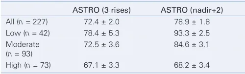
Methods:A secure on-line database was developed consisting of case report forms designed to collect relevant pre and post treatment infor-mation for patients undergoing prostate cryoablation. Data from 1608 patients who had undergone primary cryotherapy is in the registry but this analysis includes only the 690 each of whom had a minimum 24 months of follow-up. Patients were stratified according to risk groups as follows. For low risk patients all of the following were true: PSA < 10, Gleason < 7 and stage < T2b. If a patient had a PSA > 20, Gleason > 7, or stage T2c or greater they were considered high risk. All other patients were con-sidered moderate risk. Biochemical failure was defined according to both
the original ASTRO definition (3 rises) and the 2006 updated ASTRO def-inition of nadir+2. Biopsy was performed at the physician’s discretion, but most commonly if a patient had a rising or suspicious PSA. Results:The average age was 67.8 ± 7.8 years. Pre treatment PSA was 9.7 ± 10.2 ng/mL, the average Gleason was 6.5 ± 1.3. Only 19.0% of patients had low risk disease, 37.7% had moderate risk disease and 43.3% had high risk. Patients were followed for 48.3 ± 26.2 months. Five year actu-arial biochemical survivals and the number of patients at risk at 5 years are reported in the table. Notably, 27 of the 157 patients (17.2%) who failed according to the 3 rises definition did not have their PSA ever rise above 0.5 ng/mL. A total of 304 underwent post treatment biopsy. Of these, 53 showed evidence of disease resulting in a positive biopsy rate for those who underwent biopsy of 17.4%. The positive biopsy rate of the entire population was 53/690 (4.9%).
Conclusions:Cryoablation, as a primary treatment for prostate cancer practised over a wide spectrum of users provides durable biochemical and local control through 5 years.
29
Androgen replacement therapy after prostate cancer William P. Conners, III, MD; Ronald Kaufman
Albany Medical College, Albany, NY
Background:Androgen replacement therapy (ART) for hypogonadal men is widely considered to be contraindicated in patients with a history of prostate cancer, even in those who have been effectively treated and have no evidence of recurrent disease. However, there is a clear dearth of literature, and especially recent studies, to support this view. We exam-ined our experience with ART following prostate cancer treatment for safe-ty and efficacy.
Methods:A retrospective chart review was conducted for all patients receiving ART following definitive treatment for prostate cancer at our institution. The data was analyzed for tumour stage, grade, treatment modality, type and duration of ART, indication for ART, evidence of clinical or biochemical recurrence, as well as pre- and post-therapy testos-terone levels.
Results:Nine patients, with ages ranging from 57 to 80 years (mean 67) were identified. The Gleason score of those treated ranged from 6 to 9 with a mode of 7. The cancer stages ranged from cT1c to pT3a, with all being node/metastasis negative. Five were treated with surgery alone, 3 with combination external beam radiation and androgen deprivation ther-apy, and one with a combination of external beam, brachyseed, and andro-gen deprivation therapies. The interval between definitive treatment and initiation of ADT ranged from 4 to 76 months (mean 40), with a