_____________________________________________________________________________________________________ *Corresponding author: E-mail: [email protected];
www.sciencedomain.org
Bone Marrow Mast Cell Density Correlates with
Circulating Biomarkers of Bone Disease in Multiple
Myeloma
Rodanthi Vyzoukaki
1, Maria Devetzoglou
2, Constantina A. Pappa
3,
Andreas N. Antonakis
4, Samer Masud
1, Anastasia Papadopoulou
1,
George Tsirakis
5and Michael G. Alexandrakis
2*1
Laboratory of Hematology, University Hospital of Heraklion, Voutes, Heraklion, 71110, Greece.
2
Department of Hematology, University Hospital of Heraklion, Voutes, Heraklion, 71110, Greece.
3
Department of Internal Medicine, Venizelion Hospital of Heraklion, Knossos Avenue, Heraklion, Greece.
4
Department of Medicine, University of Crete, Voutes, Heraklion, 71110, Greece.
5
Department of Hematology, General Hospital of Chania, Chania, Greece.
Authors’ contributions
This work was carried out in collaboration between all authors. Authors RV and MGA designed the study, wrote the protocol and wrote the first draft of the manuscript. Authors GT and MD managed the literature searches, and performed the ELISA analysis. Authors CAP and ANA managed the experimental process. Authors SM and AP analyzed the data. All authors read and approved the final manuscript.
Article Information
DOI: 10.9734/BJMMR/2016/25402 Editor(s): (1) Rui Yu, Environmental Sciences & Engineering, Gillings School of Global Public Health, The University of North Carolina at Chapel Hill, USA. Reviewers: (1) Nikhil Gupta, Dr Mgr University, Tamil Nadu, India. (2)Burak Uz, Gazi University, Turkey. Complete Peer review History:http://sciencedomain.org/review-history/14208
Received 29th February 2016 Accepted 8th April 2016 Published 16th April 2016
ABSTRACT
Aims: There is increased mast cell density (MCD) in multiple myeloma (MM) bone marrow (BM)
that is correlated with advanced disease stage. Mast cells (MCs) produce various mediators promoting MM growth and bone metabolism. The aim of this study was to evaluate whether BM MCD correlates with markers of bone metabolism, such as circulating levels of osteoprotegerin (OPG), soluble receptor activator of nuclear factor-κB ligand (sRANKL), osteopontin (OPN) and macrophage inflammatory protein-1 alpha (MIP-1 alpha).
Study Design: This is a cross-sectional study.
Place and Duration of Study: Department of Medicine, University of Crete, Department and
Laboratory of Haematology, University Hospital of Heraklion, Department of Haematology and Internal Medicine, Venizelion Hospital of Heraklion, Department of Haematology, General Hospital of Chania, between January 2010 and December 2014.
Methodology: All parameters were analyzed by ELISA. We studied 56 active MM patients (32
males, 24 females) and 20 healthy controls. According to ISS 14 patients were in stage I, 22 in stage II and 20 in stage III. MCs were highlighted in BM immunohistochemically, using monoclonal antibody to MC tryptase.
Results: All values were higher in active MM patients compared to healthy subjects (p<0.001 for all
cases). OPG was decreasing in advanced stages (p= 0.018), whereas all other values were in parallel with ISS disease stages (p<0.001 for all cases). Moreover, BM MCD correlated positively with sRANKL, OPN and MIP1 alpha (p<0.0001 for all cases), and negatively with OPG (r= -0.279 p<0.03).
Conclusion: MCs are increased in MM BM and participate in many aspects of the disease. They
may release various cytokines increasing myeloma growth and also may participate in the skeletal disease of MM rendering them as potential targets for therapy.
Keywords: Bone disease; mast cells; multiple myeloma; osteolysis.
ABBREVIATIONS
Mast Cell Density (MCD); Multiple Myeloma
(MM); Bone Marrow (BM); Monoclonal
Gammopathy of Undetermined Significance
(MGUS); Macrophage Inflammatory
protein-1 alpha (MIP-protein-1 alpha); Receptor Activator of
Nuclear factor-κB (RANK); RANK Ligand
(RANKL); osteoprotegerin (OPG); osteopontin (OPN); Mast Cells (MCs); International Staging
System (ISS); Stem-cell Factor (SCF);
Interleukin-3 (IL-3); Microvascular density (MVD); N-terminal telopeptide (Ntx).
1. INTRODUCTION
Multiple myeloma (MM) is a debilitating B cell neoplasia, part of a spectrum of diseases, ranging from monoclonal gammopathy of undetermined significance (MGUS) to plasma
cell leukemia. This progression is based on a quite complex molecular basis, with both
genetic aberrations in MM cells and evolving interactions between different cell types. Malignant plasma cells rely heavily on their interactions with elements of the surrounding
microenvironment, in order to survive and further to expand. By these means, several cell types and cytokines are involved, playing
major roles in various aspects of MM progression, including angiogenesis and skeletal disease [1].
Myeloma skeletal disease is unique among other malignant bone diseases. It is considered to be
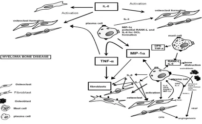
Fig. 1. Mechanisms responsible for myeloma bone disease. Myeloma cells produce factors that activate osteoclast formation. In addition they induce RANK-Ligand and IL-6 production
and enhance bone destructive process. Myeloma cells also produce IL-3, MIP-1a and VEGF and in combination with OPN produced by osteoclasts mediate the process of angiogenesis
There is also a plethora of cells implicated in the various aspects of MM progression. Among them, inflammatory cells are active participants and mast cells (MCs) possess multiple roles: they are accumulated in BM microenvironment as an initial host response, with potential tumorcidial activity. By these means, myeloma cells secrete attractants for MCs, but in turn, they secrete several factors favoring MM growth: MCs accumulation, although with an initial anti-tumor mood, aims disease progression [15,16]. There is an extensively studied participation of MCs in the angiogenic process: MCs secrete various direct and indirect angiogenic factors and also may participate in vasculogenesis mimicry [15,17,18]. We have already correlated BM MCs density (MCD) with international staging system (ISS) stages [19] and proliferation rate of myeloma plasma cells [20]. These correlations could be justified through their participation in the angiogenic process. On the other hand, since MCs may produce various versatile factors, it seems that their participation in MM growth is more complex.
The aim of the present study is to ascertain whether the degree of MCs accumulation in MM
BM is associated with bone disease. Therefore, we estimated MCD in active MM BM and correlated it with circulating levels of known factors of bone disease, such as MIP-1 alpha, OPN, IL-6, OPG, and sRANKL.
2. MATERIALS AND METHODS
2.1 Materials
matched healthy subjects (12 males and 8 females, median age 64, range 41-75 years) were recruited as controls among blood donors and people following a physical training program (over 60 years of age). All procedures performed in studies involving human participants were in accordance with the ethical standards of the institutional research committee and with the 1964 Helsinki declaration and its later amendments. Informed consent was obtained from all individual participants included in the study.
2.2 Methods
Serum samples from patients were collected before starting any myeloma-related treatment. Sera, collected from patients and controls, were aliquoted into separate vials and stored at
−70°C. All assays were performed at the end of
the study, in order to avoid interassay variability. Before starting the analysis, the samples were thawed to room temperature and mixed by vortexing in order to eliminate substances potentially affecting reproducible results. Hemolyzed or lipemic samples were excluded from the analysis. The detection of MIP-1alpha, OPN, IL-6, OPG, and sRANKL, in the serum was performed by a solid-phase sandwich ELISA, using monoclonal antibodies against MIP-1alpha, OPN, IL-6 (Quantikine, R&D systems Inc. Mineapolis, MN, USA), OPG and sRANKL antibodies (Bio Vendor-Laboratorni Medicina as Brno, Czech Republic).
MM patients and controls underwent trans iliac trephine biopsies. The histological diagnosis was made on hematoxilin-eosin-stained slides. Initially, biopsies were fixed in 10% formalin decalcified in 10% EDTA for 48h and embedded in paramat extra. Hematoxylin and eosin stained, 3 µm thick sections were examined by light microscopy. MCD was estimated immunohistochemically, as has been described previously [19]. Briefly, MCs were highlighted by immunostaining with the monoclonal antibody to MC tryptase (Ab-2 of Thermo-Scientific, Clone AA1) using the alkaline phosphatase – antialkaline phosphatase method (ultravision LP Detection System- Thermo-Scientific-TL12S-AL). After deparaffization and gradual rehydration, sections were incubated with the primary antibody (dilatation 1/3000, 30 min, room temperature without requirement of antigen retrieval, with the use of negative and positive controls (non-malignant tonsillar tissue from the files of the laboratory). In each specimen BM
MCs were quantified in three areas of neoplastic infiltration containing the highest number of MCs (hot spot) in ten high-power fields (HPF) (x400) for each hot spot separately. MCD was the mean percentage of positive staining MCs calculated in the three hot spots for each specimen and expressed as mast cells/HPF (MCs/0.0625 mm2).
2.3 Statistical Analysis
Data are presented as means ± SD. The Mann-Whitney U-test was applied to evaluate any difference in serum cytokine levels in patients and healthy individuals. Non- parametric tests were also used (Mann-Whitney and Wilcoxon for independent and paired two samples statistics). The investigation of potential correlations among variables was carried out with the Spearman’s rho correlation co-efficient. P values <0.05 were considered to be statistically significant.
3. RESULTS
Mean± SD levels of the measured parameters in active MM patients and in controls are shown in Table 1.
Table 1. Values of bone marrow mast cell (MC) density (MCD) and serum levels of osteoprotegerin (OPG), soluble receptor
activator of nuclear factor κΒ ligand (sRANKL), osteopontin (OPN) and macrophage inflammatory protein-1 alpha (MIP- 1alpha) in patients with active multiple myeloma (MM) compared to healthy controls.
(p<0.001 for all cases)
MM patients
Controls
(n= 56) (n= 20)
MCD (MCs/0.0625 mm2)
10.0±5.2 1.6±0.6
OPG (pmol/l) 13.1±11.6 6.4±3.5 sRANKL (pmol/l) 368.5±391.5 20.7±12.6 OPN (pg/ml) 78.7±58.4 40.0±32.8 MIP-1alpha (pg/ml) 46.9±29.7 21.0±2.8
Table 2. Values of bone marrow mast cell density (MCD) and serum levels of osteoprotegerin (OPG), soluble receptor
activator of nuclear factor κΒ ligand (sRANKL), osteopontin (OPN) and macrophage inflammatory protein-1 alpha (MIP-protein-1alpha) in various stages of multiple myeloma, according to International
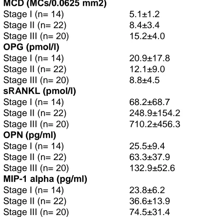
Staging System (p<0.001 for MCD, sRANKL, OPN and MIP-1 alpha and p= 0.018 for OPG)
MCD (MCs/0.0625 mm2) Stage I (n= 14) 5.1±1.2 Stage II (n= 22) 8.4±3.4 Stage III (n= 20) 15.2±4.0
OPG (pmol/l)
Stage I (n= 14) 20.9±17.8 Stage II (n= 22) 12.1±9.0 Stage III (n= 20) 8.8±4.5
sRANKL (pmol/l)
Stage I (n= 14) 68.2±68.7 Stage II (n= 22) 248.9±154.2 Stage III (n= 20) 710.2±456.3
OPN (pg/ml)
Stage I (n= 14) 25.5±9.4 Stage II (n= 22) 63.3±37.9 Stage III (n= 20) 132.9±52.6
MIP-1 alpha (pg/ml)
Stage I (n= 14) 23.8±6.2 Stage II (n= 22) 36.6±13.9 Stage III (n= 20) 74.5±31.4
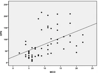
A significant negative correlation was found between MCD and OPG (r= -0.279, p<0.03), whereas positive correlations between MCD with sRANKL (r= 0.572), OPN (r= 0.605) (Fig. 1).
4. DISCUSSION
MCs are found in most tissues, serving a fundamental role within the immune system [22]. They arise from pluripotent CD34+ stem cells and differentiate along the myeloid pathway. After leaving BM, MCs progenitors are influenced by a variety of signals directing their migration into tissues, where they subsequently mature [23]. A major step is the interaction with endothelial cells and various tissue factors via adhesion molecules. MCs chemotaxis occurs in response to a variety of growth and differentiation factors, such as stem-cell factor (SCF) and interleukin-3 (IL-3), and to numerous chemokines. Among others, MIP-1-alpha, elicit migration of mouse BM MCs after Fc epsilon RI activation on laminin, vitronectin, and fibronectin
[24]. MCs can be activated after exposure to allergens or by other triggers, such as cytokines and neuropeptides. Their activation can be enhanced by SCF and IL-33 [25]. Upon activation, MCs release various mediators upon activation. These mediators can be divided into three overlapping categories: preformed and newly-synthesized lipid mediators, cytokines and chemokines. There is a large body of evidence supporting their involvement in tumor angiogenesis. Therefore, among others, MCs secrete several classical (vascular endothelial growth factor, fibroblast growth factor-2, platelet-derived growth factor beta, IL6) and nonclassical (IL8, chymase, tryptase) proangiogenic factors. Tryptase is a very powerful factor, inducing proliferation of endothelial cells and degradation of extracellular matrix components, through activation of latent metaloproteinases, releasing latent pro-angiogenic factors [16,23].
MCs accumulation has been associated with enhanced growth and invasion in various human cancers, including breast, stomach, esophagus and oral cavity [26]. There is an increasing study of MCs participation in MM progression [27,28]. The major aspect of MCs participation in MM progression is enhancing angiogenesis: BM MCD has been correlated with BM microvascular density (MVD) in MGUS, non-active and active MM [29]. It seems that MCs produce various angiogenic factors and may participate in vasculogenesis mimicry. We already have demonstrated that MM BM MCD is in parallel with ISS stages and also correlates with proliferation rate of myeloma plasma cell [19,20]. We suggest that these correlations are justified due to participation of MCs in the angiogenic process. Angiogenesis is a complex procedure, favouring direct MM growth and the implicated participants possess multiple and versatile properties.
Fig. 2. Positive correlation of mast cell (MC) density (MCD) (MCs/0.0625 mm2) with osteopontin (OPN) (pg/ml) in patients with active multiple myeloma (r= 0.605, p<0.0001)
During the last decade, immunomodulatory drugs, icluding thalidomide and lenalidomide and proteasome inhibitors, such as bortezomibe, have been increasingly used for the treatment of MM. The role of these drugs in bone metabolism has been evaluated in several studies [31]. Proteasome inhibitor bortezomibe is a potent, highly selective, and reversible proteasome inhibitor that targets 26S proteasome complex and inhibits its function [32]. The proteasome regulates key cellular processes, including modulation of transcription factors, such as NF-kB, cell cycle progression, inflammation, immune surveillance, growth arrest, and apoptosis [33]. Bortezomib has inhibitory effects on the NF-κB activity in MM cells. NF-κB is a major transcriptional factor which mediates the expression of many proteins including cytokines, chemokines, cell adhesion molecules, as well as those involved in anti-apoptosis and cellular growth control [33]. Thalidomide has a direct
tumoricidal activity, an antiangiogenic effect and modulates TNF-α signalling through direct
and/or indirect effects on the tumour microenvironment [34], reduces FGF-2, VEGF and IL-6 secretion in bone marrow stromal cells and by MM cells [35]. Lenalidomide, a derivative of thalidomide, is less toxic and more potent than the parent drug [36]. In patients with relapsed or
refractory MM, lenalidomide can overcome resistance not only to conventional chemotherapy but also to thalidomide [37,38]. The bisphosphonates are other compounds that, although originally used to reduce bone loss in MM due to an anti-osteoclast activity, have also been shown to have a direct effect on MM cells [39]. In fact, zoledronic acid has a direct cytotoxic activity on tumor cells and suppresses angiogenesis [40,41], inhibits FGF-2 - and VEGF-dependent proliferation of endothelial cells and inhibits VEGFR-2 in an autocrine loop [39] .Neridronate exerts its antiangiogenic activity through both a direct effect on endothelial cell proliferative activity and inhibitory effect on the responsivity of the endothelial cells to the proliferative stimuli mediated by angiogenic cytokines [15]. No drugs that target biomarkers have been used in this study.
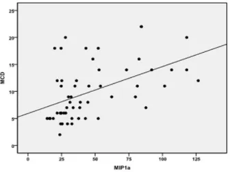
Fig. 3. Positive correlation of mast cell (MC) density (MCD) (MCs/0.0625 mm2) with macrophage inflammatory protein-1 alpha (MIP-1a) (pg/ml) in patients with active multiple myeloma
(r= 0.528, p<0.0001)
progression, which in turn is correlated with both disease activity and bone disease [28], it can be suggested that the participation of MCs in osteolytic disease is indirect. On the other hand, there are several angiogenic procedures participating in osteolytic disease, such as degradation of extracellular matrix. Our observation is enhanced by the positive correlations of MCD with levels of RANKL, MIP-1alpha and OPN. The correlation with RANKL may be partially direct, since MCs may secrete RANKL, along with IL-6, prostaglandin D2, tumor necrosis factor, and tryptase, all of which may cause bone disease in MCs disorders [23,29]. Similarly, the correlation with OPN may also be partially direct since MCs may produce OPN [42,43]. It is obvious that for both cases, MCs are not their unique source. On the other hand, the positive correlation of MCD with MIP-1alpha and the negative correlation with OPG are rather indirect and can be justified due to the modified BM microenvironment, favouring all aspects of MM growth. To the best of our knowledge, no study until now has displayed a correlation between MCD and parameters of bone disease, such as MIP-1alpha, OPG, sRANKL and OPN, in MM.
5. CONCLUSION
Our study showed that MCs play a significant role in MM growth and progression. They mainly promote angiogenesis, but it seems that they also may participate, indirectly and directly, in skeletal disease, as well. Therefore, the participation of MCs in MM biology is getting more complex and more studies are needed in order to explore the precise implicated mechanisms.
CONSENT
All authors declare that ‘written informed consent was obtained from the patient (or other approved parties) for publication of this article and accompanying images.
ETHICAL APPROVAL
COMPETING INTERESTS
Authors have declared that no competing interests exist.
REFERENCES
1. Hideshima T, Mitsiades C, Tonon G, Richardson PG. Understanding multiple myeloma pathogenesis in the bone marrow to identify new therapeutic targets. Nat Rev Cancer. 2007;7:585-98.
2. Reagan MR, Liaw L, Rosen CJ, Ghobrial IM. Dynamic interplay between bone and multiple myeloma: emerging roles of the osteoblast. Bone. 2015;75:161-9.
3. Toscani D, Bolzoni M, Accardi F, Aversa F, Giuliani N. The osteoblastic niche in the context of multiple myeloma. Ann N Y Acad Sci. 2015;1335:45-62.
4. Gerard C, Rollins BJ. Chemokines and disease. Nat Immunol. 2001;2:108-15. 5. Choi S, Cruz JC, Craig J, Anderson J,
Roodman GD, Alsina M. Macrophage inflammatory protein (MIP)-1a is a potential osteoclast stimulatory factor in myeloma. Blood. 2000;96:671-5.
6. Han JH, Choi SJ, Kurihara N, Koide M, Oba Y, Roodman GD. Macrophage inflammatory protein-1a is an osteoclastogenic factor in myeloma that is independent of receptor activator of nuclear factor kappaB ligand. Blood. 2001;97:3349-53.
7. Kukita T, Nomiyama H, Ohmoto Y, Kukita A, Shuto T, Hotokebuchi T, et al. Macrophage inflammatory protein-1a; (LD78) expressed in human bone marrow: Its role in regulation of hematopoiesis and osteoclast recruitment. Lab Invest. 1997;76:399-406.
8. Vallet S, Anderson KC. CCR1 as a target for multiple myeloma. Expert Opin Ther Targets. 2011;15:1037-47.
9. Nakagawa N, Kinosaki M, Yamaguchi K, Shima N, Yasuda H, Yano K, et al. RANK is the essential signaling receptor for osteoclast differentiation factor in osteoclastogenesis. Biochem Biophys Res Commun. 1998;253:395-400.
10. Boyle W, Simonet W, Lacey D. Osteoclast differentiation and activation. Nature. 2003;423:337-42.
11. Roodman F. Treatment strategies for bone disease. Bone marrow Transplant. 2007; 40:1139-46.
12. Lacey D, Timms E, Tan H, Kelley M, Dunstan C, Burgess T, et al. Osteoprotegerin ligand is a cytokine that regulates osteoclast differentiation and activation. Cell. 1998;93:165-76.
13. Haylock DN, Nilsson SK. Osteopontin: A bridge between bone and blood. Br J Haematol. 2006;134:467-74.
14. Sfiridaki A, Miyakis S, Pappa C, Tsirakis G, Alegakis A, Kotsis V, et al. Circulating osteopontin: A dual marker of bone destruction and angiogenesis in patients with multiple myeloma. J Hemat Oncol. 2011;4:22-4.
15. Ria R, Reale A, De Luisi A, Ferrucci A, Moschetta M, Vacca A. Bone marrow angiogenesis and progression in multiple myeloma. Am J Blood Res. 2011;1:76-89. 16. de Souza Junior DA, Santana AC, da Silva
EZ, Oliver C, Jamur MC. The role of mast cell specific chymases and tryptases in tumor angiogenesis. Biomed Res Int. 2015;2015:142359.
DOI: 10.1155/2015/142359
17. Ribatti D, Crivellato E, Roccaro AM, Ria R, Vacca A. Mast cell contribution to angiogenesis related to tumour progre-ssion. Clin Exp Allergy. 2004;34:1660-4. 18. Nico B, Mangieri D, Crivellato E, Vacca A,
Ribatti D. Mast cells contribute to vasculogenic mimicry in multiple myeloma. Stem Cells and Development. 2008;17:19-22.
19. Pappa CA, Tsirakis G, Roussou P, Xekalou A, Goulidaki N, Konsolas I, et al. Positive correlation between bone marrow mast cell density and ISS prognostic index in patients with multiple myeloma. Leuk Res. 2013;37:1628-31.
20. Pappa CA, Tsirakis G, Stavroulaki E, Kokonozaki M, Xekalou A, Konsolas I, et al. Mast cells influence the proliferation rate of myeloma plasma cells. Cancer Invest. 2015;33:137-41.
21. Greipp PR, San Miguel J, Durie BG, Crowley JJ, Barlogie B, Bladé J, et al. International staging system for multiple myeloma. J Clin Oncol. 2005;23:3412-20. 22. Lowman MA, Rees PH, Benyon RC,
23. Theoharides TC, Valent P, Akin C. Mast cells, mastocytosis, and related disorders. N Engl J Med. 2015;373:163-72. 24. Dougherty RH, Sidhu SS, Raman K, Solon M, Solberg OD, Caughey GH, et al. Accumulation of intraepithelial mast cells with a unique protease phenotype in T(H)2-high asthma. J Allergy Clin Immunol. 2010;125:1046-53.
25. Enoksson M, Lyberg K, Mueller-Westerberg C, Fallon PG, Nilsson G, Lunderius-Andersson C. Mast cells as sensors of cell injury through IL-33 recognition. J Immunol. 2011;186:2523-8. 26. Acikalin M, Oner U, Topcu I, Yasar B,
Kiper H, Colak E. Tumour angiogenesis and mast cell density in the prognostic assessment of colorectal carcinomas. Dig Liv Dis. 2005;37:162-9.
27. Ribatti D, Vacca A, Nico B, Quondamatteo F, Ria R, Minischetti M, et al. Bone marrow angiogenesis and mast cell density increase simultaneously with progression of human multiple myeloma. Brit J Cancer. 1999;79:451-5.
28. Anderson KC. Multiple myeloma: A clinical overview. Oncology (Williston Park). 2011;25(Suppl 2):3-9.
29. Arock M, Akin C, Hermine O, Valent P. Current treatment options in patients with mastocytosis: status in 2015 and future perspectives. Eur J Haematol. 2015;94: 474-90.
30. Vyzoukaki R, Tsirakis G, Pappa CA, Devetzoglou M, Tzardi M, Alexandrakis MG. The impact of mast cell density on the progression of bone disease in Multiple Myeloma patients. Int Arch Allergy Immunol. 2015;168:263-268
31. Terpos E, Dimopoulos MA, Sezer O, Roodman D, Abildgaard N, Vescio R, et al. International Myeloma working group: The use of biochemical markers of bone remodeling in multiple myeloma: A report of the International Myeloma Working Group. Leukemia. 2010;24:1700-1712. 32. Roccaro AM, Hideshima T, Raje N, Kumar
S, Ishitsuka K, Yasui H, Shiraishi N, Ribatti D, Nico B, Vacca A, Dammacco F, Richardson PG, Anderson KC. Bortezomib mediates antiangio-genesis in multiple myeloma via direct and indirect effects on endothelial cells. Cancer Res. 2006;66: 184–191.
33. Roccaro AM, Hideshima T, Richardson PG, Russo D, Ribatti D, Vacca A,
Dammacco F, Anderson KC. Bortezomib as an antitumor agent. Curr Pharmac Biothechn. 2006;7:441–448.
34. Segeren CM, Sonneveld P, van der Holt B, Vellenga E, Croockewit AJ, Verhoef GE, Cornelissen JJ, Schaafsma MR, van Oers MH, Wijermans PW, Fibbe WE, Wittebol S, Schouten HC, van Marwijk Kooy M, Biesma DH, Baars JW, Slater R, Steijaert MM, Buijt I, Lokhorst HM. Dutch-Belgian Hemato-Oncology Cooperative Study Group Overall and event-free survival are not improved by the use of myeloablative therapy following intensified chemotherapy in previously untreated patients with multiple myeloma: A prospective randomized phase 3 study. Blood. 2003; 101:2144–2151
35. Ribatti D, Vacca A. Novel therapeutic approches targeting vascular endothelial growth factor and its receptors in haematological malignancies. Curr Cancer Drug Targets. 2005;5:573–578.
36. García-Sanz R, González-Porras JR, Hernández JM, Polo-Zarzuela M, Sureda A, Barrenetxea C, Palomera L, López R, Grande-García C, Alegre A, Vargas-Pabón M, Gutiérrez ON, Rodríguez JA, San Miguel JF. The oral combination of thalidomide, cyclophosphamide and dexamethasone (ThaCyDex) is effective in relapsed/refractory multiple myeloma. Leukemia. 2004;18:856–863.
37. Richardson PG, Schlossman RL, Weller E, Hideshima T, Mitsiades C, Davies F, LeBlanc R, Catley LP, Doss D, Kelly K, McKenney M, Mechlowicz J, Freeman A, Deocampo R, Rich R, Ryoo JJ, Chauhan D, Balinski K, Zeldis J, Anderson KC. Immunomodulatory drug CC-5013 overcomes drug resistence and is well tolerated in patients with relapsed multiple myeloma. Blood. 2002;100:3063–3067. 38. Richardson PG, Blood E, Mitsiades CS,
Jagannath S, Zeldenrust SR, Alsina M, Schlossman RL, Rajkumar SV, Desikan KR, Hideshima T, Munshi NC, Kelly-Colson K, Doss D, McKenney ML, Gorelik S, Warren D, Freeman A, Rich R, Wu A, Olesnyckyj M, Wride K, Dalton WS, Zeldis J, Knight R, Weller E, Anderson KC. A randomized phase 2 study of lenalidomide therapy for patients with relapsed or relapsed and refractory multiple myeloma. Blood. 2006;108:3458–3464.
Bertieri R, Coluccia AM, Ribatti D, Dammacco F, Vacca A. Zoledronic acid affects over-angiogenic phenotype of endothelial cells in patients with multiple myeloma. Mol Cancer Ther. 2007;6:3256– 3262.
40. Clezardin P. The antitumor potential of bisphosphonates. Semin Oncol. 2002;29: 33–42.
41. Wood J, Bonjean K, Ruetz S, Bellahcène A, Devy L, Foidart JM, Castronovo V, Green JR. Novel antiangiogenic effects of
the bisphosphonates compound zoledronic acid. J Pharmacol Exp Ther. 2002;302: 1055–1061.
42. Bulfone-Paus S, Paus R. Osteopontin as a new player in mast cell biology. Eur J Immunol. 2008;38:338-41.
43. Nagasaka A, Matsue H, Matsushima H, Aoki R, Nakamura Y, Kambe N, et al. Osteopontin is produced by mast cells and affects IgE-mediated degranulation and migration of mast cells. Eur J Immunol. 2008;38:489-99.
_________________________________________________________________________________
© 2016 Vyzoukaki et al.; This is an Open Access article distributed under the terms of the Creative Commons Attribution License (http://creativecommons.org/licenses/by/4.0), which permits unrestricted use, distribution, and reproduction in any medium, provided the original work is properly cited.
Peer-review history: