1802 | P a g e
Performance Analysis of neural network based image
processing mechanism to detect lung cancer
Meenakshi
ABSTRACT
Cancer is notable disease that reasons for death in the two people and comprehend about the survival
rate of lung tumor which is to a great degree poor. To build this survival rate of destructive patient, it is
fundamentally to identify at untimely stage which empowers numerous new alternatives for the growth
treatment without chance. This paper exhibits a neural system based way to deal with identify lung tumor
from crude chest X-beam images. The author has talked about a image handling systems to denoise, to
improve, for division and edge discovery in the X-beam image to separate the territory, edge and state of
nodule. These extricated highlights are considered as the contributions of neural system to prepare and to
check whether the removed nodule is a threatening or non-dangerous.
Keywords: Neural Network, Medical Image Processing, Segmentation and MATLAB.
I. INTRODUCTION
Lung cancer was extraordinary toward the start of the twentieth century, is presently an overall problem, which
is more intermittent tumor on the planet. The Lung Cancer remains a main source of mortality. The survival rate
can be enhanced by finding the presence of growth in beginning time. Beginning period can be performed in
tenants screening. Chest projection radiography is the most widely recognized screening mode [2]. It has been
uncovered in the Early Lung Cancer Action Projects that the standard chest X-Ray utilized for the finding of
aspiratory nodules. The aspiratory nodule happens in lung as a roundly formed mass which is swoon by
neighboring anatomical structures. The multifaceted nature for spotting lung nodules in X-Ray radiographs are
nodule sizes, thickness and difference adjustment. The breadth of a nodule can be in the middle of a couple of
millimeters up to a few centimeters.
The human body experiences diverse maladies. The cancer is perilous ailment for human life. The non specific
kinds of tumor in human body are Bladder, Breast, Colon and Rectal, Endometrial, Kidney, Leukemia, Lung,
Melanoma, Non-Hodgkin Lymphoma, Pancreatic, Prostate and Thyroid growth. The more number of
individuals is experiencing and kicked the bucket lungs disease than some other tumor [1, 2]. The survival rate
of lungs cancer quiet is just 14% yet it could be expanded up to half if there is an early discovery of lungs
disease. The survival rate is fundamentally enhanced yet there is have to build this survival rate more than the
present esteem. This ought to be managed without opening the patient body. The assignment is performed in the
1803 | P a g e
from inside the body like X-beams, CT filters, MRI and so forth. The CT filter is most prescribed technique
which delivers the 3D images of the lungs [3].
Growth discovery framework has been granted another progression in early analysis of lung disease. The
proposed framework comprises of five center level:
(i) Lung region extraction
(ii) Segmentation of extracted lung
(iii) Nodule detection
(iv) Feature extraction
(v) Analysis using Neural Network.
A few nodules are preferably denser than the neighboring lung tissues. Consequently, the perceivability on a
X-Ray radiograph is decreased. The nodule can be discovered anyplace in the lung field because of difference
variety to the foundation. A tumor caused a lung growth by duplicating and creating of unusual cells. Because of
this reality, lung disease survival rate is 15% out of five years [4]. To the examination of tumor cells, a piece can
be detracted from the lungs in blood, or lymph liquid that encompasses lung tissue. Through the circulatory
system, metastasis can spread to any inaccessible organs or lymph hubs from the chest. Lung, mind, liver, bones
and adrenal organs are regular inverse inaccessible organs.
Cancer that starts in the lung is called essential lung tumor [22]. There are Small Cell Lung Cancer and Non
Small Cell Lung Cancer. Little Cell Lung Cancer is analyzed around 20 out of each 100 lung growths. These
growth cell are little in estimate frequently spreads early and is caused by smoking and is extremely uncommon
for somebody who has never smoked. Non Small Cell Lung Cancer is gathered together. It acts in a comparative
way and respond to conduct curiously to little cell lung tumor.
2. RELATED WORK
Gomathi et al. [2010] communicated that Computer Aided diagnosing framework which utilizes FPMC
calculation for division to enhance the precision. Govern based system is connected to characterize the growth
nodules after division. For its better arrangement, the learning is performed with the assistance of Extreme
Learning Machine [5].
Patil et al. [2009] passed on that image division is vital for restorative image examination. It recognizes the
nonappearance or nearness of illness in a image. The Gray Level Co-event Matrix (GLCM) procedure is utilized
to assess surface highlights. It is connected on two principle kinds of lung growth images, similar to Small-cell,
1804 | P a g e
Cancer is an infection with an anomalous cell development, its diverse kinds are grouped by the sort of at first
influenced cell, it hurts people when harmed cells partition wildly and it by and large structures tumors (Argiris,
2012). Tumors develop and meddle with human frameworks and discharge hormones that change body
capacities [7].
It is realized that tumors (Kennedy et al., 2000) are considerate or dangerous, kind tumors are under 3 mm, and
are reparable disease cases, harmful tumors are more prominent than 3 mm, and are wild [8].
Lung tumor (Miao et al., 2016) is one of the fundamental driver of cancer mortality in numerous nations,
including the United States of America (USA), lung disease causes a bigger number of passings than a joined of
bosom, prostate, and colorectal growths [9].
A more exact prescreening strategy are required CT imaging (Al Mohammad et al., 2016) is among the
powerful techniques to recognize lung tumor, as it can quantify nodule sizes, track the development of nodules,
bolster the portrayal of morphological sore and image pivotal segments of chest [10].
In 2010, the quantity of cancer cases in Jordan (Tarawneh et al., 2010; Al-Sayaideh et al., 2012) has expanded to
4921, lung tumor cases (guys and females) were among the best five growth cases 380 (7.8%) [11].
Zare et al. [2011] pronounced that the methodologies of substance based image recovery (CBIR) utilizing low
level highlights, for example, shape and surface are examined with a specific end goal to make a system that
characterize medicinal X-beam image naturally. Dark level Co-event Matrix, Canny Edge Operator, Local
Binary Pattern and pixel level data of the images go about as image based element portrayals. The execution of
image arrangement offered by joining the promising highlights expressed above is researched. Trial comes
about utilizing 116 unique classes of 11,000 X-beam images indicated 90.7% grouping exactness [12].
In Tao et al. (2011), a powerful screening technique for lung tumor utilizing a spiral premise NNs is proposed.
In Wu et al. (2011), numerous unmistakable tumor marker bunches are joined utilizing NNs to accomplish a
92.8% exactness in lung cancer recognition [13].
In Flores-Fernández et al. (2012), NNs and Principal Component Analysis (PCA) are utilized as a part of lung
growth identification and accomplished 90% precision by assessing serum biomarkers levels in lung disease
patients [14].
In (Abdulla and Shaharum, 2012), NNs classifier is utilized as a part of identifying lung cancer and
accomplished 90% precision. In Sun et al. (2013), numerous machine learning strategies utilized as a part of
1805 | P a g e
In Tariq et al. (2013), a neuro fluffy classifier is proposed to recognize lung nodules in CT images, and, lung
nodules are ordered in light of properties, for example, zone, mean, standard deviation, vitality, entropy, and
unusualness [16].
In Kuruvilla and Gunavathi (2014), another proposed preparing calculation is utilized with back spread NNs,
utilized some normal factual parameters, for example, mean, standard deviation, and so on to identify lung
growth in CT images, and, accomplished 93.3% exactness [17].
In Firmino et al. (2016), discovery and an analysis framework for aspiratory nodules on CT images is proposed
utilizing watershed and histogram of arranged inclination methods to perceive nodules, the conclusion depends
on the probability of threat. Additionally, the proposed frameworks sent help vector machine and run based
classifiers [18].
In Syed and Muhammad (2017), some adequacy growth location calculations for lung disease, a review of
nodule recognition techniques, and, a scope of highlight extraction, order, and division calculations are
displayed [19].
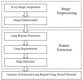
3. METHODOLOGY
The proposed Lung Cancer Detection System can distinguish the proper dangerous affected districts by applying
the accompanying advances appeared in Fig. 1. In lung X-Ray, pneumonic nodule shows up as a circularly
molded mass [22]. It can be contorted by nearby anatomical development. There are no limits on size or
spreading in lung tissue. The pneumonic nodule is arranged into a couple of gatherings; nodule is related to
pleural surface and associated with neighboring vessels by thin structure [23]. Pre-finding approaches help to
find the danger of lung cancer disease in beginning period [20].
Fig. 1: Lung Cancer Detection System X-ray Image Acquisition
Image Enhancement
Image
Preprocessing
Feature
Extraction
Lung Regions Extraction
Lung Segmentation
Edge Detection
1806 | P a g e
In the pre-symptomatic of lung tumor, some of methodologies utilized are Artificial Neural Based Learning
Process, Rule Based Learning Technique, Supervised Learning Methods, Fuzzy System, Expert System and
Genetic Algorithm [24].
IMAGE PROCESSING
In this paper, the author utilize ANN based learning technique and utilized a few unique X-Ray images of
different size and complexity for both carcinogenic and non-dangerous patients, which are gathered from
different presumed restorative organization and doctor's facilities and it is further to group the tumor as
benevolent or harmful. The analysis framework utilizes resizing, editing appeared in Fig. 2(d) and applying
middle, gaussian channels to smooth the X-Ray images and the differentiations are improved [25]. The images
of lung are portioned by applying the Otsu's limit. After twofold transformation, lung disease identification
framework works-out morphologic method to separate highlights including the edge, territory and state of
nodule. For the benefit of this task, malignant arrangement of a lungs nodule is utilized to examine those
highlights either tumor cells exist or not. Likewise, if growths cells remain alive, at that point its stage is
recognized. After fulfillment of image obtaining, it takes after that to upgrade the co-related pixel scalar
qualities in MATLAB program.
IMAGE ACQUISITION
To improve X-Ray image, the author put endeavors to denoise, to upgrade the structure and difference. When
Median, Laplacian and Gaussian channels are utilized for denoise then the procedure is adjusted to improving
the edge of image structure contains unsharp and upgrades the image differentiate by histogram balance [26].
There are diverse sorts of tissues like bone; muscle and fat have number of data in a X-Ray. Just the dark scale
image contains clamors, for example, repetitive sound, and pepper commotion and so forth. The most widely
recognized issues in image handling are repetitive sound. Here is the principle proposal of any channel is to
compute pixel weights. While the middle channel is a nonlinear basic upgrade advanced separating method for
evacuating commotion without lessening the sharpness of the image [27]. The ensuing figures appeared in Fig.
2(a), Fig. 2(b) and Fig. 2(c) : [28]
1807 | P a g e
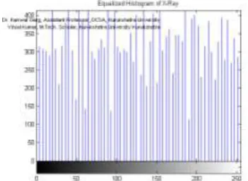
IMAGE ENHANCEMENT
It is normally connected for smoothing of the lung limits appeared in Fig. 2(d) in limit in computerized image
handling. The author connected here for sifting by allotting 5x5 pixels. The straight channel is regarding as to
expel certain sorts of clamor. Histogram activity is intense practice for X-Ray image upgrade. The interim
qualities between the base and most extreme pixel isolated into similarly dispersed containers in histogram. It
check the quantity of pixels identified with each canister. The states of histograms are relying upon the span of
interims. The whole image powers ought to be similarly isolated receptacles as appeared in Fig. 2(e) histogram
evening out [27].
The esteem k for every shine level j in the first X-Ray image is controlled by:
j Nj
k = ∑ * I max
i=0 T
Binary conversion is applied later than the process of enhancement as 8-bit X-Ray image altered into 2-bit gray
scale image. If the pixel value in image is greater than threshold value, it shows “0” (black) and if less than threshold then it show “1” (white) [20].
Fig. 2(d): Median Filtered X-ray Fig. 2(e): Equalized Histogram of X-ray
Performance Analysis using Neural Network
Artificial Neural Network is created for determination and order of competitor nodules subsequent to applying
preparing and testing process. The neural system comprises of three primary layers, i.e. input layer, hidden layer
and output layer. The Back proliferation calculation is utilized for the most part. The recommendation of this
calculation is to decrease mistakes and delivered by the distinction between genuine yield and expected outcome
1808 | P a g e
concealed hubs of system, i.e. preparing rate for preparing ANN, number of ages. When the created organize
ends up fruitful, it is then prepared for grouping process for the competitor nodule.
CONCLUSION
In this paper, the author talked about a lung tumor identification framework for early location of lung growth by
contemplating lung X-Ray images utilizing various advances. The approach begins by extricating the lung
locales from lung X-Ray image utilizing a few image handling methods in MATLAB including paired image,
disintegration, enlargement, gaussian channel and middle channel. The key focal points of Artificial Neural
Network are their capacity to find the favored data in information. The author recommends the best ANN
system with calculation utilized for arrangement of lung cancer nodules in X-beam images. This framework
encourages the radiologist and doctor to perceive the suspicious nodules that expansion the affectability of the
conclusion.
REFERENCES
[1]. Azian Azamimi Abdullah, Syamimi Mardiah Shaharum, “Lung Cancer Cell Classification Method
using Artificial Neural Network”, Information Engineering Letters, ISSN:21604114, Volume 2, Number
1, March 2012.
[2]. B. Magesh et al., “Computer Aided Diagnosis System for the Identification and Classification of Lessions in Lungs”, International Journal of Computer Trends and Technology, ISSN : 2231-2803, IJCTT
May-June 2011.
[3]. B. V. Ginneken, B. M. Romeny and M. A. Viergever, “Computer Aided Diagnosis in Chest Radiography:
A Survey”, IEEE, Transactions on Medical Imaging, Vol 20, No. 12, 2001.
[4]. S. A. Patil, Dr. V. R. Udupi, Dr. C. D. Kane, Dr. A. I. Wasif, Dr. J. V. Desai, A. N. Jadhav,
“Geometrical and Texture Features Estimation of Lung Cancer and TB Image using Chest X-Ray Database”, 978-4244-4764-0/09/$25.00 © 2009 IEEE.
[5]. Gomathi, G. Gamsu, M. Ginsberg, L. Jiang and L. H. Schwartz, “Automatic Detection of Small Lung
Nodules on CT Utilizing a Local Density Maximum Algorithm”, Journal of Applied Clinical Medical
Physics, Vol. 4, 2010.
[6]. Patil S. C., Gargano G., “Computer Aided Diagnosis for Lung CT using Artificial Life Models”, Seventh
International Symposium on Symbolic and Numeric Algorithms for Scientific Computing, SYNASC
2009.
[7]. Argiris and C. Yan, “Lung Nodules Identification Rules Extraction with Neural Fuzzy Network”, IEEE,
Neural Information Processing, Vol. 4, 2012.
[8]. Kennedy Taher, Rachid Sammouda, “Identification of Lung Cancer Based On Shape and Color”- IEEE
2000.
[9]. Miao, Dr. R. Anitha, “Supervisory Expert System Approach for Pre-Diagnosis of Lung Cancer”,
1809 | P a g e
[10].Al Mohammad, D. de. Ridder and H. Handels, “Image Processing with Neural Networks - A Review”,
Pattern Recognition 35, 2279-2301, 2016.
[11].M. Gomathi , Dr. P. Thangaraj, “A Computer Aided Diagnosis System for Detection of Lung Cancer
Nodules Using Extreme Learning Machine”, International Journal of Engineering Science And
Technology Vol.2(10), 2010.
[12].Penedo, M.G., Carreira, M.J., Mosquera, A. and Cabello, D., “Computer-Aided Diagnosis: A
Neural-Network-Based Approach to Lung Nodule Detection”, IEEE Transactions on Medical Imaging, Pp: 872 –
880, 1998.
[13].Qing-Zhu Wang, Ke Wang, Yang Guo and Xin-zhu Wang, “Automatic Detection of Pulmonary Nodules
in Multi-slice CT Based on 3D Neural Network with Adaptive Initial Weight”, DOI
10.1109/ICICTA.2010.751, 978-0-7695-4077-1/10 $26.00 ©2010 IEEE.
[14].Ramnath Takiar, Deenu Nadayil, A Nandakumar, “Projection of number of cancer cases in India
(2010-2020) by Cancer Groups”, Asian Pacific Journal of Cancer Prevention, Vol 11, 2010.
[15].R. H. Chan, C. W. Ho and M. Nikolova, “Salt and Peeper Noise Removal by Median Type Noise
Detector and Edge Preserving Regularization”, Department of Mathematics- Hong Kong, pp. 1-7,
2003.
[16].Vinod Kumar, Dr. Kanwal Garg, “Neural Network Based Approach for Detection of Abnormal Regions
of Lung Cancer in X-Ray Image”, International Journal of Engineering Research & Technology, ISSN:
2278-0181, IJERT Vol. 1 Issue 5, July 2012.
[17].Amal Farag, James Graham, Salwa Elshazly and Aly Farag, “Statistical Modeling of the Lung Nodules in
Low Dose Computed Tomography Scans of the Chest”, 17th International Conference on Image
Processing, Proceeding of IEEE September 2010.
[18].A. El-Baz, A. A. Farag, Ph.D., R. Falk, M. D. and R. L. Rocco, M. D., “Detection, Visualization and
Identification of Lung Abnormalities in Chest Spiral CT Scan : Phase I”, Information Conference on
Biomedical Engineering, Egypt, 2002.
[19].Syed and Muhammad, “Text book of lung cancer”, 2nd ed., National University Hospital, Copenhagen,
Denmark, ISBN-13: 9780415385107, 2017.
[20].Jia Tong, Zhao Da-Zhe, Wei Ying, Zhu Xin-Hua, Wang Xu, “Computer-Aided Lung Nodule Detection
Based On CT Images”, IEEE/ICME International Conference on Complex Medical Engineering, 2007.
[21].Mohammad Reza Zare, Ahmed Mueen, Woo Chaw Seng, Mohammad Hamza Awedh, “Communication
Systems and Networks Combined Feature Extraction on Medical X-ray Images” Third International
Conference on Computational Intelligence, Bali, Indonesia, July 26-July 28, 2011.
[22].Taher F, Werghi N, Al-Ahmad H and Sammouda R. 2012. Lung Cancer Detection by Using Artificial
Neural Network and Fuzzy Clustering Methods. Am J Biomed Eng., 2(3): 136-142.
[23].Tao W, Jianping L and Bingxin L. 2011Research of Lung Cancer Screening Algorithm Based on RBF
Neural Network. Proceedings of the 2011 International Conference on Computer and Management
(CAMAN), Wuhan, China, 1-4.
[24].Tarawneh M, Nimri O, Arkoob K and AL Zaghal M. 2010. Cancer Incidence in Jordan 2010. The
1810 | P a g e
[25].Chen S, Suzuki K. 2013. Computerized Detection of Lung Nodules by Means of “Virtual Dual-Energy”
Radiography. IEEE Transactions on Biomed Eng., 60(2): 369-378.
[26].Dimililer K, Ugur B and Ever Y. 2017. Tumor detection on CT lung images using image enhancement.
The Online J Sci Technol., 7(1): 133-138.
[27].Syed M and Muhammad S. 2017. Recent Developments in Computer Aided Diagnosis for Lung Nodule
Detection from CT images: A Review. Current Med Imaging Rev., 13(1): 3-19.
[28].Tajbakhsh N and Suzuki K. 2017. Comparing two classes of endto-end machine-learning models in lung