N A N O E X P R E S S
Open Access
Significantly enhanced dye removal performance
of hollow tin oxide nanoparticles via carbon
coating in dark environment and study of its
mechanism
Shuanglei Yang
1,2, Zhaohui Wu
4, LanPing Huang
1, Banghong Zhou
1, Mei Lei
3, Lingling Sun
3, Qingyong Tian
3,
Jun Pan
1, Wei Wu
2*and Hongbo Zhang
1*Abstract
Understanding the correlation between physicochemical properties and morphology of nanostructures is a prerequisite for widespread applications of nanomaterials in environmental application areas. Herein, we illustrated that the uniform-sized SnO2@C hollow nanoparticles were large-scale synthesized by a facile hydrothermal method. The size of the core-shell hollow nanoparticles was about 56 nm, and the shell was composed of a solid carbon layer with a thickness of 2 ~ 3 nm. The resulting products were characterized in terms of morphology, composition, and surface property by various analytical techniques. Moreover, the SnO2@C hollow nanoparticles are shown to be effective adsorbents for removing four different dyes from aqueous solutions, which is superior to the pure hollow SnO2nanoparticles and commercial SnO2. The enhanced mechanism has also been discussed, which can be attributed to the high specific surface areas after carbon coating.
Keywords:Tin oxide; Carbon coating; Core-shell; Dye removal
Background
With the development of society and scientific techno-logy, more attentions have been paid to environmental issues which were caused by the discharge of wastewa-ter. Oil spillage, organic solvents, and synthetic dyes dis-charged by the textile, paper, and tannery industries are primary pollutants of water sources [1]. It is estimated that more than 100,000 commercially available dyes with over 7 × 10⁵ tonnes of dyestuff are produced annually [2]. Generally, synthetic dyes have complex aromatic structures that make them stable and difficult to bio-degrade. As synthetic dyes in wastewater cannot be ef-ficiently decolorized by traditional methods (such as catalytic degradation, cation exchange membranes, and biological processes), the adsorption of synthetic dyes
on inexpensive and efficient solid supports was consi-dered as a simple and economical method for their re-moval from water and wastewater.
The most commonly used absorbent for dye removal is activated carbon, because of its capability for effi-ciently adsorbing a broad range of different types of dyes [3]. Up to now, there have been many successful meth-odologies for the fabrication of activated carbon mate-rials, such as pinewood-based activated carbon [4], coir pith activated carbon [5], rice husk-based activated carbon [6], and bamboo-based activated carbon [7]. Although, na-tural renewable resources have been widely used as raw materials for manufacturing activated carbon, the high production and treatment costs of activated carbon may still hinder its further application.
As a competitive alternative, various nanomaterials have been developed and used to remove the dyes. For example, Zhu and co-workers have prepared hierarchical NiO spheres with a high specific area of 222 m2/g as an adsorbent for removal of Congo red [8]. Mou and co-workers have fabricatedγ-Fe2O3and Fe3O4chestnut-like
* Correspondence:[email protected];[email protected]
2Laboratory of Printable Functional Nanomaterials and Printed Electronics,
School of Printing and Packaging, Wuhan University, Wuhan 430072, People's Republic of China
1
State Key Laboratory for Powder Metallurgy, Central South University, Changsha 410083, People's Republic of China
Full list of author information is available at the end of the article
hierarchical nanostructures, which can be separated sim-ply and rapidly from treated water by magnetic separation after As(V) adsorption treatment. And the As(V) removal capacity of as-obtainedγ-Fe2O3is maintained at 74% and reaches 101.4 mg/g [9]. And then, they have prepared magnetic Fe2O3 chestnut-like amorphous-core/γ -phase-shell hierarchical nanostructures with a high specific area of 143.12 m2/g and with a maximum adsorption capacity of 137.5 mg/g for As(V) adsorption treatment [10]. Liu and co-workers have prepared various bismuth oxyiodide hierarchical architectures, and their nanomaterials shown enhanced the photocatalytic performance and adsorption capabilities [11]. Recently, the carbon functionalized nano-materials have recently attracted considerable attention because of their enhanced dye removal performance. For instance, Fan and co-workers have synthesized hybridi-zation of graphene sheets and carbon-coated Fe3O4 na-noparticles as an adsorbent of organic dyes [12]. Li and co-workers have reported Mg(OH)2@reduced graphene oxide composite, which exhibited excellent adsorption be-havior for methylene blue (MB) [13].
Indeed, the adsorption technique is especially attract-ive because of its simple design, high efficiency, and easy operation, but it requires materials with large specific surface area, well-defined pore size, and shape. Hollow structured materials fit these criteria well, and they have attracted tremendous interest as a special class of mate-rials compared to other solid counterparts, owing to their higher specific surface area, lower density, and bet-ter permeation, which have been extensively considered as potential materials applied in adsorption, catalysis, chemical reactors, and various new application fields [14-16]. Therefore, design and fabrication of materials like carbon-coated hollow structure would increase the dye removal abilities.
Herein, a well-defined carbon-coated hollow SnO2 nanoparticles have been designed and fabricated by a fa-cile two-step hydrothermal method without using any surfactants. Carbon coating prepared by hydrothermal treatment of low-cost glucose has aroused much inter-est. The preparation process belongs to green chemistry as the reaction process is safe and does not incur any contamination of the environment. More importantly, the carbon layer increases the specific area of bare hol-low SnO2nanoparticles, which exhibits an enhanced dye removal performance.
Methods
Materials
Potassium stannate trihydrate (K2SnO3· 3H2O), com-mercial SnO2, rhodamine B (RhB), MB, rhodamine 6G (Rh6G), and methyl orange (MO) were purchased from Shanghai Jingchun Chemical Reagent Co., Ltd. (Shanghai, China). Urea (CO(NH2)2), ethylene glycol (EG), ethanol
(C2H5OH), and glucose (C6H12O6) were purchased from Sinopharm Chemical Reagent Co., Ltd. (Shanghai, China). All the materials were used without further purification in the whole experimental process. Deionized water was used throughout the experiments.
Synthesis of hollow SnO2nanoparticles
In a typical process, 0.6 g potassium stannate trihydrate was dissolved in 50 mL ethylene glycol through the ultrasonic method. Urea (0.4 g) was dissolved in 30 mL deionized water and then the solution was mixed to-gether and transferred into a Teflon-lined stainless steel autoclave with a capacity of 100 mL for hydrothermal treatment at 170°C for 32 h. The autoclave solution was removed from the oven was allowed to cool down to room temperature. The product was harvested by centri-fugation and washed with deionized water and ethanol and then dried at 80°C under vacuum.
Synthesis of hollow SnO2@C nanoparticles
SnO2@C hollow nanoparticles were prepared by a glu-cose hydrothermal process and subsequent carboniza-tion approach. In a typical process, 0.4 g of as-prepared hollow SnO2 nanoparticles and 4 g glucose were re-dispersed in ethanol/H2O solution. After stirring, the solution was transferred into a 100-ml Teflon-lined stain-less steel autoclave sealed and maintained at 170°C for 8 h. After the reaction was finished, the resulting black solid products were centrifuged and washed with deion-ized water and ethanol and dried at 80°C in air. Lastly, the black products were kept in a tube furnace at 600°C for 4 h under argon at a ramping rate of 5°C/min.
Characterization
Transmission electron microscopy (TEM) and high-resolution transmission electron microscopy (HRTEM) were performed with a JEOL JEM-2100 F transmission electron microscope (Tokyo, Japan) at an accelerating voltage of 200 kV, and all the samples were dissolved in ethanol by ultrasonic treatment and dropped on copper grids. Powder X-ray diffraction (XRD) patterns of the samples were recorded on a D/ruanx2550PC (Tokyo, Japan) using CuKα radiation (λ= 0.1542 nm) operated at 40 kV and 40 mA. The absorption spectra of the sam-ples were carried out on a Shimadzu UV-2550 spectro-photometer (Kyoto, Japan). Raman measurement was performed using a HORIBA Jobin Yvon LabRAM HR spectroscope (Edison, NJ, USA) using an excitation laser wavelength of 488 nm.
Adsorption experiments
stirring. The reaction solution was sampled every 15-min intervals at room temperature; 2 mL solution was sampled and centrifuged to remove the adsorbents, and the corresponding UV-visible spectra were recorded to monitor the progress of the degradation of organic dyes by a Shimadzu 2550 UV-visible spectrophotometer.
Results and discussion
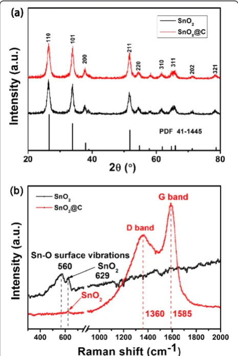
Figure 1a shows the representative XRD patterns of the as-obtained hollow SnO2 and hollow SnO2@C nano-particles. All of the diffraction peaks can be well indexed to the tetragonal rutile phase of SnO2(JCPDS card No. 41-1445). The absence of characteristic peaks corres-ponding to impurities indicates high purity of the pro-ducts [17]. The result reveals that the carbon coating process and annealing treatment will not change the structure of the SnO2. To prove the generation of the carbon layer on the as-prepared hollow SnO2seeds, the two samples were characterized by Raman spectroscopy. As shown in Figure 1b, the two peaks of 1,585 and
1,360 cm−1can be observed in the hollow SnO2@C sam-ple, which can be attributed to the E2gvibration mode of the ordered carbon layer (G band) and the A1gvibration mode of the disordered carbon layer (D band), respec-tively. The peak intensity ratio (ID/IG) (ca. 0.76) calculated is a useful index for comparing the degree of crystallinity of various carbon materials; a smaller value ratio reflects a higher degree of ordering in the carbon material. The peaks at 560 and 629 cm−1can be observed, respectively. The peak at 560 cm−1can be assigned to the Sn-O surface vibrations; the peak at 629 cm−1can be indexed to the A1g mode of SnO2. The above results reveal that the carbon has been successfully coated on the surface of the SnO2 nanoparticles, and the structure of SnO2was not change.
The structure and morphology of the as-prepared hol-low SnO2 nanoparticles are investigated by TEM and HRTEM. As shown in Figure 2a, the as-prepared sam-ples mainly consist of uniform flower-like nanoparticles. The contrast (dark/bright) between the boundary and the center of the nanoparticles confirms their hollow na-ture. The histogram of the particle diameters (inset in Figure 2a) demonstrates that the average diameter of the as-prepared hollow SnO2 nanoparticles is 53 nm. The bright rings in the selected-area electron diffraction (SAED) pattern (Figure 2b) can be well indexed to the rutile-phase SnO2. Figure 2c shows the TEM image at high magnification of the hollow SnO2 nanoparticles. It can be seen that the SnO2particles were consist of small SnO2 grains, and the surface of the SnO2 particles is rough, which means that the shell is incomplete and porous, not solid. This feature endows that the hollow SnO2nanoparticles have high surface area. As shown in Figure 2d, the HRTEM image confirms that the SnO2 particles consist of small SnO2 grains, and their size is about 3 ~ 5 nm. From the insets of Figure 2d, there are two lattice fringes with lattice spacing of about 0.334 and 0.26 nm, which can be assigned to the (110) and (101) planes of tetragonal rutile-phase SnO2 nanopar-ticles, respectively.
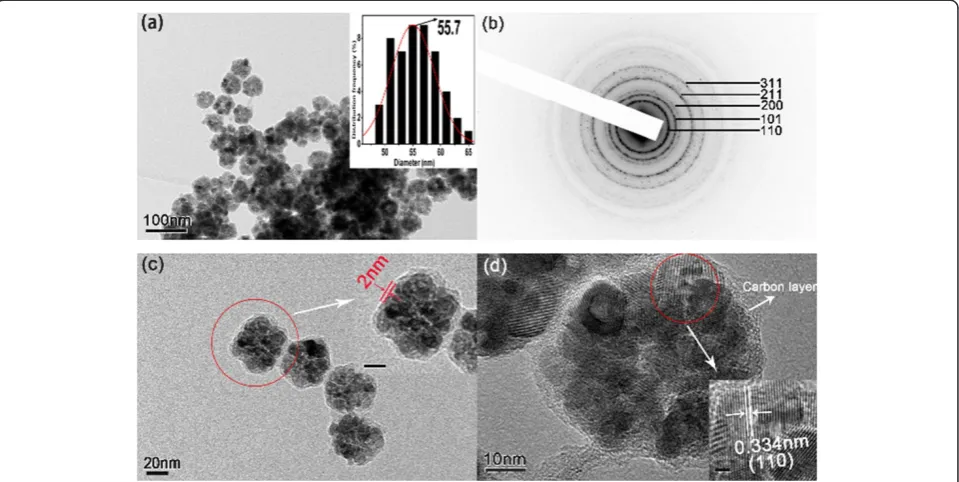
Subsequently, the morphologies of the carbon-coated hollow SnO2 nanoparticles (SnO2@C) were further stu-died by TEM and HRTEM. Figure 3a shows the TEM image of the SnO2@C nanoparticles. It can be seen that the SnO2@C nanoparticles still maintained a uniform morphology. The inset histogram diameters illustrate that the average diameter of SnO2@C nanoparticles is 55.7 nm. Compared with the naked hollow SnO2 nano-particles, the thickness of the carbon coating layer is about 2 ~ 3 nm. As shown in Figure 3b, the bright rings in the SAED pattern can be well indexed to the struc-ture of the rutile-phase SnO2, which demonstrate that the structure of SnO2is also not change by carbon coat-ing. From the magnified TEM images (Figure 3c), a thin carbon layer on the surface of the SnO2nanoparticles
[image:3.595.57.290.355.703.2]can be observed clearly, and the thermal gravimetric analysis (Additional file 1: Figure S1) illustrates that about 37% of carbon has coated the SnO2nanoparticles. The HRTEM image (Figure 3d) shows that the carbon layer is smooth, continuous, and has a thickness of
[image:4.595.58.539.90.330.2]about 2 ~ 3 nm. There are lattice fringes with lattice spacing of about 0.334 nm, which can be indexed to the (110) plane of tetragonal rutile-phase SnO2 nanoparti-cles. The above results prove that the carbon has been successfully coated on the surface of the hollow SnO2
Figure 2SAED patterns and TEM images at low and high magnifications. (a)TEM image at low magnification (the inset is the histogram of particle diameters).(b)SAED patterns and(c)TEM images at high magnification (the inset scale bar is 10 nm) of the as-prepared hollow SnO2 nanoparticles, and(d)HRTEM image of a single SnO2nanoparticle (the inset scale bar is 2 nm).
[image:4.595.59.539.454.695.2]nanoparticles, and the morphology is still maintained after the coating treatment.
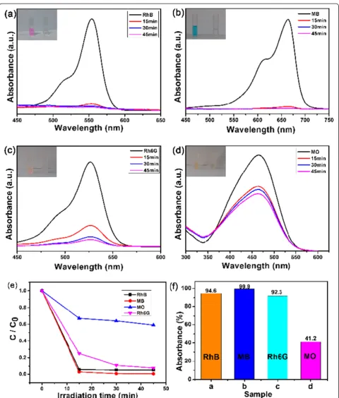
[image:6.595.306.539.85.409.2]We also investigated the potential application of the as-synthesized carbon-coated hollow SnO2 nanoparti-cles to be used as an adsorbent in wastewater treatment. Figure 4 shows the time-dependent absorption spectra for different organic dyes (include RhB, Rh6G, MB, and MO) in 45 min, and the characteristic absorption of these dyes was used to monitor the process of adsorp-tion. The used dyes are chemically stable and are common constituents of effluents in industries which demand an appropriate method to dispose them off. As shown in Figure 4a,b,c, we can see that the peak intensities at 554 nm for RhB, 664 nm for MB, and 525 nm for Rh6G decreased very quickly once the hollow SnO2@C were added. After only 45 min, these peaks became too weak to be observed, suggesting the high efficiency for removing these three dyes. Meanwhile, the insets of Figure 4a,b,c shows the change of the color of these three dyes in solution within 45 min. It can be seen that the color of the three dyes disappeared, suggesting that the chromophoric structure of RhB, MB, and Rh6G were decomposed. However, for the removal of MO, the color of the MO solutions did not disappear in 45 min (Figure 4d). This means that a part of the molecular structure of MO was not decomposed by SnO2@C and remained in the solution.
Figure 4e,f further confirms that the removal rate of RhB (10 mg/L) can reach to 94.6%. The results reveal that the as-prepared hollow SnO2@C nanoparticles ex-hibit excellent removal performance for RhB dyes. Mean-while, the hollow SnO2@C nanoparticles also showed a good removal performance for MB and Rh6G (5 mg/L); the removal rate can reach to 99.9% and 92.3%, respect-ively. However, for the MO dyes (5 mg/L), the removal rate can only reach to 41.2%, because the chromophoric structure of MO dye is different from those of RhB and MB, and this will cause a different electrostatic interaction capacity between functional groups of carbon and dye molecules [18-20]. The above results illustrate that the as-obtained hollow SnO2@C nanoparticles exhibit a good dye removal performance.
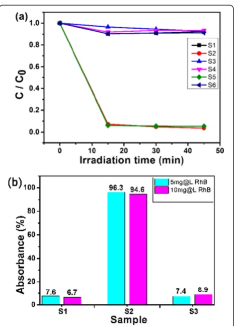
To further study the dye removal abilities of the as-prepared hollow SnO2@C nanoparticles, the dye removal performance of naked hollow SnO2 nanoparticles and commercial SnO2 nanoparticles (average size is 70 nm) was measured for comparison. Figure 5a shows the time-dependent adsorption kinetics of the samples at diffe-rent initial RhB dye concentrations. Obviously, among all the samples, the hollow SnO2@C nanoparticles (sam-ples S2 and S5) exhibit the fastest absorption abilities. As shown in Figure 5b, the removal rate of the hol-low SnO2@C nanoparticles (S2) is highest among the three samples and can reach to 96.3% and 94.6% for
the RhB dye with different concentration of 5 and 10 mg/L, respectively, which is much higher than that of S1 (naked hollow SnO2, 7.6% and 6.7%) and S3 (commercial SnO2, 7.4% and 8.9%). The above results demonstrate that carbon coating can significantly en-hance the dye removal abilities. As a comparison, the measured results of the removal performance experi-ment of carbon sphere and hydrochloric acid-treated SnO2@C nanoparticles (SnO2 has been removed) are shown in Additional file 1: Figures S2 and S3. The re-sults show that the as-prepared hollow SnO2@C nano-particles' removal dye performance is better than those of pure carbon materials.
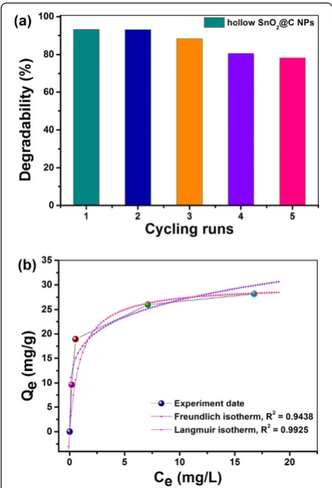
Subsequently, the stability of the as-prepared hollow SnO2@C nanoparticles has been further investigated by recycling the removal for RhB, and the results are shown in Figure 6a. The hollow SnO2@C nanoparticles
exhibited a good removal dye activity and stability; the degradation rate of RhB solution was found to be more than 78% after 5 cycles. As shown in Figure 6b and Additional file 1: Figure S4, the adsorption capacity for RhB increased with the different RhB concentra-tions. The maximum adsorption capacity in the con-centration range studied is 28.2 mg/g for RhB. The amount of the dye adsorbed was calculated using the equation: Qe= (C0−Ce) V/m, where Qe (mg/g) is the amount of RhB adsorbed onto the adsorbent at equi-librium, C0 (mg/L) and Ce (mg/L) are the initial and equilibrated RhB concentrations, respectively,V (L) is the volume of solution added, and m (g) is the mass of the adsorbent. Figure 6b shows the isotherms for RhB adsorption on the as-obtained SnO2@C nanopar-ticles. It can be found that the regression coefficient R2
obtained from the Langmuir model is much higher than that of from the Freundlich model (0.9925 > 0.9438), suggesting the Langmuir model fits better with the ex-perimental data [21].
To avoid the photocatalytic effect of SnO2and SnO2@C nanoparticles, the dye removal tests are carried out in a dark environment. And the results reveal that the carbon coating can enhance the absorption abilities. To illustrate the reason, the nitrogen adsorption iso-therms of the hollow SnO2 and SnO2@C nanoparti-cles have been measured and shown in Figure 7. The BET surface areas of the hollow SnO2 and SnO2@C nanoparticles are 60.59 and 168.33 m2/g, respectively. Both samples exhibit the type IV isotherms with a dis-tinct hysteresis loop at the relative pressure P/P0
[image:7.595.56.290.88.432.2]ran-ging from 0.5 to 0.8. Clearly, the carbon coating will greatly enhance the surface area, which can be the main reason of significant enhanced dye removal performance of hollow SnO2@C nanoparticles. The large number and array of different functional groups on the carbon layers (e.g., carboxylic, hydroxyl, carbonyl) implied the existence of many types of adsorbent-solute interaction [22]. Additionally, carbon coating has made the cova-lent bond interaction with hexagonal structure, which has a -π structure properties of aromatic ring, easy to
Figure 7Nitrogen adsorption-desorption isotherms and pore size distribution. (a)Nitrogen adsorption-desorption isotherms of the as-synthesized SnO2and hollow SnO2@C nanoparticles.(b)The pore size distribution of the hollow SnO2@C nanoparticles. Figure 6Reutilization properties.Removal performance under
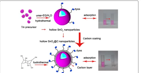
[image:7.595.305.539.368.686.2]interact with conjugated double bonds. And some of the dye structure have conjugated double bonds and easy to be adsorbed by the coating carbon [23]. As shown in Figure 8, the hollow SnO2@C nanoparticles can capture more dye molecules due to the introduced carbon layer. Indeed, relatively larger amount of water and hydroxyl groups can be adsorbed on the surface by hydrothermal process [24]. The surface chemistry of the adsorbents plays a major role in the adsorption. The ad-sorption of the reactive dye on carbon is favored, mainly due to the dispersive interactions between the deloca-lized π electrons of the carbon materials and the free electrons of the dye molecules [20]. The functional groups on the hollow SnO2@C nanoparticles' surface acted as a negative potential that provides a weak electrostatic inter-action between the organic dyes and the hollow SnO2@C nanoparticles.
Conclusions
In summary, hollow SnO2@C nanoparticles have been synthesized on a large scale through a facile hydrother-mal method. The as-prepared hollow SnO2@C nanopar-ticles show excellent adsorption capacity toward RhB, MB, and Rh6G dyes in aqueous solutions. Compared with the naked hollow SnO2 and commercial SnO2 nanopar-ticles, the adsorption capacity showed about an 89% im-provement for RhB organic dye. The porous carbonaceous shells coated on the surface of hollow SnO2nanoparticles greatly enhanced the specific area, which provides more
active sites for dye adsorption. Owing to their unique hollow structures, high surface areas and low cost, the as-obtained hollow SnO2@C nanoparticles are poten-tially applicable in wastewater treatment. Accordingly, it may be concluded that the developed SnO2@C is an ef-ficient method for the decolorization of RhB, MB, and Rh6G dyes.
Additional file
Additional file 1:Supporting information.Thermal gravimetric analysis, UV-vis absorption spectra of dyes, adsorption kinetics, and the effect of RhB dye equilibrium concentrations of SnO2@C nanoparticles.
Competing interests
The authors declare that they have no competing interests.
Authors’contributions
SY carried out the absorbance studies and drafted the manuscript. ZW, BZ, and JP participated in the dye removal analysis. LPH, ML, LS, and QT did the fabrication and characterization experiments. WW and HZ analyzed the results and participated in its design and coordination. All authors read and approved the final manuscript.
Acknowledgements
[image:8.595.57.548.88.341.2]We greatly acknowledge financial support by the NSFC (51302325, 51201115, 51471121), New Century Excellent Talents in University (NCET-12-0553), Program for Shenghua Overseas Talent (1681–7607030005) from Central South University, and the partial financial support from the Open-End Fund for the Valuable and Precision Instruments of Central South University (Grant No. CSUZC2013025) and the Fundamental Research Funds for the Central Universities.
Author details
1
State Key Laboratory for Powder Metallurgy, Central South University, Changsha 410083, People's Republic of China.2Laboratory of Printable
Functional Nanomaterials and Printed Electronics, School of Printing and Packaging, Wuhan University, Wuhan 430072, People's Republic of China.
3
Key Laboratory of Artificial Micro and Nano-structures of Ministry of Education, School of Physics and Technology, Wuhan University, Wuhan 430072, People's Republic of China.4Department of Chemical Engineering, Kyung Hee University, Seocheon-Dong, Giheung-Gu, 446-701 Yongin, Korea.
Received: 24 June 2014 Accepted: 20 August 2014 Published: 28 August 2014
References
1. Lei W, Portehault D, Liu D, Qin S, Chen Y:Porous boron nitride nanosheets for effective water cleaning.Nat Commun2013,4:1777.
2. Rafatullah M, Sulaiman O, Hashim R, Ahmad A:Adsorption of methylene blue on low-cost adsorbents: a review.J Hazard Mater2010,177:70–80. 3. Hsiu-Mei C, Ting-Chien C, San-De P, Chiang HL:Adsorption characteristics
of Orange II and Chrysophenine on sludge adsorbent and activated carbon fibers.J Hazard Mater2009,161:1384–1390.
4. Tseng RL, Wu FC, Juang RS:Liquid-phase adsorption of dyes and phenols using pinewood-based activated carbons.Carbon2003,41:487–495. 5. Namasivayam C, Kavitha D:Removal of Congo red from water by
adsorption onto activated carbon prepared from coir pith, an agricultural solid waste.Dyes Pigments2002,54:47–58.
6. Guo Y, Yang S, Fu W, Qi J, Li R, Wang Z, Xu H:Adsorption of malachite green on micro-and mesoporous rice husk-based active carbon.
Dyes Pigments2003,56:219–229.
7. Hameed B, Din AM, Ahmad A:Adsorption of methylene blue onto bamboo-based activated carbon: kinetics and equilibrium studies.
J Hazard Mater2007,141:819–825.
8. Zhu T, Chen JS, Lou XW:Highly efficient removal of organic dyes from waste water using hierarchical NiO spheres with high surface area.J Phys Chem C2012,116:6873–6878.
9. Mou F, Guan J, Ma H, Xu L, Shi W:Magnetic iron oxide chestnutlike hierarchical nanostructures: preparation and their excellent arsenic removal capabilities.ACS Appl Mater Interfaces2012,4:3987–3993. 10. Mou F, Guan J, Xiao Z, Sun Z, Shi W, Fan X:Solvent-mediated synthesis of
magnetic Fe2O3chestnut-like amorphous-core/γ-phase-shell hierarchical nanostructures with strong As(v) removal capability.J Mater Chem2011,
21:5414–5421.
11. Liu QC, Ma DK, Hu YY, Zeng YW, Huang SM:Various bismuth oxyiodide hierarchical architectures: alcohothermal-controlled synthesis, photocatalytic activities, and adsorption capabilities for phosphate in water.ACS Appl Mater Interfaces2013,5:11927–11934. 12. Fan W, Gao W, Zhang C, Tjiu WW, Pan J, Liu T:Hybridization of graphene
sheets and carbon-coated Fe3O4nanoparticles as a synergistic adsorbent of organic dyes.J Mater Chem2012,22:25108–25115. 13. Li B, Cao H, Yin G:Mg(OH)2@ reduced graphene oxide composite for
removal of dyes from water.J Mater Chem2011,21:13765–13768. 14. Duan F, Dong W, Shi D, Chen M:Template-free synthesis of ZnV2O4
hollow spheres and their application for organic dye removal.Appl Surf Sci2011,258:189–195.
15. Wu W, Xiao X, Zhang S, Li H, Zhou X, Jiang C:One-pot reaction and subsequent annealing to synthesis hollow spherical magnetite and maghemite nanocages.Nanoscale Res Lett2009,4:926–931.
16. Lou XWD, Archer LA, Yang Z:Hollow micro-/nanostructures: synthesis and applications.Adv Mater2008,20:3987–4019.
17. Wu W, Zhang S, Zhou J, Xiao X, Ren F, Jiang C:Controlled synthesis of monodisperse sub-100 nm hollow SnO2nanospheres: a template-and surfactant-free solution-phase route, the growth mechanism, optical properties, and application as a photocatalyst.Chem Eur J2011,
17:9708–9719.
18. Vinu R, Madras G:Environmental remediation by photocatalysis.J Indian Inst Sci2010,90:189–230.
19. Dutta S, Sarkar S, Ray C, Pal T:Benzoin derived reduced graphene oxide (rGO) and its nanocomposite: application in dye removal and peroxidase-like activity.RSC Advances2013,3:21475–21483.
20. Figueiredo J, Sousa J, Orge C, Pereira M, Orfao J:Adsorption of dyes on carbon xerogels and templated carbons: influence of surface chemistry.
Adsorption2011,17:431–441.
21. Kyzas GZ, Kostoglou M, Lazaridis NK:Relating interactions of dye molecules with chitosan to adsorption kinetic data.Langmuir2010,
26:9617–9626.
22. Al-Ghouti MA, Li J, Salamh Y, Al-Laqtah N, Walker G, Ahmad MNM:
Adsorption mechanisms of removing heavy metals and dyes from aqueous solution using date pits solid adsorbent.J Hazard Mater2010,
176:510–520.
23. Sun H, Cao L, Lu L:Magnetite/reduced graphene oxide nanocomposites: one step solvothermal synthesis and use as a novel platform for removal of dye pollutants.Nano Res2011,4:550–562.
24. Baiju KV, Shukla S, Biju S, Reddy MLP, Warrier KGK:Morphology-dependent dye-removal mechanism as observed for anatase-titania photocatalyst.
Catal Lett2009,131:663–671.
doi:10.1186/1556-276X-9-442
Cite this article as:Yanget al.:Significantly enhanced dye removal performance of hollow tin oxide nanoparticles via carbon coating in
dark environment and study of its mechanism.Nanoscale Research Letters
20149:442.
Submit your manuscript to a
journal and benefi t from:
7 Convenient online submission
7 Rigorous peer review
7 Immediate publication on acceptance
7 Open access: articles freely available online
7 High visibility within the fi eld
7 Retaining the copyright to your article