Simultaneous Analysis of Multiple Staphylococcal Enterotoxin
Genes by an Oligonucleotide Microarray Assay
Nikolay Sergeev,
1Dmitriy Volokhov,
2Vladimir Chizhikov,
2and Avraham Rasooly
1*
Center for Food Safety and Applied Nutrition, Food and Drug Administration, College Park, Maryland,1and
Center for Biologics Evaluation and Research, Food and Drug Administration, Rockville, Maryland2
Received 18 November 2003/Returned for modification 31 December 2003/Accepted 9 January 2004
Staphylococcal enterotoxins (SEs) are a family of 17 major serological types of heat-stable enterotoxins that are one of the leading causes of gastroenteritis resulting from consumption of contaminated food. SEs are considered potential bioweapons. ManyStaphylococcus aureusisolates contain multiple SEs. Because of the large number of SEs, serological typing and PCR typing are laborious and time-consuming. Furthermore, serological typing may not always be practical because of antigenic similarities among enterotoxins. We report on a microarray-based one-tube assay for the simultaneous detection and identification (genetic typing) of multiple enterotoxin (ent) genes. The proposed typing method is based on PCR amplification of the target region of theentgenes with degenerate primers, followed by characterization of the PCR products by microchip hybridization with oligonucleotide probes specific for eachentgene. We verified the performance of this method by using several other techniques, including PCR amplification with gene-specific primers, followed by gel electrophoresis or microarray hybridization, and sequencing of the enterotoxin genes. The assay was evaluated by analysis of previously characterized staphylococcal isolates containing 16entgenes. The microarray assay revealed that some of these isolates contained additional previously undetectedentgenes. The use of degenerate primers allows the simultaneous amplification and identification of as many as nine differententgenes in one S. aureusstrain. The results of this study demonstrate the usefulness of the oligonucleotide microarray assay for the analysis of multitoxigenic strains, which are common amongS. aureusstrains, and for the analysis of microbial pathogens in general.
Staphylococcal food-borne diseases resulting from the con-sumption of food contaminated with staphylococcal enterotox-ins (SEs) are one of the most common food-borne illnesses (1, 5, 11, 14, 31). SEs are also involved in rheumatoid arthritis (17, 38), atopic eczema (9, 10, 27), and toxic shock syndrome (16). SEs are considered potential bioweapons.
SEs belongs to a protein family called superantigens, which induce a polyclonal immune response by direct binding to class II major histocompatibility complex proteins and T-cell recep-tors on the surfaces of B and T cells without being internalized and processed like a normal antigen (3, 15, 28). These toxins may be involved in modulating the host immune response and may contribute to evasion of host defenses and bacterial
per-sistence (12). Expression of specific enterotoxin (ent) genes by
Staphylococcus aureus depends on the host tissue source and
may play a role in the adaptation of S. aureus to the host
environment (4).
There are 17 known major types of SEs (SEA to SER, respectively, with no SEF), and multiple SEs are commonly
found amongS. aureusstrains (19, 20, 32). Many of the known
staphylococcal enterotoxins (SEK to SER) were discovered recently.
The traditional method of identifying SEs by serological typing is relatively complex and time-consuming and is imprac-tical for the detection and identification of a large group of related toxins with significant antigenic similarities (23, 24).
Furthermore, the concentrations of toxins produced byS.
au-reusstrains differ when the strains are grown on various natural
substrates and laboratory media (7, 33). Other techniques have been used to identify toxin genotypes, including DNA-DNA hybridization and PCR, but these protocols were designed to detect only one or a few toxin genes (21, 35). Multiplex PCR
for detection of severalentgenes has been reported (6, 26, 29,
30, 36), but additional restriction endonuclease assays or other steps are required to ensure unambiguous identification of
ent-specific amplicons. Therefore, there is still a need for a
rapid and specific method for simultaneous detection and iden-tification of SEs for diagnostic and epidemiological purposes. Here we describe a rapid and reliable one-tube microarray-based assay for simultaneous detection and identification
(genetic typing) of almost all knownentgenes. The method
includes PCR amplification of part of the ent genes with
universal primers, followed by analysis of amplicons by
hybrid-ization withent-specific oligonucleotide probes immobilized on
the microchip.
MATERIALS AND METHODS
Bacterial strains.The strains used in this study were obtained from theS. aureuscollection of the Network on Antimicrobial Resistance inStaphylococcus aureus(NARSA; Focus Technologies, Inc., Herndon, Va.) and from the bacte-rial collection of Farukh Khambaty, Center of Food Safety and Applied Nutri-tion, Food and Drug Administration (FDA).
Total DNA preparation.DNA was extracted from freshly grown cells by phenol-chloroform extraction (34). The presence, concentration, and purity of genomic DNA in the prepared samples were detected by measuring the absor-bances at 260 and 280 nm with an Ultraspec 3000 spectrophotometer (Pharma-cia, Peapack, N.J.).
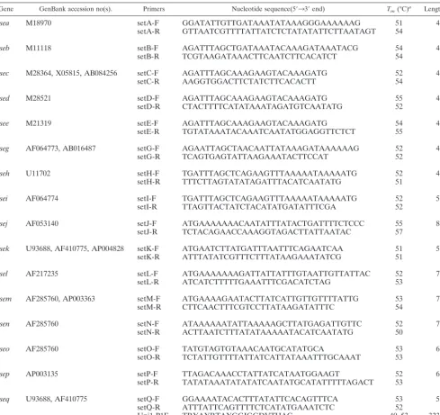
PCR amplification.Table 1 lists the primers used to amplify differentS. aureus
enterotoxin genes. Specific primers were used for amplification of individual enterotoxin genes. The standard PCR mixture (30l) contained 1.5 U of Hot-* Corresponding author. Mailing address: NIH/NCI, 6130 Executive
Blvd. EPN, Room 6035A, Rockville, MD 20852. Phone: (301) 402-4185. Fax: (301) 402-7819. E-mail: [email protected].
2134
on May 15, 2020 by guest
http://jcm.asm.org/
StarTaqDNA polymerase, 1⫻buffer supplemented with 2.0 mM MgCl2
(Qia-gen, Valencia, Calif.), 200 nM each forward and reverse primers, 200M each deoxynucleoside triphosphate (dNTP; dATP, dGTP, dCTP, and dTTP), and 100 to 300 ng of DNA template. PCR was performed with a Gene AMP PCR system 9600 thermocycler (Applied Biosystems, Foster City, Calif.) with the following cycle conditions: initial activation at 95°C for 15 min; 40 cycles at 94°C for 30 s, 55°C for 40 s, and 72°C for 60 s; and a final extension at 72°C for 7 min. The presence of amplified PCR products was detected by 2% agarose gel electro-phoresis in 1⫻Tris-acetate-EDTA or Tris-borate-EDTA buffer. The gels were stained with ethidium bromide and photographed under UV light with a digital camera (EDAS 290; Kodak, Rochester, N.Y.).
For simultaneous amplification of multiple enterotoxin genes by PCR with universal primers, the standard PCR mixture (50l) contained 5 U of HotStart
TaqDNA polymerase (Qiagen), 1⫻buffer supplemented with 3.5 mM MgCl2
(Qiagen), the universal primer mixture (700 nM forward universal primer, 1,400 nM reverse universal primer, and the seh-specific reverse primer at a
concentration of 50 nM), 200M each dNTP, and 600 to 800 ng of template (total DNA). PCR was performed with a Gene AMP PCR system 9600 thermo-cycler (Applied Biosystems) with the following conditions: initial activation of the enzyme at 95°C for 15 min; 7 cycles at 94°C for 1 min, 40°C for 1 min, and 72°C for 1 min; 35 cycles at 94°C for 1 min, 45°C for 1 min, and 72°C for 1 min; and a final extension at 72°C for 7 min. The PCR products were purified with a Qiaquick PCR purification kit (Qiagen). The concentrations of the PCR prod-ucts were estimated by measuring the absorbance at 260 nm.
[image:2.603.47.536.81.542.2]Synthesis of ssDNA.Single-stranded DNA (ssDNA) samples were synthesized by use of a primer extension (PE) reaction in the presence of only the reverse primer. The standard mixture (50l) for PE with enterotoxin gene-specific primers contained 3 U ofTaqDNA polymerase (Sigma, St. Louis, Mo.), 1⫻PCR buffer, 200 nM the corresponding reverse primer, 200M each dNTP, and 300 to 500 ng of the amplicon obtained during the previous PCR step. PE reactions were performed with a Gene AMP PCR system 9600 thermocycler (Applied Biosystems) with the following temperature conditions: initial denaturing of TABLE 1. Primers used for amplification ofS. aureus entgenes
Gene GenBank accession no(s). Primers Nucleotide sequence(5⬘33⬘end) Tm(°C)a Length (nt)b
sea M18970 setA-F GGATATTGTTGATAAATATAAAGGGAAAAAAG 51 439
setA-R GTTAATCGTTTTATTATCTCTATATATTCTTAATAGT 54
seb M11118 setB-F AGATTTAGCTGATAAATACAAAGATAAATACG 54 494
setB-R TCGTAAGATAAACTTCAATCTTCACATCT 54
sec M28364, X05815, AB084256 setC-F AGATTTAGCAAAGAAGTACAAAGATG 52 490
setC-R AAGGTGGACTTCTATCTTCACACTT 54
sed M28521 setD-F AGATTTAGCAAAGAAGTACAAAGATG 55 481
setD-R CTACTTTTCATATAAATAGATGTCAATATG 52
see M21319 setE-F AGATTTAGCAAAGAAGTACAAAGATG 54 473
setE-R TGTATAAATACAAATCAATATGGAGGTTCTCT 55
seg AF064773, AB016487 setG-F AGAATTAGCTAACAATTATAAAGATAAAAAAG 52 496
setG-R TCAGTGAGTATTAAGAAATACTTCCAT 52
seh U11702 setH-F TGATTTAGCTCAGAAGTTTAAAAATAAAAATG 52 466
setH-R TTTCTTAGTATATAGATTTACATCAATATG 51
sei AF064774 setI-F TGATTTAGCTCAGAAGTTTAAAAATAAAAATG 52 505
setI-R TTAGTTACTATCTACATATGATATTTCGA 52
sej AF053140 setJ-F ATGAAAAAAACAATATTTATACTGATTTTCTCCC 55 807
setJ-R TCTACAGAACCAAAGGTAGACTTATTAATAC 57
sek U93688, AF410775, AP004828 setK-F ATGAATCTTATGATTTAATTTCAGAATCAA 51 545
setK-R ATTTATATCGTTTCTTTATAAGAAATATCG 51
sel AF217235 setL-F ATGAAAAAAAGATTATTATTTGTAATTGTTATTAC 52 723
setL-R ATCATCTTTTTGAAATTTCGACATCTAG 53
sem AF285760, AP003363 setM-F ATGAAAAGAATACTTATCATTGTTGTTTTATTG 53 720
setM-R CTTCAACTTTCGTCCTTATAAGATATTTC 54
sen AF285760 setN-F ATAAAAAATATTAAAAAGCTTATGAGATTGTTC 52 777
setN-R ACTTAATCTTTATATAAAAATACATCAATATG 50
seo AF285760 setO-F TATGTAGTGTAAACAATGCATATGCA 53 685
setO-R TCTATTGTTTTATTATCATTATAAATTTGCAAAT 53
sep AP003135 setP-F TTAGACAAACCTATTATCATAATGGAAGT 52 618
setP-R TATATAAATATATATCAATATGCATATTTTTAGACT 53
seq U93688, AF410775 setQ-F GGAAAATACACTTTATATTCACAGTTTCA 53 539
setQ-R ATTTATTCAGTTTTCTCATATGAAATCTC 52
Uni1 P1F TRYAYRTAYGGIGGDVTHAC 40–53 323–377
Uni2 P2R AHHRTTYTATTRTCWYYRTAHA 36–51
aThe basic melting temperature (T
m) was calculated with the Oligonucleotide Properties calculator (http://www.basic.nwu.edu/biotools/oligocalc.html). bAmplicon size (in nucleotides [nt]).
on May 15, 2020 by guest
http://jcm.asm.org/
DNA at 94°C for 2 min, followed by 40 cycles each of 94°C for 30 s, 52°C for 40 s, and 72°C for 1 min and a final extension at 72°C for 7 min.
The PE reaction mixture for multipleentgenes with a universal reverse primer (700 nM) andsehreverse primer (50 nM) was the same as described above, except that the amount of DNA template (PCR amplicons) was increased to 800 ng to 1g. The cycling conditions were the following: initial activation of the enzyme at 95°C for 15 min; 40 cycles at 94°C for 30 s, 45°C for 40 s, and 72°C for 60 s; and a final extension at 72°C for 7 min. The ssDNA was purified with a Qiaquick PCR purification kit (Qiagen) and dried under vacuum.
Chemical labeling of ssDNA.The dry ssDNA was reconstituted in 20l of water and chemically labeled with a fluorescent dye (cyanine 5 [Cy5]) with a MicroMax labeling kit (Perkin-Elmer, Boston, Mass.), according to the protocol of the manufacturer. Nonincorporated dye was removed from the DNA by purification through Centrisep columns (Princeton Separations, Adelphia, N.J.). The amount of the Cy5 dye incorporated into ssDNA was monitored by mea-suring the ratio of the absorbance at 649 to the absorbance at 260 nm. The typical ratio of649/260was about 0.15 to 0.25, which corresponds to 1.5 to 3 dye
moieties per 100 nucleotides of ssDNA.
Design of PCR primers and enterotoxin gene-specific microarray oligonucle-otide probes.Searches with the BLAST program were used to find and retrieve the sequences of the availableentgenes. The retrieved sequences were aligned by using ClustalX software (37). Sequences of highly conserved regions among all alleles of eachentgene were selected to design toxin-specific primers for accurate detection and identification of each target toxin gene. Toxin-specific
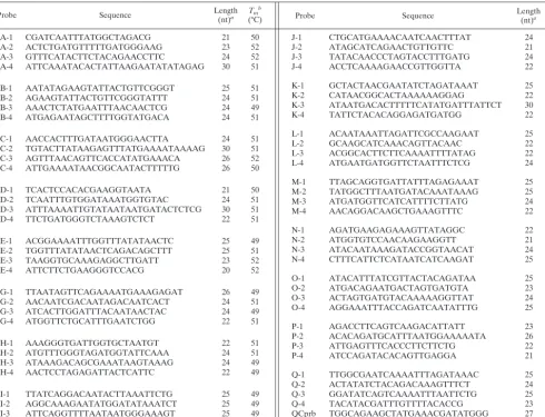
oligonucleotide probes were designed by using highly conserved regions for alleles of eachentgene within the region flanked by primers. The oligonucleo-tides selected are summarized in Table 2. The 5⬘end of the amino acid sequence of each oligonucleotide probe was modified during the synthesis (Qiagen) to enable the immobilization of the oligonucleotide to silylated (aldehyde) slides (ArrayIt, Sunnyvale, Calif.).
Microchip design and fabrication.To increase the confidence in the results of the microarray analysis, four individual oligonucleotide probes were selected for eachentgene. To facilitate interpretation of the microarray data, all oligonucle-otide probes specific for one gene were placed on a separate row of the array. Microchips were printed by use of a contact microspotting robotic system (PIXSYS 5500; Cartesian Technologies, Inc., Irvine, Calif.). The average size of the spots was 250m. The concentrations of the oligonucleotide probes were adjusted to 100M in 50% dimethyl sulfoxide before they were printed on the slides. A quality control oligonucleotide probe (39) of nonbacterial origin was added to each oligonucleotide probe at a concentration of 10M to enable monitoring of the spotting and hybridization steps of the microarray assay. Printed slides were incubated for at least 10 min at 85°C to evaporate the dimethyl sulfoxide completely, followed by 15 min of incubation in a freshly prepared 0.25% NaBH4solution in water. The slides were washed once for 5 min
[image:3.603.48.539.82.457.2]with 0.1% sodium dodecyl sulfate in water and five times for 1 min each time with distilled water to remove unbound oligonucleotides. Control spots used to mark the array position on the slide were generated by using 1⫻Spotting Solution (ArrayIt) in 0.25 M acetic acid.
TABLE 2. Oligonucleotide probes for detection and discrimination amongentgenes
Probe Sequence Length(nt)a Tm
b
(°C) Probe Sequence Length(nt)a (°C)Tmb
A-1 CGATCAATTTATGGCTAGACG 21 50
A-2 ACTCTGATGTTTTTGATGGGAAG 23 52
A-3 GTTTCATACTTCTACAGAACCTTC 24 52
A-4 ATTCAAATACACTATTAAGAATATATAGAG 30 51
B-1 AATATAGAAGTATTACTGTTCGGGT 25 51
B-2 AGAAGTATTACTGTTCGGGTATTT 24 51
B-3 AAACTCTATGAATTTAACAACTCG 24 49
B-4 ATGAGAATAGCTTTTGGTATGACA 24 51
C-1 AACCACTTTGATAATGGGAACTTA 24 51
C-2 TGTACTTATAAGAGTTTATGAAAATAAAAG 30 51
C-3 AGTTTAACAGTTCACCATATGAAACA 26 52
C-4 ATTGAAAATAACGGCAATACTTTTTG 26 50
D-1 TCACTCCACACGAAGGTAATA 21 50
D-2 TCAATTTGTGGATAAATGGTGTAC 24 51
D-3 ATTTAAAATTGTATAATAATGATACTCTCG 30 51
D-4 TTCTGATGGGTCTAAAGTCTCT 22 51
E-1 ACGGAAAATTTGGTTTATATAACTC 25 49
E-2 TGGTTTATATAACTCAGACAGCTTT 25 51
E-3 TAAGGTGCAAAGAGGCTTGATT 23 52
E-4 ATTCTTCTGAAGGGTCCACG 20 52
G-1 TTAATAGTTCAGAAAATGAAAGAGAT 26 49
G-2 AACAATCGACAATAGACAATCACT 24 51
G-3 ATCACTTGGATTTACAATAACTAC 24 49
G-4 ATGGTTCTGCATTTGAATCTGG 22 51
H-1 AAAGGGTGATTGGTGCTAATGT 22 51
H-2 ATGTTTGGGTAGATGGTATTCAAA 24 51
H-3 ATAAAGACAGCGAAATAAGTAAAG 24 49
H-4 AACTCCTAGAGATTACTCATTC 22 49
I-1 TTATCAGGACAATACTTAAATTCTG 25 49
I-2 AGGCAAAGAATATGGATATAAATCT 25 49
I-3 ATTCAGGTTTTAATAATGGGAAAGT 25 49
I-4 GCCTGTAAGTTTTTTGAAAATTTATGAA 28 51
aAmplicon size (in nucleotides [nt]). bThe basic melting temperature (T
m) was calculated with the Oligonucleotide Properties Calculator (http://www.basic.nwu.edu/biotools/oligocalc.html). The references for the oligonucleotide probe are cited in Materials and Methods.
J-1 CTGCATGAAAACAATCAACTTTAT 24 49
J-2 ATAGCATCAGAACTGTTGTTC 21 49
J-3 TATACAACCCTAGTACCTTTGATG 24 52
J-4 ACCTCAAAAGAACCGTTGGTTA 22 51
K-1 GCTACTAACGAATATCTAGATAAAT 25 49
K-2 CATAACGGCACTAAAAAAGGAG 22 51
K-3 ATAATGACACTTTTTCATATGATTTATTCT 30 51
K-4 TATTCTACACAGGAGATGATGG 22 51
L-1 ACAATAAATTAGATTCGCCAAGAAT 25 49
L-2 GCAAGCATCAAACAGTTACAAC 22 51
L-3 ACGGCACTTCTTCAAAATTTTATAG 22 51
L-4 ATGAATGATGGTTCTAATTTCTCG 24 51
M-1 TTAGCAGGTGATTATTTAGAGAAAT 25 49
M-2 TATGGCTTTAATGATACAAATAAAG 25 48
M-3 ATGATGGTTCATCATTTTCTTATG 24 49
M-4 AACAGGACAAGCTGAAAGTTTC 22 51
N-1 AGATGAAGAGAAAGTTATAGGC 22 49
N-2 ATGGTGTCCAACAAGAAGGTT 21 50
N-3 ATACAATAAAGATACCGGTAACAT 24 49
N-4 CTTTCATTCTCATAATCATCAAGAT 25 49
O-1 ATACATTTATCGTTACTACAGATAA 25 48
O-2 ATGACAGAATGACTAGTGATGTA 23 50
O-3 ACTAGTGATGTACAAAAAGGTTAT 24 49
O-4 AGGAAATTTACCAGATCAATATTTG 25 49
P-1 AGACCTTCAGTCAAGACATTATT 23 50
P-2 ACACAGATGCATTTAATGGAAAAATA 26 50
P-3 ATTGAGTTTCACCCTTCTTCTG 22 51
P-4 ATCCAGATACACAGTTGAGGA 21 50
Q-1 TTGGCGAATCAAAATTTAGATAAAC 25 49
Q-2 ACTATATCTACAGACAAAGTTTCT 24 49
Q-3 GGATATCAGTCAAAATTTAATTCTG 25 49
Q-4 TACATACGATTTGTTTTACACCG 23 50
QCprb TGGCAGAAGCTATGAAACGATATGGG 27 58
Cy3-QC CCCATATCGTTTCATAGCTTCTGCCA 26 58
on May 15, 2020 by guest
http://jcm.asm.org/
Hybridization conditions.Hybridization of the fluorescently labeled DNA samples to the microarray was performed in 1⫻hybridization buffer (5⫻ Den-hardt’s solution, 6⫻SSC buffer [1⫻SSC is 0.15 M NaCl plus 0.015 M sodium citrate], 0.1% Tween 20) at 45°C for 45 min. Before hybridization, 2 to 3l of Cy5-labeled DNA sample was mixed with an equal volume of 2⫻hybridization buffer containing 0.1M Cy3 quality control probe, followed by denaturation at 95°C for 3 min and chilling on ice. Each sample was placed on the microchip and covered with a glass coverslip (6 by 15 mm) to prevent evaporation of the probe during incubation. After the hybridization, the coverslips were washed away with 6⫻SSC containing 0.2% Tween 20 at room temperature. The slides were washed in a stepwise manner with 6⫻SSC buffer, 2⫻SSC buffer, and 1⫻SSC buffer for 2 min each and dried by airflow.
Microarray scanning.Fluorescent images of the microarrays were taken by scanning the slides with a ScanArray 5000 instrument (Perkin-Elmer). The flu-orescent signals from each spot were measured and compared by using Quan-tArray software (Perkin-Elmer).
Sequencing.We sequenced some enterotoxin genes, includingseb,sed,see, and
seq. The PCR-amplified DNA fragments were purified by agarose gel electro-phoresis, extracted with a QIAquick gel extraction kit (Qiagen) according to the protocol of the manufacturer, and sequenced with an ABI Prism 310 Genetic Analyzer System (PE Applied Biosystems, Foster City, Calif.).
Nucleotide sequence accession numbers.The accession numbers of the se-quences deposited in GenBank are AY518386 forsebof strain ATCC 14458, AY518387 forsedof strain NTCC10656, AY518772 forsemof strain ATCC 19095, and AY518388 and AY518389 forseeandseqof strain ATCC 27664, respectively.
RESULTS
Enterotoxin gene-specific PCR primers and microarray oli-gonucleotide probe designs. For our genotyping scheme for simultaneous detection and identification (genetic typing) of
multiple SE (ent) genes, we developed PCR amplification
as-says for 16 of the 17 known enterotoxin genes (seatoseeand
segtoseq) and an oligonucleotide microarray for the
identifi-cation of the PCR amplicons.
To develop toxin-specific PCR primers and microarray oli-gonucleotide probes, we performed multiple-sequence
align-ment analysis of theentgenes using sequence data from
Gen-Bank. As shown in Fig. 1, the analysis identified conserved regions flanking variable regions. The conserved regions were used to design universal primers for simultaneous
amplifica-tion of multipleent genes. The genetically divergent regions
were used to design individual PCR primers specific for each
entgene and to design gene-specific oligonucleotide probes to
discriminate among the 16entgenes (Fig. 1).
Our sequence data analysis revealed discrepancies in the
nomenclatures of several ent genes from the published
quences of strains MW2 and MU50. For example, the
se-quence named segin strain MW2 has 98% similarity to seq
(GenBank accession number AF410775) and a lower degree of
homology to another sequence of seg (GenBank accession
number AF064773). Similarly,sepfrom strain MU50 has only
83% similarity to a different reportedsepgene (GenBank
ac-cession number NC002745), whereas it has 98% similarity to
the sea enterotoxin gene (GenBank accession number
M18970).
For the amplification of eachentgene, gene-specific primers
were selected on the basis of unique sequences common to all alleles of each toxin gene determined by the multiple-sequence alignment (Fig. 1). To minimize cross-amplification between different toxin genes, the primers selected contained five and more mismatches with homologous toxins. However, in the
case ofs, we decided not to develop allele-specific
oligonucle-FIG. 1. Multiple-sequence alignment analysis of ent genes. The DNA sequences of 16 major SEs were retrieved from GenBank and aligned by using ClustalX software. The alignment results were presented by using GeneDoc software. Relatively conserved regions that were used for universal primer design are marked with arrows. The sequences of the oligonucleotide probes used for discrimination of the ent genes were selected from within the variable region flanked by the conserved regions. Gray background indicates similar sequences, and black background indicates conserved sequences.
on May 15, 2020 by guest
http://jcm.asm.org/
otide probes for the three described alleles,s, sec1, andsec2, because of their sequence similarities. In the assay, all three
are treated as a single gene, thesgene.
To design the oligonucleotide probes for the microarray, toxin-specific sequences were selected from the variable region identified by the multiple-sequence alignment (Fig. 1). Four individual oligonucleotide probes (21 to 30 nucleotides in length, with an average melting temperature of 50°C) were
designed to represent the sequence of each targetentgene
(Table 2). To minimize cross-hybridization with other ent
genes, oligonucleotide probes whose sequences had at least three mismatches with the sequence of the genetically closest
entgenes were selected.
Microarray analysis ofent genes in reference strains.For validation of the selected gene-specific primers and
oligonu-cleotide probes, we used three well-characterized S. aureus
sequencing strains, N315, MU50 (22), and MW2 (2), which
contain most of the known staphylococcal toxin genes (sea,s,
seg,seh,sei,sek,sel,sem,sen,seo, andsep). For the four toxins
not coded for by these strains, we used three additional
refer-ence strains: ATCC 14458 forseb, NTCC10656 forsedandsej,
and ATCC 27664 forseeandseq.
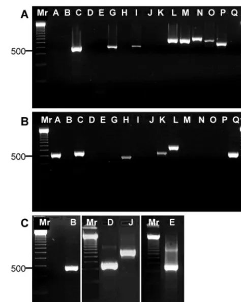
Genomic DNA from the five reference strains (N315, MW2, ATCC 14458, NTCC10656, and ATCC 27664) was amplified
with the ent-specific primers. The sizes of the PCR products
generated withent-specific primers varied from 466 to 807 bp,
depending on theentgene (Fig. 2A to C), and there was good
agreement between the observed and the predicted sizes of the amplicons. We unambiguously identified the presence of all 16
toxin genes previously shown in these strains: the toxin geness,
seg,sei,sel, sem,sen, seo, andsepwere found in strain N315
(Fig. 2A); the toxin genessea,s,seh,sek,sel, andseqgenes were
found in strain MW2 (Fig. 2B); and the toxin gene sebwas
amplified from ATCC 14458, the toxin genessedandsejwere
amplified from NTCC10656, and the toxin geneseewas
am-plified from ATCC 27664 (Fig. 2C). The toxin genessea,s,seg,
sei,sel,sem,sen, andseowere found in strain MU50 (data not
shown). We confirmed the identities of theseb,sed,see, andseq
amplicons by sequencing using the corresponding toxin-spe-cific primers. The GenBank accession numbers of the depos-ited sequences are presented above, in Materials and Methods.
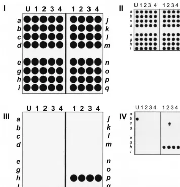
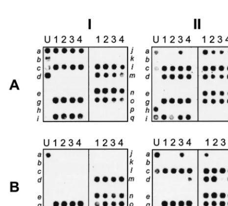
Theentmicroarray was prepared by immobilizing the four
oligonucleotides specific to each of the 16entgenes in separate
rows (shown schematically in Fig. 3I). The left-hand rows
con-tain probes specific for sea to sei, and the right-hand rows
contain probes specific forsejtoseq. The quality control scan
of the array (Fig. 3II) shows the actual scan of such an array. In our quality control procedure (40), each spot was printed with a quality control oligonucleotide, in addition to the spe-cific oligonucleotide probe. Each Cy5-labeled target ssDNA was spiked with quality control Cy3-labeled reference material complementary to the quality control oligonucleotide. The Cy3 quality control scan provides a control image of the entire microarray, which allows validation of the fabrication and hy-bridization steps.
The chip contains 10 identical microarrays for simultaneous analysis of five different microbial samples, each with two du-plicates (data not shown).
For microarray analysis of the ent-specific primers
ampli-cons, DNAs from S. aureus reference strains (strains N315,
MU50, MW2 ATCC 14458, NTCC10656, and ATCC 27664)
were amplified by using the ent gene-specific primers, and
fluorescently labeled ssDNA was synthesized from each of the
entamplicons by PE of the PCR products (see Materials and
Methods).
The microarray accurately detected each toxin gene. For
example, the detection of sepis shown schematically in Fig.
3III, and the actual results from microarray hybridization are shown in Fig. 3IV. It is noteworthy that occasional
cross-re-acting spots are observed, such as aselspot (Fig. 3IV, spot L2),
which cross-reacted with sep amplicons because of sequence
similarity. However, because four oligonucleotide probes were
used to detect eachentgene, a small number of cross-reacting
spots did not interfere with toxin identification. Figure 4 shows
the results of microarray analysis of all 16entgenes (seatoseq)
amplified by specific primers.
Overall, the results demonstrate the ability of the microarray
to amplify eachentgene with the gene-specific primers and to
unambiguously discriminate among theentgenes.
[image:5.603.304.541.68.364.2]Development of one-tube assay for typing ent genes. Our attempts to combine all 16 primer pairs in one tube for a
FIG. 2. PCR amplification of staphylococcalentgenes with gene-specific primers. Total DNA from reference strains N315, MW2, ATCC 14458, NTCC10656, and ATCC 27664 was amplified with gene-specific primers, and the resulting products were separated by electro-phoresis on a 2% agarose gel. Lanes: Mr, molecular size markers (indicated on the left in nucleotides); A,sea; B,seb; C,s; D,sed; E,see; G,seg; H,seh; I,sei; J,sej; K,sek; L,sel; M,sem; N,sen; O,seo; P,sep; Q,seq. (A) Strain N315 DNA amplification with all 16 gene-specific primers; (B) strain MW2 DNA amplification with all 16 gene-specific primers; (C) amplification ofsebfor strain ATCC 14458,sedandsejfor NTCC10656, andseefor ATCC 27664.
on May 15, 2020 by guest
http://jcm.asm.org/
multiplex PCR assay did not succeed in amplifying all target
ent genes. Several amplicons were not represented; and the
yields ofseo,sen,seh, andsejwere too low to be detected by
microarray hybridization (data not shown). This is likely due to interference between primers that significantly reduces the lev-els of amplification of particular toxin genes (data not shown). We overcame this problem by developing degenerate prim-ers (univprim-ersal primprim-ers) corresponding to the highly conserved
regions ofent(Table 1) and by adding a primer specific for the
underrepresented seh gene to the universal primer set. This
combination significantly improved the representation of all 16
entgenes. Control DNA and the five individual strains used in
the mixture are presented in three-by-two matrix composite
image (Fig. 5). All 16entgenes in the mixture were detected in
this analysis (Fig. 5AI). Interestingly, ATCC 14458 (Fig. 5BIII),
which is known to code for seb, was found to contain the
recently discoveredsekandseqgenes. Similarly, NTCC10656
was found to encode theseg,sei,sej,sem,sen, andseogenes, in
addition to thesedgene, the presence of which in NTCC10656
was already known (Fig. 5BI); and theseqgene was
unexpect-edly found in ATCC 27664 (Fig. 5BII).
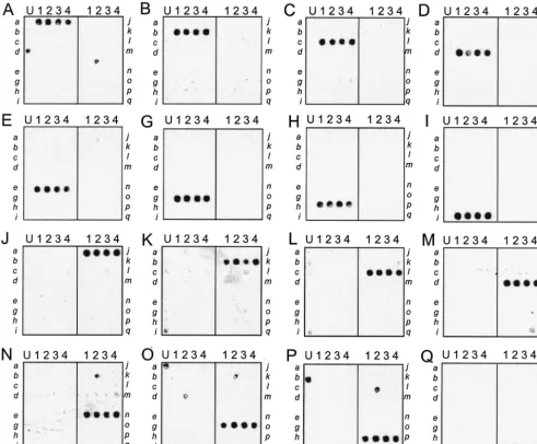
Presence of newly discovered ent genes in staphylococcal isolates analyzed previously.As shown above, our microarray
assay detects multiple ent genes simultaneously and detects
additional entgenes in isolates analyzed previously. We used
the microarray to analyze additionalS. aureusisolates analyzed
previously and found that other isolates contained multiple genes in several different combinations (Fig. 6). Strain FRI109,
listed in the NARSA collection as coding forsec2, also contains
sed, seg, sei, sej, sel, sem, sen, and seo (Fig. 6AII). Strain
A900322, which was thought to contain only the enterotoxin
[image:6.603.114.471.69.440.2]gene cluster withseg,sei,sen,seo, andsem(20), also contains
FIG. 3. Layout of staphylococcalentgenes on the microarray. Each microarray is composed of four sections, and each section contains four different oligonucleotide probes specific for each of four differententgenes in the section. (I) Schematic illustration of theentgene microarray. Genesseatoseiare in the rows on the left, and genessejtoseqare in the rows on the right, with each gene represented by four spots (columns). The first columns of each array are printed with spotting solution (U) to assist with array orientation. (II) Quality control scan of an array. Each spot of the array is visualized because each spot contains a small amount of quality control oligonucleotide probe QCprb, which hybridizes with a Cy3-labeled oligonucleotide, Cy3-QC, which is used to spike each target mixture. The spot is visualized with a 543-nm laser. (III) Schematic showing microarray detection ofsep. (IV) Microarray analysis ofsepgenomic DNA from strain N315, which was amplified withsep-specific primers. The resulting PCR product was fluorescently labeled with Cy5 and hybridized to theentgene array, which was then scanned with a 632-nm laser.
on May 15, 2020 by guest
http://jcm.asm.org/
thesepgene (Fig. 6BI). Strain MNDON, which was reported in
the NARSA catalogue to code for sec1, seg, seh, and sei, is
shown here to also encodesel,sem,sen, andseo(Fig. 6BII).
DISCUSSION
The traditional method of identifying SEs is serological typ-ing with antibodies. In general, serological typtyp-ing is less sensi-tive to small variations among SEs than DNA-based methods.
Several PCR-based methods are available forS. aureustoxin
typing (21, 35). Most require several separate reactions to
distinguish among several entgenes. More recently, methods
forS. aureustoxin typing by multiplex PCR have been reported
(6, 26, 29). These PCR methods are based on combinations of
entgene-specific primers or a combination of universal forward
primers and specific reverse primers (36).
One problem with all present PCR-based methods is that
novel or unexpected toxin genes can lead to false-positive or
-negative results. For example, we observed that allsea
gene-specific primers described in the literature can be used for
successful amplification ofsepas well. This might lead to the
mistaken conclusion that a strain encodes seaand incorrect
data about the distributions ofentgenes and their roles in food
poisoning. Since the relationship between the presence of a specific enterotoxin (or a combination of enterotoxins) and human food poisoning is not clearly understood, there is a need for a reliable and universal method for unambiguous
identification of knownentgenes and for detection of novelent
genes.
We describe here a combination PCR-microarray assay for
detection and identification ofentgenes. The analysis is based
on PCR amplification of a variable region of almost all known
ent genes with a single set of degenerate primers whose
[image:7.603.48.540.71.477.2]se-quences correspond to those of the flanking highly conserved
FIG. 4. Microarray-based detection of 16 staphylococcalentgenes with gene-specific primers. Total DNAs from fiveS. aureusreference strains (strains N315, MW2, ATCC 14458, NTCC10656, and ATCC 27664) were amplified with theentgene-specific primers, with the panel labels A to E and G to Q corresponding to the lanes described in the legend to Fig. 2. The ssDNAs derived from the PCR products were labeled with Cy5 and hybridized to theentgene microarray (Fig. 3), which was then scanned with a 632-nm laser.
on May 15, 2020 by guest
http://jcm.asm.org/
regions. The amplicons are then identified by analysis on the oligonucleotide microarray. This combined method takes ad-vantage of the strengths of each technique. PCR amplification is highly sensitive, detecting target genes from genomic DNA even when they are present at low concentrations. DNA-DNA hybridization on the microarray increases the specificity of the assay and allows parallel analysis of multiple sequences simul-taneously. In addition, the nonspecific amplicons often seen in PCRs have no effect on the hybridization of the targets with specific oligonucleotide probes.
Microarrays are not in common use in average laboratories today. However, like any new technology, as more applications are developed for the microarray technology, it will become more practical and may well become widely used. In the work
described here, the presence of genes for each of 16entgenes
was analyzed by four methods: PCR amplification with primers specific for each of the 16 enterotoxin genes, followed by anal-ysis by gel electrophoresis (Fig. 2); PCR amplification with specific primers (Fig. 4), followed by analysis by the microarray assay; amplification of the 16 genes with universal primers, followed by microarray analysis (Fig. 5); and sequencing of the enterotoxin genes to verify their identities. Thus, using two amplification methods (methods with specific primers and uni-versal primers) as well as three DNA analysis methods (gel electrophoresis, DNA sequencing, and DNA microarray anal-ysis), we verified the performance of the method. Using this array, we have shown that some strains previously analyzed by
immunological methods contain additional entgenes not
de-tected by the original assays (Fig. 5 and 6).
[image:8.603.304.537.401.612.2]In our microarray system, we used relatively short oligonu-cleotides (21 to 30 nuoligonu-cleotides) for three reasons. First, shorter
FIG. 5. One-tube microarray-based detection of 16 staphylococcalent genes with universal primers. Genomic DNAs from five S. aureus reference strains (strains N315, MW2 ATCC 14458, NTCC10656, and ATCC 27664) were amplified with a mixture ofentgene-specific universal primers supplemented with the primers specific forseh. The resulting PCR amplicons were subjected to PE with a mixture of the reverseent gene-specific universal primers supplemented with the reverse primers specific forseh, followed by Cy5 chemical labeling. The labeled targets were hybridized to theentgene microarray, which was then scanned with a 632-nm laser. A control array which contains a mixture of DNA representing all genes (from strains N315, MW2 ATCC 14458, NTCC10656, and ATCC 27664) was included for demonstration of the ability of the assay to detect 16entgenes in a single sample. AI, strain control array; AII, strain N315; AIII, MW2; BI, NTCC10656; BII, ATCC 27664; BIII, ATCC 14458.
FIG. 6. One-tube microarray-based analysis of previously analyzed S. aureusstrains. Genomic DNA from four strains analyzed previously (strains Mu50, FRI361, A90322, and MNDON) was amplified by using a mixture ofentgene-specific universal primers supplemented with the primer specific forseh. The resulting PCR amplicons were subjected to PE with a mixture of the reverseentgene-specific universal primers supplemented with the reverse primer specific forseh, followed by Cy5 chemical labeling, and were then hybridized to theentgene micro-array. The resulting image was scanned with a 632-nm laser. The analysis of the four strains is presented in a two-by-two matrix com-posite image. AI, strain Mu50; AII, strain FRI361; BI, A90322; BII, MNDON.
on May 15, 2020 by guest
http://jcm.asm.org/
oligonucleotide probe sequences (⬍25 bp) are often capable of detecting a single-nucleotide mismatch between the target ssDNA and the oligonucleotide probe, which allows detection of minor genetic variants in target genes in a bacterial popu-lation. Second, the shorter oligonucleotide probes allow inde-pendent testing of several species-specific regions of each gene, enabling effective coverage of the target sequence with more (but shorter) oligonucleotide probes. This reduces the proba-bility of misidentification. Third, short oligonucleotides reduce the cost of chip production.
The redundancy of the testing (the number of spots repre-senting each gene) is one way to reduce the risk of SE mis-identification. While only one portion of each SE gene (the variable region shown in Fig. 1) is used for the analysis, leaving open the possibility of significant sequence variation in other parts of the genes, such variation is not common.
The main disadvantage of simultaneous PCR amplification of multiple targets is that different copy numbers of the genes in a cell result in different signal intensities on the array. This might be overcome by use of supplemental specific primers to improve the detection of underrepresented amplicons (Fig. 5 and 6).
Many of the known SEs have been discovered only in the last
few years, including some, such as sep, that were discovered
only through the sequencing of the S. aureus N315 genome
(22). Given the genetic variability and the spread of theent
gene family, it is possible that there are other, as yet unknown,
entgenes. However, the similarity in the conserved regions of
these genes (Fig. 1) suggests that any additional gene family members will share those conserved sequences. Thus, it is possible that this assay might lead to the discovery of
addi-tionalentgenes in new strains. The amplicons of the novelent
genes can be discernible as amplicons that hybridize to
com-mon spots but not to anyentgene-specific spots.
Multiple SE genes are commonly found inS. aureusstrains
(19, 20, 32). Among 198 S. aureus isolates implicated in S.
aureus infections in France, 85.4% expressed multiple SEs
(19). Our analysis of these data suggests that the majority
(92%) contain multiple SEs, especially theegccluster (seg,sei,
sen,seo, andsem). Among theS. aureusisolates implicated in
food-poisoning episodes in Japan, 93% expressed SEs, while only 72.2% of isolates from healthy people expressed SEs (32). One explanation for the presence of multiple toxins in most strains is that these genes are often structurally linked. Several
pathogenicity islands have been reported inS. aureus,
includ-ing one encodinclud-ing the toxic shock syndrome toxin (tst) and
s-andsel-like proteins (13) and another encoding SE serotypes
B, K, and Q (41). Others have reported pathogenicity islands
containing the tstgene and an open reading frame with
se-quence similarity to those encoding SEs (25) and a region contains enterotoxins D and J (42). In addition, as noted
above, a group of five toxin genes (seg,sei,sen,seo, andsem) is
encoded by the enterotoxin gene cluster, egc(20).
Interest-ingly, in the study of 198 clinical isolates by Jarraud et al. (19), half of the 14 strains that carried only a single enterotoxin gene
had the seagene, perhaps because it has been shown to be
associated with a structurally unstable, possibly mobile,
dis-crete genetic element (8) that is not part of theegccluster.
In terms of the functionalities of SEs, multiple toxins with diverse spectra of activities may offer the pathogen versatility
in terms of the host range. For example, it has been shown that different types of SEs have different emetic response activities in house musk shrews, although it was thought that there are no differences in the emetic response activities of SEA, SEB, SEC, SED, and SEE in humans and primates (18). In addition,
S. aureus expression of specificentgenes may depend on the
host tissue and may play a role in the adaptation ofS. aureusto
the host environment (4). Some have speculated that some cases of food poisoning result from the simultaneous
expres-sion of several enterotoxins in a single pathogenic S. aureus
strain rather than from the expression of a single toxin. However, it is unclear whether all the toxins are actually expressed and what the biological and clinical effects of mul-tiple toxins might be. The method presented here can detect
ent genes but does not determine whether the gene is
ex-pressed or whether the encoded protein is functional. The levels of correlation between the presence of genes that code for the production of SE (as determined by PCR) and the expression of these genes (as determined by enzyme-linked immunosorbent assay) were 100% for SEA and SEE, 86% for SEC, 89% for SED, and 47% for SEB (30). Thus, the actual presence of the toxin needs to be assessed by an immunological or activity assay.
In summary, the PCR-microarray method described here is
a potentially powerful tool for the analysis ofS. aureusstrains.
We used this method to test clinical isolates analyzed previ-ously and found that these isolates frequently carry the genes for numerous toxins, including some of the newly discovered SEs. More studies need to be done to understand the biolog-ical regulation and the biologbiolog-ical and clinbiolog-ical effects of multiple enterotoxins. Our method has great potential for application in
high-throughput screening and accurate genotyping ofentgenes,
which are especially important in epidemiological studies.
ACKNOWLEDGMENTS
We thank Farukh Khambaty, FDA Center of Food Safety and Ap-plied Nutrition, and the NARSAS. aureuscollection (Focus Technol-ogies, Inc.) for providing the strains used in this study.
This work was supported in part by USDA grant 0013000 and fund-ing provided by the FDA Office of Science.
REFERENCES
1. Archer, D. L., and F. E. Young.1988. Contemporary issues: diseases with a food vector. Clin. Microbiol. Rev.1:377–398.
2. Baba, T., F. Takeuchi, M. Kuroda, H. Yuzawa, K. Aoki, A. Oguchi, Y. Nagai, N. Iwama, K. Asano, T. Naimi, H. Kuroda, L. Cui, K. Yamamoto, and K. Hiramatsu.2002. Genome and virulence determinants of high virulence community-acquired MRSA. Lancet359:1819–1827.
3. Balaban, N., and A. Rasooly.2000. Staphylococcal enterotoxins. Int. J. Food Microbiol.61:1–10.
4. Banks, M. C., N. S. Kamel, J. B. Zabriskie, D. H. Larone, D. Ursea, and D. N. Posnett.2003. Staphylococcus aureus express unique superantigens depending on the tissue source. J. Infect. Dis.187:77–86.
5. Bean, N. H., J. S. Goulding, C. Lao, and F. J. Angulo.1996. Surveillance for foodborne-disease outbreaks—United States, 1988–1992. Morb. Mortal. Wkly. Rep. CDC Surveill. Summ.45:1–66.
6. Becker, K., R. Roth, and G. Peters.1998. Rapid and specific detection of toxigenicStaphylococcus aureus: use of two multiplex PCR enzyme immu-noassays for amplification and hybridization of staphylococcal enterotoxin genes, exfoliative toxin genes, and toxic shock syndrome toxin 1 gene. J. Clin. Microbiol.36:2548–2553.
7. Bergdoll, M. S.1995. Importance of staphylococci that produce nanogram quantities of enterotoxin. Zentbl. Bakteriol. Parasitenkd. Infektkrankh. Hyg. Abt. 1 Orig.282:1–6.
8. Betley, M. J., S. Lofdahl, B. N. Kreiswirth, M. S. Bergdoll, and R. P. Novick.
1984. Staphylococcal enterotoxin A gene is associated with a variable genetic element. Proc. Natl. Acad. Sci. USA81:5179–5183.
on May 15, 2020 by guest
http://jcm.asm.org/
9. Breuer, K., M. Wittmann, B. Bosche, A. Kapp, and T. Werfel.2000. Severe atopic dermatitis is associated with sensitization to staphylococcal entero-toxin B (SEB). Allergy55:551–555.
10. Bunikowski, R., M. Mielke, H. Skarabis, U. Herz, R. L. Bergmann, U. Wahn, and H. Renz.1999. Prevalence and role of serum IgE antibodies to the Staphylococcus aureus-derived superantigens SEA and SEB in children with atopic dermatitis. J. Allergy Clin. Immunol.103:119–124.
11. Bunning, V. K., J. A. Lindsay, and D. L. Archer.1997. Chronic health effects of microbial foodborne disease. World Health Stat. Q.50:51–56. 12. Ferens, W. A., and G. A. Bohach.2000. Persistence of Staphylococcus aureus
on mucosal membranes: superantigens and internalization by host cells. J. Lab. Clin. Med.135:225–230.
13. Fitzgerald, J. R., S. R. Monday, T. J. Foster, G. A. Bohach, P. J. Hartigan, W. J. Meaney, and C. J. Smyth.2001. Characterization of a putative patho-genicity island from bovineStaphylococcus aureusencoding multiple super-antigens. J. Bacteriol.183:63–70.
14. Garthright, W. E., D. L. Archer, and J. E. Kvenberg.1988. Estimates of incidence and costs of intestinal infectious diseases in the United States. Public Health Rep.103:107–115.
15. Herman, A., J. W. Kappler, P. Marrack, and A. M. Pullen.1991. Superan-tigens: mechanism of T-cell stimulation and role in immune responses. Annu. Rev. Immunol.9:745–772.
16. Herz, U., R. Bunikowski, M. Mielke, and H. Renz.1999. Contribution of bacterial superantigens to atopic dermatitis. Int. Arch. Allergy Immunol.
118:240–241.
17. Howell, M. D., J. P. Diveley, K. A. Lundeen, A. Esty, S. T. Winters, D. J. Carlo, and S. W. Brostoff.1991. Limited T-cell receptor beta-chain hetero-geneity among interleukin 2 receptor-positive synovial T cells suggests a role for superantigen in rheumatoid arthritis. Proc. Natl. Acad. Sci. USA88:
10921–10925.
18. Hu, D. L., K. Omoe, Y. Shimoda, A. Nakane, and K. Shinagawa.2003. Induction of emetic response to staphylococcal enterotoxins in the house musk shrew (Suncus murinus). Infect. Immun.71:567–570.
19. Jarraud, S., C. Mougel, J. Thioulouse, G. Lina, H. Meugnier, F. Forey, X. Nesme, J. Etienne, and F. Vandenesch.2002. Relationships between Staph-ylococcus aureusgenetic background, virulence factors,agrgroups (alleles), and human disease. Infect. Immun.70:631–641.
20. Jarraud, S., M. A. Peyrat, A. Lim, A. Tristan, M. Bes, C. Mougel, J. Etienne, F. Vandenesch, M. Bonneville, and G. Lina.2001. egc, a highly prevalent operon of enterotoxin gene, forms a putative nursery of superantigens in Staphylococcus aureus. J. Immunol.166:669–677.
21. Johnson, W. M., S. D. Tyler, E. P. Ewan, F. E. Ashton, D. R. Pollard, and K. R. Rozee.1991. Detection of genes for enterotoxins, exfoliative toxins, and toxic shock syndrome toxin 1 inStaphylococcus aureusby the polymerase chain reaction. J. Clin. Microbiol.29:426–430.
22. Kuroda, M., T. Ohta, I. Uchiyama, T. Baba, H. Yuzawa, I. Kobayashi, L. Cui, A. Oguchi, K. Aoki, Y. Nagai, J. Lian, T. Ito, M. Kanamori, H. Matsumaru, A. Maruyama, H. Murakami, A. Hosoyama, Y. Mizutani-Ui, N. K. Taka-hashi, T. Sawano, R. Inoue, C. Kaito, K. Sekimizu, H. Hirakawa, S. Kuhara, S. Goto, J. Yabuzaki, M. Kanehisa, A. Yamashita, K. Oshima, K. Furuya, C. Yoshino, T. Shiba, M. Hattori, N. Ogasawara, H. Hayashi, and K. Hira-matsu.2001. Whole genome sequencing of methicillin-resistant Staphylo-coccus aureus. Lancet357:1225–1240.
23. Lee, A. C., R. N. Robbins, and M. S. Bergdoll.1978. Isolation of specific and common antibodies to staphylococcal enterotoxins A and E by affinity chro-matography. Infect. Immun.21:387–391.
24. Lee, A. C., R. N. Robbins, R. F. Reiser, and M. S. Bergdoll.1980. Isolation of specific and common antibodies to staphylococcal enterotoxins B, C1, and C2. Infect. Immun.27:431–434.
25. Lindsay, J. A., A. Ruzin, H. F. Ross, N. Kurepina, and R. P. Novick.1998.
The gene for toxic shock toxin is carried by a family of mobile pathogenicity islands in Staphylococcus aureus. Mol. Microbiol.29:527–543.
26. Mehrotra, M., G. Wang, and W. M. Johnson.2000. Multiplex PCR for detection of genes forStaphylococcus aureusenterotoxins, exfoliative toxins, toxic shock syndrome toxin 1, and methicillin resistance. J. Clin. Microbiol.
38:1032–1035.
27. Mempel, M., G. Lina, M. Hojka, C. Schnopp, H. P. Seidl, T. Schafer, J. Ring, F. Vandenesch, and D. Abeck.2003. High prevalence of superantigens asso-ciated with the egc locus in Staphylococcus aureus isolates from patients with atopic eczema. Eur. J. Clin. Microbiol. Infect. Dis.22:306–309.
28. Misfeldt, M. L.1990. Microbial “superantigens.” Infect. Immun.58:2409– 2413.
29. Monday, S. R., and G. A. Bohach.1999. Use of multiplex PCR to detect classical and newly described pyrogenic toxin genes in staphylococcal iso-lates. J. Clin. Microbiol.37:3411–3414.
30. Najera-Sanchez, G., R. Maldonado-Rodriguez, P. Ruiz Olvera, and L. M. de la Garza.2003. Development of two multiplex polymerase chain reactions for the detection of enterotoxigenic strains of Staphylococcus aureus isolated from foods. J. Food Prot.66:1055–1062.
31. Olsen, S. J., L. C. MacKinnon, J. S. Goulding, N. H. Bean, and L. Slutsker.
2000. Surveillance for foodborne-disease outbreaks—United States, 1993– 1997. Morb. Mortal. Wkly. Rep. CDC Surveill. Summ.49:1–62.
32. Omoe, K., M. Ishikawa, Y. Shimoda, D. L. Hu, S. Ueda, and K. Shinagawa.
2002. Detection ofseg,seh, andseigenes inStaphylococcus aureusisolates and determination of the enterotoxin productivities ofS.aureusisolates harboringseg,seh, orseigenes. J. Clin. Microbiol.40:857–862.
33. Pereira, J. L., S. P. Salzberg, and M. S. Bergdoll.1991. Production of staphylococcal enterotoxin D in foods by low-enterotoxin-producing staph-ylococci. Int. J. Food Microbiol.14:19–25.
34. Sambrook, J., E. F. Fritsch, and T. Maniatis.1989. Molecular cloning: a laboratory manual, 2nd ed. Cold Spring Harbor Laboratory, Cold Spring Harbor, N.Y.
35. Schmitz, F. J., M. Steiert, B. Hofmann, J. Verhoef, U. Hadding, H. P. Heinz, and K. Kohrer.1998. Development of a multiplex-PCR for direct detection of the genes for enterotoxin B and C and toxic shock syndrome toxin-1 in Staphylococcus aureus isolates. J. Med. Microbiol.47:335–340.
36. Sharma, N. K., C. E. Rees, and C. E. Dodd.2000. Development of a single-reaction multiplex PCR toxin typing assay forStaphylococcus aureusstrains. Appl. Environ. Microbiol.66:1347–1353.
37. Thompson, J. D., D. G. Higgins, and T. J. Gibson.1994. CLUSTAL W: improving the sensitivity of progressive multiple sequence alignment through sequence weighting, position-specific gap penalties and weight matrix choice. Nucleic Acids Res.22:4673–4680.
38. Uematsu, Y., H. Wege, A. Straus, M. Ott, W. Bannwarth, J. Lanchbury, G. Panayi, and M. Steinmetz.1991. The T-cell-receptor repertoire in the syno-vial fluid of a patient with rheumatoid arthritis is polyclonal. Proc. Natl. Acad. Sci. USA88:8534–8538.
39. Volokhov, D., V. Chizhikov, K. Chumakov, and A. Rasooly.2003. Microarray analysis of erythromycin resistance determinants. J. Appl. Microbiol.95:787– 798.
40. Volokhov, D., A. Rasooly, K. Chumakov, and V. Chizhikov.2002. Identifi-cation ofListeriaspecies by microarray-based assay. J. Clin. Microbiol.40:
4720–4728.
41. Yarwood, J. M., J. K. McCormick, M. L. Paustian, P. M. Orwin, V. Kapur, and P. M. Schlievert.2002. Characterization and expression analysis of Staphylococcus aureus pathogenicity island 3. Implications for the evolution of staphylococcal pathogenicity islands. J. Biol. Chem.277:13138–13147. 42. Zhang, S., J. J. Iandolo, and G. C. Stewart.1998. The enterotoxin D plasmid
of Staphylococcus aureus encodes a second enterotoxin determinant (sej). FEMS Microbiol. Lett.168:227–233.