pH-responsive magnesium- and carbonate-substituted
apatite nano-crystals for efficient and cell-targeted delivery
of transgenes
Ezharul Hoque Chowdhury
Faculty of Medicine, Nursing and Health Sciences, Jeffrey Cheah School of Medicine and Health Sciences, Monash University Sunway Campus, Jalan Lagoon Selatan, Bandar Sunway, Malaysia
Email: [email protected]
Received 27 February 2013; revised 29 March 2013; accepted 16 May 2013
Copyright © 2013 Ezharul Hoque Chowdhury. This is an open access article distributed under the Creative Commons Attribution License, which permits unrestricted use, distribution, and reproduction in any medium, provided the original work is properly cited.
ABSTRACT
The short half-lives due to the enzymatic degradation in blood, the lack of tissue targetability and the inca- pability to passively diffuse across the plasma mem- brane and smoothly traffic across the harsh intracel- luar environment are the major shortcomings for nu- cleic acid-based potential therapeutics, such as recom- binant plasmid and antisense oligonucleotides or small interferring RNA (siRNA). Plasmid DNA con- taining a gene of interest could have immense impact as a promising therapeutic drug for treating genetic as well as acquired human diseases at the molecular level with high level of efficacy and precision. Thus both viral and non-viral synthetic vectors have been developed in the past decades to address the afore- mentioned challenges of naked DNA. While in the viral particles plasmid DNA is integrated into the vi- ral genome, in most non-viral cases the DNA being anionic in nature is electrostatically associated with a cationic lipid or polymer forming lipoplex or polyplex, respectively, or a cationized inorganic gold, silica or iron oxide particle. Due to the potential immunoge- nicity and carcinogenicity issues with the viral parti- cles, non-viral vectors have drawn much more atten- tion for the clinical evaluation. However, the main concern of using non-biodegradable particles, spec- ially the inorganic ones, is the adverse effects owing to their long term interactions with body components. We have recently developed biodegradable pH-sensi- tive inorganic nanoparticles of Mg/CaPi and carbon- ate apatite for efficient transgene delivery to primary, cancer and embryonic stem cells, by virtue of their high affinity binding with the DNA, ability to contact the cell membrane by ionic or ligand-receptor inter- actions and fast dissolution kinectis in endosomal aci-
dic pH facilitating release of the DNA from the dis- solving particles and also from the endosomes.
Keywords:Gene Therapy; Nanoparticles; Particle Dissolution; Ca/Mgpi; Carbonate Apatite; Endosome, Nucleus; Gene Expression
1. INTRODUCTION
Extensive research in the past decades on genomics and proteomics has led to the comprehensive understanding of the functional roles of proteins in the signal transduc- tion pathways for phenotype regulation. Since both gene- tic and acquired human diseases are generally associated with up- or down-regulation of various genes and conse- quential over-expression or suppression of gene products, such as mRNA and proteins, treatment strategies could virtually target any of the three cellular macromolecules (genes, mRNAs and proteins) either by inhibiting or re- storing their functions within the cells. However, treat- ment of a disease at the genetic level by either supplying a functional gene into the nucleus in order to be trans- cribed into a functional mRNA (i.e., replacement of a
RNA-based genetic materials.
Viral systems are by far the most effective means of DNA delivery to mammalian cells, but some major limi- tations including toxicity, immunogenicity, restricted tar- geting of specific cell types, limited DNA carrying ca- pacity, production and packaging problems, recombina- tion and high cost, hamper their successful applications in basic research and clinical medicine. The effective- ness of a viral particle is the result of its highly evolved and specialized structure basically composed of a protein coat surrounding a nucleic acid core. Such a highly orga- nized structure can prevent viral particles from unwanted interactions with serum components, while promoting subsequent internalization by the cells, escaping from the endosomes, and releasing genetic material from the par- ticle either before or after entering the nucleus [6-10]. Development of a nonviral approach having the benefi- cial virus-like properties and lacking the disadvantageous ones would emerge as the most attractive one for im- plementation in research laboratories and gene therapy.
Here, we report on the current progress and develop- ment of pH-sensitive inorganic nanoparticles of Mg2+- substituted calcium phosphate (CaPi) and carbonate apa- tite for efficient and targeted delivery of transgenes into primary, cancerous and embryonic stem cells.
2. MECHANISM OF DNA DELIVERY BY
CLASSICAL CaPi PARTICLES
CaPi precipitation has been one of the most widely used methods due to its simplicity, cost effectiveness and effi- cacy, for delivery of plasmid DNA to mammalian cells expressing a desirable transgene (known as “transfec- tion”) since the technique was developed in 1973 [11]. Despite being one of the oldest transfection methods, the procedure has remained almost unchanged, basically com- posed of either direct or drop-wise mixing of two solu- tions, one containing DNA and calcium chloride (CaCl2) and the other possessing inorganic phosphate (Pi) (either NaH2PO4 or Na2HPO4) in order to induce “supersatu- ration” of the final solution with respect to CaCl2 and Pi, resulting in precipitation of microscopically visible and invisible particles with the DNA being adsorbed through electrostatic interactions [12-14]. Although the particles have net negative charge, they are capable of binding anionic DNA presumably through the Ca2+-rich domains instead of the -rich sites existing on their surfaces. X-ray diffraction and FT-IR analysis of the precipitated particles established them as “hydroxyapatite” with the molecular formula of Ca10(PO4)6(OH)2 [12-14]. The re- sulting DNA/CaPi co-precipitates are usually dispersed onto the cultured cells from several hours to overnight in the incubator allowing them to be taken up by the cells via endocytosis. The internalized DNA is thus entrapped into the acidic compartments of endosomes which can
subsequently be fused with the lysosomes of relatively lower pH, where the DNA is usually fragmented by hy- drolytic enzymes (nucleases). However, treatment with glycerol [12,15] or chloroquine [15,16] can help release functional DNA from the acidic vesicles to the cytosol in return for some extent of cell toxicity. Depending on whether the cell is dividing, like a cancer cell or non- dividing like most of the normal cells in our body, cy- tosolic plasmid DNA can enter the nucleus at the time when the nuclear membrane is disrupted during mitosis or slowly pass through the nuclear pores by passive dif- fusion, respectively [5]. As a result, like other non-viral vectors, transcription of the nuclear translocated DNA and subsequent translation of the resulting mRNA to a particular protein are substantially high in cancer cells compared to nonmitotic or slowly dividing cells.
3 4
PO
3. APPROACHES FOR REGULATION OF
PARTICLE GROWTH: INFLUENCES
ON CELLULAR UPTAKE OF DNA
fection [10]. However, the transfection in 10% serum- supplemented medium was significantly higher than in serum-free medium probably due to the particles of lar- ger size formed in the latter case, since endocytosis is usu- ally more effective for the particles of smaller size [18].
4. DEVELOPMENT OF Ca-Mg
PHOSPHATE (Ca/MgPi)
NANOPARTICLES FOR EFFICIENT
DELIVERY OF DNA
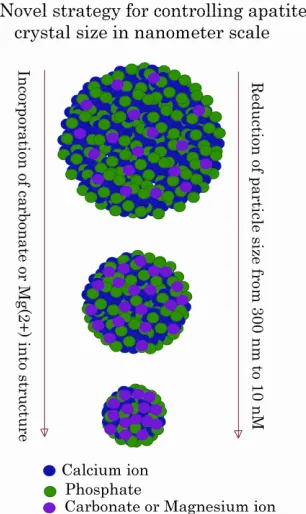
In spite of the great efforts made in the past for controll- ing the growth kinetics of CaP precipitation by optimiz- ing the parameters of reactant concentrations, pH, incu- bation period and temperature, the first modification of the particles at the molecular level in order to delicately regulate the particle size was achieved by partially sub- stituting Ca2+ with another divalent cation, Mg2+. The re- sulting particles of Ca/MgPi precipitates, like CaPi ones, adsorb DNA, but unlike the latter, could prevent the growth of the precipitates to a significant extent, leading to huge uptake of DNA and consequential efficient trans- gene expression, which is 10 to 100 times higher than the classical CaPi co-precipitation method in HeLa and NIH3T3 cells [13].
Mixing of a 300 µl of aqueous solution containing in- creasing doses of Mg2+ along with 250 mM of Ca2+, with a 300 ul of 2XHBS (pH 7.05) having 1.5 mM of Pi re- sulted in a dramatic decline in particle diameter from 2.5 um up to 500 nm for 30 min incubation at room tempe- rature, depending on the concentrations of initially added Mg2+. Clearly, with an increase in the amount of Mg2+ substituting Ca2+ in the apatite structure, the particle size decreased transforming the particle diameter from 500 nm to below 100 nM at 80 mM of Mg2+ for 1 min incu- bation. The estimated molar ratios of Ca, Mg and P pre- sent in the precipitates of Ca/MgPi indicated formation hydroxyapatite with the molecular formula of Ca10−XMgX(PO4)6(OH)2 for 0.58 and 1.03 percentage of Mg and octacalcium phosphate (OCP) with the formula of Ca4−XMgX(PO4)3 for 1.76 to 3.16 percentage of Mg substituted in the particles [13]. The uptake of fluore- scence-labeled plasmid in HeLa cells indicated size-de- pendent endocytosis of the Ca/MgPi particles with the particles of the smaller size resulting in higher uptake of the DNA compared to those of bigger size, implying in agreement with the notion that internalization of apatite particles is indeed size-dependent [18,19].
5. ESTABLISHMENT OF CARBONATE
APATITE NANOPARTICLES AS
SMART DNA CARRIERS
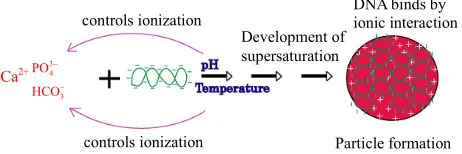
Bicarbonate-buffered medium is widely used for cell cul- ture. Addition of relatively low amount of Ca2+(3 - 5 mM)
to such medium (pH 7.5) containing endogenous Pi, fol- lowed by incubation at 37˚C, resulted in microscopically visible particles which were apparently absent in the in- cubated HBS containing the same doses of total Ca2+ and Pi, implicating that bicarbonate is one of the reactants in generation of those particles. Elemental analysis and FT- IR of the lyophilized powder of the sedimented particles suggested the structure of typical carbonate apatite, a major component of hard tissues in our body [20,21]. X- ray diffraction patterns demonstrated the widening of the peaks as indicative of poor crystallinity unlike those for highly crystalline hydroxyapatite as formed with CaPi precipitation [20,21]. Thus, the chemical reaction for particle formation takes place among Ca2+, 3
4
PO and 3
HCO, and DNA can electrostatically be associated with the cationic (Ca2+-rich)domains of the particles [20, 21]. The two crucial factors for determining transfection- potency, namely, the number and the average size of the particles are dramatically influenced by the concentra- tions of calcium, phosphate and bicarbonate, the pH of the medium and the incubation period and temperature (Figure 1). Increasing the concentration of any of the reactants (calcium, phosphate and bicarbonate) accele- rates particle growth by providing a stronger driving force for the reaction while the other parameters (pH, in- cubation time and temperature) are constant. On the other hand, an increase in pH and temperature (or incubation period) mainly shifts the ionization equilibrium of phos- phate towards the forward direction, thereby favoring the particle generation by increasing the reaction rate, ac- companied by growth and aggregation of the particles [20,21]. However, like Mg2+ in Ca/MgPi, bicarbonate as a minor component of the final apatite product with the molecular formula of Ca10(PO4)6−X(CO3)X(OH)2, prevents the aggregation and generates smaller crystals in a dose- dependent manner (Figure 2). Finally, due to the small size of the crystal having an average diameter of 50 - 300 nM, and strong binding affinity towards the DNA, car- bonate apatite can effectively be transferred into the cell with the embedded DNA through endocytosis, following electrostatic interactions with the plasma membrane. The efficiency of cellular uptake of DNA was estimated to beat least 10-times higher than that for classical CaPi method [20,21].
controls ionization
Development of supersaturation
controls ionization Particle formation
DNA binds by ionic interaction
Ca2+ 34 3
PO HCO
[image:3.595.309.540.627.704.2]
lower transgene expression than the viral counterpart in terms of the total number of plasmid copies initially given. Among the other major barriers are inability of escaping the endosomes and difficulty in nuclear trans- location. Depending on the type of carriers, DNA can be released from the particles in the acidic compartments, cytosol or nucleus mostly by interacting with the cellular macromolecules. Endosomal escape of the DNA either in free form or in complexation with the particles is guided by distinct mechanisms. For expression of the DNA car- ried by carbonate apatite, acidic environment of the en- dosomes (or lysosomes) was found essential, since inhi- bition of v-ATPase, a proton pump involved in the aci- dification process, resulted in almost complete inhibition of the expression. Moreover, modifying carbonate apatite to the higher state of crystallinity (i.e., with lower acid
[image:4.595.96.249.85.342.2]solubility) by incorporating fluoride or strontium ions into the apatite structure, transgene expression could sur- prisingly be prevented despite the uptake of the DNA by 100% of the treated cells. We therefore propose that once the particles are inside the endosomes, exposure to an in- creasingly acidic environment results in the consumption of the excess H+ by phosphate and carbonate ions of the particle, leading to the particle dissolution, the swelling and rupture of the endosomes following passive chloride influx as a consequence of the potential difference across the endosomal membrane, and the resulting release of the DNA in cytosol (Figure 3). Therefore, inorganic crystals with higher acid solubility or lower crystallinity would enable quicker DNA release in endosomes than the Figure 2. A schematic presentation of con-
trolling particle size in accordance with the ini- tially adjusted doses of Mg2+ and
3
HCO .
Transport of nanoparticle-associated plasmid DNA across the cell membrane is the first step of cellular traffic of the DNA which is usually subjected to massive degradation by lysosomal nucleases accounting for much
ATP H+ ADP+Pi
N u c l e u s
ATP H+ ADP+Pi
Proton pump
Incorporation of DNA, siRNA, proteins and drugs
nano-particle
ligand-coated nano-particle
Nucleus
Cl-Na+ H2O
Crystal dissolution Endosome lysis
Endosome swelling & lysis
Release of therapeutics
[image:4.595.100.497.458.706.2]crystals with lower solubility or higher crystallinity. Fi- nally, the released DNA can enter the nucleus either through the nuclear pore or during cell division (Figure 3), promoting high level of transgene expression both in primary and cancer cell lines with an efficiency 5- to 100-times higher than with the conventional CaPi co-pre- cipitation method or lipofectamine in serum-supplemen- ted media [20,21].
6. TARGETING pH-SENSITIVE
INORGANIC NANOPARTICLES FOR
RECEPTOR-SPECIFIC TRANSGENE
DELIVERY
The major goals for delivering a transgene(s) to a se- lected cell type are to increase the expression efficacy in those particular cells and prevent the side effects owing to its expression in other cells. A common strategy in non-viral cases involves the attachment of a targeting moi- ety to a polycationic backbone of lipid or polymer which subsequently condenses the DNA through ionic interac- tions. Polylysine, the first cationic plomer used for gene delivery, was conjugated to a diverse set of cell-targeting ligands, such as asialoorosomucoid, transferrin, EGF, man- nose, fibroblast growth factor (FGF) and antibodies for receptor-specific delivery into hepatocytes via asialogly- co protein receptors, transferrin receptor-positive cells, EGF receptor-carrying cells, macrophages through mem- brane lectins, FGF receptor-bearing cells and lympho- cytes via surface-bound antigens, respectively [4].
The fascinating surface properties of carbonate apatite due to the existence of two unique Ca2+- and 3
4
PO/
-rich domains, can facilitate binding of either ani- onic or cationic macromolecules to the particles by elec- trostatic interactions. In an innovative approach, the par- ticle surface area was successfully coated sequentially with a cell-recognizable protein, such as asislofetuin for targeting asialoglycoprotein receptors present on hepato- cytes or transferrin for transferrin receptors on several cancer cell lines and a highly hydrophilic protein, such as serum albumin for blocking non-specific interactions of the particles with other cell membrane-anchored or free serum proteins as well as preventing aggregation with other neighboring particles [10]. The functionalized par- ticles with dual surface properties were shown to accele- rate both transgene delivery and expression solely in the particular receptor-bearing cells.
2 3
CO
In addition, by mimicking the natural mineralization process, extracellular matrix (ECM) proteins, such as col- lagen or fibronectin having strong affinity for the apatite particles, were successfully immobilized onto the nano- crystals for integrin-specific delivery and expression of a transgene [22]. Similarly, fibronectin-coated nanoparti- cles of Ca/MgPi were successfully utilized for transgene expression in the corresponding integrin expressing cell
line [23].
A notable success was achieved in transfecting em- bryonic stem cells which were resistant to the interac- tions with the particles alone resulting in low uptake and expression of a reporter gene. However, when the parti- cles were complexed with a naturally occurring fibronec- tion and a genetically engineered E-cadherin-Fc in pre- sence of DNA, a synergistic effect on the uptake of the DNA led to a dramatic enhancement in transgene expres- sion in mouse embryonic stem cells which possess both transmembrane fibronectin-specific integrin and E-cad- herin [24-26]. A further enhancement in transgene ex- pression with the same bio-functional nanoparticles was observed by activating protein kinase C (PKC) in the same cells, since PKC in “inside-out” signaling cascade can enhance integrin affinity toward ECM proteins, pro- moting cell adhesion and spreading and also, up-regulate endocytosis and recycling of E-cadherin [27].
In a human T leukemia cell line (Jurkat), nano-crystals of carbonate apatite in association with the surface-em- bedded fibronectin and/or E-cadherin-Fc, could also en- hance transgene delivery and the expression efficacy was dramatically accelerated up to 150 times by selectively disrupting the actinfilaments [28].
7. CONCLUSION
Development of pH-sensitive nanoparticles of Ca/MgPi and carbonate apatite has basically led to the creation of a new branch of therapeutic delivery tools based on bio- compatible and bio-mimicking inorganic materials with huge potential for extensive pre-clinical and clinical ap- plications. In spite of the current availability of a good number of organic or inorganic non-viral vectors and their derivatives, an ideal system in terms of the efficacy, tissue targetability and safety is still missing for clinical implementations. The dual surface charges and pH res- ponsiveness of Mg2+- and -substituted apatite are two amazing features conferring the binging affinity for potential therapeutics, flexibility in surface modification for cell targetability and dissolution of desirable kinetics for effective intracellular drug release. These novel ap- proaches therefore could pave the way to the wide and fruitful applications in nanomedicine delivery from labo- ratories to clinical medicine.
2 3
CO
REFERENCES
[1] Chowdhury, E.H. and Akaike, T. (2005) Advances in fa- brication of calcium phosphate nano-composites for Smart Delivery of DNA and RNA to mammalian cells. Current Analytical Chemistry, 2, 187-192.
doi:10.2174/1573411054021592
nano-medicine delivery for 21st century.
Current Gene Therapy, 5, 669-676. doi:10.2174/156652305774964613 [3] Chowdhury, E.H., Kutsuzawa, K. and Akaike, T. (2005)
Designing Smart nano-apatite Composites: The Emerging era of non-viral gene delivery. Gene Therapy & Molecu- lar Biology, 9, 301-316.
[4] Chowdhury, E.H. (2007) pH-sensitive nano-crystals of carbonate apatite for smart and cell-specific transgene de- livery. Expert Opinion on Drug Delivery, 4, 193-196. doi:10.1517/17425247.4.3.193
[5] Chowdhury, E.H. (2009) Nuclear targeting of viral and non-viral DNA. Expert Opinion on Drug Delivery, 6, 697-703. doi:10.1517/17425240903025744
[6] Chowdhury, E.H. and Akaike, T. (2005) Integrin-targeted gene delivery: A common approach for advanced viral and non-viral vectors. Gene Therapy & Molecular Biology, 9, 431-444.
[7] Chowdhury, E.H. and Akaike, T. (2007) pH-sensitive in- organic nano-particles and their precise cell targetbility: An efficient gene delivery and expression system. Cur- rent Chemical Biology, 1, 201-213.
[8] Chowdhury, E.H. (2008) Self-assembly of DNA and cell- adhesive proteins onto pH-sensitive inorganic crystals for precise and efficient transgene delivery. Current Phar- maceutical Design, 14, 2212-2228.
doi:10.2174/138161208785740207
[9] Chowdhury, E.H. (2011) Strategies for tumor-directed de- livery of siRNA. Expert Opinion on Drug Delivery, 8, 389-401. doi:10.1517/17425247.2011.554817
[10] Chowdhury, E.H. and Akaike, T. (2005) A Bio-recogni- tion device developed onto nano-crystals of carbonate apatite for cell-targeted gene delivery. Biotechnology and Bioengineering, 90, 414-421. doi:10.1002/bit.20398 [11] Graham, F.L. and van der Eb, A.J. (1973) A new techni-
que for the assay of infectivity of human adenovirus 5 DNA. Virology, 52, 456-467.
doi:10.1016/0042-6822(73)90341-3
[12] Batard, P., Jordan, M. and Wurm, F. (2001) Transfer of high copy number plasmid into mammalian cells by calcium phosphate transfection. Gene, 270, 61-68.
doi:10.1016/S0378-1119(01)00467-X
[13] Chowdhury, E.H., Kunou, M., Nagaoka, M., Kundu, A.K., Hoshiba, T. and Akaike, T. (2004) High-efficiency gene delivery for expression in mammalian cells by nanopre- cipitates of Ca-Mg phosphate. Gene, 341, 77-82. doi:10.1016/j.gene.2004.07.015
[14] Okazaki, M., Yoshida, Y., Yamaguchi, S., Kaneno, M. and Elliott J.C. (2001) Affinity binding phenomena of DNA onto apatite crystals. Biomaterials, 22, 2459-2464. doi:10.1016/S0142-9612(00)00433-6
[15] Hasan, M.T., Subbaroyan, R. and Chang, T.Y. (1991) High- efficiency stable gene transfection using chloroquine- treated Chinese hamster ovary cells. Somatic Cell and Molecular Genetics, 17, 513-517.
doi:10.1007/BF01233175
[16] Luthman, H. and Magnusson, G. (1983) High efficiency po- lyoma DNA transfection of chloroquine treated cells. Nu- cleic Acids Research, 11, 1295-1308.
doi:10.1093/nar/11.5.1295
[17] Jordan, M., Schallhorn, A. and Wurm, F.M. (1996) Trans- fecting mammalian cells: Optimization of critical para- meters affecting calcium-phosphate precipitate formation. Nucleic Acids Research, 24, 596-601.
doi:10.1093/nar/24.4.596
[18] Chowdhury, E.H., Sasagawa, T., Nagaoka, M., Kundu, A. K. and Akaike, T. (2003) Transfecting mammalian cells by DNA/calcium phosphate precipitates: Effect of tem- perature and pH on precipitation. Analytical Biochemis- try, 314, 316-318. doi:10.1016/S0003-2697(02)00648-6 [19] Chowdhury, E.H., Megumi, K., Harada, I., Kundu, A.K.
and Akaike, T. (2004) Dramatic effect of Mg(2+) on tran- fecting mammalian cells by DNA/calcium phosphate pre- cipitates. Analytical Biochemistry, 328, 96-97.
doi:10.1016/j.ab.2004.01.009
[20] Chowdhury, E.H., Maruyama, A., Nagaoka, M., Hirose, S., Megumi, K. and Akaike, T. (2006) pH-sensing nano- crystals of carbonate apatite: Effects on intracellular de- livery and release of DNA for efficient expression into mammalian cells. Gene, 376, 87-94.
doi:10.1016/j.gene.2006.02.028
[21] Chowdhury, E.H. and Akaike, T. (2007) High perform- ance DNA nano-carriers of carbonate apatite: Multiple factors in regulation of particle synthesis and transfection efficiency. International Journal of Nanomedicine, 2, 101-106. doi:10.2147/nano.2007.2.1.101
[22] Chowdhury, E.H., Nagaoka, M., Ogiwara, K., Zohra, F.T., Kutsuzawa, K., Tada, S., Kitamura, C. and Akaike, T. (2005) Integrin-supported fast rate intracellular delivery of plasmid DNA by ECM protein embedded-calcium pho- sphate complexes. Biochemistry (USA), 44, 12273-12278. doi:10.1021/bi050595g
[23] Chowdhury, E.H. and T. Akaike. (2006) Fibronectin-coat- ed nano-precipitates of calcium-magnesium phosphate for integrin-targeted gene delivery. Journal of Controlled Re- lease, 116, 68-69. doi:10.1016/j.jconrel.2006.09.054 [24] Kutsuzawa, K., Chowdhury, E.H., Nagaoka, M., Maruya-
ma, K., Akiyama, Y. and Akaike T. (2006) Surface func- tionalization of inorganic nano-crystals with fibronectin and E-cadherin chimera synergistically accelerate trans- gene delivery into embryonic stem cells. Biochemical and Biophysical Research Communications (BBRC), 350, 514- 520. doi:10.1016/j.bbrc.2006.09.081
[25] Kutsuzawa, K., Maruyama, K., Akiyama, T., Akaike, T. and Chowdhury, E.H. (2008) Efficient transfection of mouse embryonic stem cells with cell-adhesive protein-embed- ded inorganic nano-carrier. Analytical Biochemistry, 372, 122-124. doi:10.1016/j.ab.2007.06.033
[26] Kutsuzawa, K., Akaike, T. and Chowdhury, E.H. (2008) The influence of the cell adhesive proteins E-cadherin and fibronectin embedded in carbonate-apatite DNA car- rier on transgene delivery and expression in a mouse em- bryonic stem cell line. Biomaterials, 29, 370-376. doi:10.1016/j.biomaterials.2007.09.011
chemistry, 371, 116-117. doi:10.1016/j.ab.2007.05.029 [28] Kutsuzawa, K., Tada, S., Hossain, S., Fukuda, K., Maru-
yama, K., Akiyama, Y., Akaike, T. and Chowdhury, E.H. (2009) Disrupting actin filaments promote efficient trans-